REGULAR PAPER Lingyun Yu • Joost Ouwerling • Pjotr Svetachov • Frans h. j. van Hoesel • Peter M. A. van Ooijen • Jiri Kosinka VeLight: A 3D virtual reality tool for CT-based anatomy teaching and training Received: 8 July 2021 / Accepted: 23 July 2021 Ó The Visualization Society of Japan 2021 Abstract For doctors and other medical professionals, the human body is the focus of their daily practice. A solid understanding of how it is built up, that is, the anatomy of the human body, is essential to ensure safe medical practice. Current anatomy education takes place either using text books or via dissecting human cadavers, with text books being the most traditional way to learn anatomy due to the cost of the alternatives. However, printed media offer only a 2D perception of a part of the human body. Although dissection of human cadavers can give a more direct observation and interaction with human bodies, it is extremely costly because of the need of preserving human bodies and maintaining dissection rooms. To solve this issue, we developed VeLight, a system with which students can learn anatomy based on CT datasets using a 3D Virtual Reality display (zSpace). VeLight offers simple and intuitive interactions, and allows teachers to design their own courses using their own material. The system offers an interactive, depth-perceptive learning experience and improves the learning process. We conducted an informal user study to validate the effectiveness of VeLight. The results show that participants were able to learn and remember how to work with VeLight very quickly. All participants reported enthusiasm for the potential of VeLight in the domain of medical education. Keywords 3D manipulation Á Spatial interaction Á Anatomy teaching Á Medical education Supplementary Information The online version contains supplementary material available at https://doi.org/10.1007/s12650- 021-00790-y. L. Yu (&) Department of Computing, Xi’an Jiaotong-Liverpool University, Suzhou, China E-mail: [email protected] L. Yu Á J. Ouwerling Á J. Kosinka Bernoulli Institute of Mathematics, Computer Science and Artificial Intelligence, University of Groningen, Groningen, Netherlands E-mail: [email protected] J. Kosinka E-mail: [email protected] P. M. A. van Ooijen Department of Radiation Oncology and the Data Science Center in Health, University Medical Center Groningen, University of Groningen, Groningen, Netherlands E-mail: [email protected] P. Svetachov Á F. h. j. van Hoesel Center for Information Technology, University of Groningen, Groningen, Netherlands E-mail: [email protected] F. h. j. van Hoesel E-mail: [email protected] J Vis https://doi.org/10.1007/s12650-021-00790-y

Welcome message from author
This document is posted to help you gain knowledge. Please leave a comment to let me know what you think about it! Share it to your friends and learn new things together.
Transcript

REGULAR PAPER
Lingyun Yu • Joost Ouwerling • Pjotr Svetachov • Frans h. j. van Hoesel • Peter M. A. van Ooijen •
Jiri Kosinka
VeLight: A 3D virtual reality tool for CT-basedanatomy teaching and training
Received: 8 July 2021 / Accepted: 23 July 2021� The Visualization Society of Japan 2021
Abstract For doctors and other medical professionals, the human body is the focus of their daily practice. Asolid understanding of how it is built up, that is, the anatomy of the human body, is essential to ensure safemedical practice. Current anatomy education takes place either using text books or via dissecting humancadavers, with text books being the most traditional way to learn anatomy due to the cost of the alternatives.However, printed media offer only a 2D perception of a part of the human body. Although dissection ofhuman cadavers can give a more direct observation and interaction with human bodies, it is extremely costlybecause of the need of preserving human bodies and maintaining dissection rooms. To solve this issue, wedeveloped VeLight, a system with which students can learn anatomy based on CT datasets using a 3DVirtual Reality display (zSpace). VeLight offers simple and intuitive interactions, and allows teachers todesign their own courses using their own material. The system offers an interactive, depth-perceptivelearning experience and improves the learning process. We conducted an informal user study to validate theeffectiveness of VeLight. The results show that participants were able to learn and remember how to workwith VeLight very quickly. All participants reported enthusiasm for the potential of VeLight in the domainof medical education.
Keywords 3D manipulation � Spatial interaction � Anatomy teaching � Medical education
Supplementary Information The online version contains supplementary material available at https://doi.org/10.1007/s12650-021-00790-y.
L. Yu (&)Department of Computing, Xi’an Jiaotong-Liverpool University, Suzhou, ChinaE-mail: [email protected]
L. Yu � J. Ouwerling � J. KosinkaBernoulli Institute of Mathematics, Computer Science and Artificial Intelligence, University of Groningen, Groningen,NetherlandsE-mail: [email protected]
J. KosinkaE-mail: [email protected]
P. M. A. van OoijenDepartment of Radiation Oncology and the Data Science Center in Health, University Medical Center Groningen,University of Groningen, Groningen, NetherlandsE-mail: [email protected]
P. Svetachov � F. h. j. van HoeselCenter for Information Technology, University of Groningen, Groningen, NetherlandsE-mail: [email protected]
F. h. j. van HoeselE-mail: [email protected]
J Vishttps://doi.org/10.1007/s12650-021-00790-y

1 Introduction
Anatomy is considered to be one of the most essential courses for medical students to develop their clinicalskills (Fig. 1). A solid understanding includes, for instance, how the human skeleton is built up, the spatialrelationships between body parts, the features of different organs, etc. Traditionally, anatomy is taught eitherby dissecting human cadavers or using text books. Dissection is considered as the most powerful means topresenting and learning the human body since it gives a direct observation and interaction with the humanbodies. However, there are a few disadvantages of this method. First of all, preserving human bodies andmaintaining dissection rooms in accordance with the European laws is extremely costly (Turney 2007).Besides, due to the limited space around a dissection table, only a small group of students can learn with onecadaver. However, the limited number of available cadavers does not allow a large number of dissections,which limits the opportunities for students to get sufficient anatomy education. Finally, the limitations of theregulations and number of cadavers does not enable self-directed learning, which is identified as an essentialskill for medical graduates (Ramnarayan and Hande 2005). Therefore, most teaching of anatomy is stillperformed from books and online materials that are usually easy to reach and cheap. Another benefit is thatall teaching materials in available books and other supporting materials can be found from given referencesor other online resources, which makes self-directed learning possible. So this method is easy for teachers toorganize courses and teaching materials and for students to systematically acquire the required knowledge.However, most of the traditional text books focus on 2D images which is not sufficient to replace humandissection. 3D perception, compared to 2D, provides a better view of depth information of visualized data,which is very important in understanding the spatial location and relationships of human bodies and theircomposition (Tam et al. 2009; Drake et al. 2010). Another disadvantage is the fact that books show mostlyonly a specific, and typically a perfect, example of the human body. Therefore, it is not possible to learnfrom real use cases. Although these are sometimes enhanced with 3D images, for instance, the existing 3Daugmented reality books (Wu et al. 2013) can pop up a 3D virtual object on top of the presented material,the interactive 3D experience for students is very limited.
Interactive 3D visualizations have made a significant impact in many research fields. Being intuitive andeasy to use, these systems are getting more and more popular. An overview of integrating visualization andinteraction in scientific data exploration is given in Keefe (2010) and Keefe and Isenberg (2013). With therapid growths of advanced VR and AR technologies, many interactive visualization techniques using VRand AR techniques have been developed (Bach et al. 2016; Kreylos et al. 2006). These technologies providea 3D immersive view and direct interactions with the presented data, which greatly supports researchers inunderstanding and further exploring their datasets. This trend has been confirmed recently by a great numberof effective data exploration techniques, such as the data exploration in medical imaging (Johnson et al.2016; Kim et al. 2017; Sotiropoulos et al. 2012; Jackson et al. 2013; astronomy Yu et al. 2012; 2016; Fuet al. 2010), molecular biology, experimental particle physics, etc. Though expert evaluations and userstudies for the fundamental exploration task, all these works showed that, compared to the traditionalinteraction techniques, the interactive visualization based on VR and AR techniques can give more efficientand effective results.
However, there are two research questions that have to be discussed before integrating VR/AR tech-nologies into scientific exploration.
Q1: what exploration tasks in the work practice can be improved by integrating VR/AR techniques?Q2: how VR/AR techniques support students’ data exploration needs?
These questions are very essential for designing interaction methods in fundamental studies since the newinteractive visualization system/techniques may affect the traditional learning system.
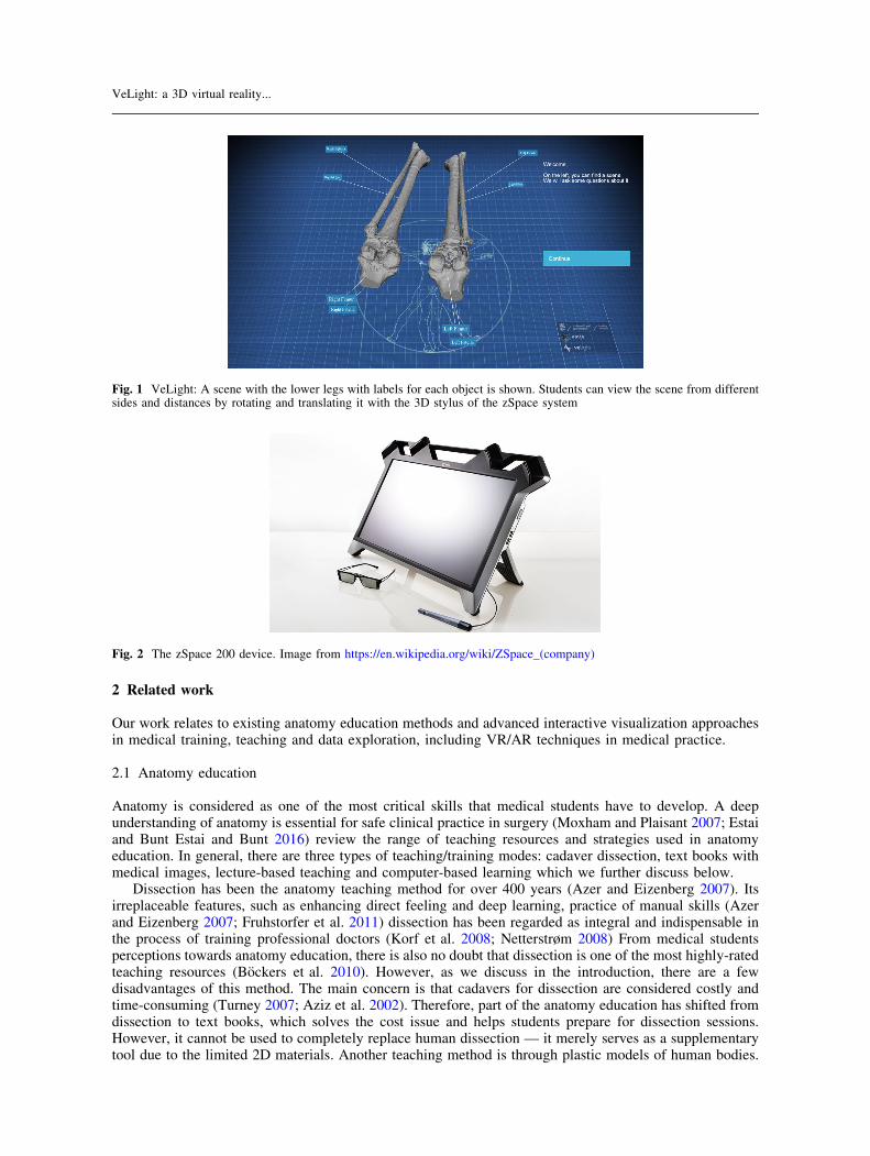
In this application paper, we focus on VR techniques since they provide simple interaction techniques for3D manipulations with 3D spaces and objects. We developed an interactive visualization tool for computedtomography scan (CT) based anatomy teaching and training, with which students can study anatomy using a3D VR display with intuitive interaction methods. The VR device that we use is zSpace 1. It consists of astereographic screen, head tracked eyewear and a stylus, as shown in Fig. 2. With the help of the headtracking system and the 3D glass, the zSpace system understands the correct position of the user andprovides 3D perception to the user. Similar to the glasses, the stylus is also tracked in 6DOF, with which theuser is able to have full 6DOF interaction with virtual objects.
1 https://zspace.com/.
L. Yu et al.
2 Related work
Our work relates to existing anatomy education methods and advanced interactive visualization approachesin medical training, teaching and data exploration, including VR/AR techniques in medical practice.
2.1 Anatomy education
Anatomy is considered as one of the most critical skills that medical students have to develop. A deepunderstanding of anatomy is essential for safe clinical practice in surgery (Moxham and Plaisant 2007; Estaiand Bunt Estai and Bunt 2016) review the range of teaching resources and strategies used in anatomyeducation. In general, there are three types of teaching/training modes: cadaver dissection, text books withmedical images, lecture-based teaching and computer-based learning which we further discuss below.
Dissection has been the anatomy teaching method for over 400 years (Azer and Eizenberg 2007). Itsirreplaceable features, such as enhancing direct feeling and deep learning, practice of manual skills (Azerand Eizenberg 2007; Fruhstorfer et al. 2011) dissection has been regarded as integral and indispensable inthe process of training professional doctors (Korf et al. 2008; Netterstrøm 2008) From medical studentsperceptions towards anatomy education, there is also no doubt that dissection is one of the most highly-ratedteaching resources (Bockers et al. 2010). However, as we discuss in the introduction, there are a fewdisadvantages of this method. The main concern is that cadavers for dissection are considered costly andtime-consuming (Turney 2007; Aziz et al. 2002). Therefore, part of the anatomy education has shifted fromdissection to text books, which solves the cost issue and helps students prepare for dissection sessions.However, it cannot be used to completely replace human dissection — it merely serves as a supplementarytool due to the limited 2D materials. Another teaching method is through plastic models of human bodies.
Fig. 1 VeLight: A scene with the lower legs with labels for each object is shown. Students can view the scene from differentsides and distances by rotating and translating it with the 3D stylus of the zSpace system
Fig. 2 The zSpace 200 device. Image from https://en.wikipedia.org/wiki/ZSpace_(company)
VeLight: a 3D virtual reality...
Although it can solve the 2D perception issue, the model can only represent an example of the human body.In the medical practice, many different models of body parts and different real use cases are needed to bestudied in different levels of detail (Korf et al. 2008). Thus, Fruhstorfer et al. (2011) conclude that plas-tinated prosections are an adequate resource for the early stages of undergraduate training, but the study ofwet cadaveric material is still needed for further learning experience. Estai and Bunt (2016) propose that themost effective method to motivate students in modern anatomy education is through multimodal teachingparadigms — by combining multiple pedagogical resources to complement one another. Finally,3D4Medical 2 is a cross-platform anatomy education application for helping people understand anatomythrough intuitive visualization and interactivity. Users are supported to rotate, zoom, pan and select 3Dmodels through different platforms. An interesting feature is that students can view the 3D model and sharetheir views in real-time in the multi-users AR environment. In the next session, we will focus on thediscussion of VR/AR visualizations in the medical education.
2.2 VR/AR visualizations in medical education
Computer-based learning is another popular method in medical training and teaching. There are two mainreasons: first of all, the use of medical imaging in anatomy education makes it possible for the students tolearn from real cases, instead of the given examples in the text books; second, it increases students’ interestin studying anatomy (Pabst 2009; Reeves et al. 2004) Yet, there is no clear proof that computer-basedlearning is a better approach than traditional teaching methods (McNulty et al. 2003; Khot et al. 2013; Estaiand Bunt 2016; Research Tam et al. 2010) shows that computer-based learning can be used as a greatsupplement rather than replacement of the traditional teaching methods.
Data visualization aims to help researchers to understand data through visual representations. This isachieved by visualization methods that are intuitive, can easily enhance important structures, and improvethe iterative, interactive procedure of exploration and analysis. Concerning these advantages, most com-puter-based learning methods in medical education are based on 3D visualizations of anatomical structures.Data visualization also encompasses user interaction and analysis (Ware 2004) With the rapid developmentof VR/AR technologies, interactive visualizations with these technologies have made a significant impact inmany different disciplines. Many studies (McNulty et al. 2003; Rizzolo and Stewart 2006) have confirmedthe usefulness of VR in medical education. By using touch-based surface, Fyfe et al. (2013) present a virtual3D dissection platform to explore anatomical body structures. In CINCH Akers 2006, Akers et al. usebimanual input (a trackball and a tablet PC pen) to manipulate and explore 3D models.
Recently, Besancon et al. (2021) present a survey of the state-of-the-art in spatial interfaces for 3Dvisualization. Their work focuses on the visualization tasks as well as interaction paradigms, such astangible interaction, mid-air gestures, haptic interfaces and hybrid interaction paradigms. By using advanced3D VR head mounted displays, for instance Oculus Rift or HTC VIVE, users are supported to see, analyzeand explore complex data visualizations in immersive 3D virtual environment. Ye et al. (2021) present animmersive analytics system to assist experts in exploring and analyzing movement trajectory. Their systemcombines 2D and 3D visualizations and provide users first person view of trajectory data in badminton.Users can also use the VR controller to select and analyze 3D trajectories. Similarly, VR (3D representa-tions, intuitive 3D interactions) can also offer benefits in analyzing and exploring other sports data, such asdata visualizations for soccer (Xie et al. 2021) and table tennis matches (Wang et al. 2021; Mathur Mathur2015) introduces a VR application, by using Oculus Rift in conjunction with Razer Hydra hand controllers,for medical training and instruction purposes. Park et al. (2014) design and evaluate the efficacy of a low-cost VR system for training surgical skills for novice trainees and their results show the preliminaryevidence of the efficacy of the VR system. Sousa et al. (2017) use a VR setup and interactive surface tosupport radiologists to explore 3D data. With hand tracking system, such as Leap motion, Theart et al.(2017) propose the gesture-based method for microscopic data analysis in data scaling and rotation tasks.Khadka et al. (2018) propose a tangible multimodule interface for immersive visualizations. They usetangible props worn around the wrist to represent individual slices of data. Users are supported to add andremove slices from the visualization by manipulating the props.
AR is another immersive 3D visualization technology. Different from VR, the novel aspect of AR is tocombine the virtual object with the real environment, which shows a great potential use in the operatingroom. Various surgical fields including craniomaxillofacial surgery, orthopaedics, spine surgery,
2 https://3d4medical.com/.
L. Yu et al.

neurosurgery, laparoscopy surgery and biopsy procedures are exploring the potential of AR Mischkowskiet al. (2005; Kalavakonda et al. 2019) propose that AR could be used for aiding tumor resection in skull-based surgery. They use Microsoft Hololens 3 to enable visualization of Computed Tomography (CT)imaging superimposed in 3D on the patient. Meulstee et al. (2019) present an AR image-guided surgerysystem (IGS) and compare it to the accuracy of a conventional IGS system. Moreover, Glas et al. (2021)explore the possibility of translating the virtual surgical plan to the operating room. The results of thepreliminary study show that AR-based technique enables typical navigation tasks to be performed faster andmore accurately compared the traditional method. A challenge is the manipulation of 3D data and cuttingplanes with axis-based constrains (Blum et al. 2012). For the medical visualizations, users often need tomanipulate/zoom 3D, which often required to be constrained along a single direction. Another interestingapproach for our work is the lightweight tangible 3D interface for exploration of thin fiber structurespresented by Jackson et al. (2013). In their work, they employ a depth sensing camera to track small-scalegestures with a paper prop and then they map the movement of the tracked prop into the manipulations of3D bioimaging datasets. Their approach of data manipulation interests us since the zSpace stylus, which weuse to interact with virtual objects, has the same shape as their paper prop.
However, VR and AR head-mounted displays are usually costly, which limits their use in daily edu-cation. Furthermore, VR and AR visualization and interaction methods are very case-oriented, for instance,it is difficult to answer 2D text questions by writing in the pure 3D environment provided by these head-mounted displays. Thus, before integrating VR/AR technology in our work, we first discuss the two researchquestions, Q1 and Q2, proposed in the introduction.
3 System design
In order to learn the necessity of introducing the advanced VR/AR technology to the medical education, weconducted an initial interview with medical experts and teachers. We aimed to learn better the generalrequirements and the fundamental tasks in medical training and teaching, based on which we can discuss ourresearch questions (Q1 and Q2) and design our system.
3.1 Participants
We invited three medical experts in the interview. All of them are currently working in the local UniversityMedical Center. Two of them had more than 10 years clinical experience in orthopedics and the other has 15years research experience in Medical Informatics. In the interview, firstly, the medical experts were asked topresent the general tasks and requirements, including their workflow in the medical education, the teachingmaterials as well as the question types. After that, we had a discussion on their tasks and approaches.
3.2 General requirements and tasks
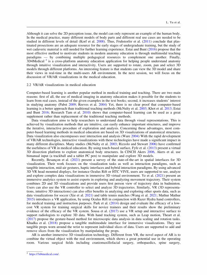
Based on the interview, we list the most fundamental tasks which are required in the medical training andteaching in Table 1. We now explain some of these tasks in more details.
Visualization Data visualizations in text books are usually provided by static images. This only gives avery limited view of a prefect example of the human body. Thus, we need our system to give a 3Dperception of data, which can easily reveal relevant structures and improve the iterative, interactive pro-cedure of analysis. Our system should support users in observing both volumetric scans (CT, MRI, PET as aform of DICOM data (Mildenberger et al. 2002) (V2) and the inner structure of the body, which is usually asubset of 3D meshes (V1 and V3). Moreover, volume rendering is typically used to render volumetric dataand a transfer function is used to assign RGB and alpha values for every voxel in the volume. Users can editit in order to look at different anatomical structures, such as the skin, the skull or the vessels. Thus, thesystem should provide a colormap for the transfer function (V6) and the editing function on it (I6).
Interaction Interactivity supports users in learning the required knowledge while they are ‘‘playing’’ withthe data. Users receive immediate feedback and improve their ability to understand the presented data.Therefore, our system should provide intuitive interactions for 3D data manipulations. For instance, users
3 https://www.microsoft.com/en-us/hololens.
VeLight: a 3D virtual reality...
are able to highlight dedicated objects by a single click on the object (I4), and pick up an object and put it atthe right position and orientation in the scene (I3).
Modes As a tool for teachers and a training system for students, the system should have two modes: aself-assessment mode (M1) and an examination mode (M2). In both modes, teachers should be supported toarrange questions (or tasks) and set specific configuration options for the questions. For instance, they canprovide possible answers in multiple choice tasks and specify the correct position of the target object in theobject selection tasks. Different from the examination mode, in the self-assessment mode, students should beable to receive more feedback on their actions and should have the possibility to retry the tasks.
Course management As a teaching tool, teachers should have a large degree of freedom in designingtheir courses (C1), by using the provided elements. The system should have different questions/tasks types,such as object selection (T3) and placement (T4). Teachers who using the system in their classes should havethe freedom to design their courses as they want. Pre-designed courses would limit the flexibility of thesystem. Moreover, this flexibility may come in handy when the system potentially expands to other dis-ciplines, such as visualizing surgery procedures to patients. To provide teachers with this flexibility, theapplication should be able to read a specification file with what kind of question types the program shouldask.
3.3 Discussion with domain experts
In the interview, the domain experts were impressed by the default demo provided by the zSpace system,which shows anatomical objects and supports users to explore human body systems. They further expressedthe potential needs for such a novel interactive visualization tool in the medical practice, not only forteaching and training, but also for surgery planning. We discussed the exploration tasks in their dailypractice or in teaching, which could potentially benefit from learning from a virtual environment (Q1). Themedical experts commented that understanding and memorizing the spatial relationships of the anatomicalstructure is strongly required in medical studies. However, 2D representations of the body structures givenby the images in text books are unintuitive, which requires students’ strong imagination in order tounderstand the spatial structure in 3D. To this point, the 3D perception of depth can indeed help them tounderstand the data structure. Supported by the tracking system, VR systems also provide the ability to lookaround the presented data and always show the precise perspective. In addition, because of the intuitiveinteraction methods, a large degree of freedom can be provided in designing tasks for the same purpose. Forinstance, for the same purpose of studying the position of a dedicated object, we can either ask students toselect the object, or let them pick up the object and put it at the right place and orientation in the body. Thus,we see the potential value in integrating VR interactions in understanding spatial anatomical structures.
The other question is how VR techniques support researchers’ data exploration tasks (Q2). Morespecifically, we want to make sure the proposed 3D interaction techniques can support users to study and
Table 1 General requirements and tasks as specified by domain experts
Category Tasks
Visualization V1: show a sceneV2: show a volumeV3: show a subset of objectsV4: show the labels with the name of objectsV5: show the slicerV6: show a colormap for the transfer function of the volume rendering
Interaction I1: highlight dedicated objects/regions of interestI2: rotate the 3D sceneI3: manipulate an object (translation, rotation)I4: select an objectI5: manipulate the slicer planeI6: edit the colormap (the transfer function)
Questions/tasks for students T1: multiple choicesT2: open questionsT3: object selectionT4: object placement
Modes M1: self-assessment modeM2: examination mode
Courses management C1: courses/questions design
L. Yu et al.

memorize the spatial structures. First of all, one benefit of the zSpace, and also of most of VR technologies,is that the 3D view and depth perception can greatly help to understand the data. Second, the zSpace stylus isheld like a pen and users can move their hand naturally to check an object from all directions. This is a veryintuitive design that we believe most users can remember how to use without any difficulties. However, wedo not want to limit the interaction choices. Instead, we want to provide more natural interaction techniques(such as the lightweight tangible 3D interface (Jackson et al. 2013) in case those can assist users to betterunderstand their data. Another design choice is that, in order to avoid memory overload, we will use onlyone button on the stylus.
4 VeLight
In the design of our system and the interaction techniques, we were guided by several complementary goals.We designed our system to:
G1: support the focus on data exploration,G2: support the manipulation of the scene space as well as the selected object,G3: encapsulate all six degrees of freedom for 3D interaction,G4: provide both large-scale and precise manipulations,G5: be intuitive and require little learning time,G6: be flexible to apply different visualization methods on the target objects, andG7: be easily extensible with different task types, courses and teaching materials.
As an interactive visualization tool for medical education, we specifically suggest to leave a big space on theinterface for presenting the visualized data (G1). This is a crucial design decision since a big visualizationcan provide more details of the data, which assists users with fully concentrating on data exploration. Wealso want to enable users to manipulate both the whole scene space and the selected object (G2). Forinstance, students can view the whole body, including the 3D volumetric CT scan as well as the bones, by alarge-scale interaction. However, precise interaction is often needed (G4), especially in the task T4, when anobject is requested to be moved to the correct location. They can also select and manipulate a dedicatedobject in the scene. A specific task in this case is tasks I2 - I4 in Table 1. Moreover, we want to enable usersto control all degrees of freedom (G3) in an intuitive manner (G4) so that our interaction technique can takeadvantage of the setup of the VR device. As discussed above, we want to keep the interaction techniquesimple and use only one button on the stylus, so that little learning time is required (G5). The button is usedfor holding an object. Furthermore, we want to provide a large freedom in designing the courses (G7). As welisted in Table 1, a variety of questions should be given to explore anatomy by a variety of objectmanipulation tasks and multiple-choice questions. Those tasks should be possibly included in the system.Finally, VeLight needs to load both volume and mesh data and visualize the data on the zSpace screen.Thus, we also strive for our visualization to have different visualization ways (G6). The users are able toadjust the colormap of the transfer function to get different visualization effects (see Fig. 3).
Fig. 3 The users can adjust the colormap of the transfer function to look at different anatomical structures
VeLight: a 3D virtual reality...

4.1 User interface
Figure 4 presents the interface of VeLight. The left part of the interface shows the scene, including thevolume data, the objects and highlighted target objects. The scene is visualized by the support of stereorendering of the graphics library and the correct projection is used based on the spatial location of the user,which is tracked by the zSpace 3D glasses. The right side of the screen is dedicated to a 2D user interface(UI), which explains the task that the user has to perform and provides the necessary UI elements. Forpreparation of the teaching materials, the system provides various types of questions/tasks, such as multiplechoice questions, object selection and placement. Teachers are supported to design their courses, teachingand assessment forms. They can upload their own datasets, edit questions, and arrange the course/lecturestructure. Students can attend the course and interact with teaching materials (3D data), through which theycan understand and remember the body parts, the features of different organs, etc. They are also supported todo a self-assessment (M1) and receive immediate feedback. In the examination mode (M2), when studentscomplete a task, the application should record their answers,so teachers can extract scores of students fromthe system.
4.2 Interaction techniques
VeLight offers interaction with the data through the zSpace stylus. Here we apply the concept of an adaptivelength stylus in the interaction technique. The stylus is visualized as a ray emerging from the stylus tip in the
Fig. 4 The interface: the right part of the screen is the UI Area, the left part is the Scene Area. In this particular task, the userhas to select the highlighted object in the volume rendering. The UI Area contains the question, the transfer function of thevolume, the multiple choices and a submit button
Fig. 5 The user is moving the bone in the direction of its correct position. a The red outline indicates that the object is not yetconsidered to be correctly placed; b The user moves the bone to the correct position in the scene. The green outline indicatesthis to the user
L. Yu et al.
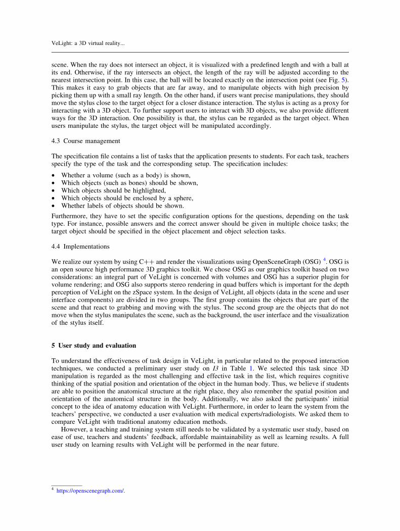
scene. When the ray does not intersect an object, it is visualized with a predefined length and with a ball atits end. Otherwise, if the ray intersects an object, the length of the ray will be adjusted according to thenearest intersection point. In this case, the ball will be located exactly on the intersection point (see Fig. 5).This makes it easy to grab objects that are far away, and to manipulate objects with high precision bypicking them up with a small ray length. On the other hand, if users want precise manipulations, they shouldmove the stylus close to the target object for a closer distance interaction. The stylus is acting as a proxy forinteracting with a 3D object. To further support users to interact with 3D objects, we also provide differentways for the 3D interaction. One possibility is that, the stylus can be regarded as the target object. Whenusers manipulate the stylus, the target object will be manipulated accordingly.
4.3 Course management
The specification file contains a list of tasks that the application presents to students. For each task, teachersspecify the type of the task and the corresponding setup. The specification includes:
• Whether a volume (such as a body) is shown,• Which objects (such as bones) should be shown,• Which objects should be highlighted,• Which objects should be enclosed by a sphere,• Whether labels of objects should be shown.
Furthermore, they have to set the specific configuration options for the questions, depending on the tasktype. For instance, possible answers and the correct answer should be given in multiple choice tasks; thetarget object should be specified in the object placement and object selection tasks.
4.4 Implementations
We realize our system by using C?? and render the visualizations using OpenSceneGraph (OSG) 4. OSG isan open source high performance 3D graphics toolkit. We chose OSG as our graphics toolkit based on twoconsiderations: an integral part of VeLight is concerned with volumes and OSG has a superior plugin forvolume rendering; and OSG also supports stereo rendering in quad buffers which is important for the depthperception of VeLight on the zSpace system. In the design of VeLight, all objects (data in the scene and userinterface components) are divided in two groups. The first group contains the objects that are part of thescene and that react to grabbing and moving with the stylus. The second group are the objects that do notmove when the stylus manipulates the scene, such as the background, the user interface and the visualizationof the stylus itself.
5 User study and evaluation
To understand the effectiveness of task design in VeLight, in particular related to the proposed interactiontechniques, we conducted a preliminary user study on I3 in Table 1. We selected this task since 3Dmanipulation is regarded as the most challenging and effective task in the list, which requires cognitivethinking of the spatial position and orientation of the object in the human body. Thus, we believe if studentsare able to position the anatomical structure at the right place, they also remember the spatial position andorientation of the anatomical structure in the body. Additionally, we also asked the participants’ initialconcept to the idea of anatomy education with VeLight. Furthermore, in order to learn the system from theteachers’ perspective, we conducted a user evaluation with medical experts/radiologists. We asked them tocompare VeLight with traditional anatomy education methods.
However, a teaching and training system still needs to be validated by a systematic user study, based onease of use, teachers and students’ feedback, affordable maintainability as well as learning results. A fulluser study on learning results with VeLight will be performed in the near future.
4 https://openscenegraph.com/.
VeLight: a 3D virtual reality...

5.1 Participants
Eight students from different research background (four from medical field) joined the user study. Six ofthem reported as right handed. However, hand preference does not really make a difference in our designsince the stylus can be used in both hands. Seven medical experts/radiologists joined the user evaluation.
5.2 Apparatus
The zSpace setup consists of a stereographic screen, head-tracked eyewear and a stylus. The screen is a FullHD (1920 � 1080 pixels) stereo-enabled display, with a refresh rate of 120 HZ. The display was positionedso that the center of the display was at a height of about 30 degrees below participants’ eye (see Fig.7). Allparticipants reported that it was the best view for them to finish the tasks.
5.3 Tasks
We tested two reposition tasks (see Fig. 6). The first task was to place an object from the correct position,with the outline of the correct location shown to the participants. The second task was similar, but withoutthe correct position outlined. The reposition task is regarded as a low-level actions that controls position andorientation of the 3D object. However, it requires cognitive thinking of the spatial position and orientation ofthe object in the human body.
Each task had four trials. At the start of each trial, the objects were repositioned, to prevent the learningeffect. The participants were asked to reposition the object as quickly and precisely as possible. In bothtasks, there was no indication whether the object had been positioned at the right position. So that theparticipants had the control of the time that they could stop the current trial and start the next one. Finally,after finishing all trials, the participants were asked to fill in a questionnaire to comment on the experience ofusing the new interaction techniques and their ideas about the prospects of VeLight.
In the user evaluation, we did not set any specific task, instead, we let our participants to use the VeLightsystem to examine/study a prepared course — the leg anatomy. We asked them to examine the data andquestions for each task and to report any interesting aspects they noticed.
5.4 Results
We do not compare the results of the two tasks. Instead, we want to test whether continuous use of theinteraction technique in VeLight improves users’ understanding. Although the task completion times andposition and rotation offsets during the study were logged, we did not notice anything interesting from thelog data. The reason is that the sample was still too small to generate any learning effects. However, fromthis preliminary study we do not expect to learn how long the participants spent on each trail and howprecisely they performed the task. We aim to determine an initial idea whether the VR interaction techniquecan improve users’ understanding and whether users accept the new studying method. Based on the results
Fig. 6 User study tasks: Repositioning an object at the correct position, with the correct location outlined a; and without theoutline b
L. Yu et al.

of the preliminary study we can further discuss how to run a more detailed user study for the learning effectsin the future.
From the observations and discussion, we noticed that the participants were able to learn and work withVeLight easily and quickly: after a short introduction students were capable to work with the systemindependently. Response to the 3D visualization and interaction was very positive. The use of the stylus forinteractions was intuitive. When we asked whether VeLight can replace the current used methods (e.g.,textbooks with 2D visualizations) for learning anatomy, with the scale from 1 (fully disagree) to 5 (fullyagree), half of the participants voted for 4 and the other half voted for 5. However, note that, the aim of ourwork is not replacing the traditional method with our new interactive system. We asked this question onlyfor understanding whether and to which extent the interactive system can be an additional education methodfor anatomy study. One participant commented that ‘‘With VeLight and its interaction methods, you canpractice in 3D and see the connection between structures.’’ Another participant reported ‘‘If the user wasable to upload specific anatomical structures and play around at will, I think it may be more insightful thanjust looking at pictures or descriptions.’’ All participants believed that the 3D perception and interactionwith the scene gave them a better impression of how the scene is structured.
In the interview with medical experts, we asked them to compare VeLight with traditional anatomyeducation methods. In general, we got very positive comments — all participants reacted enthusiastically tothe potential of VeLight. Most participants found the highlighted contour very helpful in determining thecorrect position. However, one medical expert mentioned ‘‘Without the contour, when you have to make thecomplete bone structure with the separate bones, you have to think about the orientation and position,instead of ‘somewhere there’.’’, which we think is very challenging and requires users to have a clear ideaabout the spatial structure of the data. They also liked the task to grab and move an object to the correctposition. On the other hand, some participants commented that VeLight should be regarded as an additionaltool instead of a replacement for the currently used methods.
6 Discussion
We now look back to the two research questions that we proposed in the introduction. The initial interviewwith the medical experts suggested that anatomy teaching and training may be improved by advanced VR/AR technologies. Our preliminary user study and evaluation clearly showed the potential of such a systemfor anatomy education. Moreover, VeLight provides 3D perception and 6DOF interaction with 3D medicaldatasets, facilitating a deeper understanding of anatomical data in an intuitive manner. Thus, we believe thatVR/AR techniques have advantages for medical students’ exploration. These advantages include 3Dvisualization, intuitive interactions, iterative analysis as well as data and image augmentation facilitated byadvanced visualization methods. We also agree with the participants who gave the comments that VeLightshould be used as an additional tool in the anatomy education, instead of being replacement. The reason isthat text books, as a written language, give the most precise description of learning materials and students
Fig. 7 A participant in the user study using our system
VeLight: a 3D virtual reality...

can always find a specific definition and interpretation. By dissecting human cadavers, students can get adirect experience from the actual cases, which would not be possible from any indirect way or any virtual/artificial objects. Thus, we believe VeLight is a strong supplement for the current anatomy educationsystem.
However, there are some improvements that can be considered in further development. First of all, someparticipants in the user study complained about the fatigue issues in the user study. One participant reportedthat at the end of the study his wrist got a bit tired of twisting and changing positions, which caused thereposition result to be not very accurate. We also noticed that the rotation component was very sensitive forsome participants, especially when the manipulated object is cylinder-shape. While the grabbing, movingand rotating interaction is already very intuitive, we have further explored new interaction techniques,inspired by Jackson et al. (2013). We believe a combination of interaction approaches would allow users tochoose the technique to engage depending on the target dataset.
The user study presented in Sect. 5 was only focused on the interaction of students with the zSpace andtheir initial impression of the system. Although this gives an impression about the intuitiveness of VeLight,it does not provide insight in if and how the system improves the learning results. To get insight in this data,an advanced user study on learning results needs to be carried out. Thus, we will need to conduct acontrolled user study to compare the VeLight interaction with traditional interaction methods, for instance,mouse and keyboard input. Another interesting study is to deploy VeLight to the classroom.
7 Conclusion
In this application paper, we present VeLight, a VR system for medical education. Its development wasbased on two research questions and an informal study and interviews with medical domain experts. Welearn that advanced VR technologies can improve users’ understanding, in the tasks which requires spatialattention. Thus, we focus on anatomy training which requires users to have a good understanding ofanatomical structures and relationships. The zSpace on which the VeLight is based provides an interactiveenvironment to implement VR applications. With the development of VeLight we have opened this platformfor use in medical teaching and training based on CT datasets. We conducted a preliminary user study andan evaluation to learn users’ initial impression of the system and the interaction method. In general, we gotpositive comments on VeLight. In the future, we will further improve the user interface and include moremedical materials. Furthermore, formal user validation to determine the effect on learning anatomy has to beperformed.
Supplementary InformationThe online version contains supplementary material available at https://doi.org/10.1007/s12650-021-00790-y.
Acknowledgements The authors wish to thank all the participants who joined in the preliminary user study and the discus-sions. L. Yu is supported by XJTLU Research Development Funding RDF-19-02-11.
References
Akers D 2006. Cinch: A cooperatively designed marking interface for 3d pathway selection. In Proceedings of the 19th AnnualACM Symposium on User Interface Software and Technology, UIST ’06, pp. 33–42. ACM, New York, NY, USA, https://doi.org/10.1145/1166253.1166260
Azer SA, Eizenberg N (2007) Do we need dissection in an integrated problem-based learning medical course? perceptions offirst- and second-year students. Surg Radiol Anat 29:173–180
Aziz MA, Mckenzie JC, Wilson JS, Cowie RJ, Ayeni SA, Dunn BK (2002) The human cadaver in the age of biomedicalinformatics. Anatom Record 269(1):20–32. https://doi.org/10.1002/ar.10046
Bach B, Dachselt R, Carpendale S, Dwyer T, Collins C, Lee B 2016. Immersive analytics: Exploring future interaction andvisualization technologies for data analytics. In Proceedings of the 2016 ACM International Conference on InteractiveSurfaces and Spaces, ISS ’16, pp. 529–533. ACM, New York, NY, USA, https://doi.org/10.1145/2992154.2996365
Besancon L, Ynnerman A, Keefe DF, Yu L, Isenberg T (2021) The state of the art of spatial interfaces for 3d visualization.Comput Graphics Forum 40(1):293–326. https://doi.org/10.1111/cgf.14189
Blum T, Kleeberger V, Bichlmeier C, Navab N 2012. mirracle: An augmented reality magic mirror system for anatomyeducation. In IEEE Virtual Reality Workshops, pp. 115–116, https://doi.org/10.1109/VR.2012.6180909
Bockers A, Jerg-Bretzke L, Lamp C, Brinkmann A, Traue HC, Bockers TM (2010) The gross anatomy course: an analysis ofits importance. Anatom Sci Edu 3(1):3–11. https://doi.org/10.1002/ase.124
Drake RL, McBride JM, Lachman N, Pawlina W (2009) Medical education in the anatomical sciences: the winds of changecontinue to blow. Anatom Sci Edu 2(6):253–259. https://doi.org/10.1002/ase.117
L. Yu et al.

Estai M, Bunt S (2016) Best teaching practices in anatomy education: a critical review. Ann Anatom - Anatomischer Anzeiger208:151–157. https://doi.org/10.1016/j.aanat.2016.02.010
Fruhstorfer B, Palmer J, Brydges S, Abrahams P (2011) The use of plastinated prosections for teaching anatomy-the view ofmedical students on the value of this learning resource. Clin Anatom 24(2):246–252. https://doi.org/10.1002/ca.21107
Fu C.-W., Goh W.-B., Ng J. A. 2010. Multi-touch techniques for exploring large-scale 3d astrophysical simulations. InProceedings of the SIGCHI Conference on Human Factors in Computing Systems, CHI ’10, pp. 2213–2222. ACM, NewYork, NY, USA, https://doi.org/10.1145/1753326.1753661
Fyfe G, Fyfe S, Dye D, Crabb H (2013) Use of Anatomage tables in a large first year core unit. In: 30th Annual conference onAustralian Society for Computers in Learning in Tertiary Education, ASCILITE 2013, pp 298–302
Glas H, Kraeima J, van Ooijen P, Spijkervet F, Yu L, Witjes M (2021) Augmented reality visualization for image-guidedsurgery: a validation study using a three-dimensional printed phantom. J Oral Maxillofac Surg. https://doi.org/10.1016/j.joms.2021.04.001
Jackson B, Lau TY, Schroeder D, Toussaint KC, Keefe DF (2013) A lightweight tangible 3d interface for interactivevisualization of thin fiber structures. IEEE Trans Visual Comput Graph 19(12):2802–2809. https://doi.org/10.1109/TVCG.2013
Johnson S, Jackson B, Tourek B, Molina M, , Erdman A. G., Keefe D. F. 2016 Immersive analytics for medicine: Hybrid 2d/3dsketch-based interfaces for annotating medical data and designing medical devices. Immersive Analytics: ExploringFuture Interaction and Visualization Technologies for Data Analytics, Workshop at IIS 2016,
Kalavakonda N, Sekhar L, Hannaford B 2019. Augmented reality application for aiding tumor resection in skull-base surgery.In 2019 International Symposium on Medical Robotics (ISMR), pp. 1–6, https://doi.org/10.1109/ISMR.2019.8710203
Keefe DF (2010) Integrating visualization and interaction research to improve scientific workflows. IEEE Comput Graph Appl30:8–13. https://doi.org/10.1109/MCG.2010.30
Keefe DF, Isenberg T (2013) Reimagining the scientific visualization interaction paradigm. Computer 46(5):51–57. https://doi.org/10.1109/MC.2013.178
Khadka R, Money J, Banic A 2018. Body-prop interaction: Augmented open discs and egocentric body-based interaction forexploring immersive visualizations. In Proc. ISS, pp. 327–332. ACM, New York, https://doi.org/10.1145/3279778.3281458
Khot Z, Quinlan K, Norman GR, Wainman B (2013) The relative effectiveness of computer-based and traditional resources foreducation in anatomy. Anatom Sci Edu 6(4):211–215. https://doi.org/10.1002/ase.1355
Kim K, Lawrence R. L., Kyllonen N, Ludewig P. M., Ellingson A. M., Keefe D. F. 2017 Poster: Anatomical 2d/3d shape-matching in virtual reality: A user interface for quantifying joint kinematics with radiographic imaging. IEEE Symposiumon 3D User Interfaces, March
Korf H-W, Wicht H, Snipes RL, Timmermans J-P, Paulsen F, Rune G, Baumgart-Vogt E (2008) The dissection course -necessary and indispensable for teaching anatomy to medical students. Ann Anatom - Anatomischer Anzeiger190(1):16–22. https://doi.org/10.1016/j.aanat.2007.10.001
Kreylos O, Bawden G, Bernardin T, Billen M. I., Cowgill E. S., Gold R. D., Hamann B, Jadamec M, Kellogg L. H., StaadtO. G., Sumner D. Y. 2006. Enabling scientific workflows in virtual reality. In Proceedings of the 2006 ACM InternationalConference on Virtual Reality Continuum and Its Applications, VRCIA ’06, pp. 155–162. ACM, New York, NY, USA,https://doi.org/10.1145/1128923.1128948
Mathur A. S. 2015. Low cost virtual reality for medical training. In 2015 IEEE Virtual Reality (VR), pp. 345–346, Marchhttps://doi.org/10.1109/VR.2015.7223437
McNulty JA, Halama J, Espiritu B (2003) Evaluation of computer-aided instruction in the medical gross anatomy curriculum.Clin Anatom 17(1):73–78. https://doi.org/10.1002/ca.10188
McNulty JA, Sonntag B, Sinacore JM (2009) Evaluation of computer-aided instruction in a gross anatomy course: a six-yearstudy. Anatom Sci Edu 2(1):2–8. https://doi.org/10.1002/ase.66
Meulstee JW, Nijsink J, Schreurs R, Verhamme LM, Xi T, Delye HHK, Borstlap WA, Maal TJJ (2019) Toward holographic-guided surgery. Surg Innov 26(1):86–94. https://doi.org/10.1177/1553350618799552
Mildenberger P, Eichelberg M, Martin E (2002) Introduction to the dicom standard. Euro Radiol 12(4):920–927. https://doi.org/10.1007/s003300101100
Mischkowski R, Zinser M, Kubler A, Seifert U, Zoller J 2005. Clinical and experimental evaluation of an augmented realitysystem in cranio-maxillofacial surgery. International Congress Series, 1281(Supplement C):565 – 570, CARS 2005:Computer Assisted Radiology and Surgery. https://doi.org/10.1016/j.ics.2005.03.277
Moxham B, Plaisant O (2007) Perception of medical students towards the clinical relevance of anatomy. Clin Anatom20(5):560–564. https://doi.org/10.1002/ca.20453
Netterstrom I, Kayser L (2008) Learning to be a doctor while learning anatomy. Anatom Sci Edu 1(4):154–158. https://doi.org/10.1002/ase.31
Pabst R (2009) Anatomy curriculum for medical students: What can be learned for future curricula from evaluations andquestionnaires completed by students, anatomists and clinicians in different countries? Ann Anatom - AnatomischerAnzeiger 191(6):541–546. https://doi.org/10.1016/j.aanat.2009.08.007
Park C. H., Wilson K. L., Howard A. M. 2014. Pilot study: Supplementing surgical training for medical students using a low-cost virtual reality simulator. In 2014 IEEE 27th International Symposium on Computer-Based Medical Systems,pp. 125–127, May https://doi.org/10.1109/CBMS.2014.74
Ramnarayan K, Hande S (2005) Thoughts on self directed learning in medical schools: Making students more responsible. NewHorizons 11(3)
Reeves RE, Aschenbrenner JE, Wordinger RJ, Roque RS, Sheedlo HJ (2004) Improved dissection efficiency in the humangross anatomy laboratory by the integration of computers and modern technology. Clin Anatom 17(4):337–344. https://doi.org/10.1002/ca.10245
Rizzolo LJ, Stewart WB (2006) Should we continue teaching anatomy by dissection when...? Anatom Record Part B NewAnatom 289B(6):215–218. https://doi.org/10.1002/ar.b.20117
VeLight: a 3D virtual reality...

Sotiropoulos F, Erdman AG, Borazjani I, Malbraaten N, Le TB, Coffey D, Keefe DF (2012) Interactive slice wim: navigatingand interrogating volume data sets using a multisurface, multitouch VR interface. IEEE Trans Visual Comput Graph18:1614–1626. https://doi.org/10.1109/TVCG.2011.283
Sousa M, Mendes D, Paulo S, Matela N, Jorge J, Lopes D. S. o. 2017. VRRRRoom: Virtual reality for radiologists in thereading room. In Proc. CHI, pp. 4057–4062. ACM, New York, https://doi.org/10.1145/3025453.3025566
Tam M, Hart A, Williams S, Holland R, Heylings D, Leinster S (2010) Evaluation of a computer program (‘disect’) toconsolidate anatomy knowledge: a randomised-controlled trial. Med Teacher 32(3):e138–e142. https://doi.org/10.3109/01421590903144110
Theart RP, Loos B, Niesler TR (2017) Virtual reality assisted microscopy data visualization and colocalization analysis. BMCBioinform 18(2):1–16. https://doi.org/10.1186/s12859
Turney BW (2007) Anatomy in a modern medical curriculum. Ann Royal College Surg Engl 89(2):104–107. https://doi.org/10.1308/003588407X168244
Wang J, Wu J, Cao A, Zhou Z, Zhang H, Wu Y (2021) Tac-miner: Visual tactic mining for multiple table tennis matches. IEEETrans Vis Comput Graph 27(6):2770–2782. https://doi.org/10.1109/TVCG.2021.3074576
Ware C (2004) Inform Visual: Percep Des. Morgan Kaufmann Publishers Inc., San FranciscoWu H-K, Lee SW-Y, Chang H-Y, Liang J-C (2013) Current status, opportunities and challenges of augmented reality in
education. Comput Edu 62:41–49. https://doi.org/10.1016/j.compedu.2012.10.024Xie X, Wang J, Liang H, Deng D, Cheng S, Zhang H, Chen W, Wu Y (2021) Passvizor: Toward better understanding of the
dynamics of soccer passes. IEEE Trans Vis Comput Graph 27(2):1322–1331. https://doi.org/10.1109/TVCG.2020.3030359
Ye S, Chen Z, Chu X, Wang Y, Fu S, Shen L, Zhou K, Wu Y (2021) Shuttlespace: Exploring and analyzing movementtrajectory in immersive visualization. IEEE Trans Vis Comput Graph 27(2):860–869
Yu L, Efstathiou K, Isenberg P, Isenberg T (2012) Efficient structure-aware selection techniques for 3D point cloudvisualizations with 2DOF input. IEEE Trans Vis Comput Graph 18(12):2245–2254. https://doi.org/10.1109/TVCG.2012.217
Yu L, Efstathiou K, Isenberg P, Isenberg T (2016) CAST: Effective and efficient user interaction for context-aware selection in3D particle clouds. IEEE Trans Vis Comput Graph 22(1):886–895. https://doi.org/10.1109/TVCG.2015.2467202
Publisher’s Note Springer Nature remains neutral with regard to jurisdictional claims in published maps and institutional
affiliations.
L. Yu et al.
Related Documents