Vein Patching Reduces Neointimal Thickening Associated with Prosthetic Graft Implantation Andrew T. Gentile, MD, Joseph L. Mills, MD, Michael A. Gooden, MD, R. Daniel Hagerty, MS, Scott S. Berman, MD, John D. Hughes, MD, Leigh B. Kleinert, Stuart K. Williams, PhD, Tucson, Arizona PURPOSE: Modified anastomotic techniques utiliz- ing autogenous vein-cuffs or patches have been devised with the hope of improving prosthetic graft patency. The mechanisms of the presumed improvement in patched anastomoses have never been elucidated and remain speculative. We characterized the healing response of the Taylor vein patch in prosthetic arteriovenous fis- tulae in a canine model of intimal hyperplasia. METHODS: Six adult dogs underwent placement of bilateral (6 patched, 6 control) 4-mm diameter expanded polytetrafluoroethylene loop femoral artery-vein fistulae. Serial duplex ultrasound ex- aminations confirmed graft patency until explant at 6 weeks. Differential light microscopy with computerized image analysis was performed on serial 5-mm sections. Intimal thickness through the venous anastomosis and outflow veins of Taylor patch and control (nonpatched) grafts were compared. Cell type-specific immunocyto- chemical antibody stains for smooth muscle cells (a SMC actin) and endothelial cells (von Willebrand factor) were performed. RESULTS: Eleven of 12 grafts remained patent for 6 weeks, 1 control graft thrombosed. Mean du- plex-derived peak systolic velocities of patched (96 cm/sec) and control (108 cm/sec) grafts were similar. Microscopy revealed more intimal pan- nus anastomotic suture line ingrowth in controls (mean thickness 5 178 mm) than Taylor patched grafts (mean 147 mm, p 5 0.0002). Significantly less intimal thickening was present in the out- flow vein of patched (mean thickness 5 90 mm) versus control grafts (mean 195 mm, P <0.0001). The intima maintained a single cell layer of vWF 1 endothelial cells, while the majority of the cells comprising the lesion expressed a SMC ac- tin. CONCLUSION: Perianastomotic pannus is primarily composed of intimal smooth muscle cells. Neo- intimal thickening is significantly reduced in prosthetic arteriovenous fistulae created with the Taylor vein patch in a canine model. Reduction in perianastomotic intimal thickening may ex- plain the reported clinical improvement in pros- thetic bypass graft patency when modified with vein patch techniques. Am J Surg. 1998;176: 601– 607. © 1998 by Excerpta Medica, Inc. I n an effort to increase the patency of vascular recon- structions requiring the use of prosthetic bypass con- duits, various graft configurations and anastomotic modifications have been proposed and applied clinically in several small human trials. The most common reasons infrainguinal prosthetic grafts are placed are lack or insuf- ficient length of usable greater saphenous vein. Although results of lower extremity revascularization with autoge- nous vein are clearly superior to prosthetics with at least a 30% difference in primary 5-year patency, 1 approximately 10% to 30% of patients do not have suitable saphenous vein for lower extremity arterial bypass. 2,3 The failure of prosthetic grafts in distal reconstructions is thought to result largely from fibrointimal hyperplasia at the distal anastomosis. 4 The concept of a prosthetic/autogenous composite was developed in response to the generally poor patency achieved with prosthetic grafts for distal arterial bypass in the setting of lack of suitable vein. Prosthetic- vein composite graft patencies are generally intermediate between those of all-autogenous and all prosthetic bypass grafts, 5 with reported mean 2-year cumulative patencies ranging from 30% to 64%. 6,7 Theories abound concerning the pathogenesis of the in- timal lesions responsible for prosthetic, as well as autoge- nous graft-anastomotic failures. Various operative adjuncts, including vein cuffs, collars, and patches, have been placed in humans to modify these perianastomotic factors and improve graft patency. The mechanisms of the presumed improvement in patched anastomoses have never been elucidated and remain speculative. The common denomi- nator in prosthetic graft failure appears to be related to the perianastomotic healing response of the recipient vessel. This is true not only for arterial reconstructions but also for prosthetic arteriovenous fistuae with venous outflow steno- sis responsible for the majority of hemodialysis fistula fail- ure. In order to characterize the healing response of a modified prosthetic anastomosis, Taylor vein patched arte- riovenous fistulae were implanted in a canine model of intimal hyperplasia. From the Division of Vascular Surgery (ATG, JLM, MAG, RDH, SSB, JDH) and the Department of Surgical Research (LBK, SKW), University of Arizona Health Sciences Center, Tucson, Arizona. This work was supported in part by a Research Development Grant from the Impra Pharmaceuticals Company, Tempe, Ari- zona. Requests for reprints should be addressed to Joseph L. Mills, MD, Professor of Surgery, Chief, Division of Vascular Surgery, AHSC PO Box 245072, 1501 N. Campbell Avenue, Tucson, Ari- zona 85724-5072. Presented at the 50th Annual Meeting of the South western Surgical Congress, San Antonio, Texas, April 19 –22, 1998. © 1998 by Excerpta Medica, Inc. 0002-9610/98/$19.00 601 All rights reserved. PII S0002-9610(98)00286-4

Welcome message from author
This document is posted to help you gain knowledge. Please leave a comment to let me know what you think about it! Share it to your friends and learn new things together.
Transcript

Vein Patching Reduces Neointimal ThickeningAssociated with Prosthetic Graft Implantation
Andrew T. Gentile, MD, Joseph L. Mills, MD, Michael A. Gooden, MD, R. Daniel Hagerty, MS,Scott S. Berman, MD, John D. Hughes, MD, Leigh B. Kleinert, Stuart K. Williams, PhD, Tucson, Arizona
PURPOSE: Modified anastomotic techniques utiliz-ing autogenous vein-cuffs or patches have beendevised with the hope of improving prostheticgraft patency. The mechanisms of the presumedimprovement in patched anastomoses havenever been elucidated and remain speculative.We characterized the healing response of theTaylor vein patch in prosthetic arteriovenous fis-tulae in a canine model of intimal hyperplasia.
METHODS: Six adult dogs underwent placement ofbilateral (6 patched, 6 control) 4-mm diameterexpanded polytetrafluoroethylene loop femoralartery-vein fistulae. Serial duplex ultrasound ex-aminations confirmed graft patency until explantat 6 weeks. Differential light microscopy withcomputerized image analysis was performed onserial 5-mm sections. Intimal thickness throughthe venous anastomosis and outflow veins ofTaylor patch and control (nonpatched) graftswere compared. Cell type-specific immunocyto-chemical antibody stains for smooth musclecells (a SMC actin) and endothelial cells (vonWillebrand factor) were performed.
RESULTS: Eleven of 12 grafts remained patent for6 weeks, 1 control graft thrombosed. Mean du-plex-derived peak systolic velocities of patched(96 cm/sec) and control (108 cm/sec) grafts weresimilar. Microscopy revealed more intimal pan-nus anastomotic suture line ingrowth in controls(mean thickness 5 178 mm) than Taylor patchedgrafts (mean 147 mm, p 5 0.0002). Significantlyless intimal thickening was present in the out-flow vein of patched (mean thickness 5 90 mm)versus control grafts (mean 195 mm, P <0.0001).The intima maintained a single cell layer ofvWF 1 endothelial cells, while the majority of thecells comprising the lesion expressed a SMC ac-tin.
CONCLUSION: Perianastomotic pannus is primarilycomposed of intimal smooth muscle cells. Neo-intimal thickening is significantly reduced inprosthetic arteriovenous fistulae created with theTaylor vein patch in a canine model. Reductionin perianastomotic intimal thickening may ex-plain the reported clinical improvement in pros-thetic bypass graft patency when modified withvein patch techniques. Am J Surg. 1998;176:601–607. © 1998 by Excerpta Medica, Inc.
In an effort to increase the patency of vascular recon-structions requiring the use of prosthetic bypass con-duits, various graft configurations and anastomotic
modifications have been proposed and applied clinically inseveral small human trials. The most common reasonsinfrainguinal prosthetic grafts are placed are lack or insuf-ficient length of usable greater saphenous vein. Althoughresults of lower extremity revascularization with autoge-nous vein are clearly superior to prosthetics with at least a30% difference in primary 5-year patency,1 approximately10% to 30% of patients do not have suitable saphenousvein for lower extremity arterial bypass.2,3 The failure ofprosthetic grafts in distal reconstructions is thought toresult largely from fibrointimal hyperplasia at the distalanastomosis.4 The concept of a prosthetic/autogenouscomposite was developed in response to the generally poorpatency achieved with prosthetic grafts for distal arterialbypass in the setting of lack of suitable vein. Prosthetic-vein composite graft patencies are generally intermediatebetween those of all-autogenous and all prosthetic bypassgrafts,5 with reported mean 2-year cumulative patenciesranging from 30% to 64%.6,7
Theories abound concerning the pathogenesis of the in-timal lesions responsible for prosthetic, as well as autoge-nous graft-anastomotic failures. Various operative adjuncts,including vein cuffs, collars, and patches, have been placedin humans to modify these perianastomotic factors andimprove graft patency. The mechanisms of the presumedimprovement in patched anastomoses have never beenelucidated and remain speculative. The common denomi-nator in prosthetic graft failure appears to be related to theperianastomotic healing response of the recipient vessel.This is true not only for arterial reconstructions but also forprosthetic arteriovenous fistuae with venous outflow steno-sis responsible for the majority of hemodialysis fistula fail-ure. In order to characterize the healing response of amodified prosthetic anastomosis, Taylor vein patched arte-riovenous fistulae were implanted in a canine model ofintimal hyperplasia.
From the Division of Vascular Surgery (ATG, JLM, MAG, RDH,SSB, JDH) and the Department of Surgical Research (LBK, SKW),University of Arizona Health Sciences Center, Tucson, Arizona.
This work was supported in part by a Research DevelopmentGrant from the Impra Pharmaceuticals Company, Tempe, Ari-zona.
Requests for reprints should be addressed to Joseph L. Mills,MD, Professor of Surgery, Chief, Division of Vascular Surgery,AHSC PO Box 245072, 1501 N. Campbell Avenue, Tucson, Ari-zona 85724-5072.
Presented at the 50th Annual Meeting of the South westernSurgical Congress, San Antonio, Texas, April 19–22, 1998.
© 1998 by Excerpta Medica, Inc. 0002-9610/98/$19.00 601All rights reserved. PII S0002-9610(98)00286-4

MATERIALS AND METHODSSix adult mongrel dogs of either gender each weighing 24
to 28 kg were used in this study. Study design and researchmethods were approved by the Institutional Animal Careand Use Committee at the University of Arizona HealthSciences Center. All animal experiments were performedin accordance with the “Principles of Laboratory AnimalCare and the Guide for the Care and Use of LaboratoryAnimals” (NIH Publication no. 80-23, 1985).
Graft ImplantationDogs underwent preoperative general medical examina-
tion and were found fit to undergo femoral artery to veinprosthetic graft implantation. Prior to surgery, each animalreceived 50 mg dipyridamole orally twice per day for 4 daysand 325 mg aspirin orally once per day for 1 day. Thesemedications were continued during the entire postopera-tive period. All animals received perioperative antibiotics.On the day of surgery, animals were sedated with a mixtureof atropine, ketamine, and acepromazine injected intra-muscularly. General anesthesia was induced using intrave-nous sodium pentothal, and the animal was intubated andmaintained on mechanical ventilation with a mixture ofinhaled halothane and oxygen. Grafts were prepared usingstandard, 4-mm internal diameter, expanded polytetrafluo-roethylene (e-PTFE; IMPRA Inc. Tempe, Arizona). Graftimplantation was performed through diagonal, femoral in-cisions with standard vascular surgical techniques. End-graft to side-femoral arterial and venous anastomoses wereperformed (6 study and 6 controls). A single segment ofnative internal jugular vein was harvested, flushed, opened,and cut to a diamond shape for autogenous Taylor patch aspreviously described.8 Six Taylor vein-patch anastomoseswere constructed for the modified venous-outflow anasto-mosis. All anastomoses were performed with 6-0 Prolenesuture (Ethicon Inc., Somerville, New Jersey).
Graft ExplantSerial duplex ultrasonography was performed to confirm
graft patency and measure graft blood flow velocities atimplant, 3 weeks, and immediately preceding graft explantat 6 weeks. At explant, animals received identical preop-erative sedation and general anesthesia. Dogs were system-ically heparinized, and the grafts were explanted in-totoalong with approximately 3 cm of inflow artery and outflowvein segments. Grafts were gently flushed with heparinizedsaline and pressure fixed at 80- to 100 mm Hg with 10%neutral buffered formalin. After fixation, grafts were se-quentially sectioned in the direction of blood flow throughboth anastomoses. Graft segments were dehydrated andparaffin embedded. Five-micron cross sections were floatedon glass slides, deparaffinized, and stained with hematoxy-lin & eosin, and Masson’s trichrome.
Using the Nikon Optiphot microscope with image trans-fer through a DAGE SIT-66 low light level camera, serialmicrograph cross sections of the venous anastomoses werecompared at 20 to 403 magnification. Analysis of thehistologic characteristics and neointimal layer thicknesswas performed using digitized computerized image analysis(MetaMorph Image Analysis Systems) program linkedthrough Microsoft Windows Applications. Serial cross sec-
tions were compared through the venous anastomosis ofboth control and Taylor patched grafts. The image analysissoftware graphics allows calculation of neointimal area andaverage neointimal thickness was obtained for each graftsegment (Figure 1). The average intimal thicknessthroughout the entire venous anastomosis as well as theintimal thickness in a clearly defined segment of outflowvein just distal to the anastomosis were compared betweengroups (Figure 2). All measurements were electronicallytransferred from MetaMorph to Microsoft Excel spread-sheets. Statistical comparison between control and studyvenous anastomosis and outflow vein segments were per-formed using the Student’s t test for continuous variableson standard PC compatible software (StatView; AbacusConcepts, Berkeley, California). A P value of #0.05 wasconsidered statistically significant.
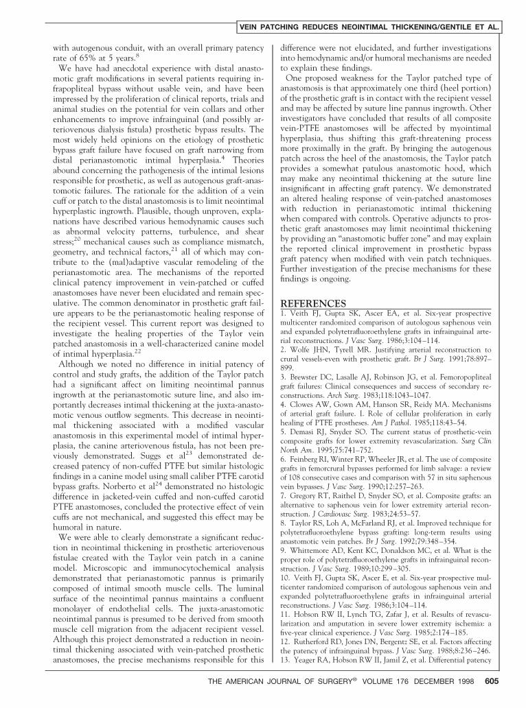
Figure 1. Cross section of a well-incorporated (light, outer ad-ventitial covering) explanted ePTFE graft (dark middle layer)with computerized image analysis for determination of meanthickness of the neointimal lining (light, hatched inner luminallayer; 320).
Figure 2. Schematic drawing of Taylor-patched arteriovenousanastomosis denoting multiple sample sites used to measureneointimal thickness at cross sections (narrow lines) through theanastomosis and separate sample area at outflow vein segments(wide line).
VEIN PATCHING REDUCES NEOINTIMAL THICKENING/GENTILE ET AL.
602 THE AMERICAN JOURNAL OF SURGERY® VOLUME 176 DECEMBER 1998

Immunohistologic AnalysisPhotomicrographs of cross sections were analyzed for
morphologic features and neointimal pannus ingrowth.Single-label immunocytochemistry was performed afterdeparaffinization with xylene and rehydration in gradedethanol solutions. Endogenous peroxidase activity wasblocked with 3% hydrogen peroxide, and primary antibod-ies to either von Willebrand factor (vWF; kindly providedby Dr. James Catalfano, New York State Health Depart-ment, Albany, New York) or a smooth muscle cell actin(Sigma Co., Bushfield, Tennessee) were incubated at roomtemperature for 60 minutes. The sections were rinsed withphosphate buffered saline, a biotinylated secondary anti-body was applied for 30 minutes, followed by a streptavidinperoxidase conjugate (Dako Corp. Carpinteria, Califor-nia). Standard peroxidase enzyme substrate, 3939-diamino-benzidine or alkaline phosphatase substrate were added toyield a brown or red reaction product. Nuclei were lightlycounterstained with dilute methyl green.
RESULTSThis canine study compared the patency and healing
characteristics of standard and a modified anastomotic con-figuration of small caliber prosthetic arteriovenous fistulae.All dogs tolerated the operative procedures without ad-verse events. Eleven of 12 grafts remained patent for 6weeks, 1 control graft thrombosed. Mean duplex-derivedperianastomotic peak systolic velocities of patched (96 622 cm/sec) and control (108 6 24 cm/sec) grafts weresimilar. At explant, all grafts were well incorporated with-out signs of infection. The grafts were implanted as smoothU-shaped loops; however, during healing the grafts oftenformed tightly associated proximal and distal segments(Figure 3). Gross examination of the luminal surface of thegrafts at the time of explant revealed clean body andmidgraft segments without an obvious inner lining or as-sociated blood components. The perianastomotic areas re-vealed only very small amounts of a glistening neointimallayer without adherence of thrombus or platelets.
Microscopic ResultsMorphologic analysis of grafts from both groups revealed
very clean luminal surfaces throughout most of the grafts.
The Taylor patch segments were well incorporated andwithout microscopic signs of infection. The patch re-mained viable as evidenced by normal appearing veinhistology consisting of endothelium, organized intimallayer bounded by an inner elastic lamina and muscularmedial layer surrounded by loose connective tissue andadventitia. There was minimal change in the intimal layermorphology of the Taylor patched segment when com-pared with normal internal jugular vein specimens (Figure4A).
The perianastomotic areas had obvious neointimal pan-nus ingrowth most often at the cut edge of the vein andanastomotic suture line. This perianastomotic neointimalpannus was qualitatively more prominent in control graftsthan in the Taylor-patched grafts (Figure 4B). The anas-tomotic neointima contained a layer of well-organizedsmooth muscle cells covered by a single layer of polygonal-shaped endothelial cells. Since grafts were sectioned trans-versely, representative cross sections were analyzed toquantify the neointima within the inner surface of thegrafts through the anastomosis and at the outflow veinsegments. Microscopy revealed distinct areas of neointimalingrowth starting at the heel of the PTFE with neointimalingrowth at the margins of the graft into the recipientvessel. Quantitatively there was more intimal pannus anas-tomotic suture line ingrowth in controls (mean thick-ness 5 178 6 129 mm) than Taylor patched grafts (mean147 6 103 mm, P 5 0.0002; Table). In addition, signif-icantly less intimal thickness was present in the outflowvein of patched (mean thickness 5 90 6 62 mm) versuscontrol grafts (mean 195 6 107 mm, P ,0.0001).
Immunohistologic ResultsSingle-label immunocytochemical staining produced pos-
itive staining of anti-vWf antibody in areas of neointimacovered by endothelial cells and staining for anti-a SMCactin in subendothelial regions. The neointima maintainedan organized morphology with a confluent single cell layerof vWF 1 endothelial cells along the neointima lining theluminal surface of the grafts, (Figure 4C) while the major-ity of the cells comprising the neointimal lesion expresseda SMC actin consistent with smooth muscle cell ingrowth.(Figure 4D)
COMMENTSPolytetrafluoroethylene grafts have been used extensively
for infrainguinal vascular reconstructions either as the con-duit of choice or as an alternative conduit when saphenousvein is unavailable. Although studies have reported accept-able early patency rates, the long-term efficacy of thesegrafts has been less satisfactory, especially in the infrapop-liteal position. Results of nearly all studies concur thatinfrainguinal PTFE prostheses provide significantly inferiorresults when compared with autogenous vein.1,9–11 Ruth-erford et al12 analyzed the factors affecting the patency ofinfrainguinal bypass grafts and confirmed the often helddictum that the quality of graft conduit and the anasto-motic level (below-knee versus above-knee) are two im-portant variables affecting bypass performance. Becauseresults for infrapopliteal revascularization with prostheticgrafts have been so disappointing, some authors have con-sidered primary amputation preferable to infrapopliteal ar-
Figure 3. Representative gross photograph at graft explant re-vealing a well-healed, tightly looped 4-mm ePTFE arteriovenousfistula. Vascular loops encircle inflow arterial (dark loops) andoutflow vein (light loops) anatomy.
VEIN PATCHING REDUCES NEOINTIMAL THICKENING/GENTILE ET AL.
THE AMERICAN JOURNAL OF SURGERY® VOLUME 176 DECEMBER 1998 603

tery bypass with PTFE in patients without usable autoge-nous vein.13 Recently, however, when faced with the morefrequent clinical dilemma of elderly patients requiring sec-ondary extremity revascularizations and lacking adequateautogenous conduit, investigators have applied varioustechnical adjuncts in hopes of improving prosthetic bypasspatency rates and limb salvage. These techniques includevarious vein patches, cuffs, or collars, and distal arterio-venous fistulae.14
Early uncontrolled studies of patency rates of PTFE withvein cuffs in the femoral-distal position reported encour-aging initial results. Miller et al (1984)15 and Tyrell andWolfe (1991)16 reported 72% and 78% 8 to 12 monthpatencies, respectively. Subsequent work on the influenceof vein cuffs on PTFE femoral-popliteal bypass by Raptis
and Miller17 retrospectively reported no difference inabove-knee PTFE patency with or without vein cuffs at 36months (68% versus 69%), but a significant improvementin below-knee PTFE patency was demonstrated with theuse of adjuvant vein cuffs (57% versus 29%). In a recentrandomized, prospective trial reported by Stonebridge etal,18 vein cuffed below-knee prosthetic bypass produced a65% 2-year patency rate compared with 29% patency inuncuffed grafts, with a sixfold greater early graft failure ratein the uncuffed grafts (13%) versus cuffed grafts (2%). Themost promising results of a vein cuff clinical series, byPappas et al,19 report 62% cumulative 2-year patency for23 infrapopliteal cuffed prosthetic grafts. Reported resultsof uncontrolled series of infrainguinal bypasses using theTaylor vein patch approach those of revascularizations
Figure 4. Photomicrographs demonstrating perianastomotic intimal pannus. A. Cross section through a Taylor-patched anastomosis at103 reveals a normal appearing vein patch segment (arrow) and outflow vein segment (*) with minimal intimal thickening. B. At highermagnification (Trichrome stain, 320) neointimal pannus ingrowth into the PTFE is observed at anastomotic heel regions and alongprolene suture lines. C. Immunocytochemical staining for anti-vWF antibody demonstrates dark staining endothelial monolayer. D. Themajority of the neointimal lesion is composed of cells staining positive for anti-a SMC actin antibody.
TABLECanine Taylor Patch Mean Intimal Thickness Measurements
Graft Venous Anastomosis Venous Outflow
Taylor patch (n 5 6) 147 6 103 mm (318 counts)* 90 6 62 mm (31 counts)Control (n 5 5) 178 6 129 mm (588 counts) 195 6 107 mm (64 counts)Significance t test P 5 0.0002 P ,0.0001
* Counts 5 individual thickness measurements.
VEIN PATCHING REDUCES NEOINTIMAL THICKENING/GENTILE ET AL.
604 THE AMERICAN JOURNAL OF SURGERY® VOLUME 176 DECEMBER 1998
with autogenous conduit, with an overall primary patencyrate of 65% at 5 years.8
We have had anecdotal experience with distal anasto-motic graft modifications in several patients requiring in-frapopliteal bypass without usable vein, and have beenimpressed by the proliferation of clinical reports, trials andanimal studies on the potential for vein collars and otherenhancements to improve infrainguinal (and possibly ar-teriovenous dialysis fistula) prosthetic bypass results. Themost widely held opinions on the etiology of prostheticbypass graft failure have focused on graft narrowing fromdistal perianastomotic intimal hyperplasia.4 Theoriesabound concerning the pathogenesis of the intimal lesionsresponsible for prosthetic, as well as autogenous graft-anas-tomotic failures. The rationale for the addition of a veincuff or patch to the distal anastomosis is to limit neointimalhyperplastic ingrowth. Plausible, though unproven, expla-nations have described various hemodynamic causes suchas abnormal velocity patterns, turbulence, and shearstress;20 mechanical causes such as compliance mismatch,geometry, and technical factors,21 all of which may con-tribute to the (mal)adaptive vascular remodeling of theperianastomotic area. The mechanisms of the reportedclinical patency improvement in vein-patched or cuffedanastomoses have never been elucidated and remain spec-ulative. The common denominator in prosthetic graft fail-ure appears to be the perianastomotic healing response ofthe recipient vessel. This current report was designed toinvestigate the healing properties of the Taylor veinpatched anastomosis in a well-characterized canine modelof intimal hyperplasia.22
Although we noted no difference in initial patency ofcontrol and study grafts, the addition of the Taylor patchhad a significant affect on limiting neointimal pannusingrowth at the perianastomotic suture line, and also im-portantly decreases intimal thickening at the juxta-anasto-motic venous outflow segments. This decrease in neointi-mal thickening associated with a modified vascularanastomosis in this experimental model of intimal hyper-plasia, the canine arteriovenous fistula, has not been pre-viously demonstrated. Suggs et al23 demonstrated de-creased patency of non-cuffed PTFE but similar histologicfindings in a canine model using small caliber PTFE carotidbypass grafts. Norberto et al24 demonstrated no histologicdifference in jacketed-vein cuffed and non-cuffed carotidPTFE anastomoses, concluded the protective effect of veincuffs are not mechanical, and suggested this effect may behumoral in nature.
We were able to clearly demonstrate a significant reduc-tion in neointimal thickening in prosthetic arteriovenousfistulae created with the Taylor vein patch in a caninemodel. Microscopic and immunocytochemical analysisdemonstrated that perianastomotic pannus is primarilycomposed of intimal smooth muscle cells. The luminalsurface of the neointimal pannus maintains a confluentmonolayer of endothelial cells. The juxta-anastomoticneointimal pannus is presumed to be derived from smoothmuscle cell migration from the adjacent recipient vessel.Although this project demonstrated a reduction in neoin-timal thickening associated with vein-patched prostheticanastomoses, the precise mechanisms responsible for this
difference were not elucidated, and further investigationsinto hemodynamic and/or humoral mechanisms are neededto explain these findings.
One proposed weakness for the Taylor patched type ofanastomosis is that approximately one third (heel portion)of the prosthetic graft is in contact with the recipient vesseland may be affected by suture line pannus ingrowth. Otherinvestigators have concluded that results of all compositevein-PTFE anastomoses will be affected by myointimalhyperplasia, thus shifting this graft-threatening processmore proximally in the graft. By bringing the autogenouspatch across the heel of the anastomosis, the Taylor patchprovides a somewhat patulous anastomotic hood, whichmay make any neointimal thickening at the suture lineinsignificant in affecting graft patency. We demonstratedan altered healing response of vein-patched anastomoseswith reduction in perianastomotic intimal thickeningwhen compared with controls. Operative adjuncts to pros-thetic graft anastomoses may limit neointimal thickeningby providing an “anastomotic buffer zone” and may explainthe reported clinical improvement in prosthetic bypassgraft patency when modified with vein patch techniques.Further investigation of the precise mechanisms for thesefindings is ongoing.
REFERENCES1. Veith FJ, Gupta SK, Ascer EA, et al. Six-year prospectivemulticenter randomized comparison of autologous saphenous veinand expanded polytetrafluoroethylene grafts in infrainguinal arte-rial reconstructions. J Vasc Surg. 1986;3:104–114.2. Wolfe JHN, Tyrell MR. Justifying arterial reconstruction tocrural vessels-even with prosthetic graft. Br J Surg. 1991;78:897–899.3. Brewster DC, Lasalle AJ, Robinson JG, et al. Femoropoplitealgraft failures: Clinical consequences and success of secondary re-constructions. Arch Surg. 1983;118:1043–1047.4. Clowes AW, Gown AM, Hanson SR, Reidy MA. Mechanismsof arterial graft failure. I. Role of cellular proliferation in earlyhealing of PTFE prostheses. Am J Pathol. 1985;118:43–54.5. Demasi RJ, Snyder SO. The current status of prosthetic-veincomposite grafts for lower extremity revascularization. Surg ClinNorth Am. 1995;75:741–752.6. Feinberg RI, Winter RP, Wheeler JR, et al. The use of compositegrafts in femorcrural bypasses performed for limb salvage: a reviewof 108 consecutive cases and comparison with 57 in situ saphenousvein bypasses. J Vasc Surg. 1990;12:257–263.7. Gregory RT, Raithel D, Snyder SO, et al. Composite grafts: analternative to saphenous vein for lower extremity arterial recon-struction. J Cardiovasc Surg. 1983;24:53–57.8. Taylor RS, Loh A, McFarland RJ, et al. Improved technique forpolytetrafluoroethylene bypass grafting: long-term results usinganastomotic vein patches. Br J Surg. 1992;79:348–354.9. Whittemore AD, Kent KC, Donaldson MC, et al. What is theproper role of polytetrafluoroethylene grafts in infrainguinal recon-struction. J Vasc Surg. 1989;10:299–305.10. Veith FJ, Gupta SK, Ascer E, et al. Six-year prospective mul-ticenter randomized comparison of autologous saphenous vein andexpanded polytetrafluoroethylene grafts in infrainguinal arterialreconstructions. J Vasc Surg. 1986;3:104–114.11. Hobson RW II, Lynch TG, Zafar J, et al. Results of revascu-larization and amputation in severe lower extremity ischemia: afive-year clinical experience. J Vasc Surg. 1985;2:174–185.12. Rutherford RD, Jones DN, Bergentz SE, et al. Factors affectingthe patency of infrainguinal bypass. J Vasc Surg. 1988;8:236–246.13. Yeager RA, Hobson RW II, Jamil Z, et al. Differential patency
VEIN PATCHING REDUCES NEOINTIMAL THICKENING/GENTILE ET AL.
THE AMERICAN JOURNAL OF SURGERY® VOLUME 176 DECEMBER 1998 605

and limb salvage for polytetrafluoroethylene and autogenous saphe-nous vein in severe lower extremity ischemia. Surgery. 1982;91:99–103.14. Webb T. Operative adjuncts for distal revascularization. SurgClin North Am. 1995;75:753–758.15. Miller JH, Foreman RK, Ferguson L, Faris I. Interposition veincuff for anastomosis of prosthesis to small arteries. Aust NZ J Surg.1984;54:283–285.16. Tyrell MR, Wolfe JHN. New prosthetic venous collar anasto-motic technique: combining the best of the other procedures. Br JSurg. 1991;78:1016–1017.17. Raptis S, Miller JH. Influence of a vein cuff on polytetrafluo-roethylene graft for primary femoropopliteal bypass. Br J Surg.1995;82:487–491.18. Stonebridge PA, Prescott RJ, Ruckley CV. Randomized trialcomparing infrainguinal polytetrafluoroethylene bypass graftingwith and without vein interposition cuff at the distal anastomosis.J Vasc Surg. 1997;26:543–550.
19. Pappas PJ, Hobson RW II, Meyers MG, et al. Patency ofinfrainguinal polytetrafluoroethylene bypass grafts with distal inter-position vein cuffs. Cardio Vasc Surg. 1998;6:19–26.20. Binns RL, Ku DN, Stewart MT, et al. Optimal graft diameter:effect of wall shear stress on vascular healing. J Vasc Surg. 1989;10:326–337.21. Abbott WM, Megerman J, Hasson JE, et al. Effect of compli-ance mismatch on vascular graft patency. J Vasc Surg. 1987;5:376–381.22. Williams SK, Jarrell BE, Kleinert LB. Endothelial cell trans-plantation onto polymeric arteriovenous grafts evaluated using acanine model. J Invest Surg. 1994;7:503–517.23. Suggs WD, Henriques HF, DePalma RG. Vein cuff interposi-tion prevents juxta-anastomotic neointimal hyperplasia. Ann Surg.1988;207:717–723.24. Norberto JJ, Sidawy AN, Trad KS, et al. The protective effectof vein cuffed anastomoses in not mechanical in origin. J Vasc Surg.1995;21:558–566.
DISCUSSIONB. Timothy Baxter, MD (Omaha, Nebraska): In some
senses, this is a preliminary study, because the end pointsinvolve really only looking at neointimal thickness andlooking at one time point. However, I think the study isvery important, and it’s also very timely for all of us,because we’re quite interested in the fact that using a veinpatch at the end of a distal prosthetic graft may help toimprove patency. The initial reports of this were uncon-trolled trials from Europe, where they have a lot moreexperience with it. But we know now from control trials inbelow-knee prosthetic femoropopliteal bypasses, that thevein collars do improve patency, and there’s a strong sug-gestion that that’s also true in the tibial vessels. We’re alsoall encountering more patients in which we’re having amore difficult time finding enough vein to do the redo-redoprocedures.
This study also included immunohistochemical analysisof the cell types in the pannus. The investigators report adecrease in thickness in the Taylor patch group comparedwith the control group. In both areas where the measure-ments were made, the differences were significant, al-though the actual thickness was quite small.
I have several questions for the investigators. First of all,a methodologic question: Why were more counts done inthe control group than in the Taylor patch group, and withthe measurements made in the venous outflow, were theydone in a blinded fashion? The second question is, did youlook at proliferation in some way? The third question, howdid you choose the time frame used in your study? Fourthquestion; the patch would appear that it might change theangle of the anastomosis. Did you correct for this in thecontrol group?
Larry W. Kraiss, MD (Salt Lake City, Utah): The veinpatch is already being used clinically in prosthetic lowerextremity bypasses. This was an a-v fistula model. Has yourgroup applied the vein patch to the venous outflow anas-tomoses of hemodialysis access grafts, and if so, do you haveany impression on the effect on patency that the patchmight have?
Glenn C. Hunter, MD (San Antonio, Texas): I wasintrigued by the last immunohistochemistry slide. All thesmooth muscle cells seemed to be aligned closer to the
luminal surface, with a relatively acellular zone betweenthe graft and the smooth muscle cells, what appeared to belike matrix. Where do you think the smooth muscle cellsoriginate?
Richard C. Pennell, MD (St. Louis, Missouri): When Iwas doing my vascular research fellowship, one of thethings that we found was that arterial and venous endo-thelium do have different characteristics. Certainly, yourexperiment is designed to analyze the way things interactin the venous endothelium. Do you have any plans toperform ligated femoral artery bypasses in which you wouldtry and test this same process compared with arterial en-dothelium rather than venous?
CLOSINGAndrew T. Gentile, MD: Dr. Baxter, you had several
good questions regarding methodology of the analysis.There were just quantitatively many more sites of intimalthickening within the control grafts than in the patchedgrafts themselves. So statistically there was much moreability to look at the intimal thickening from the centralgrafts. The actual measurements were at every single spotthat I was able to find intimal thickening. That’s why therewas a difference in the number of data points for compar-ison.
The power of the statistics is provided by multiple obser-vations. There were nearly a thousand data points throughthe venous anastomoses and almost a hundred data pointsthrough the venous outflow segment.
You asked about proliferative activity in these grafts.Proliferation assays were not specifically performed in thismodel for several reasons. We have done some studies withPCNA, proliferating cell nuclear antigen, in human mod-els of explanted vein graft stenotic segments, and that hasshown that there is a very low level of proliferative activity.
In other experimental projects, using rat vein graft ste-nosis models, there is a moderate increase in proliferativeactivity in these particular lesions. In the dog model, it’sunknown. There happens to be fewer monoclonal antibod-ies available for canine studies, and we haven’t been able tosuccessfully cross-react the antibodies of the human withthe canine model.
You asked about the time frame of the explant. This was
VEIN PATCHING REDUCES NEOINTIMAL THICKENING/GENTILE ET AL.
606 THE AMERICAN JOURNAL OF SURGERY® VOLUME 176 DECEMBER 1998

chosen based on other previous studies, both at our insti-tution and others, with a similar flow model. In the dogA-V fistula model, the intimal thickness increases andprobably peaks somewhere between 5 and 6 weeks. Thereis believed to be a leveling off or a plateauing of the intimalthickening, and then actually there may be some reductionin intimal thickening with time. I think it’s a good sugges-tion to do another series and follow them out longer andsee if this process continues.
I think the origin of the cells that are present most likelyis from the pannus. It has been shown years ago that therecipient vessel does promote healing at the area of the cutedge, and this pannus was demonstrated. It appears to becomposed mostly of smooth muscle cells or cells that ex-press smooth muscle cell alpha actin antibody. And therewere areas of some myxoid type of matrix within the grafts,but other immunochemical stains for matrix componentswere not performed.
The angle of the anastomosis is probably a critical factor,not only in the velocity turbulence measurements, but alsoin how the graph will lie. Some investigators from JosephArchie’s lab have shown in an engineering computer-basedflow model that an angle of the heel coming down on therecipient vessel of about 10 to 15 degrees may be theoptimal angle.
This angle is almost reproduced with the Taylor patch. Bypatching a piece of vein over the anastomosis, you are ableto really bring a much smoother tapered anastomosis andget approximately that same angle. I think this does bringup several more questions, which we’re looking at.
The angles for the control grafts were not modified in anyway. They were just standard anastomoses.
Dr. Kraiss asked about our experience with Taylor patchin human patients. We’ve had only anecdotal experiencewith several patients undergoing repeat leg bypass withoutuseable lengths of saphenous or other alternative sources ofvein. We don’t really have any data to support that, but wehave proposed that A-V fistula Taylor patched grafts beplaced and followed up in selected patients.
Dr. Hunter questioned about the smooth muscle cellorigin. I have to presume it’s from the recipient vessel.
Dr. Pennell also asked about if this has been done in anarterial model. In our experience, the A-V fistula model isa pretty good representation of the intimal thickening froma flow-induced model. We haven’t done them in arteriallesions. It can be done. Several other animal projects havebeen reported in the last couple of years looking at carotidinterposition grafts with patched grafts, and they show verysimilar histology and morphology to the canine A-V fistulamodel.
VEIN PATCHING REDUCES NEOINTIMAL THICKENING/GENTILE ET AL.
THE AMERICAN JOURNAL OF SURGERY® VOLUME 176 DECEMBER 1998 607
Related Documents