966 Am J Clin Pathol 2007;128:966-973 966 DOI: 10.1309/FP0N3LC8MBJUFFA6 © American Society for Clinical Pathology Hematopathology / VEGF AND MVD IN PH– CMPDS VEGF Expression Correlates With Microvessel Density in Philadelphia Chromosome–Negative Chronic Myeloproliferative Disorders Umberto Gianelli, MD, 1* Claudia Vener, MD, 2* Paola Rafaniello Raviele, MD, 1 Federica Savi, MD, 1 Francesco Somalvico, 2 Rossella Calori, MD, 2 Alessandra Iurlo, MD, 3 Franca Radaelli, MD, 3 Elisa Fermo, PhD, 3 Paolo Bucciarelli, MD, 4 Silvano Bosari, MD, 1 Guido Coggi, MD, 1 and Giorgio Lambertenghi Deliliers, MD 2 Key Words: Vascular endothelial growth factor; VEGF; Microvessel density; Ph– chronic myeloproliferative disorders DOI: 10.1309/FP0N3LC8MBJUFFA6 Abstract We examined microvessel density (MVD) and immunohistochemical expression of vascular endothelial growth factor (VEGF) in the bone marrow biopsy specimens of 98 patients with Philadelphia chromosome–negative (Ph–) chronic myeloproliferative disorders (CMPDs). There were significantly more MVD “hot spots” in chronic idiopathic myelofibrosis (CIMF; mean ± SD, 25.6 ± 6.3) and polycythemia vera (PV; 20.7 ± 10.2) cases than in essential thrombocythemia (ET) cases (10.1 ± 4.5) and normal control (NC) samples (7.5 ± 3.6) (P < .05). Similar results were found using a semiquantitative method (P < .0001). A calculated VEGF index (VEGF (i) ) was higher in CIMF (0.29 ± 0.15) and PV (0.28 ± 0.20) cases than in ET (0.12 ± 0.05) and NC (0.08 ± 0.04) cases (P < .0001). MVD and VEGF (i) were higher in the myelofibrotic phases of CIMF and PV. There was a direct correlation between VEGF (i) and MVD when considering the Ph– CMPDs together (r = 0.67; P < .001) and when considering PV (r = 0.79; P < .001) and CIMF (r = 0.40; P = .013) as individual entities. Our data could provide a rationale for directly targeting VEGF or endothelial cells in CIMF and PV. Angiogenesis is the multistep process of new capillary formation from preexisting blood vessels and has an important role in favoring the growth, dissemination, and metastatic capacity of solid tumors. 1 It has also been shown that it may be a crucial pathophysiologic mechanism in the development of hematologic malignancies. Increased microvessel density (MVD), which was first described in breast carcinoma, 2,3 has been demonstrated in many hematologic disorders, such as acute and chronic leukemia, myelodysplastic syndrome (MDS), systemic mastocytosis, and multiple myeloma. 4-6 The few studies that have investigated bone marrow angiogenesis in Philadelphia chromosome–negative (Ph–) chronic myeloproliferative disorders (CMPDs) have reported increased MVD in myelofibrosis with myeloid metaplasia (MMM). 7-9 It has also been reported that MVD is more pro- nounced in chronic idiopathic myelofibrosis (CIMF) than in essential thrombocythemia (ET) 10 and that it represents an adverse predictor of survival in CIMF. 11 It is widely accepted that the induction of angiogenesis and consequent tumor progression depends on the balance between proangiogenic and antiangiogenic factors. The most important of the multitude of proangiogenic factors is vascu- lar endothelial growth factor (VEGF), which is capable of stimulating mitogenic activity and the proliferation of endothelial cells, increasing vascular permeability, and induc- ing vasodilation. 12 High VEGF levels have been reported in a variety of hematologic malignancies, and their clinical significance has been directly related to increased MVD in acute myeloid leukemia, MDS, and multiple myeloma. 5,13-15 However, the data regarding VEGF in CMPDs are contradictory: Lundberg et al 7 found an increased number of VEGF+ cells

Welcome message from author
This document is posted to help you gain knowledge. Please leave a comment to let me know what you think about it! Share it to your friends and learn new things together.
Transcript

966 Am J Clin Pathol 2007;128:966-973966 DOI: 10.1309/FP0N3LC8MBJUFFA6
© American Society for Clinical Pathology
Hematopathology / VEGF AND MVD IN PH– CMPDS
VEGF Expression Correlates With Microvessel Density in Philadelphia Chromosome–Negative ChronicMyeloproliferative Disorders
Umberto Gianelli, MD,1* Claudia Vener, MD,2* Paola Rafaniello Raviele, MD,1 Federica Savi, MD,1
Francesco Somalvico,2 Rossella Calori, MD,2 Alessandra Iurlo, MD,3 Franca Radaelli, MD,3
Elisa Fermo, PhD,3 Paolo Bucciarelli, MD,4 Silvano Bosari, MD,1 Guido Coggi, MD,1
and Giorgio Lambertenghi Deliliers, MD2
Key Words: Vascular endothelial growth factor; VEGF; Microvessel density; Ph– chronic myeloproliferative disorders
DOI: 10.1309/FP0N3LC8MBJUFFA6
A b s t r a c t
We examined microvessel density (MVD) andimmunohistochemical expression of vascularendothelial growth factor (VEGF) in the bone marrowbiopsy specimens of 98 patients with Philadelphiachromosome–negative (Ph–) chronic myeloproliferativedisorders (CMPDs).
There were significantly more MVD “hot spots” inchronic idiopathic myelofibrosis (CIMF; mean ± SD,25.6 ± 6.3) and polycythemia vera (PV; 20.7 ± 10.2)cases than in essential thrombocythemia (ET) cases(10.1 ± 4.5) and normal control (NC) samples (7.5 ±3.6) (P < .05). Similar results were found using asemiquantitative method (P < .0001). A calculatedVEGF index (VEGF(i)) was higher in CIMF (0.29 ±0.15) and PV (0.28 ± 0.20) cases than in ET (0.12 ±0.05) and NC (0.08 ± 0.04) cases (P < .0001). MVDand VEGF(i) were higher in the myelofibrotic phases ofCIMF and PV. There was a direct correlation betweenVEGF(i) and MVD when considering the Ph– CMPDstogether (r = 0.67; P < .001) and when considering PV(r = 0.79; P < .001) and CIMF (r = 0.40; P = .013) asindividual entities.
Our data could provide a rationale for directlytargeting VEGF or endothelial cells in CIMF and PV.
Angiogenesis is the multistep process of new capillaryformation from preexisting blood vessels and has an importantrole in favoring the growth, dissemination, and metastaticcapacity of solid tumors.1 It has also been shown that it maybe a crucial pathophysiologic mechanism in the developmentof hematologic malignancies. Increased microvessel density(MVD), which was first described in breast carcinoma,2,3 hasbeen demonstrated in many hematologic disorders, such asacute and chronic leukemia, myelodysplastic syndrome(MDS), systemic mastocytosis, and multiple myeloma.4-6
The few studies that have investigated bone marrowangiogenesis in Philadelphia chromosome–negative (Ph–)chronic myeloproliferative disorders (CMPDs) have reportedincreased MVD in myelofibrosis with myeloid metaplasia(MMM).7-9 It has also been reported that MVD is more pro-nounced in chronic idiopathic myelofibrosis (CIMF) than inessential thrombocythemia (ET)10 and that it represents anadverse predictor of survival in CIMF.11
It is widely accepted that the induction of angiogenesisand consequent tumor progression depends on the balancebetween proangiogenic and antiangiogenic factors. The mostimportant of the multitude of proangiogenic factors is vascu-lar endothelial growth factor (VEGF), which is capable ofstimulating mitogenic activity and the proliferation ofendothelial cells, increasing vascular permeability, and induc-ing vasodilation.12
High VEGF levels have been reported in a variety ofhematologic malignancies, and their clinical significance hasbeen directly related to increased MVD in acute myeloidleukemia, MDS, and multiple myeloma.5,13-15 However, thedata regarding VEGF in CMPDs are contradictory:Lundberg et al7 found an increased number of VEGF+ cells

Am J Clin Pathol 2007;128:966-973 967967 DOI: 10.1309/FP0N3LC8MBJUFFA6 967
© American Society for Clinical Pathology
Hematopathology / ORIGINAL ARTICLE
correlating with MVD in MMM, whereas 2 more recentstudies found no difference in VEGF expression betweenMMM and normal control (NC) cases,16,17 and Ni et al18
reported increased VEGF levels in the megakaryocytes inCIMF cases.
We examined MVD and the immunohistochemicalexpression of VEGF in bone marrow biopsy (BMB) speci-mens from a large series of patients with Ph– CMPDs diag-nosed on the basis of the World Health Organization classifi-cation19 and looked for correlations between the 2 angiogenicparameters.
Materials and Methods
Cases
The study population included 98 consecutive patients(60 men and 38 women; M/F ratio, 1.6/1; median age, 61years; range, 18-85 years) seen between January 1994 and
December 2005 and followed up for a median of 21 months(range, 6-134 months). All patients gave informed consent.On the basis of clinical data, BMB specimens, and peripher-al blood smears, 29 patients were given a diagnosis of ET, 30of polycythemia vera (PV), and 39 of CIMF, according to theWorld Health Organization classification.19
In agreement with the European consensus parameterson grading bone marrow fibrosis,20 11 CIMF cases weregraded as CIMF-0, 11 as CIMF-1, 7 as CIMF-2, and 10 asCIMF-3. Among the PV cases, 20 were classified as being inthe polycythemic phase (pp-PV) and 10 in the postpoly-cythemic myelofibrotic phase (post-PV MF).
The NC samples consisted of 20 BMB specimensobtained for staging purposes, which were determined to befree of neoplasia and other abnormalities by histologic andimmunohistochemical examinations. They were selectedonly from patients in the fifth and sixth decades of life whoseclinical and hematologic data were within the normal range.
Clinical data at diagnosis were available for all patientsand are summarized in ❚Table 1❚.
❚Table 1❚Clinical Data at Diagnosis for 98 Patients With Philadelphia Chromosome–Negative Chronic Myeloproliferative Disorder*
CIMF Grade
pp-PV Post-PV MF ET (n = 29) 0 (n = 11) 1 (n = 11) 2 (n = 7) 3 (n = 10) (n = 20) (n = 10)
Age (y) 46 (18-82) 59 (27-75) 55 (29-73) 71 (61-78) 69 (60-85) 63 (38-83) 64 (45-85)M/F (ratio) 17/12 (1.4) 6/5 (1.2) 8/3 (2.7) 5/2 (2.5) 8/2 (4) 9/11 (0.8) 6/4 (1.5)Hemoglobin (g/dL)
Males 14.2 (11.7-16.3) 13.8 (12.1-15.8) 12.5 (10.8-15.1) 10.8 (7.8-11.7) 10.3 (7.6-13.8) 19.2 (18.5-20.9) 11.9 (8.8-15.3)Females 13.3 (11.3-14.8) 13.8 (12.5-14.5) 10.6 (7.6-13.3) 10.7 (9.6-11.8) 13 (11.8-14.1) 17.2 (16.5-20.1) 12.2 (9.8-14)
RBC count (× 1012/L)Males 4.6 (3.28-5.44) 4.6 (3.53-5.25) 4.3 (3.11-5.39) 3.5 (2.5-4.28) 3.3 (2.46-4.5) 7.5 (5.8-9.9) 4.4 (2.58-6.96)Females 4.5 (3.61-5.27) 4.8 (4.28-5.05) 3.4 (2.39-4.39) 3.6 (3.37-3.89) 4.5 (4.13-4.84) 6.5 (5.6-7.7) 4.1 (3.09-5.19)
Hematocrit (%)Males 41.8 (34-48.6) 41.7 (35.1-46.9) 37.3 (31.3-42.9) 30.1 (22.8-35.7) 30.7 (21.3-40.7) 56 (51.6-59.1) 36.9 (26.3-49.5)Females 40.1 (32.7-44.1) 41.3 (38-44.8) 30.8 (20.3-39.8) 31.7 (28.9-34.5) 38.3 (34.5-42.1) 52.4 (49.8-59.7) 37.6 (30.4-43.9)
MCV (fL)Males 91.8 (81.9-117.4) 89.8 (85.4-99.4) 87.9 (74-100.6) 86.3 (82.6-91.1) 93.2 (81.1-107) 82.8 (69.8-93.6) 87.9 (67-102.1)Females 89.9 (72-104) 86.2 (83.3-89.5) 88.9 (85.1-91.7) 87.3 (85.8-88.8) 85.1 (83.4-86.9) 81.1 (70.5-89.7) 93.6 (73-106)
Platelet count 771 (602-1,404) 727 (289-1,099) 602 (112-1,170) 409 (105-890) 201 (17-489) 566 (132-1,028) 323 (84-903)(× 109/L)
WBC count 7.4 (4.2-11.1) 9.9 (6.4-22) 10.4 (4.3-19.5) 10.7 (1.9-25.2) 14.7 (1.9-39.4) 10.2 (5.4-17) 10.4 (7-19.8) (× 109/L)
CD34+ circulating — 3.48 (1.14-23.52) 14 (0.03-114.4) 38.5 (0-101.6) 152 (19.6-439.6) — 76.8 (33.6-273) cells (absolute number, mmc)
LDH (U/L) 391 (270-529) 471 (324-877) 899 (282-2,150) 777 (386-1,080) 1,620 (548-3,426) 481 (271-729) 926 (416-1,774)Spleen tip (cm)† 0.6 (0-3) 0.8 (0-3) 2.6 (0-6) 6.3 (2-11) 8.2 (5-11) 1.1 (0-6) 8.6 (0-23)Liver tip (cm)† 0.4 (0-3) 2 (0-9) 1.1 (0-2) 3.3 (1-5) 4.5 (2-9) 1.2 (0-4) 3.4 (0-7)Bone marrow
Aspirate blast 1.49 (0.25-4) 0.89 (0-1.75) 0.96 (0.50-2) 0.67 (0.25-1.25) 1.60 (0-5.25) 3 (0-3) 0.89 (0.25-1.50) count (%)
Cellularity (%) 45 (35-60) 55 (40-80) 66 (35-90) 65 (35-90) 45 (15-80) 75 (40-85) 70 (10-80)
CIMF, chronic idiopathic myelofibrosis; ET, essential thrombocythemia; LDH, lactate dehydrogenase; MCV, mean corpuscular volume; pp-PV, polycythemic phase of PV; PV,polycythemia vera; post-PV MF, postpolycythemic myelofibrotic phase.
* Data are given as mean (range). Values for hemoglobin, hematocrit, and LDH are given in conventional units; conversions to Système International (SI) units are as follows:hemoglobin (g/L), multiply by 10.0; hematocrit (proportion of 1.0), multiply by 0.01; and LDH (µkat [microkatal]/L), multiply by 0.0167. Values for the RBC, platelet, andWBC counts and MCV are given in SI units; conversions to conventional units are as follows: RBC count (× 106/µL), divide by 1.0; platelet count, (× 103/µL), divide by 1.0;WBC count (/µL), divide by 0.001; and MCV (µm3), divide by 1.0.
† Normal-sized spleen or liver.
968 Am J Clin Pathol 2007;128:966-973968 DOI: 10.1309/FP0N3LC8MBJUFFA6
© American Society for Clinical Pathology
Gianelli et al / VEGF AND MVD IN PH– CMPDS
Methods
Formalin-fixed, paraffin-embedded BMB specimensobtained from the posterior superior iliac spine were availablefor all patients.
The BMB specimens were decalcified using an EDTA-based solution (33.27 g of EDTA and 10 mL of hydrochloricacid diluted in 1 L of distilled water) for 4 hours. Sections fromeach block were stained with H&E, Giemsa, Gomori silverimpregnation, and Masson trichrome. Immunohistochemicalanalysis was performed using the automated Genomix i-6000staining system (BioGenex, San Ramon, CA) with CD34(clone QB-END/10, dilution 1:100; DAKO, Glostrup,Denmark), VEGF (rabbit polyclonal antibody, dilution 1:400;Santa Cruz Biotechnology, Santa Cruz, CA), myeloperoxidase(rabbit polyclonal antibody, dilution 1:3,000; DAKO), andCD61 (clone Y2/51, dilution 1.30; DAKO). Heat-induced anti-gen was retrieved using a 0.01 mol/L citrate solution, pH 6.0,in a microwave oven at 750 W (2 cycles for 5 minutes each).The reaction was revealed by means of the DAKO ChemMateEnVision Detection Kit (DAKO) in accordance with the man-ufacturer’s instructions. The negative control slides were incu-bated with normal goat serum.
Bone Marrow MVD and VEGF Evaluation
Immunohistochemical expression for MVD and VEGF inbone marrow was evaluated in each biopsy specimen by 2 expe-rienced pathologists (U.G. and P.R.R.), who were blinded to theclinical data and diagnosis, using a double-headed microscope.
MVD was evaluated by means of CD34 immunostainingusing 2 methods.21 The first was the so-called hot spots method(MVD-HS),22 in which each BMB specimen was first totallyscanned at low magnification (10×) to identify 3 hot spot zones.MVD-HS was then estimated at high magnification (40×) bycounting all positively stained endothelial cells or endothelialcell aggregates that were clearly separated from adjacentmicrovessels. Sinusoid-like structures (but not arterioles) werealso counted. Megakaryocyte precursors and clusters ofmyeloid precursors expressing the CD34 antigen (respectivelyidentified on the basis of their different morphologic featuresand CD61 and myeloperoxidase expression) were excludedfrom the evaluation. The second was a semiquantitative method(MVD-SQ)8 based on the following scale: grade 1, the vascu-larity in an NC sample; grade 2, slightly increased MVD incomparison with an NC sample; grade 3, moderately increasedMVD in comparison with an NC sample; and grade 4, marked-ly increased MVD in comparison with an NC sample.
To avoid bias related to variations in the cellularity ofBMB specimens in CMPD cases, we calculated a VEGFexpression index (VEGF(i)), defined as the cellularity of theBMB specimen multiplied by the fraction of VEGF+ cells andexpressed as a number between 0 and 1 [(% of BMBSpecimen Cellularity × % of VEGF+ Cells)/104].23
JAK2 Mutational StatusGenomic DNA was extracted from peripheral whole
blood or purified granulocytes using standard manual meth-ods.24 The JAK2V617F mutation was detected by means ofallele-specific polymerase chain reaction according to the pro-tocol of Baxter et al.25 Mutational status was confirmed bydirect sequencing (ABI PRISM 310 Genetic Analyzer,Applied Biosystems, Warrington, England) using the Big DyeTerminator Cycle Sequencing Kit (Applied Biosystems).
Statistical Analysis
The data were statistically evaluated using the SPSS 11statistical package (SPSS, Chicago, IL). The hypothesis testswere 2-sided, and the other statistical tests were performed atthe 5% significance level (P < .05). The demographic andanamnestic data were analyzed descriptively (frequencies andpercentages for qualitative variables; means, medians, and SD[± SD] for quantitative variables).
The homogeneity of the population was verified by usingthe χ2 test for qualitative parameters and 1-way analysis ofvariance (ANOVA) for quantitative data. The MVD andVEGF expression findings in the Ph– CMPDs and the sub-groups of each Ph– CMPD were evaluated by means of themultiple range test (Bonferroni modified test).
Results
MVD and VEGF Index in Ph– CMPDs
The MVD-HS and VEGF(i)
results are summarized in❚Table 2❚. The MVD-HS method revealed statistically signifi-cant differences between NC (7.5 ± 3.6), ET (10.1 ± 4.5), PV
❚Table 2❚Microvessel Density “Hot Spots” and VEGF Index in 98Philadelphia Chromosome–Negative Cases of ChronicMyeloproliferative Disorders and 20 Control Cases*
Microvessel Density Hot Spots VEGF Index
Normal control cases 7.5 ± 3.6 0.08 ± 0.04Essential thrombocythemia 10.1 ± 4.5 0.12 ± 0.05Chronic idiopathic myelofibrosis 25.6 ± 6.3 0.29 ± 0.15
Chronic phase 24.8 ± 6.5 0.25 ± 0.15Myelofibrotic phase 26.0 ± 6.3 0.32 ± 0.16Grade
0 24.8 ± 6.5 0.25 ± 0.151 26.1 ± 6.4 0.33 ± 0.172 25.3 ± 4.9 0.37 ± 0.173 26.3 ± 7.6 0.27 ± 0.14
PV 20.7 ± 10.2 0.28 ± 0.20pp-PV 15.2 ± 5.9 0.19 ± 0.11Post-PV MF 31.9 ± 7.4 0.49 ± 0.2
pp-PV, polycythemic phase of PV; PV, polycythemia vera; post-PV MF,postpolycythemic myelofibrotic phase; VEGF, vascular endothelial growth factor.
* Data are given as mean ± SD.

Am J Clin Pathol 2007;128:966-973 969969 DOI: 10.1309/FP0N3LC8MBJUFFA6 969
© American Society for Clinical Pathology
Hematopathology / ORIGINAL ARTICLE
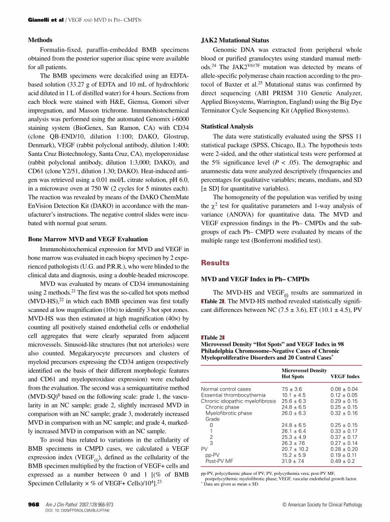
(20.7 ± 10.2), and CIMF (25.6 ± 6.3) cases ❚Figure 1A❚ (P <.0001; ANOVA), being significantly greater in PV and CIMFcases than in ET and NC cases (P < .05; multiple range testanalysis, Bonferroni LSD) ❚Image 1❚. The MVD-SQ resultswere similar (P < .0001; Kruskal-Wallis 1-way ANOVA).
A significant difference was also found when thechronic and myelofibrotic phases of PV and CIMF wereevaluated separately ❚Figure 1B❚ (P < .0001; ANOVA), withhigher MVD-HS levels found in the fibrotic phases of thediseases and a statistically significant difference when com-paring pp-PV (15.2 ± 5.9) and post-PV MF (31.9 ± 7.4) (P< .05; multiple range test analysis, Bonferroni LSD). Thesefindings were confirmed by MVD-SQ (P < .0001; Kruskal-Wallis 1-way ANOVA; and P < .05; multiple range testanalysis, Bonferroni LSD). Statistical analysis of CIMF
cases did not reveal significant differences between the dif-ferent histologic grades.
For VEGF, we first examined protein expression in nor-mal bone marrow cells and found moderately to markedlyintense VEGF immunoreactivity in the myeloid lineage at allstages of maturation, in megakaryocytes (variable expression),in histiocytes, and in plasma cells. The VEGF(i) revealed dif-ferent levels of protein expression between NC (0.08 ± 0.04),ET (0.12 ± 0.05), CIMF (0.29 ± 0.15), and PV (0.28 ± 0.20)cases ❚Figure 1C❚ (P < .0001; ANOVA), with higher levels inCIMF and PV cases than in ET and NC cases (P < .05; mul-tiple range test analysis, Bonferroni LSD) ❚Image 2❚.
VEGF(i) demonstrated higher levels of protein expres-sion in the fibrotic phases of CIMF and PV, particularly inPV (pp-PV, 0.19 ± 0.11 vs post-PV MF, 0.49 ± 0.2) (P < .05;
0.00
11.15
22.30
NCs ET CIMF
MV
D-H
S
PV
33.45
44.60
0.00
0.16
0.32
c-CIMF f-CIMF pp-PV
VE
GF
Ind
ex
Post PV-MF
0.48
0.64
0.00
0.16
0.32
NCs ET CIMF
VE
GF
Ind
ex
PV
0.48
0.64
0.00
11.15
22.30
c-CIMF f-CIMF pp-PV
MV
D-H
S
Post PV-MF
33.45
44.60
A B
C D
❚❚Figure 1❚❚ A and B, Microvessel density “hot spots” (MVD-HS) in cases of Philadelphia chromosome–negative chronicmyeloproliferative disorders (A; Ph– CMPDs) and the chronic (c-CIMF) and myelofibrotic (f-CIMF) phases of chronic idiopathicmyelofibrosis (CIMF) and the polycythemic (pp-PV) and postpolycythemic myelofibrosis (post-PV MF) phases of polycythemiavera (B; PV). C and D, Vascular endothelial growth factor (VEGF) index in cases of Ph– CMPD (C) and c-CIMF, f-CIMF, pp-PV, andpost-PV MF (D). The box-whisker plots indicate the minimum value, first quartile, mean value, third quartile, and maximum value.

970 Am J Clin Pathol 2007;128:966-973970 DOI: 10.1309/FP0N3LC8MBJUFFA6
© American Society for Clinical Pathology
Gianelli et al / VEGF AND MVD IN PH– CMPDS
multiple range test analysis, Bonferroni LSD) ❚Figure 1D❚.There were no significant differences in VEGF levelsbetween the grades of CIMF.
Correlation Between VEGF Index and MVD-HS in Ph–CMPDs
A direct correlation was found between the VEGF(i) andMVD-HS in the BMB specimens of Ph– CMPD cases con-sidered as a whole (r = 0.67; P < .001) ❚Figure 2❚.Furthermore, there was a statistically significant correlationin the BMB specimens of PV cases (r = 0.79; P < .001)❚Figure 3❚ and CIMF cases (r = 0.40; P = .013) consideredas individual entities.
JAK2 and MVD in Ph– CMPDsJAK2 mutational status was studied in a subset of 38
cases. The JAK2V617F mutation was found in 9 (56%) of 16cases of CIMF (3 homozygous and 6 heterozygous), 13 (87%)of 15 cases of PV (5 homozygous and 8 heterozygous), and 4(57%) of 7 cases of ET (1 homozygous and 3 heterozygous).We found no significant correlation between JAK2 mutation-al status and MVD or VEGF(i).
Discussion
In this study, we examined MVD and immunohistochemicalexpression of VEGF in BMB specimens obtained from patients
A B
C D
❚❚Image 1❚❚ Microvessel density (MVD) in Philadelphia chromosome–negative chronic myeloproliferative disorders evaluated usingCD34 antibody. MVD was not significantly different between the normal control samples (A) and essential thrombocythemia (B)but was greater in polycythemia vera (C) and chronic idiopathic myelofibrosis (D) (A-D, CD34 immunostaining,diaminobenzidine chromogen, ×10).

Am J Clin Pathol 2007;128:966-973 971971 DOI: 10.1309/FP0N3LC8MBJUFFA6 971
© American Society for Clinical Pathology
Hematopathology / ORIGINAL ARTICLE
with Ph– CMPDs. In comparison with NC and ET cases, PV andCIMF cases showed significantly higher MVD as evaluated bythe MVD-HS and MVD-SQ methods. Interestingly, we found nosignificant difference in MVD between the NC and ET cases.
MVD was significantly higher in myelofibrotic than inchronic phases of CIMF and PV, a difference that was statisti-cally significant in PV cases. In contrast, there were no signif-icant differences between the CIMF cases with different gradesof bone marrow fibrosis.
Our MVD data are in line with those of previous studiesdescribing increased angiogenesis in CMPDs.8-11,18,21
Interestingly, we found the highest levels of MVD in PV, par-ticularly in post-PV MF.
The published data regarding proangiogenic VEGFexpression in CMPDs are controversial. Increased serumlevels have been documented in idiopathic myelofibro-sis,15 and some positive immunohistochemical evidencehas been reported. An increased number of VEGF+ cells inMMM correlates with MVD,7 higher VEGF levels havebeen found in the bone marrow of patients with myelopro-liferative disorders,26 and there is a recent description ofstrong immunohistochemical VEGF expression in themegakaryocytes of idiopathic myelofibrosis.18 In contrast,2 recent studies found no differences in the distributionand intensity of VEGF immunostaining between MMMand control cases.16,17
A B
❚❚Image 2❚❚ Vascular endothelial growth factor (VEGF) immunohistochemical expression in Philadelphia chromosome–negativechronic myeloproliferative disorders. VEGF expression was lower in the normal control samples (A) and essentialthrombocythemia (B) but higher in polycythemia vera (C) and chronic idiopathic myelofibrosis (D) (A-D, VEGF immunostaining,diaminobenzidine chromogen, ×10).
C D

972 Am J Clin Pathol 2007;128:966-973972 DOI: 10.1309/FP0N3LC8MBJUFFA6
© American Society for Clinical Pathology
Gianelli et al / VEGF AND MVD IN PH– CMPDS
We found that the immunohistochemical expression ofVEGF parallels the increase in MVD. The VEGF(i) was signif-icantly higher in CIMF and PV cases than in ET and NCcases. It was also higher in the myelofibrotic phases of CIMFand PV, but the difference was significant only in PV. Therewas no difference between cases with different degrees ofbone marrow fibrosis.
Our findings are in line with those of Lundberg et al7 andWrobel et al26 but not with those of Chou et al16 or Ho et al.17
There may be various reasons for the discrepancies, the mostimportant of which are likely to be the use of different CMPDclassification schemes and the different method of evaluating
immunohistochemical VEGF expression. Regarding the latterquestion, we decided to use a VEGF(i) to avoid bias related tothe variability in bone marrow cellularity that frequently char-acterizes CMPDs. The index was calculated in a manner sim-ilar to that used to calculate the hairy cell index, which is usedto evaluate the entity of bone marrow involvement in hairy cellleukemia.23 In a previous study evaluating survivin expressionin MDS, Gianelli et al27 demonstrated the usefulness of thismethod of quantifying protein expression in BMB specimensand its correlation with messenger RNA levels.
To the best of our knowledge, this study is the first to docu-ment a direct correlation between VEGF levels and MVD in alarge series of Ph– CMPD cases (r = 0.67; P < .001). This corre-lation was more evident in the BMB specimens of PV cases (r =0.79; P < .001), but can also be seen in CIMF cases (r = 0.40; P= .013). A direct correlation between VEGF and MVD has beenreported in other hematologic malignancies such as acutemyeloid leukemia, MDS, and multiple myeloma.5,15 In particular,it has been postulated that VEGF may act in a paracrine mannerin contributing to the increased MVD in these malignant condi-tions, and a similar mechanism may stimulate the proliferation ofnew vessels in the bone marrow of patients with Ph– CMPDs.
It is well known that ET differs from other CMPDs interms of reticulin fiber content, and we recently reported a sta-tistically significant absence of trabecular bone remodeling inthe BMB specimens of ET cases in comparison with CIMFcases.28 Moreover, our MVD and VEGF data indicate that,also in relation to the angiogenic process, ET cases seem to bemore similar to NC cases than to the other Ph– CMPD cases.
In contrast, our results show that PV and CIMF are char-acterized by increased angiogenesis and the increased expres-sion of proangiogenic VEGF. These considerations may be ofsome relevance in the context of new therapeutic approachesaimed at directly targeting endothelial cells or VEGF. In partic-ular, it is interesting to note that thalidomide, thalidomide ana-logues, and new agents acting on the angiogenic process viathe VEGF pathway are under clinical investigation in MMM.29
Finally, we also studied JAK2 mutational status in a sub-set of Ph– CMPD cases. We found no significant correlationsbetween the JAK2V617F mutation and MVD or VEGF(i) but,given the limited number of observations, further studies areneeded to verify this finding.
Our study demonstrates that patients with Ph– CMPDs(particularly PV and CIMF) have increased MVD and VEGFlevels. The increased MVD parallels the expression of VEGF,and there is a direct correlation between the 2 angiogenicparameters. Our data could offer a rationale for clinical trialsof drugs aimed directly at target endothelial cells or VEGF inCIMF and patients with PV. Further studies of larger numbersof patients are needed to study the correlation between JAK2mutational status and bone marrow angiogenesis.
5.60.00500 0.16375 0.32250 0.48125 0.64000
14.2
22.8
VEGF Index
MV
D-H
S
31.4
40.0
3.3000.00500 0.16375 0.32250 0.48125 0.64000
13.625
23.950
VEGF Index
MV
D-H
S
34.275
44.600
❚❚Figure 2❚❚ Linear correlation between the vascular endothelialgrowth factor (VEGF) index and microvessel density “hotspots” (MVD-HS) in 98 cases of Philadelphia chromosome–negative chronic myeloproliferative disorders. r = 0.67555; P < .001.
❚❚Figure 3❚❚ Linear correlation between the vascular endothelialgrowth factor (VEGF) index and microvessel density “hotspots” (MVD-HS) in 30 cases of polycythemia vera. r = 0.79235; P < .001.

Am J Clin Pathol 2007;128:966-973 973973 DOI: 10.1309/FP0N3LC8MBJUFFA6 973
© American Society for Clinical Pathology
Hematopathology / ORIGINAL ARTICLE
From the 1Pathology Unit, Department of Medicine, Surgery andOdontology, San Paolo Hospital, and Policlinico IRCCS Hospital,Mangiagalli and Regina Elena Foundation, University of Milan,Italy; 2Hematology I–Bone Marrow Transplant Unit, PoliclinicoIRCCS Hospital, Mangiagalli and Regina Elena Foundation,University of Milan; 3Hematology II–Policlinico IRCCS Hospital,Mangiagalli and Regina Elena Foundation, University of Milan;4Internal Medicine Unit, Angelo Bianchi Bonomi Hemophilia andThrombosis Center, Policlinico IRCCS Hospital, Mangiagalli andRegina Elena Foundation, University of Milan.
Address reprint requests to Dr Gianelli: II Cattedra diAnatomia Patologica, Dipartimento di Medicina, Chirurgia eOdontoiatria, Università degli Studi di Milano, A.O. San Paolo,Via Di Rudini’ 8, 20142 Milano, Italy.
* Drs Gianelli and Vener contributed equally to the study.
References1. Folkman J. Angiogenesis in cancer, vascular, rheumatoid and
other disease. Nat Med. 1995;1:27-31.2. Weidner N, Semple JP, Welch WR, et al. Tumor angiogenesis
and metastasis correlation in invasive breast carcinoma. NEngl J Med. 1991;324:1-8.
3. Bosari S, Lee AK, DeLellis RA, et al. Microvessel quantitationand prognosis in invasive breast carcinoma. Hum Pathol.1992;23:755-761.
4. Perez-Atayde AR, Sallan SE, Tedrow U, et al. Spectrum oftumor angiogenesis in the bone marrow of children with acutelymphoblastic leukemia. Am J Pathol. 1997;150:815-821.
5. Aguayo A, Kantarjian H, Manshouri T, et al. Angiogenesis inacute and chronic leukemias and myelodysplastic syndromes.Blood. 2000;96:2240-2245.
6. Pruneri G, Ponzoni M, Ferreri AJ, et al. Microvessel density,a surrogate marker of angiogenesis, is significantly related tosurvival in multiple myeloma patients. Br J Haematol.2002;118:817-820.
7. Lundberg LG, Lerner R, Sundelin P, et al. Bone marrow inpolycythemia vera, chronic myelocytic leukemia, andmyelofibrosis has an increased vascularity. Am J Pathol.2000;157:15-19.
8. Mesa RA, Hanson CA, Rajkumar SV, et al. Evaluation andclinical correlations of bone marrow angiogenesis inmyelofibrosis with myeloid metaplasia. Blood. 2000;96:3374-3380.
9. Panteli K, Zagorianakou N, Bai M, et al. Angiogenesis inchronic myeloproliferative diseases detected by CD34expression. Eur J Haematol. 2004;72:410-415.
10. Florena AM, Tripodo C, Iannitto E, et al. Value of bonemarrow biopsy in the diagnosis of essential thrombocythemia.Haematologica. 2004;89:911-919.
11. Ponzoni M, Savage DG, Ferreri AJ, et al. Chronic idiopathicmyelofibrosis: independent prognostic importance of bonemarrow microvascular density evaluated by CD105 (endoglin)immunostaining. Mod Pathol. 2004;17:1513-1520.
12. Giles FJ. The vascular endothelial growth factor (VEGF)signaling pathway: a therapeutic target in patients withhematologic malignancies. Oncologist. 2001;6(suppl 5):32-39.
13. Aguayo A, Estey E, Kantarjian H, et al. Cellular vascularendothelial growth factor is a predictor of outcome in patientswith acute myeloid leukemia. Blood. 1999;94:3717-3721.
14. Salven P, Orpana A, Teerenhovi L, et al. Simultaneouselevation in the serum concentrations of the angiogenicgrowth factors VEGF and βFGF is an independent predictor ofpoor prognosis in non-Hodgkin lymphoma: a single-institutionstudy of 200 patients. Blood. 2000;96:3712-3718.
15. Di Raimondo F, Azzaro MP, Palumbo G, et al. Angiogenicfactors in multiple myeloma: higher levels in bone marrowthan in peripheral blood. Haematologica. 2000;85:800-805.
16. Chou JM, Li CY, Tefferi A. Bone marrowimmunohistochemical studies of angiogenic cytokines andtheir receptors in myelofibrosis with myeloid metaplasia. Leuk Res. 2003;27:499-504.
17. Ho CL, Arora B, Hoyer JD, et al. Bone marrow expression of vascular endothelial growth factor in myelofibrosis withmyeloid metaplasia. Eur J Haematol. 2005;74:35-39.
18. Ni H, Barosi G, Hoffman R. Quantitative evaluation of bonemarrow angiogenesis in idiopathic myelofibrosis. Am J ClinPathol. 2006;126:241-247.
19. Vardiman JW, Brunning RD, Harris NL, et al. Chronicmyeloproliferative diseases. In: Jaffe ES, Harris NL, Stein H, etal, eds. Pathology and Genetics of Tumours of Haematopoietic andLymphoid Tissues. Lyon, France: IARC Press; 2001:15-41.World Health Organization Classification of Tumours.
20. Thiele J, Kvasnicka HM, Facchetti F, et al. Europeanconsensus on grading bone marrow fibrosis and assessment of cellularity. Haematologica. 2005;90:1128-1132.
21. Kvasnicka HM, Thiele J. Bone marrow angiogenesis: methodsof quantification and changes evolving in chronicmyeloproliferative disorders. Histol Histopathol. 2004;19:1245-1260.
22. Fox SB, Leek RD, Weekes MP, et al. Quantitation andprognostic value of breast cancer angiogenesis: comparison of microvessel density, Chalkley count, and computer imageanalysis. J Pathol. 1995;177:275-283.
23. Golomb HM, Vardiman JW. Response to splenectomy in 65patients with hairy cell leukemia: an evaluation of spleenweight and bone marrow involvement. Blood. 1983;61:349-352.
24. Sambrook J, Fritsch EF, Maniatis T. In: Molecular Cloning: A Laboratory Manual. New York, NY: Cold Spring HarborLaboratory Press; 1989.
25. Baxter EJ, Scott LM, Campbell PJ, et al. Acquired mutation of the tyrosine kinase JAK2 in human myeloproliferativedisorders. Lancet. 2005;365:1054-1061.
26. Wrobel T, Mazur G, Surowiak P, et al. Increased expression ofvascular endothelial growth factor (VEGF) in bone marrow ofpatients with myeloproliferative disorders (MPD). Pathol OncolRes. 2003;9:170-173.
27. Gianelli U, Fracchiolla NS, Cortelezzi A, et al. Survivinexpression in “low-risk” and “high-risk” myelodysplasticsyndromes. Ann Hematol. 2007;86:185-189.
28. Gianelli U, Vener C, Raviele PR, et al. Essentialthrombocythemia or chronic idiopathic myelofibrosis? asingle-center study based on hematopoietic bone marrowhistology. Leuk Lymphoma. 2006;47:1774-1781.
29. Hennessy BT, Thomas DA, Giles FJ, et al. New approaches in the treatment of myelofibrosis. Cancer. 2005;103:32-43.
Related Documents