Vascular Connective Tissue

Welcome message from author
This document is posted to help you gain knowledge. Please leave a comment to let me know what you think about it! Share it to your friends and learn new things together.
Transcript
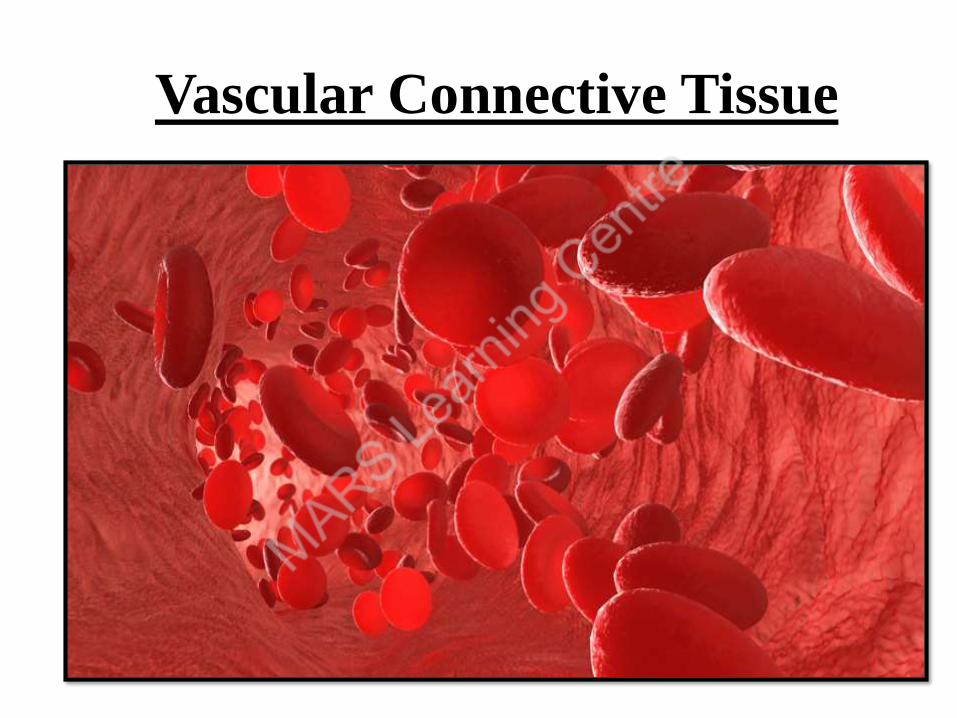
Vascular Connective Tissue
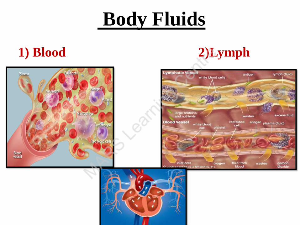
Body Fluids
1) Blood 2)Lymph
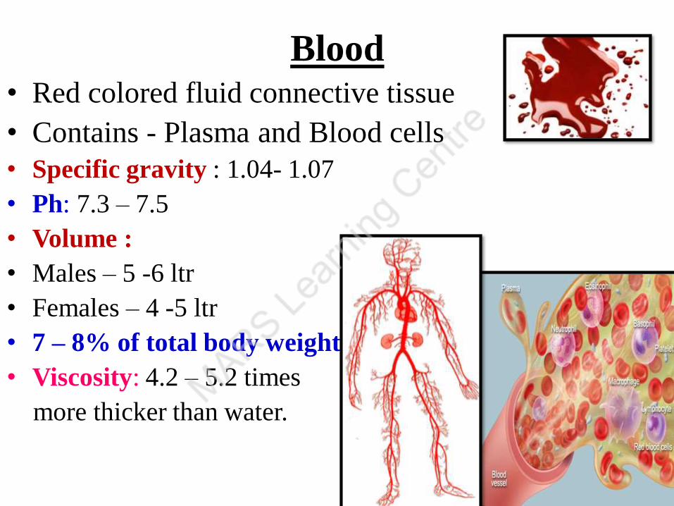
Blood • Red colored fluid connective tissue
• Contains - Plasma and Blood cells
• Specific gravity : 1.04- 1.07
• Ph: 7.3 – 7.5
• Volume :
• Males – 5 -6 ltr
• Females – 4 -5 ltr
• 7 – 8% of total body weight
• Viscosity: 4.2 – 5.2 times
more thicker than water.
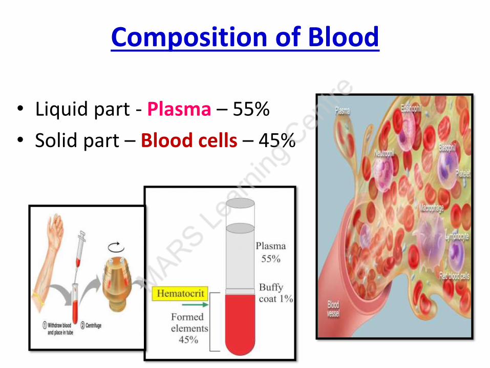
Composition of Blood
• Liquid part - Plasma – 55%
• Solid part – Blood cells – 45%
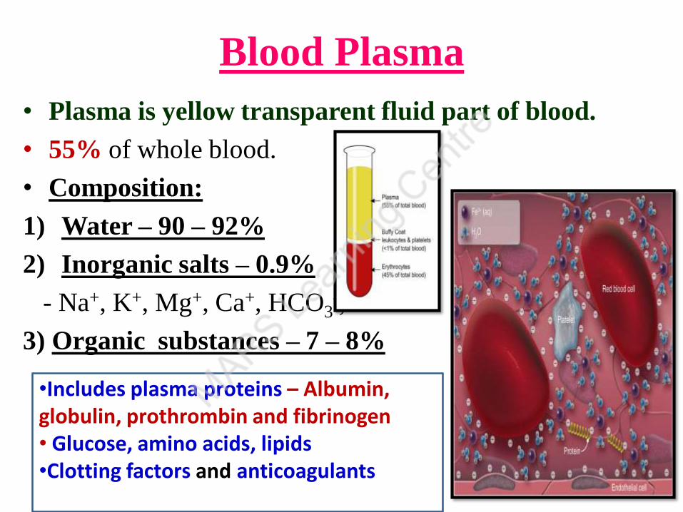
Blood Plasma
• Plasma is yellow transparent fluid part of blood.
• 55% of whole blood.
• Composition:
1) Water – 90 – 92%
2) Inorganic salts – 0.9%
- Na+, K+, Mg+, Ca+, HCO3-, Cl-
3) Organic substances – 7 – 8%
•Includes plasma proteins – Albumin, globulin, prothrombin and fibrinogen• Glucose, amino acids, lipids •Clotting factors and anticoagulants
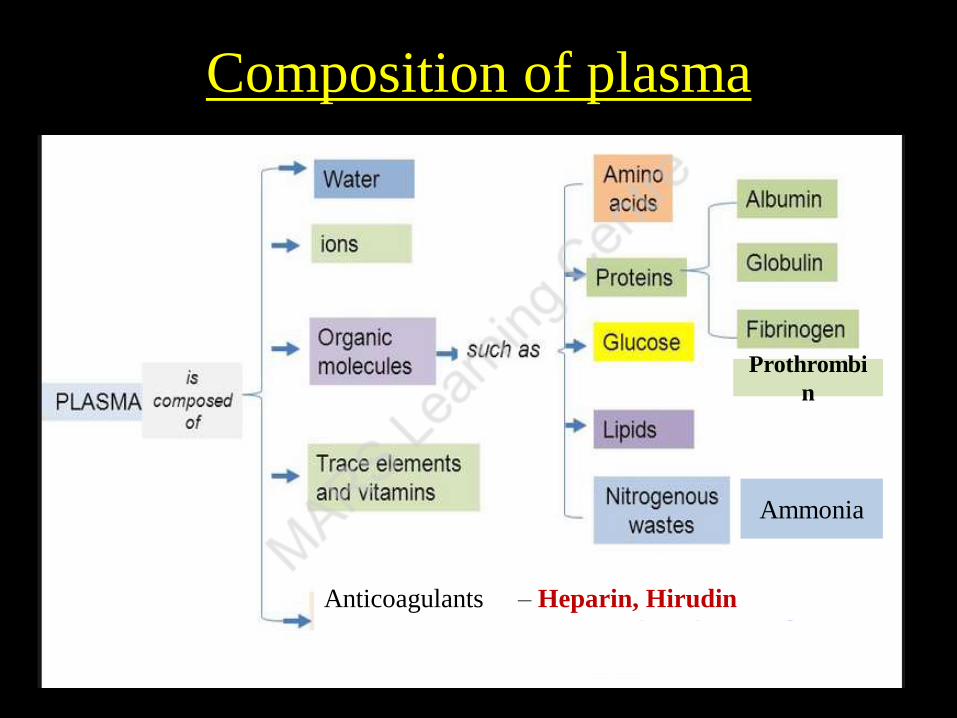
Composition of plasma
Prothrombi
n
Ammonia
Anticoagulants – Heparin, Hirudin
- Coumarin/ Dicumarol
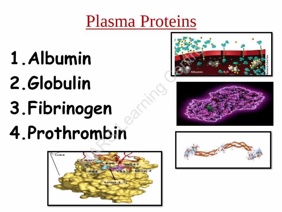
Plasma Proteins
1.Albumin
2.Globulin
3.Fibrinogen
4.Prothrombin

Plasma Proteins
• Types• Albumin: Synthesized by liver
- 4- 7% of total plasma proteins
- Maximum among plasma proteins
• Function:
- Maintains colloid osmotic pressure
- Helps in tissue filtration
- Helps in taking back water from tissues
- Maintains blood volume and pressure
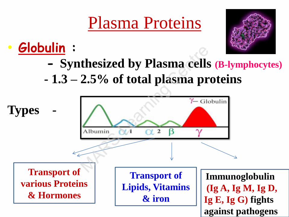
Plasma Proteins
• Globulin : - Synthesized by Plasma cells (B-lymphocytes)
- 1.3 – 2.5% of total plasma proteins
Types -
Transport of
various Proteins
& Hormones
Transport of
Lipids, Vitamins
& iron
Immunoglobulin
(Ig A, Ig M, Ig D,
Ig E, Ig G) fights
against pathogens
Plasma Proteins
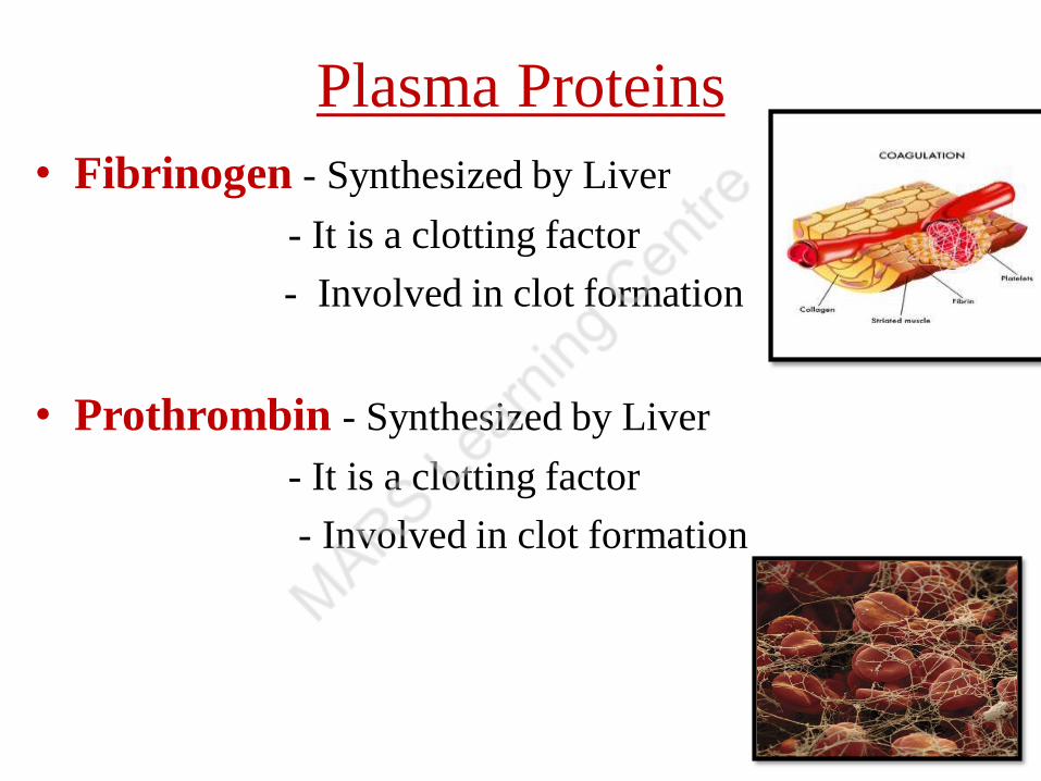
• Fibrinogen - Synthesized by Liver
- It is a clotting factor
- Involved in clot formation
• Prothrombin - Synthesized by Liver
- It is a clotting factor
- Involved in clot formation
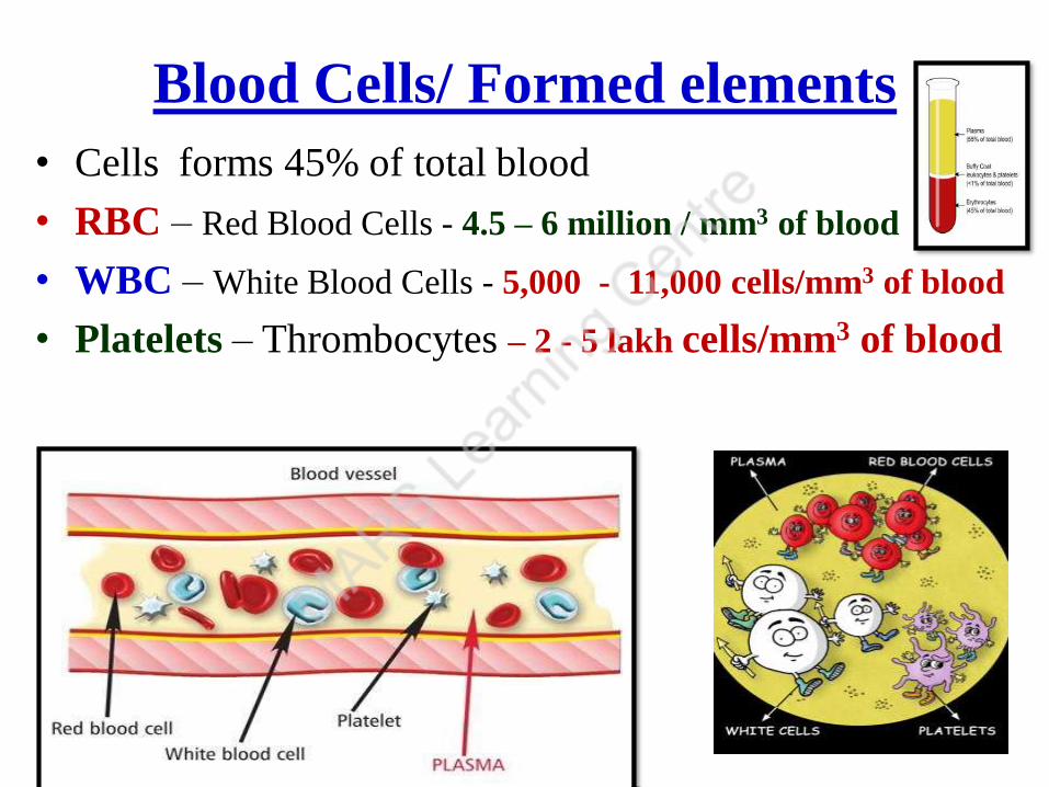
Blood Cells/ Formed elements
• Cells forms 45% of total blood
• RBC – Red Blood Cells - 4.5 – 6 million / mm3 of blood
• WBC – White Blood Cells - 5,000 - 11,000 cells/mm3 of blood
• Platelets – Thrombocytes – 2 - 5 lakh cells/mm3 of blood

Red Bone marrow
12
Is the site of production of all blood cells
13
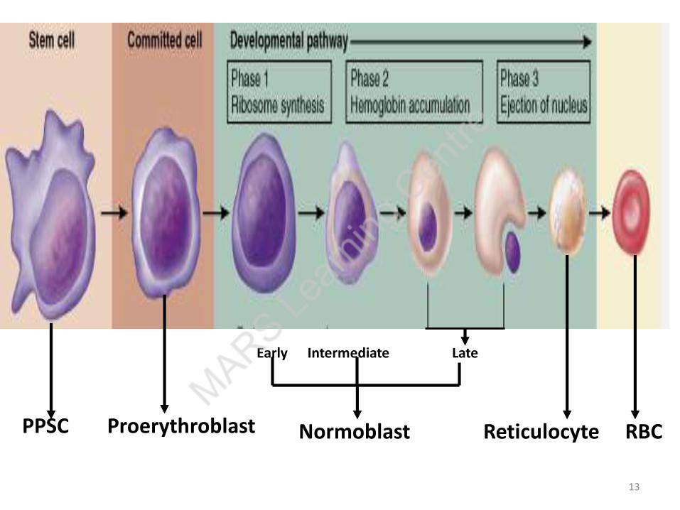
ProerythroblastPPSC
Early Intermediate Late
Reticulocyte RBCNormoblast
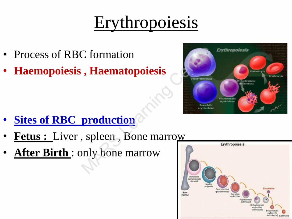
Erythropoiesis
• Process of RBC formation
• Haemopoiesis , Haematopoiesis
• Sites of RBC production
• Fetus : Liver , spleen , Bone marrow
• After Birth : only bone marrow

• 5 yrs – 25 yrs – Bone marrow of long bones
• Later - from red bone marrow of
flar bones - Skull, Vertebrae, Ribs, Sternum, Pelvis

RBC / Erythrocytes
• Discovered by Leeuvenhoek in 1674.
• No nucleus , Biconcave / Disc shaped
• Provides more surface area for diffusion of
O2 & CO2
• Cell membrane of RBC is Donnan’s membrane
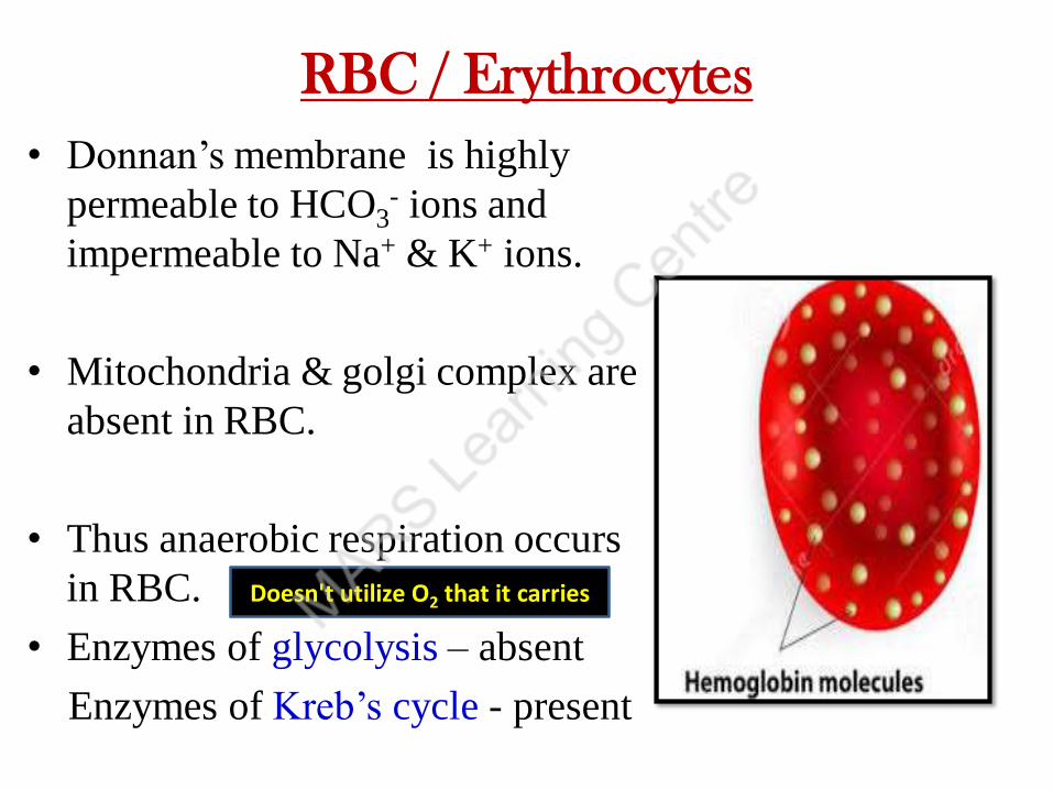
RBC / Erythrocytes
• Donnan’s membrane is highly
permeable to HCO3- ions and
impermeable to Na+ & K+ ions.
• Mitochondria & golgi complex are
absent in RBC.
• Thus anaerobic respiration occurs
in RBC.
• Enzymes of glycolysis – absent
Enzymes of Kreb’s cycle - present
Doesn't utilize O2 that it carries
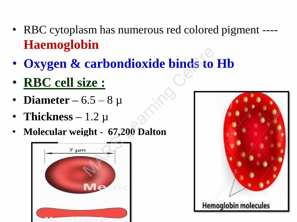
• RBC cytoplasm has numerous red colored pigment ----
Haemoglobin
• Oxygen & carbondioxide binds to Hb
• RBC cell size :
• Diameter – 6.5 – 8 µ
• Thickness – 1.2 µ
• Molecular weight - 67,200 Dalton

• Normal RBC count
• Males : 5.5 – 6 million / mm3 of blood
• Females : 4.5 – 5 million / mm3 of blood
• Composition : 64% water, 20% Hb, 7% lipids
• Erythrocytes of different
size & shape are called
Poikilocytes.

Shape of RBC
• Normal – Biconcave shape
• Sickle cell anemia – Sickle shape
• Iron deficiency anemia – RBC small
• Pernicious anemia/ Megaloblastic anemia –
RBC big
Lack of Hb – Microcytic cells
Immaturity of RBC – Macrocytic / megaloblastic cells

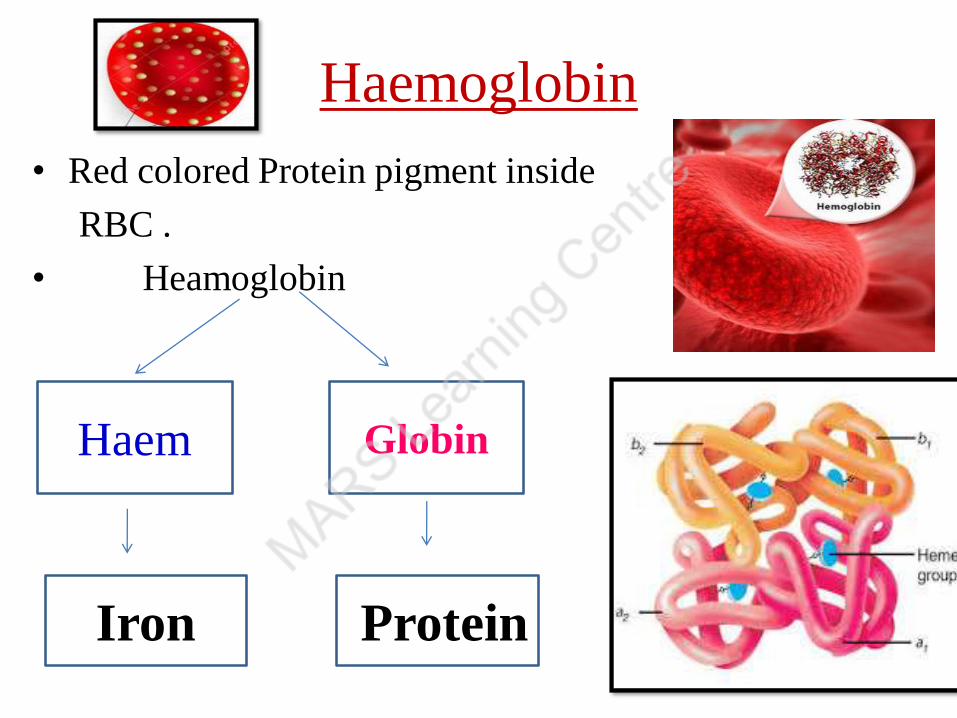
Haemoglobin
• Red colored Protein pigment inside
RBC .
• Heamoglobin
Haem Globin
Iron Protein

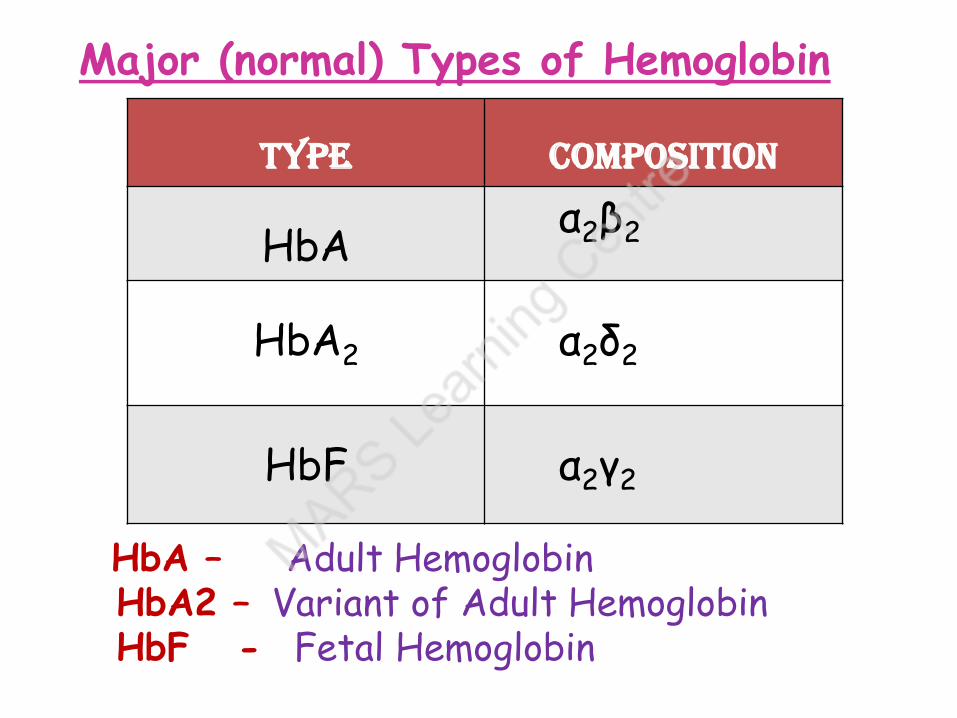
Major (normal) Types of Hemoglobin
Type Composition
HbAα2β2
HbA2 α2δ2
HbF α2γ2
• HbA – Adult Hemoglobin • HbA2 – Variant of Adult Hemoglobin • HbF - Fetal Hemoglobin

carbonic anhydrase
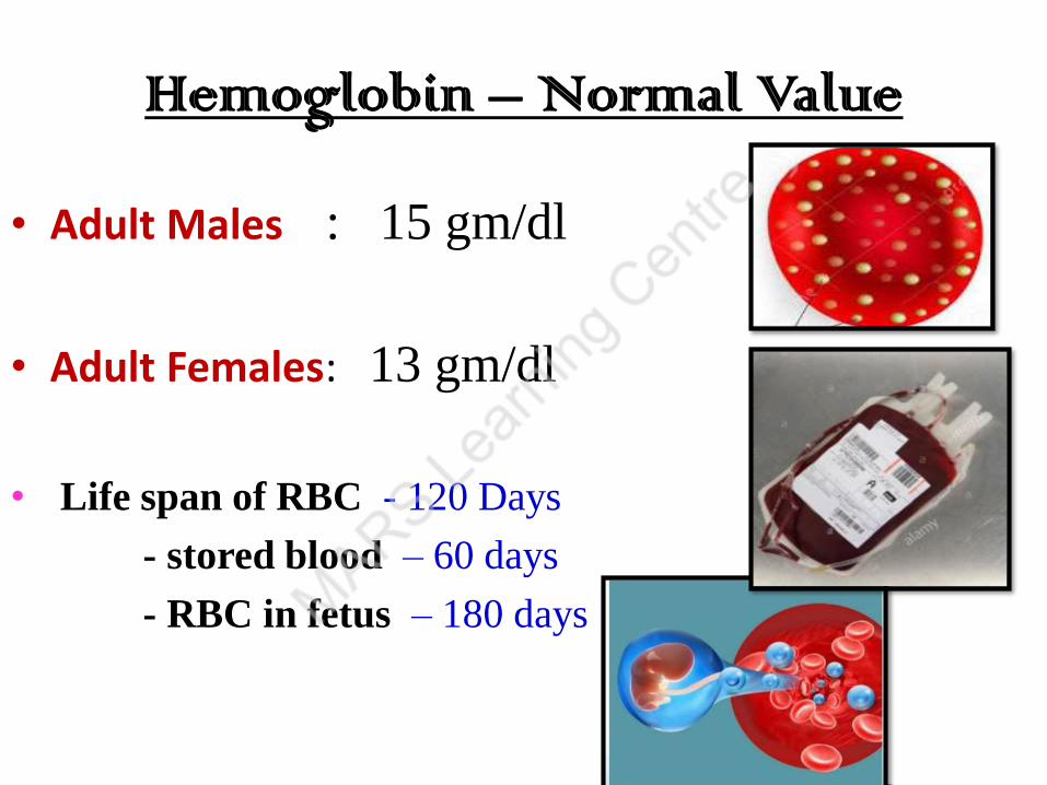
Hemoglobin – Normal Value
• Adult Males : 15 gm/dl
• Adult Females: 13 gm/dl
• Life span of RBC - 120 Days
- stored blood – 60 days
- RBC in fetus – 180 days
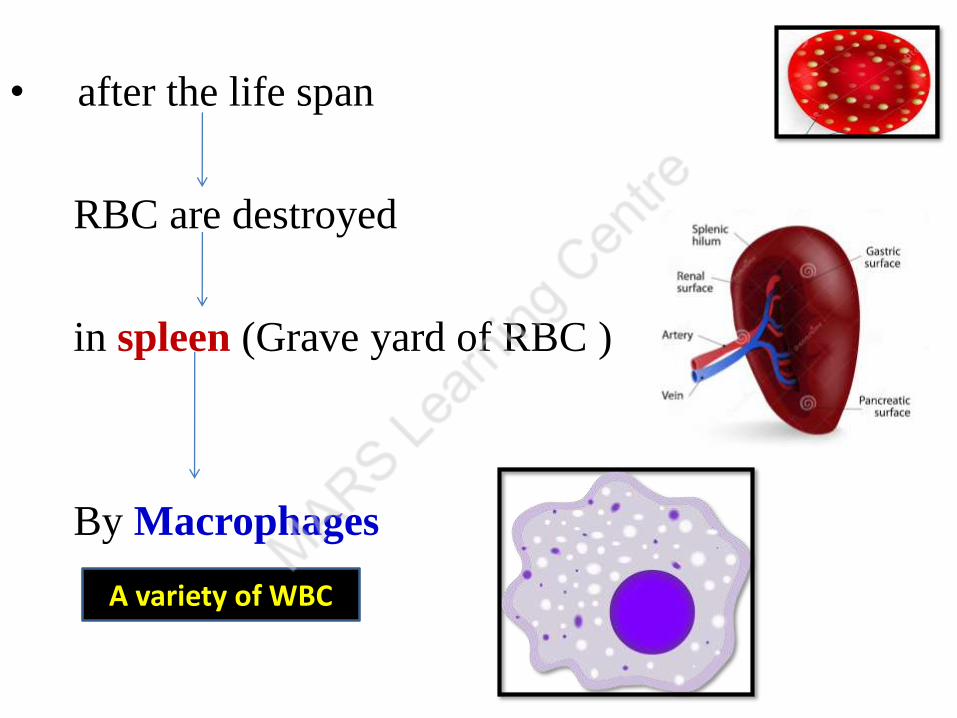
• after the life span
RBC are destroyed
in spleen (Grave yard of RBC )
By Macrophages
A variety of WBC
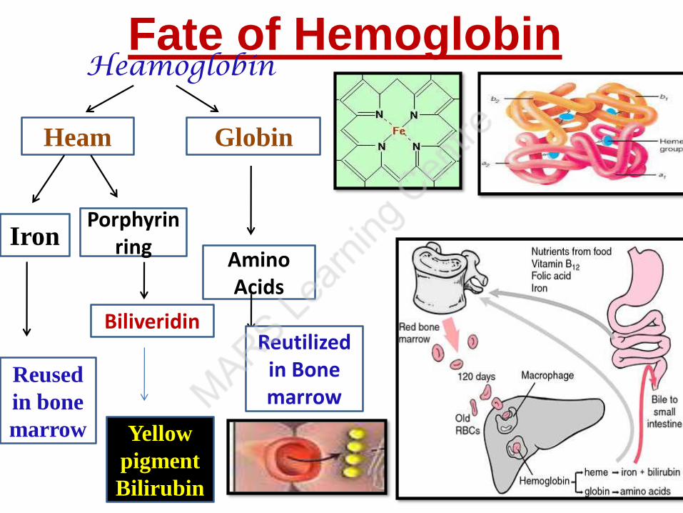
Fate of Hemoglobin Heamoglobin
Heam Globin
Amino Acids
Reutilized in Bone marrow
Iron Porphyrin
ring
Reused
in bone
marrow
Biliveridin
Yellow
pigment
Bilirubin

• Haemoglobin count
- Heamometer
• RBC count
- Haemocytometer
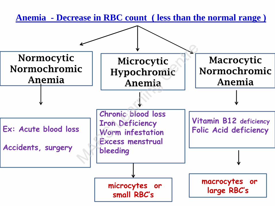
Normocytic
Normochromic
Anemia
Microcytic
Hypochromic
Anemia
Macrocytic
Normochromic
Anemia
Ex: Acute blood loss
Accidents, surgery
Chronic blood loss Iron Deficiency Worm infestationExcess menstrual bleeding
Vitamin B12 deficiency Folic Acid deficiency
Anemia - Decrease in RBC count ( less than the normal range )
microcytes or small RBC’s
macrocytes or large RBC’s
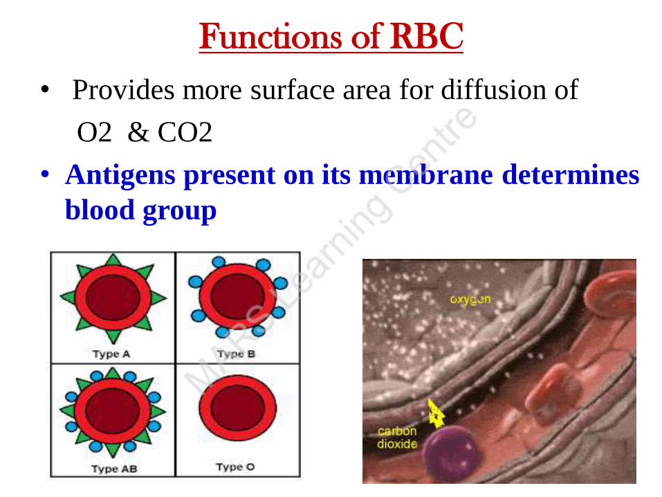
Functions of RBC
• Provides more surface area for diffusion of
O2 & CO2
• Antigens present on its membrane determines
blood group
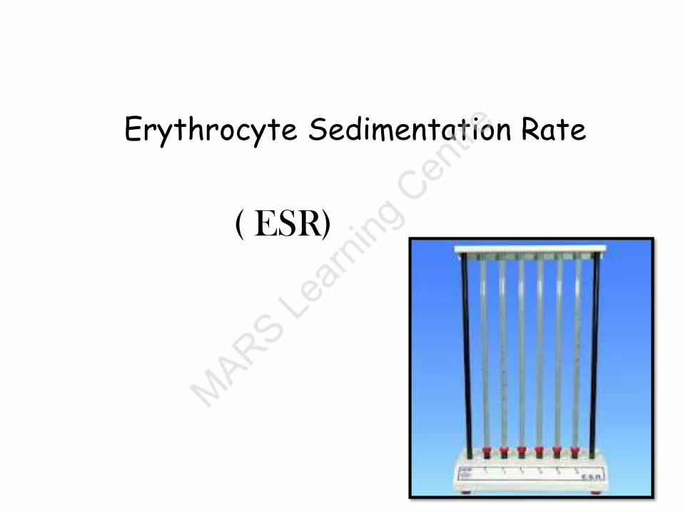
Erythrocyte Sedimentation Rate
( ESR)

• Defn of E S R :
The rate at which the erythrocytes sediment to
bottom when blood mixed with anticoagulant is
held in a vertical tube.
• Reason for sedimentation
- Erythrocytes are heavier than plasma
- Piling of RBC
( Rouleaux Formation)
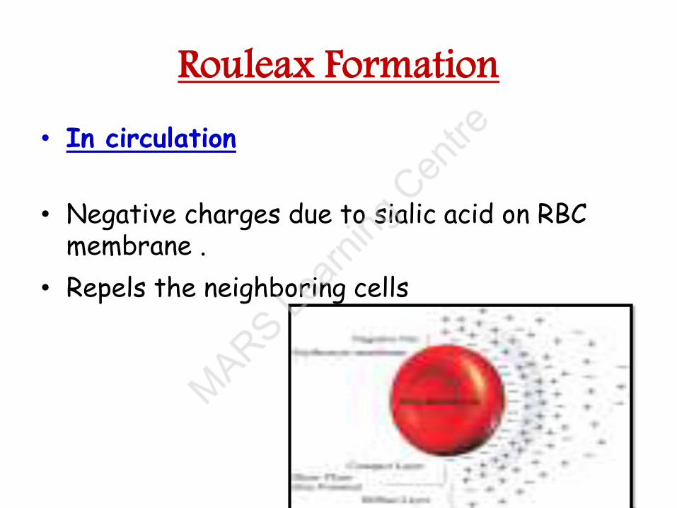
Rouleax Formation
• In circulation
• Negative charges due to sialic acid on RBC membrane .
• Repels the neighboring cells
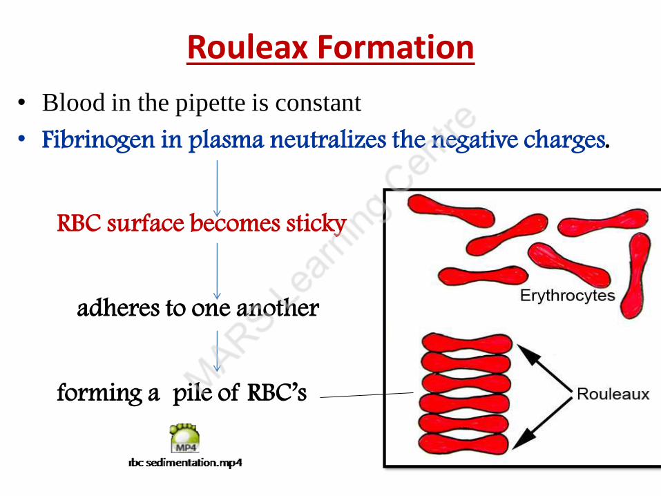
Rouleax Formation
• Blood in the pipette is constant
• Fibrinogen in plasma neutralizes the negative charges.
RBC surface becomes sticky
adheres to one another
forming a pile of RBC’s

• Normal values :
• Males – 0 -10mm/hr
• Females – 0 – 20 mm/hr
• Significance of ESR
• To diagnose various diseases.
• High – infections, arthritis, TB,
Anemia
• Low - Polycythemia ,
sickle cell anemia
Due to less RBC count

Ghost Cells
• RBC post haemolysis
• RBC’s loose their inner contents and have only
cell membrane
• Empty RBC covers are called Ghost cells
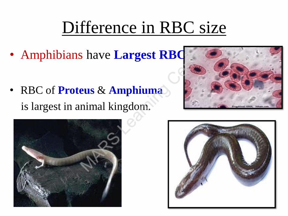
Difference in RBC size
• Amphibians have Largest RBC
• RBC of Proteus & Amphiuma
is largest in animal kingdom.
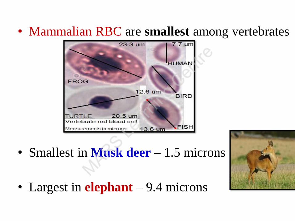
• Mammalian RBC are smallest among vertebrates
• Smallest in Musk deer – 1.5 microns
• Largest in elephant – 9.4 microns

WBC / Leucocytes
• Colorless, nucleated cells
• Larger than RBC
• Count : 5,000 - 11,000 cells/mm3 of blood
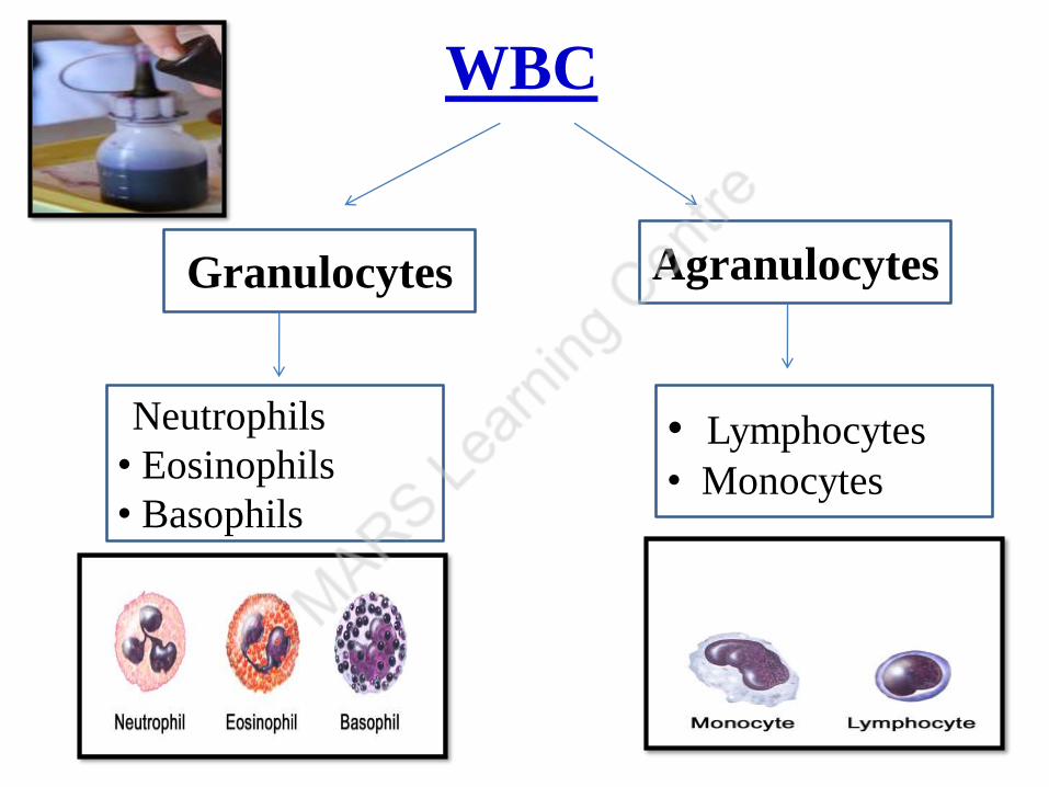
WBC
Granulocytes Agranulocytes
•Neutrophils
• Eosinophils
• Basophils
• Lymphocytes
• Monocytes
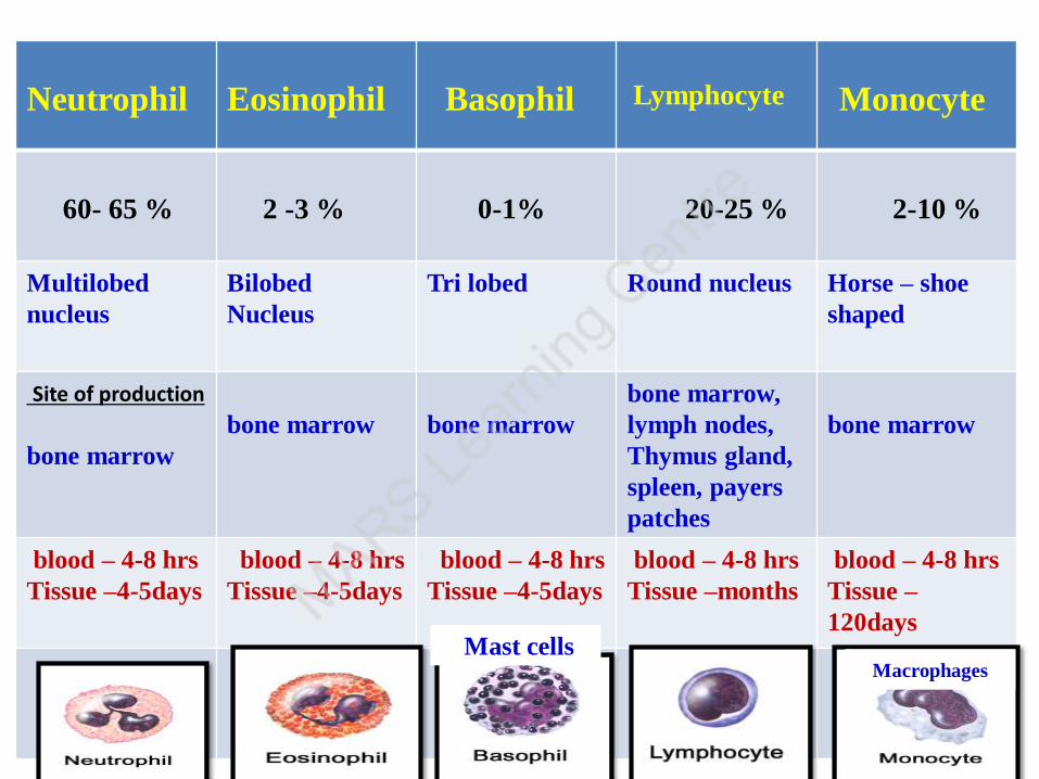
Neutrophil Eosinophil Basophil Lymphocyte Monocyte
60- 65 % 2 -3 % 0-1% 20-25 % 2-10 %
Multilobed
nucleus
Bilobed
Nucleus
Tri lobed Round nucleus Horse – shoe
shaped
Site of production
bone marrow
bone marrow bone marrow
bone marrow,
lymph nodes,
Thymus gland,
spleen, payers
patches
bone marrow
blood – 4-8 hrs
Tissue –4-5days
blood – 4-8 hrs
Tissue –4-5days
blood – 4-8 hrs
Tissue –4-5days
blood – 4-8 hrs
Tissue –months
blood – 4-8 hrs
Tissue –
120days
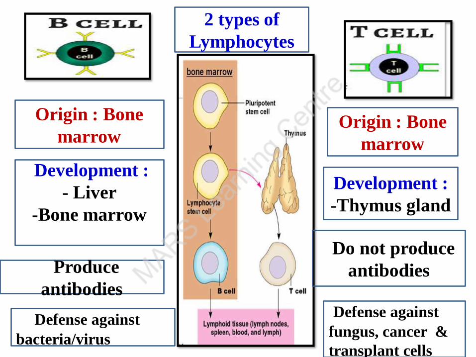
MacrophagesMast cells

Origin : Bone
marrow Origin : Bone
marrow
Development :
- Liver
-Bone marrow
Development :
-Thymus gland
Produce
antibodies
Do not produce
antibodies
Defense against
bacteria/virus
Defense against
fungus, cancer &
transplant cells
2 types of
Lymphocytes
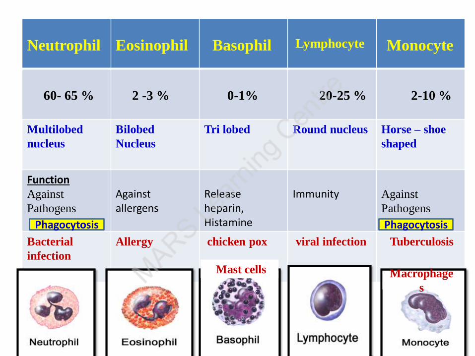
Neutrophil Eosinophil Basophil Lymphocyte Monocyte
60- 65 % 2 -3 % 0-1% 20-25 % 2-10 %
Multilobed
nucleus
Bilobed
Nucleus
Tri lobed Round nucleus Horse – shoe
shaped
FunctionAgainst
Pathogens
Against allergens
Release heparin, Histamine
Immunity Against
Pathogens
Bacterial
infection
Allergy chicken pox viral infection Tuberculosis
Macrophage
s
Mast cells
Phagocytosis Phagocytosis
Origin : Bone
marrow Origin : Bone
marrow
Development :
- Liver
-Bone marrow
Development :
-Thymus gland
Produce
antibodies
Do not produce
antibodies
Defense against
bacteria/virus
Defense against
fungus, cancer &
transplant cells
2 types of
Lymphocytes

Phagocytosis
The process of engulfment of microrganisms by
neutrophils and monocyte cells
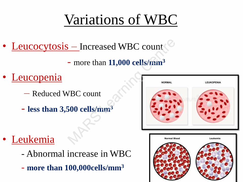
Variations of WBC
• Leucocytosis – Increased WBC count
- more than 11,000 cells/mm3
• Leucopenia
– Reduced WBC count
- less than 3,500 cells/mm3
• Leukemia
- Abnormal increase in WBC
- more than 100,000cells/mm3
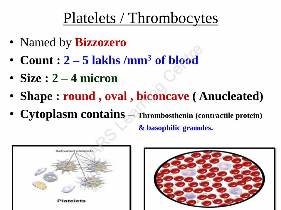
Platelets / Thrombocytes
• Named by Bizzozero
• Count : 2 – 5 lakhs /mm3 of blood
• Size : 2 – 4 micron
• Shape : round , oval , biconcave ( Anucleated)
• Cytoplasm contains – Thrombosthenin (contractile protein)
& basophilic granules.

Platelets
• Production : Megakaryocytes of bone marrow
• Cytoplasm of Megakaryocytes pinches off to form
platelets.
• Life span: 7 – 10 days

• Thrombocytopenia – Reduced platelet count
• Critical Platelet count - 40,000 cells/mm3
• Below this leads to spontaneous bleeding and red
spots on the skin called Purpura
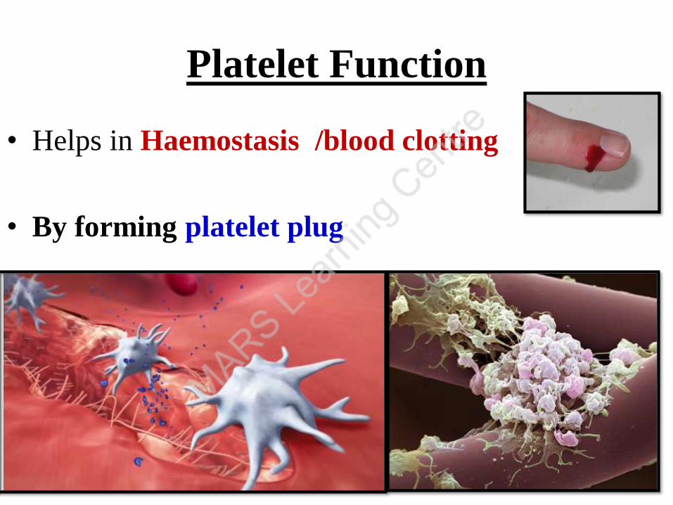
Platelet Function
• Helps in Haemostasis /blood clotting
• By forming platelet plug

fully activated
Following injury to a blood vessel – platelets attach to injured site get activated ----- There is a
change in its shape – spicules develop around platelets -----
surface become sticky ---- attract thousands of platelets to
site ---- platelet plug – temporarily stops bleeding
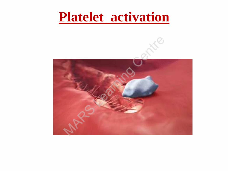
Platelet activation
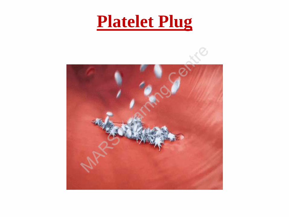
Platelet Plug
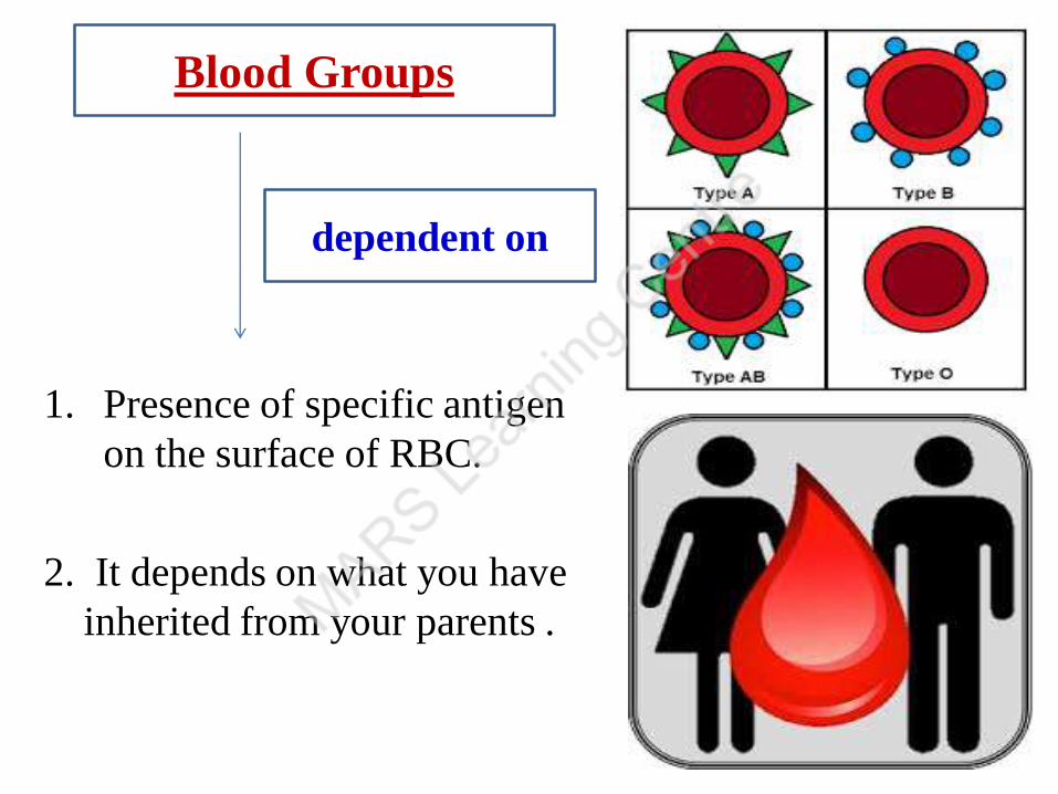
1. Presence of specific antigen
on the surface of RBC.
2. It depends on what you have
inherited from your parents .
Blood Groups
dependent on
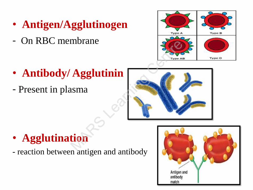
• Antigen/Agglutinogen
- On RBC membrane
• Antibody/ Agglutinin
- Present in plasma
• Agglutination
- reaction between antigen and antibody
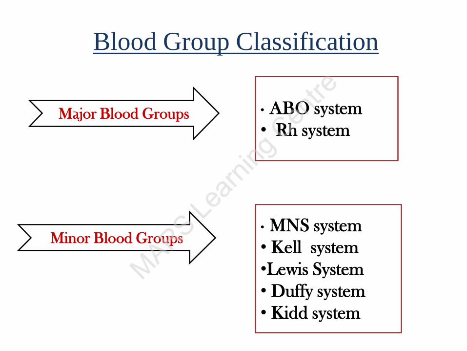
Blood Group Classification
Major Blood Groups
Minor Blood Groups
• ABO system
• Rh system
• MNS system
• Kell system
•Lewis System
• Duffy system
• Kidd system
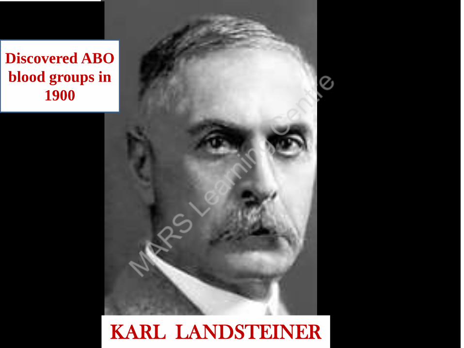
KARL LANDSTEINER
Discovered ABO
blood groups in
1900

Land-Steiner’s law of blood grouping
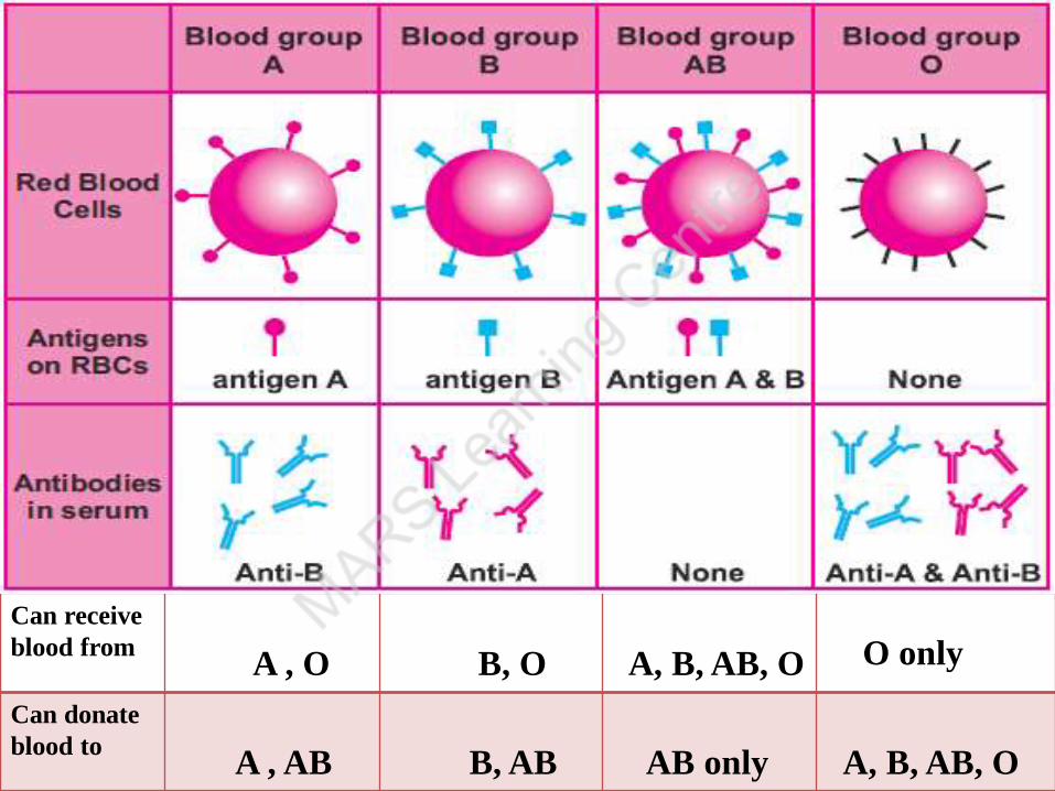
Can receive
blood fromA , O B, O A, B, AB, O O only
Can donate
blood to A , AB B, AB AB only A, B, AB, O
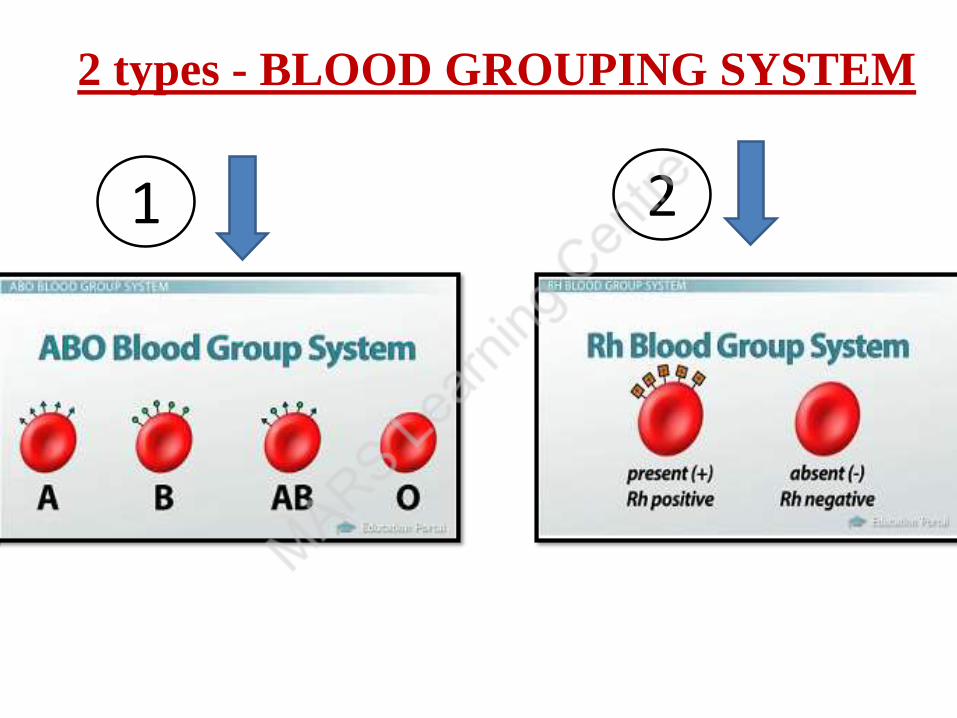
2 types - BLOOD GROUPING SYSTEM
21
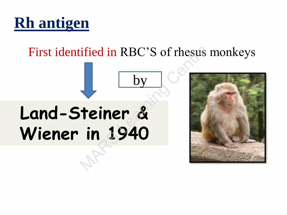
Rh antigen
First identified in RBC’S of rhesus monkeys
Land-Steiner & Wiener in 1940
by
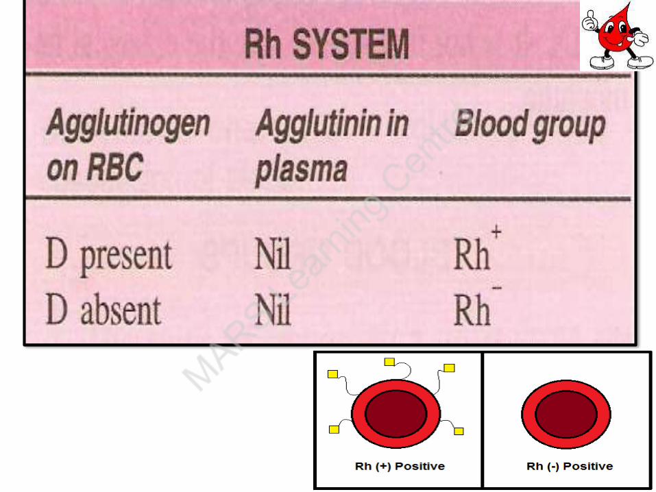
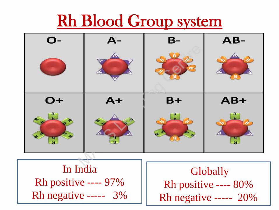
Rh Blood Group system
In India
Rh positive ---- 97%
Rh negative ----- 3%
Globally
Rh positive ---- 80%
Rh negative ----- 20%
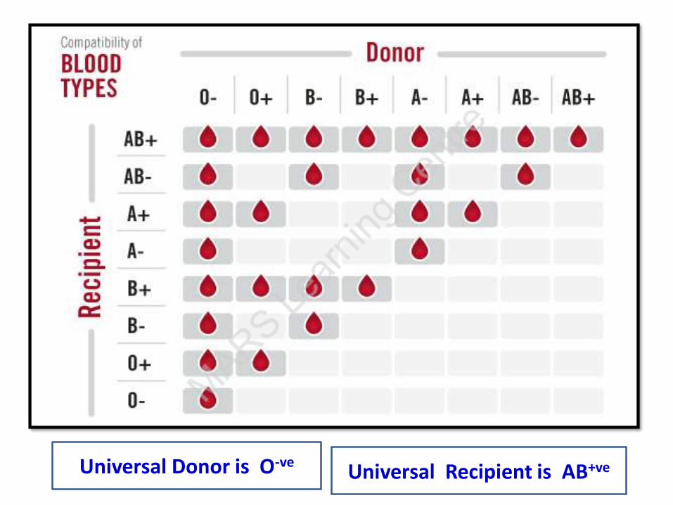
Universal Donor is O-veUniversal Recipient is AB+ve

Rh ANTIBODIES
Ig G immunoglobulins
do not occur naturally
produced when Rh – ve person infused Rh+ve blood
OR
Rh-ve mother carries a Rh+ve fetus
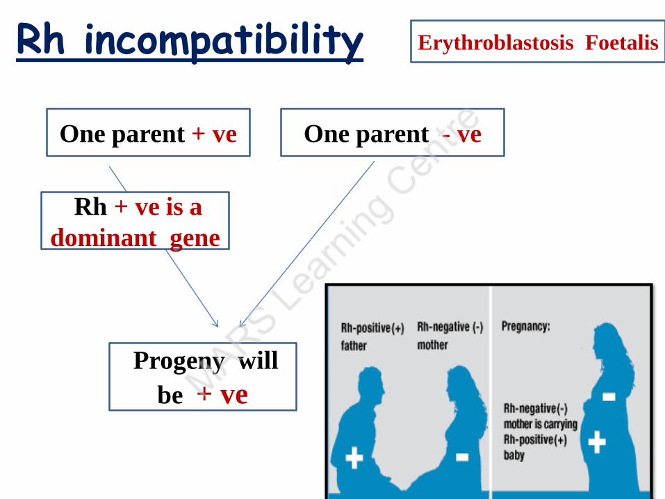
Erythroblastosis FoetalisRh incompatibility
One parent + ve One parent - ve
Progeny will
be + ve
Rh + ve is a
dominant gene

• Rh negative Mother carries Rh positive fetus
Small amount of fetal blood enters into
maternal circulation at the time of delivery
Erythroblastosis fetalis
Mothers blood develops antibodies against
Rh + antigens of the fetus and that remains in
maternal circulation for years
In second pregnancy -- If fetus is again Rh+
Erythroblastosis fetalis
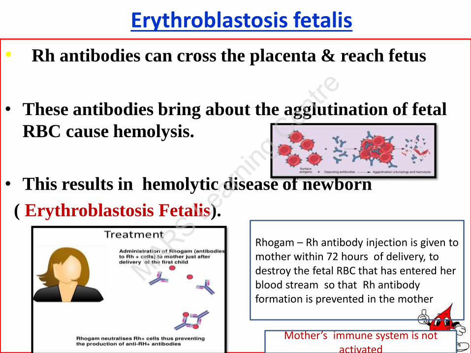
• Rh antibodies can cross the placenta & reach fetus
• These antibodies bring about the agglutination of fetal
RBC cause hemolysis.
• This results in hemolytic disease of newborn
( Erythroblastosis Fetalis).
Mother’s immune system is not activated
Rhogam – Rh antibody injection is given to mother within 72 hours of delivery, to destroy the fetal RBC that has entered her blood stream so that Rh antibody formation is prevented in the mother .
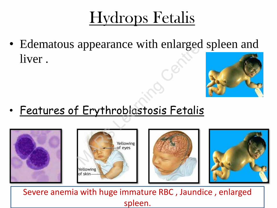
Hydrops Fetalis
• Edematous appearance with enlarged spleen and
liver .
• Features of Erythroblastosis Fetalis
Severe anemia with huge immature RBC , Jaundice , enlarged spleen.
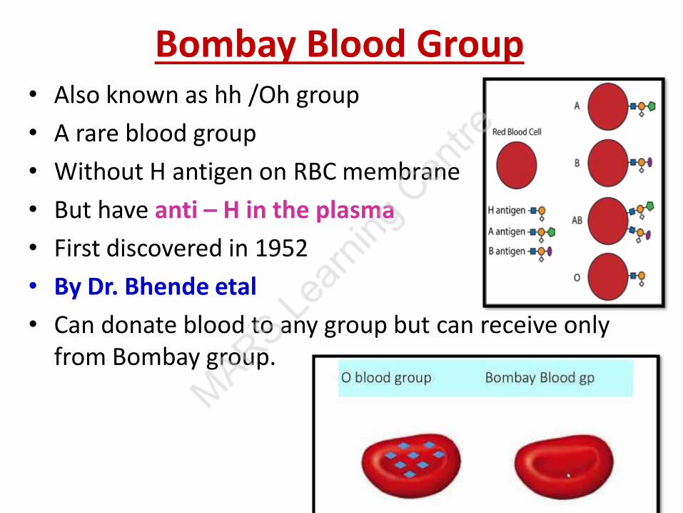
Bombay Blood Group• Also known as hh /Oh group
• A rare blood group
• Without H antigen on RBC membrane
• But have anti – H in the plasma
• First discovered in 1952
• By Dr. Bhende etal
• Can donate blood to any group but can receive only from Bombay group.

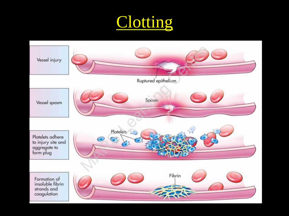
Blood clotting
• When bleeding occurs from an injured blood vessel.
• In few minutes blood gets converted to semisolid jelly like
mass .
•
Process is called Clotting
Clotting starts after platelet plug formation
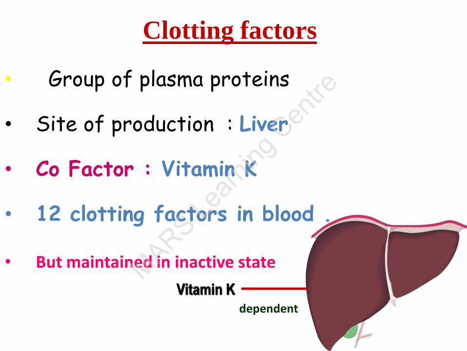
Clotting factors
• Group of plasma proteins
• Site of production : Liver
• Co Factor : Vitamin K
• 12 clotting factors in blood .
• But maintained in inactive state
Vitamin Kdependent
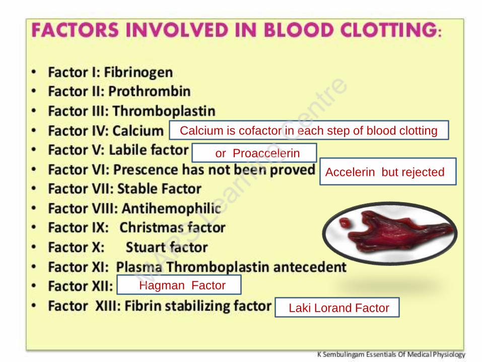
Calcium is cofactor in each step of blood clotting
Laki Lorand Factor
Hagman Factor
or Proaccelerin
Accelerin but rejected
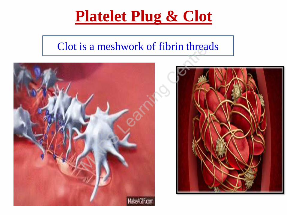
Platelet Plug & Clot
Clot is a meshwork of fibrin threads
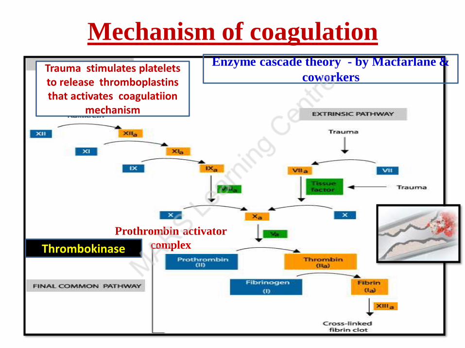
Mechanism of coagulation
Prothrombin activator
complex
Enzyme cascade theory - by Macfarlane &
coworkers
Thrombokinase
Trauma stimulates platelets to release thromboplastinsthat activates coagulatiion
mechanism
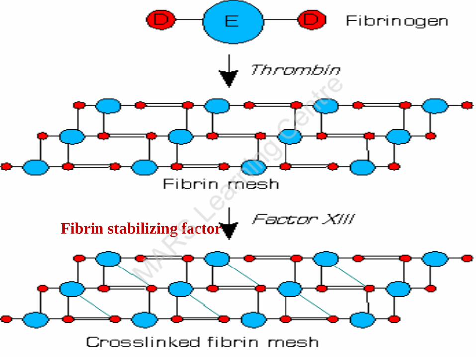
FACTOR XIII OR FIBRIN STABILIZING FACTOR
Fibrin stabilizing factor
83
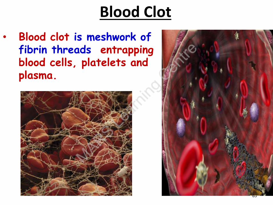
Blood Clot
• Blood clot is meshwork of fibrin threads entrapping blood cells, platelets and plasma.
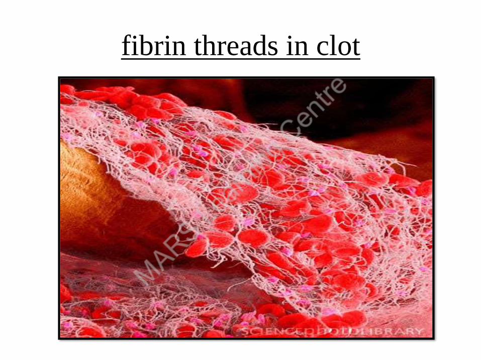
fibrin threads in clot
Clotting

Mechanism of coagulation (source ALLEN)
• There are Three steps in coagulation
1)Releasing of Thromboplastin
- Injured tissues release exothromboplastin and platelets release
endothromboplastin.
- Both these react with clotting factors along with calcium ions to
form Thrombokinase / Prothrombinase enzyme.
- This enzyme inactivates anticoagulant heparin.
2) Conversion of Prothrombin to Thrombin
- Prothrombinase enzyme converts inactive Factor 2 ( Prothrombin )
to active factor Thrombin. In the presence of ca2+.
3) Conversion of Fibrinogen to Fibrin
- Thrombin converts inactive Factor 1 ( Fibrinogen ) to active factor
Fibrin and forms a mesh work

• Blood minus blood cells = Plasma
• Plasma minus Fibrinogen and = Serum
large plasma proteins
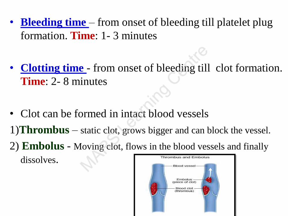
• Bleeding time – from onset of bleeding till platelet plug
formation. Time: 1- 3 minutes
• Clotting time - from onset of bleeding till clot formation.
Time: 2- 8 minutes
• Clot can be formed in intact blood vessels
1)Thrombus – static clot, grows bigger and can block the vessel.
2) Embolus - Moving clot, flows in the blood vessels and finally
dissolves.

• Anticoagulants are substances that prevent clotting of blood.
• Heparin , Antithrombin III, Protein C – natural anticoagulants
• Sodium citrate, Double oxalate, EDTA – used in lab
• Significance :
Has an important role in maintaining the fluidity of blood in circulation.
Anticoagulants

Heparin
• Released from basophils , mast cells
Mechanism of action :
• Inhibits conversion of Prothrombin ---- thrombin
• Combines with Antithrombin III and it
neutralizes the action of thrombin, inactivates
Factors 9,10,11 & 12
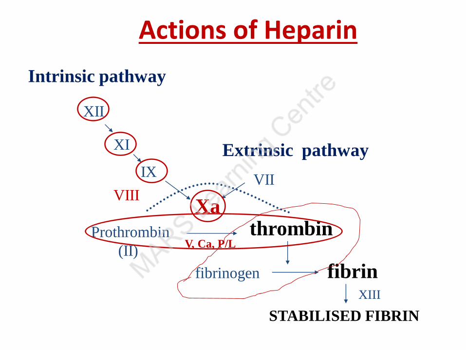
Actions of Heparin
XII
XI
IX
XaVIII
Prothrombin
(II)
thrombin
fibrinogen fibrin
STABILISED FIBRIN
V, Ca, P/L
VII
Intrinsic pathway
Extrinsic pathway
XIII
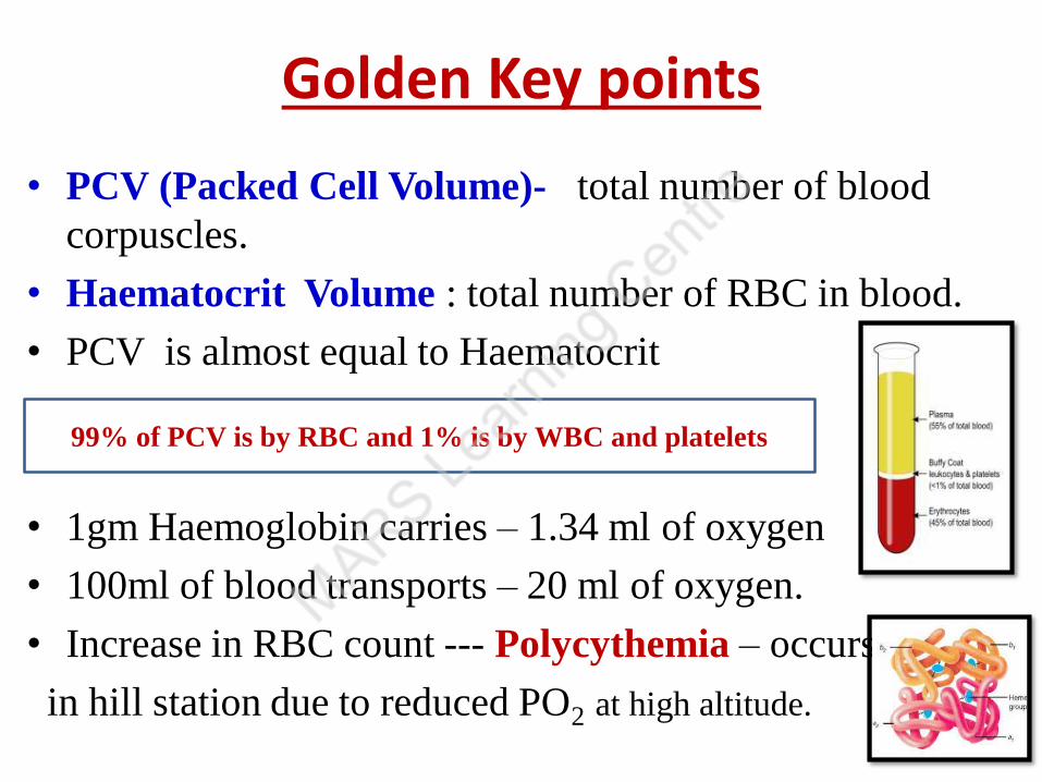
Golden Key points
• PCV (Packed Cell Volume)- total number of blood
corpuscles.
• Haematocrit Volume : total number of RBC in blood.
• PCV is almost equal to Haematocrit
• 1gm Haemoglobin carries – 1.34 ml of oxygen
• 100ml of blood transports – 20 ml of oxygen.
• Increase in RBC count --- Polycythemia – occurs
in hill station due to reduced PO2 at high altitude.
99% of PCV is by RBC and 1% is by WBC and platelets
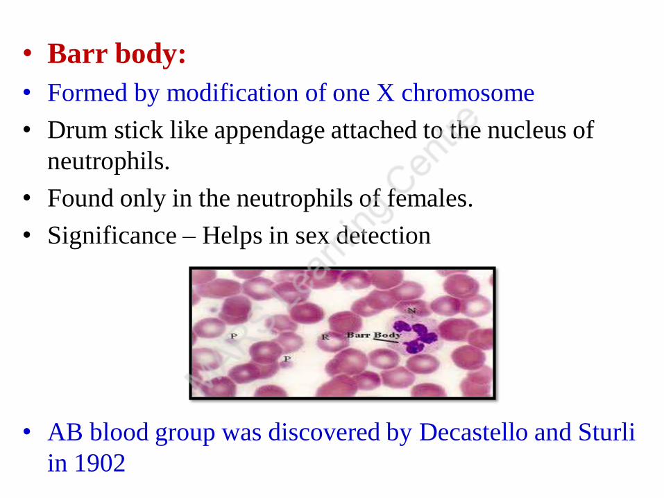
• Barr body:
• Formed by modification of one X chromosome
• Drum stick like appendage attached to the nucleus of
neutrophils.
• Found only in the neutrophils of females.
• Significance – Helps in sex detection
• AB blood group was discovered by Decastello and Sturli
in 1902
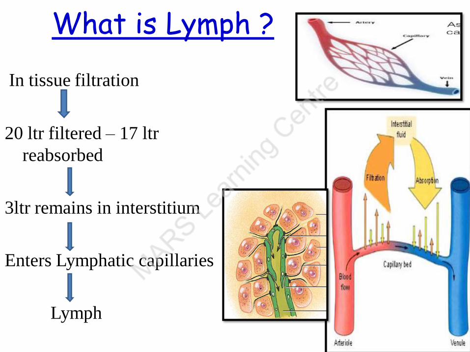
What is Lymph ?
In tissue filtration
20 ltr filtered – 17 ltr
reabsorbed
3ltr remains in interstitium
Enters Lymphatic capillaries
Lymph
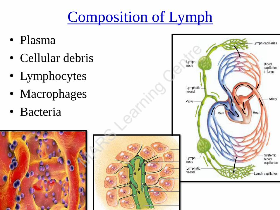
Composition of Lymph
• Plasma
• Cellular debris
• Lymphocytes
• Macrophages
• Bacteria
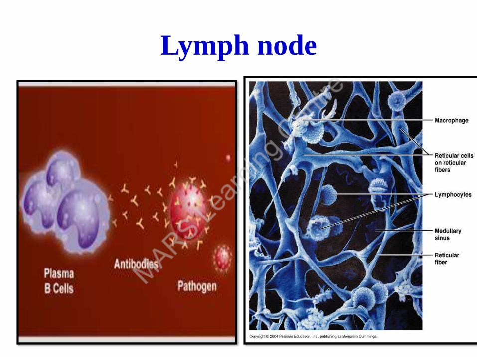
Lymph node

Lymph nodes
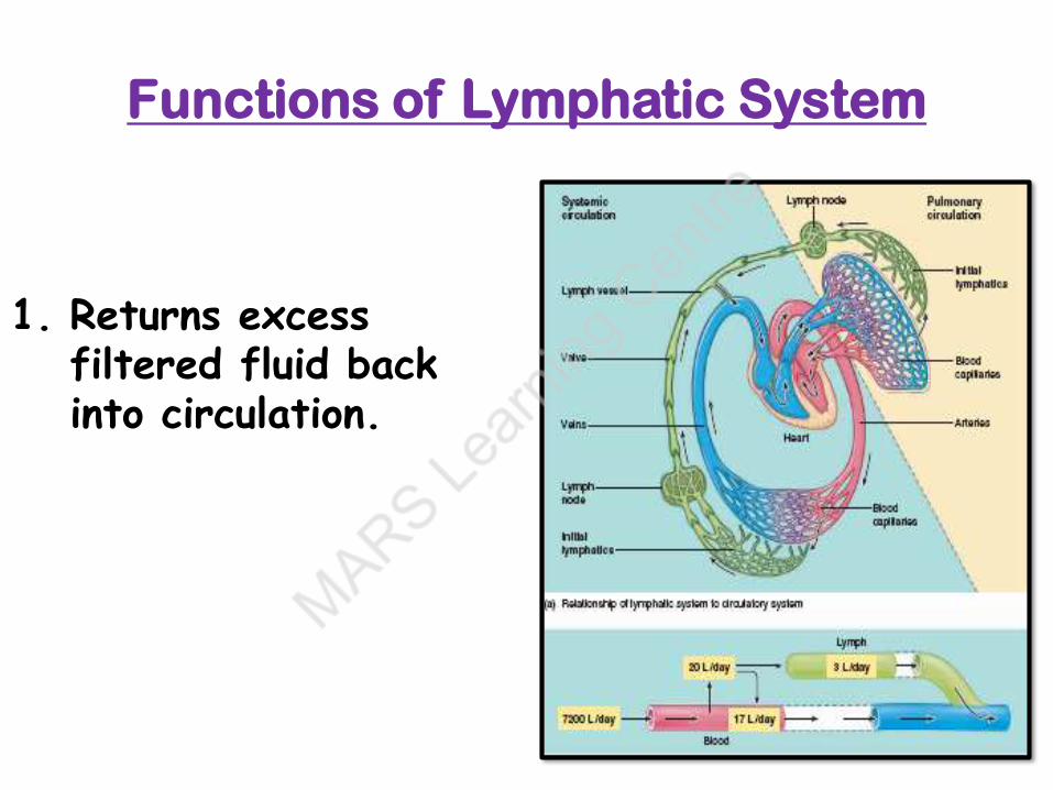
Functions of Lymphatic System
1. Returns excess filtered fluid back into circulation.

3. Returns filtered proteins
Proteins leaked in filtration
Not reabsorbed by capillaries
Enters lymphatic capillaries

2. Defense against diseases
Phagocytes (Lymphocytes)
in Lymph Nodes
Destroys pathogens in lymph
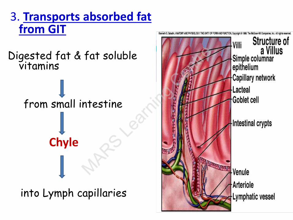
3. Transports absorbed fat from GIT
Digested fat & fat soluble vitamins
from small intestine
into Lymph capillaries
Chyle
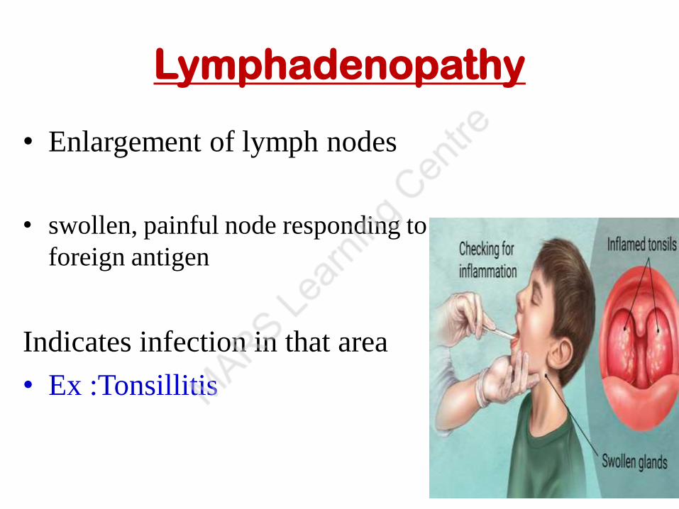
Lymphadenopathy
• Enlargement of lymph nodes
• swollen, painful node responding to
foreign antigen
Indicates infection in that area
• Ex :Tonsillitis

Swelling Elephantiasis

Related Documents