Module : Session: Advanced Care Paramedicine Advanced Care Paramedicine Vascular Anatomy 7 1

Vascular Anatomy
Jan 02, 2016
Vascular Anatomy. 7. 1. Objectives. Understand the historical progression of knowledge regarding the components and function of the cardiovascular system. Identify the blood vessels of the body used for intravenous cannulation and phlebotomy. - PowerPoint PPT Presentation
Welcome message from author
This document is posted to help you gain knowledge. Please leave a comment to let me know what you think about it! Share it to your friends and learn new things together.
Transcript
Module:
Session:
Advanced Care ParamedicineAdvanced Care Paramedicine
Vascular Anatomy
7
1
Objectives
Understand the historical progression of knowledge regarding the components and function of the cardiovascular system.
Identify the blood vessels of the body used for intravenous cannulation and phlebotomy.
Recognize the anatomical components of the vasculature.
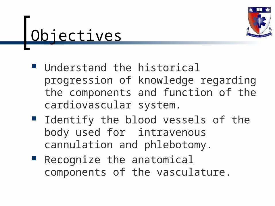
"The arteries have no sensation, for they even are without blood, nor do they all contain the breath of life; and when they are cut only the part of the body concerned is paralyzed...the veins spread underneath the whole skin, finally ending in very thin threads, and they narrow down into such an extremely minute size that the blood cannot pass through them nor can anything else but the moisture passing out from the blood in innumerable small
drops which is called sweat."
Pliny the Elder, Rome 23-79 AD
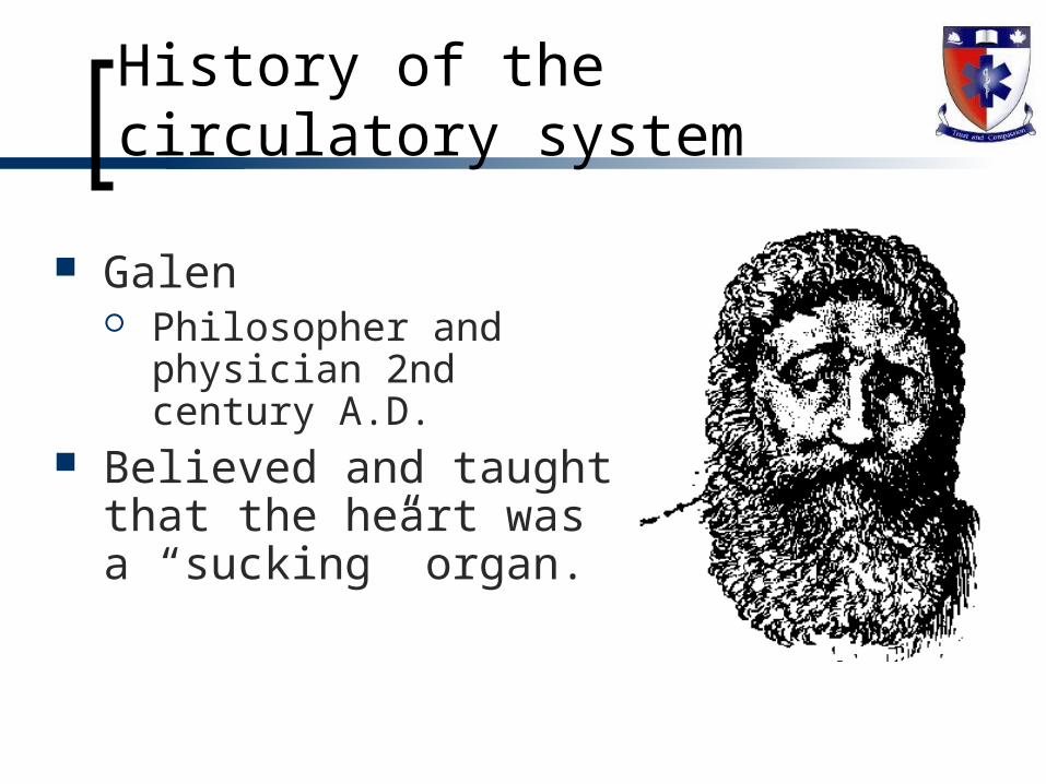
Galen Philosopher and
physician 2nd century A.D.
Believed and taught that the heart was a “sucking” organ.
History of the circulatory system
Galen
Also believed and taught that there were two distinct types of blood.
‘nutritive blood’ was thought to be made by the liver (transformed from food) and carried through veins to the organs, where it was consumed.
‘vital blood’ was thought to be made by the heart and pumped through arteries to carry the “ vital spirits.”
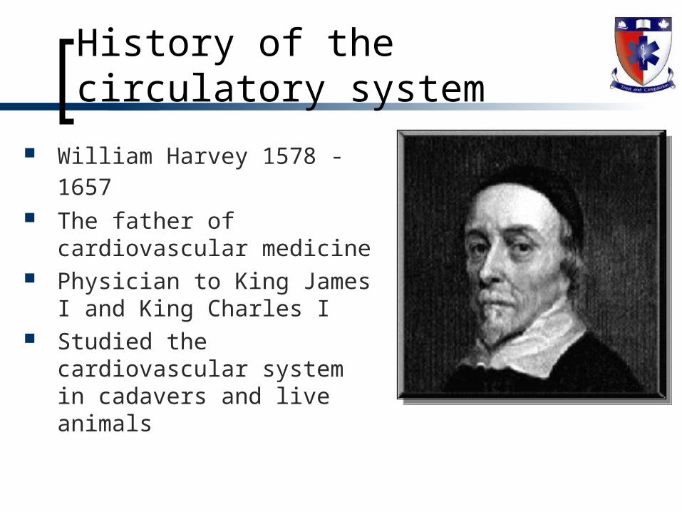
William Harvey 1578 - 1657 The father of cardiovascular
medicine Physician to King James I and
King Charles I Studied the cardiovascular
system in cadavers and live animals
History of the circulatory system
History
Discovered the veins and arteries in the septum - disputing the previous concepts that there were perforations between the ventricles
Harvey also theorized that arteries and veins were connected by capillaries - thus creating a closed circuit, but lacked a microscope to confirm his theory
History
Harvey’s “On the Movement of the Heart and Blood in Animals” - 1628
Identified heart circulates the same blood physically impossible to eat/drink enough to replace blood
volume daily Was not published for thirteen years due to fear Was not accepted for more than twenty years More questions raised than answered
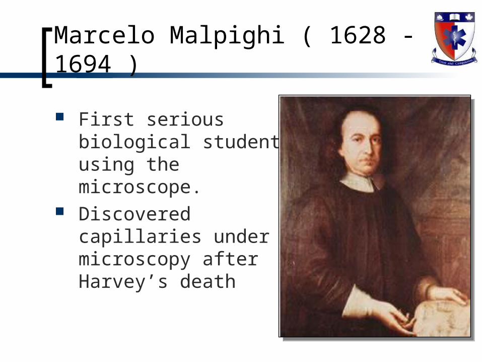
First serious biological student using the microscope.
Discovered capillaries under microscopy after Harvey’s death
Marcelo Malpighi ( 1628 - 1694 )
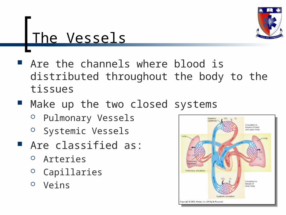
The Vessels
Are the channels where blood is distributed throughout the body to the tissues
Make up the two closed systems Pulmonary Vessels Systemic Vessels
Are classified as: Arteries Capillaries Veins
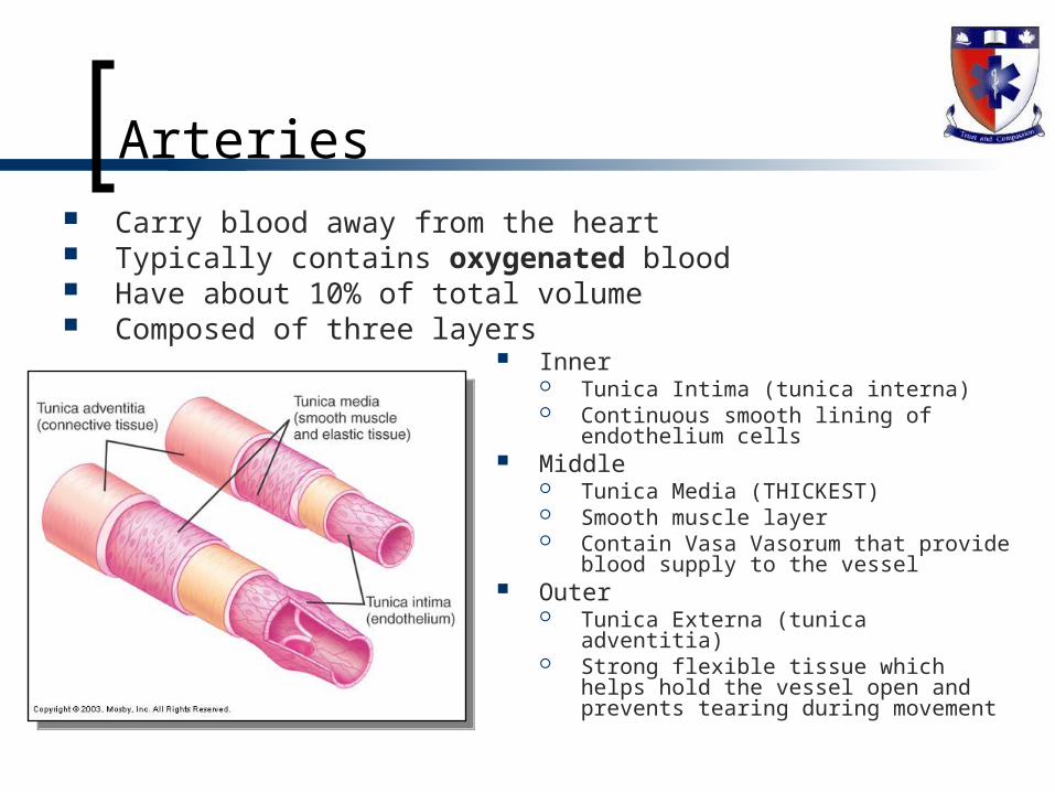
Arteries Carry blood away from the heart Typically contains oxygenated blood Have about 10% of total volume Composed of three layers
Inner Tunica Intima (tunica interna) Continuous smooth lining of
endothelium cells Middle
Tunica Media (THICKEST) Smooth muscle layer Contain Vasa Vasorum that provide
blood supply to the vessel Outer
Tunica Externa (tunica adventitia) Strong flexible tissue which helps hold
the vessel open and prevents tearing during movement
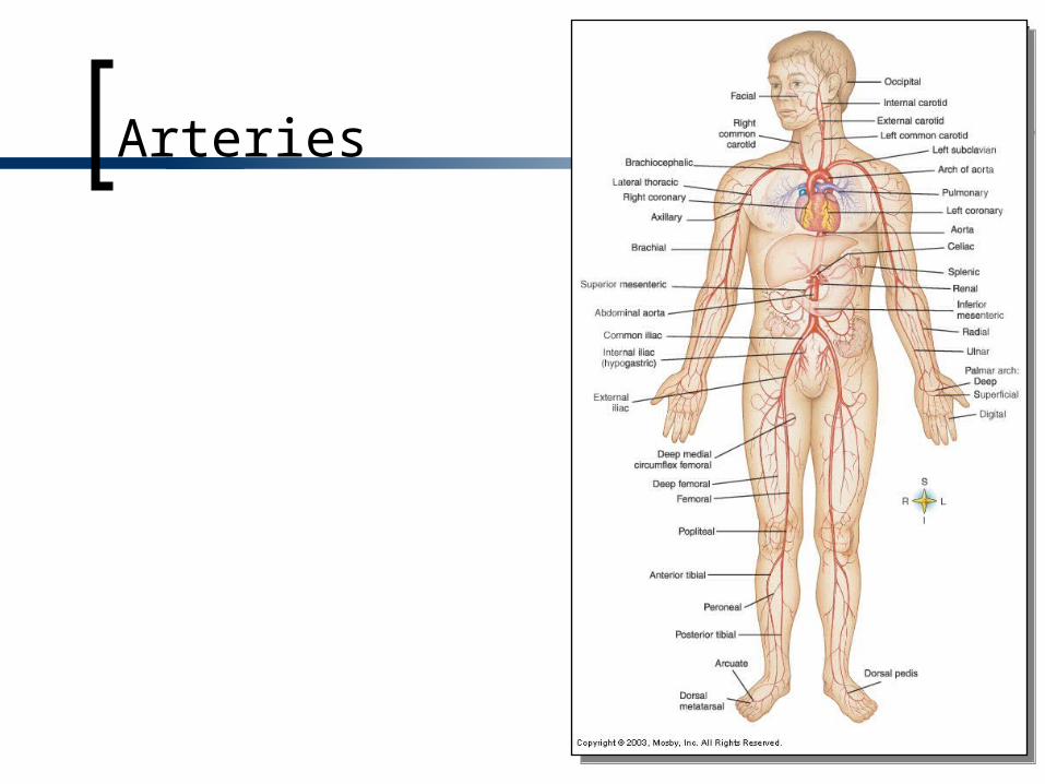
Arteries
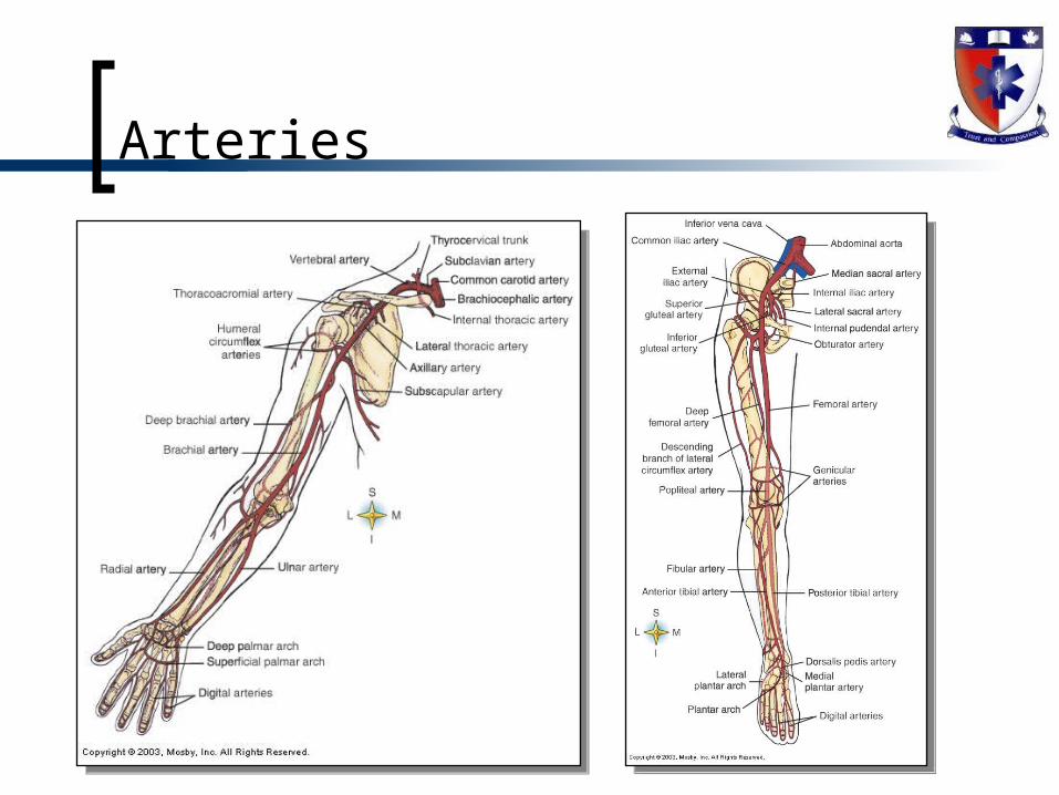
Arteries
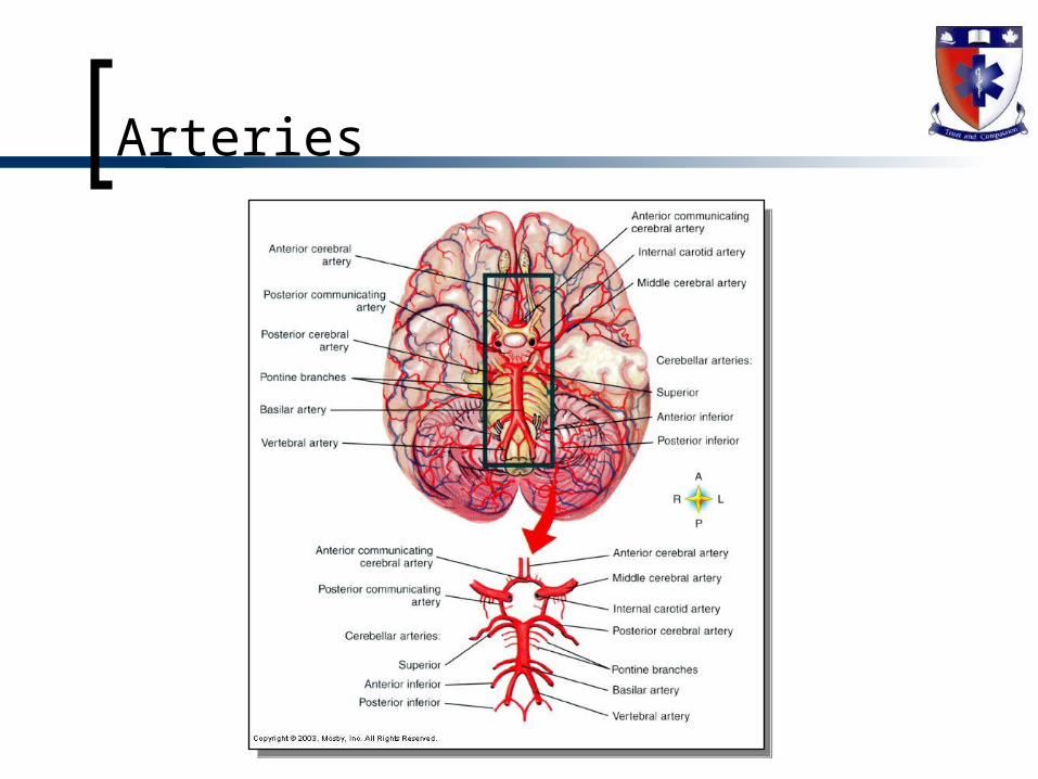
Arteries
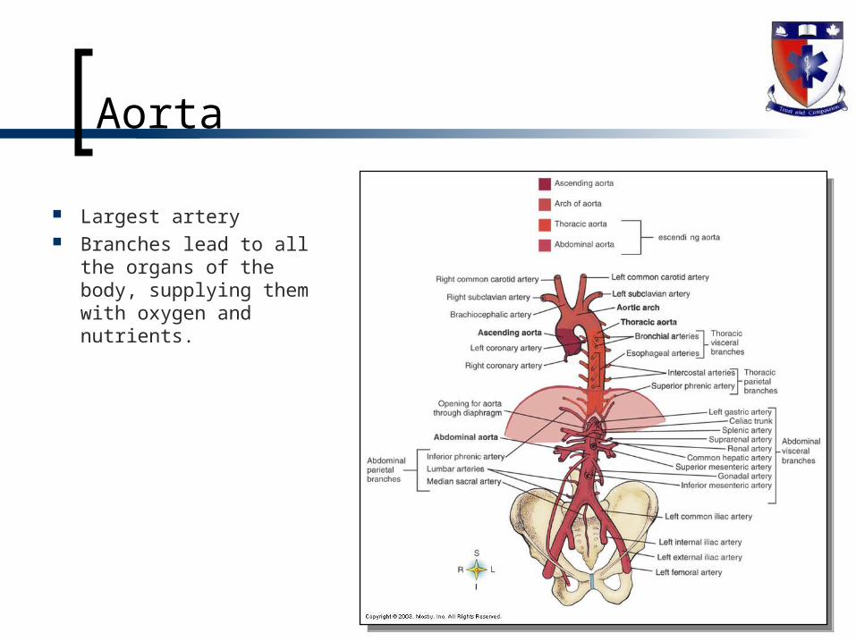
Aorta
Largest artery Branches lead to all
the organs of the body, supplying them with oxygen and nutrients.
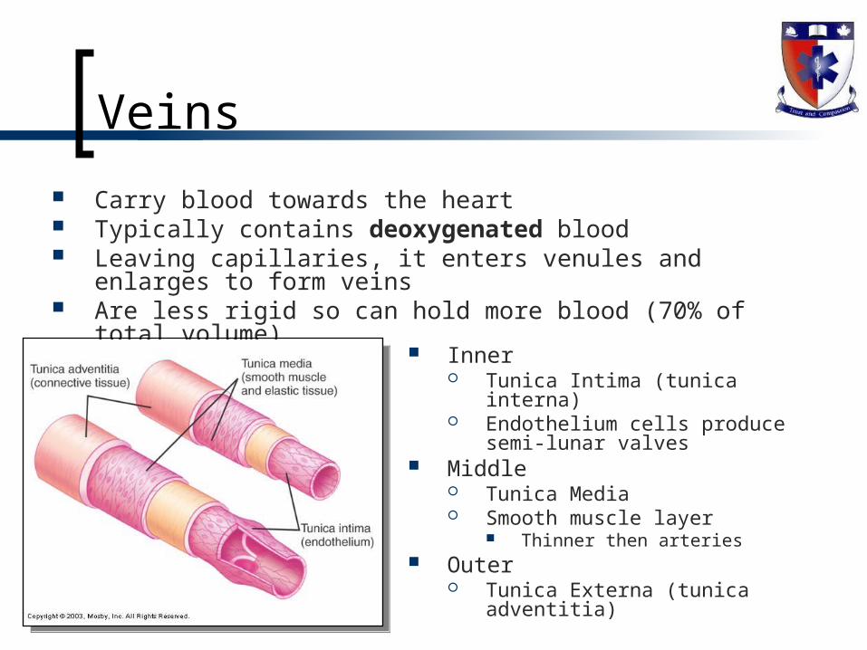
Veins
Carry blood towards the heart Typically contains deoxygenated blood Leaving capillaries, it enters venules and enlarges to form veins Are less rigid so can hold more blood (70% of total volume)
Inner Tunica Intima (tunica interna) Endothelium cells produce semi-
lunar valves Middle
Tunica Media Smooth muscle layer
Thinner then arteries Outer
Tunica Externa (tunica adventitia)

Venous Blood Reservoir
Have great capacity to stretch (capacitance)
Allows for accommodation of large amounts of blood with no change in BP
Allows for venous circulation based on pressure from valve below
Veins

Veins
Veins
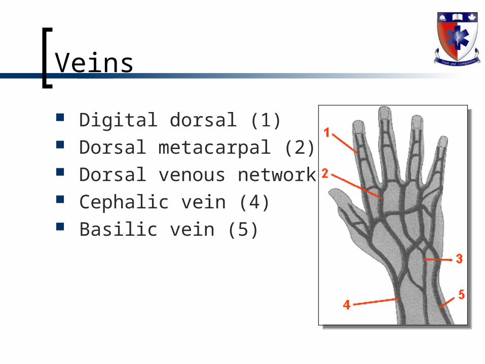
Digital dorsal (1) Dorsal metacarpal (2) Dorsal venous network (3) Cephalic vein (4) Basilic vein (5)
Veins
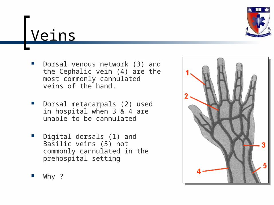
Dorsal venous network (3) and the Cephalic vein (4) are the most commonly cannulated veins of the hand.
Dorsal metacarpals (2) used in hospital when 3 & 4 are unable to be cannulated
Digital dorsals (1) and Basilic veins (5) not commonly cannulated in the prehospital setting
Why ?
Veins
Veins
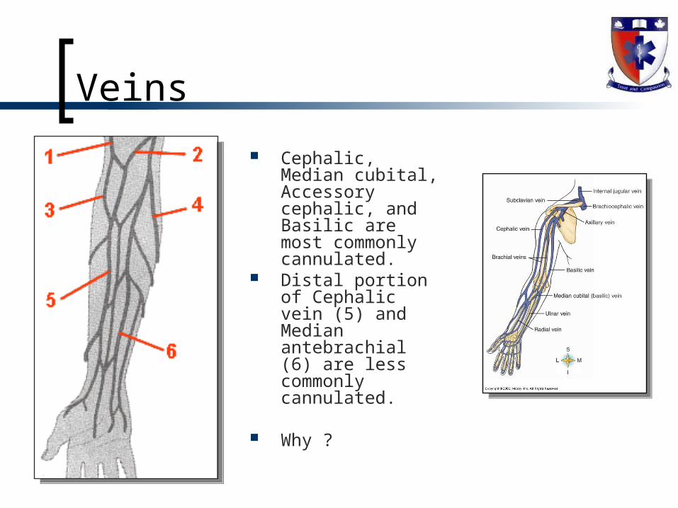
Cephalic, Median cubital, Accessory cephalic, and Basilic are most commonly cannulated.
Distal portion of Cephalic vein (5) and Median antebrachial (6) are less commonly cannulated.
Why ?
Capillaries Smallest and most numerous
Contain about 5% of total volume
Are the connection between the arteries to the veins Are composed of only the endothelium
Interesting facts Are typically only ½ inch in length If all capillaries placed end to end would reach 100,000 km Estimated that 1 cm3 of muscle contains 100,000
Distribution is based on metabolic needs Liver, muscle, kidneys have extensive network Epidermis, lens and cornea have none
Capillaries
Have vital role in exchange of gases, nutrients and waste between blood and tissue Thin wall (one cell thick) with fenestrations Provide the slowest rate of speed of blood in the system Tissues are surrounded by extracellular fluid called interstitial
fluid
Blood flow into capillaries is regulate by smooth muscle (pre-capillary sphincters) If constricted blood is directed through metarterioles
(arteriovenous anastomoses or AV shunts)
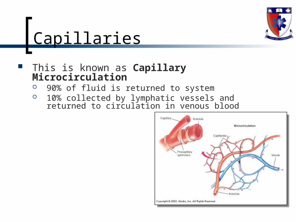
Capillaries
This is known as Capillary Microcirculation 90% of fluid is returned to system 10% collected by lymphatic vessels and returned to circulation
in venous blood
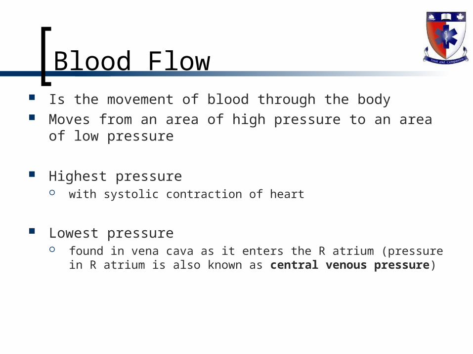
Blood Flow Is the movement of blood through the body Moves from an area of high pressure to an area of low
pressure
Highest pressure with systolic contraction of heart
Lowest pressure found in vena cava as it enters the R atrium (pressure in R
atrium is also known as central venous pressure)
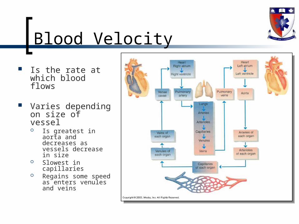
Blood Velocity
Is the rate at which blood flows
Varies depending on size of vessel Is greatest in aorta
and decreases as vessels decrease in size
Slowest in capillaries
Regains some speed as enters venules and veins
Venous Blood Flow
Very little pressure in veins Venous return is dependant on:
Muscle action Muscle contracts, thickens and squeezes veins next to it
Respiratory movements As diaphragm contracts changes thoracic pressure causing
abdominal blood to move Contraction of veins
Sympathetic reflexes cause constriction
Related Documents