Vascular Access Workshop BC Vascular Access Educators Group August 2010 (Revised June 2015) Workshop Developed by: Rick Luscombe, Vascular Access Nurse, Providence Health Care Laurie Bates, Renal Educator, Interior Health Authority

Welcome message from author
This document is posted to help you gain knowledge. Please leave a comment to let me know what you think about it! Share it to your friends and learn new things together.
Transcript

Vascular Access Workshop
BC Vascular Access Educators Group
August 2010 (Revised June 2015)
Workshop Developed by: Rick Luscombe, Vascular Access Nurse, Providence Health Care
Laurie Bates, Renal Educator, Interior Health Authority

Outline
• VA Case Studies & More
– AV Fistulas (AVFs)
– Central Venous Catheters (CVCs)
– AV Grafts (AVGs)

Vascular Access (VA) Case Studies & More
We are the Lifeline Keepers!

VA Case Studies & More
Application of VA knowledge to a case study (Mr. Kline)
Divided into 5 modules: 1. Arteriovenous (AV) Fistula Creation 2. Central Venous Catheters (CVC) &
Complications 3. AV Fistula Maturation, Needling &
Complications 4. CVC Complications cont’d 5. AV Grafts

Module #1:
AV Fistula Creation » AVF Pre-creation Assessment
» AVF Pre-operative Teaching
» AVF Creation
» AVF Post-creation Complications

AVF Pre-creation Assessment:
Case Study Mr. Kline is a 75-year-old male.
He has Diabetes Mellitus (DM) type 2, hypertension & peripheral vascular disease.
He has been attending the kidney care clinic for the past three years. His glomerular filtration rate is now about 14.
The care clinic nurses have convinced him that a CVC is not an appropriate long-term option for HD due to complications of infection, & stenosis & a higher mortality risk.
He has agreed to go for an AV fistula assessment.

AVF Pre-creation Assessment:
Case Study
Mr. Kline is referred to the VA Clinic.
His left arm is assessed for an AVF (he is right hand dominant).
Allen’s test is negative – his hand is slightly cool to the touch but pink, & he says he has some numbness of his fingers.
There are no visible or palpable veins in the forearm or upper arm when a tourniquet is applied.
U/S demonstrates a cephalic vein in the forearm. 3 mm in diameter but does not continue to the antecubital fossa.

AVF Pre-creation Assessment
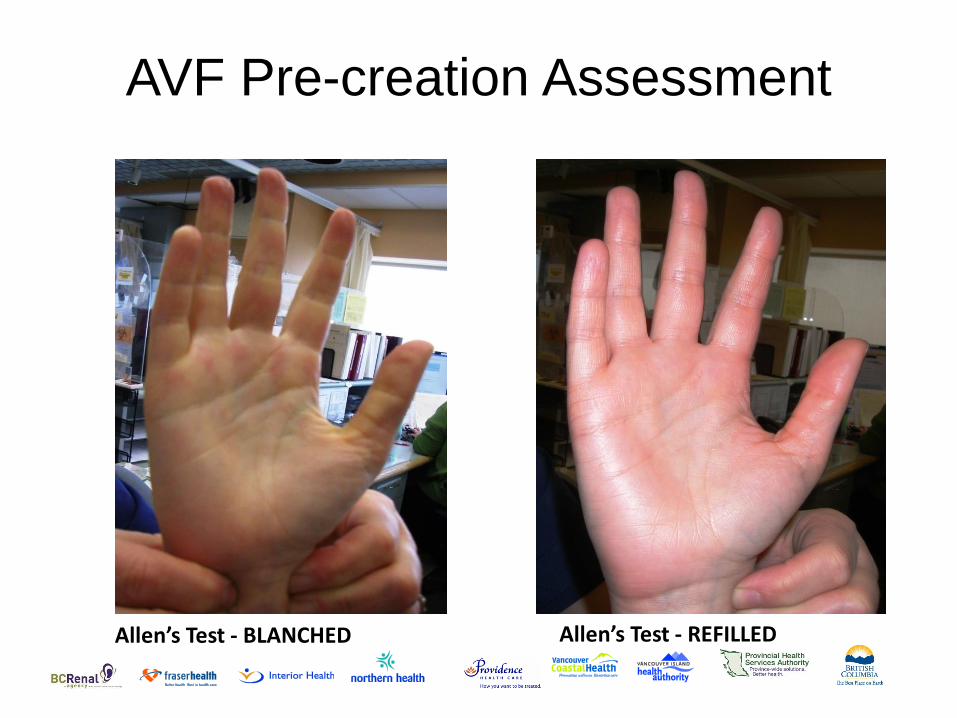
ALLEN’S TEST – What is it?
1. Elevate Mr. Kline’s arm & ask him to make a fist for about 30 seconds.
2. Apply pressure over the ulnar & the radial arteries so as to occlude both of them.
3. Still elevated, ask Mr. Kline to open his hand. It should appear blanched (pallor can be observed in the finger nails).
4. Release ulnar pressure. Colour should return in 3-5 seconds.
5. Repeat the same steps releasing the radial pressure. Colour should return in 3-5 seconds.
A slow return of colour upon release of either artery is a red flag to suggest that the artery may be impaired and not suitable for creation of an AVF in the wrist.
Adapted from: http://en.wikipedia.org/wiki/Allen's_test
AVF Pre-creation Assessment
Allen’s Test - BLANCHED Allen’s Test - REFILLED

AVF Pre-creation Assessment:
Case Study
Returning to the case…..
A left arm venogram is performed in radiology to confirm the cephalic vein is patent & rule out any central vein stenosis.
The venogram demonstrates occlusion of the cephalic vein in the mid-forearm but a patent cephalic vein & basilic vein in the upper arm.
There is minor narrowing at the brachio-cephalic/subclavian junction but the radiologist deems the narrowing to be insignificant.

AVF Pre-operative Teaching:
Case Study
A decision is made to proceed with creation of a left upper arm brachio-cephalic fistula (L BC AVF).
What patient teaching will Mr. Kline require?

AVF Pre-operative Teaching Mr. Kline will need to know…
About the surgery:
•Not to eat or drink after midnight •Hold blood thinners 3-5 days before the procedure (consult with surgeon ) •Approximately 90 min •Will be done as a day or short stay surgery; non-dominant arm preferred.
After the surgery:
•Keep arm straight & raised on a pillow, even while sleeping. •Monitor discomfort. If mild to moderate, take pain medication regularly. If this doesn’t work, tell doctor or nurse. •Rest arm for a few days, then use gently. Avoid lifting, vacuuming or excessive bending until the swelling is resolved and sutures have been removed. Sutures will be removed between 10-14 days •Keep dressing clean & dry. Tub bath is best.
• If regular sutures or clips are in place, dressing is usually worn until sutures are removed (up to 3 weeks post-op). If steri-strips are in place, allow to fall off.
• If the wound is dry & healing, the dressing can be taken off & the area kept clean & dry. May wish to wear light dressing until incision is completely healed to protect from irritation due to clothing rub.

AVF Pre-operative Teaching Mr. Kline will need to know…
After the surgery cont’d:
• Monitor colour & warmth of fingers on fistula hand. Should be the same as other hand. Severe numbness or pain is not normal.
• Check fistula every morning & evening for thrill (feel for buzzing sensation) & bruit (hold anastomosis site to the ear & listen for bruit).
• Keep fingers moving. Squeeze soft ball to encourage vein development
• Follow-up appointments in 2 & 6 weeks (centre-specific).

AVF Pre-operative Teaching Mr. Kline will need to know… Ongoing monitoring: • Check fistula for thrill & bruit every morning & evening &/or after an
episode of hypotension. • Avoid heavy lifting. • Avoid putting pressure on fistula (even when sleeping). Do not wear
restrictive jewelry. • Stay active, but avoid contact sports. • Save fistula arm for dialysis & let others know this. No BP, blood work or
IV. Wear Medical Alert bracelet. When to call the nephrologist or dialysis unit: • Can’t feel the thrill &/or bruit sounds different than usual. • Redness, warmth or pain in fistula arm. • Oozing or drainage from fistula. • Noticeable swelling or itching in fistula arm. • Difficulty moving fingers in fistula arm. • Fever or episodes of chills/rigor.

AVF Creation: Case Study
Returning to the case…..
• Mr. Kline is scheduled for surgery.
• He returns from the O.R. with a left upper arm brachio-cephalic fistula.
• He is assessed by the VA nurse 3 hours post-creation.
• He is given post-op instructions & discharged home.

AVF Creation: 1st Choice - Radial-cephalic
thumb

AVF Creation: 2nd Choice - Brachio-cephalic

AVF Creation: 3rd Choice - Brachio-basilic

AVF Creation: 3rd Choice - Brachio-basilic

AVF Creation List 3 goals of post op assessment.
1. Patency: – Post-op BP is adequate to support AVF blood flow. – AVF blood flow is assessed as present. – Thrill is palpable & bruit is audible.
2. Complications: – No decreased circulation to the hand (i.e. no steal syndrome). – Minimal bleeding or swelling at incision site. – No evidence of infection.
3. Patient response to surgery: – Manage pain. – Monitor emotional response. – Provide follow-up information/patient teaching.

AVF Creation
What tools are required for post-op assessment?
• Hemo RNs
– Eyes LOOK
– Hands FEEL
– Stethoscope LISTEN
• VA Nurse &/or Advanced Cannulators
– All the above, plus…
– Ultrasound with doppler
L L F
The VA Cartouche!

Assessing Fistula/Graft Sounds
Abnormal AVF Sounds
Abnormal AVG Sounds
Stenosis Whistle
Stenosis Whistle
Normal Bruit

AVF Creation
Where might you find information about Mr. Kline’s surgery?
• Verbally: – Discussion with daycare/ short-stay nurse regarding the
report they received from the surgeon. – Surgeon directly, if possible. – Mr. Kline, as to his understanding of the procedure.
• Health Record (chart & electronic): – OR progress note. – PROMIS: “VA” or “documents” re: OR report. – Site-specific e-documentation.

AVF Post-creation Complications
2 week post op assessment
• Mark developing vessel with felt pen for mapping purposes.
• Mark division between arterial & venous needling areas with “X.”

AVF Post-creation Complications
List the 2 most common post-creation complications.
• Thrombosis at time of creation, or later.
• Failure to mature (e.g. inadequate vein size, stenosis, occlusions, calcified vessels).

AVF Post-creation Complications
Other possible complications: • Steal syndrome • Venous hypertension • Stenosis (module 3)
• Infiltration (module 3) • Infection (module 3) • Aneurysm (module 3) • Skin breakdown (module 3)
AVF Post-creation Complications: Case Study
Returning to the case…..
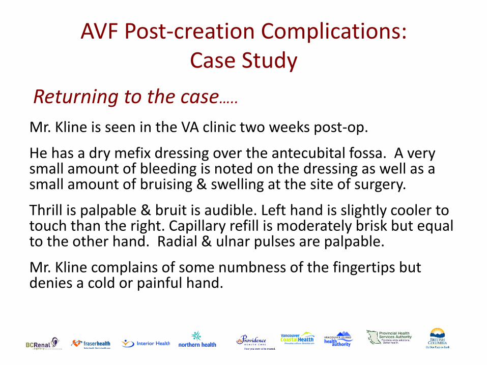
Mr. Kline is seen in the VA clinic two weeks post-op.
He has a dry mefix dressing over the antecubital fossa. A very small amount of bleeding is noted on the dressing as well as a small amount of bruising & swelling at the site of surgery.
Thrill is palpable & bruit is audible. Left hand is slightly cooler to touch than the right. Capillary refill is moderately brisk but equal to the other hand. Radial & ulnar pulses are palpable.
Mr. Kline complains of some numbness of the fingertips but denies a cold or painful hand.
AVF Post-creation Complications
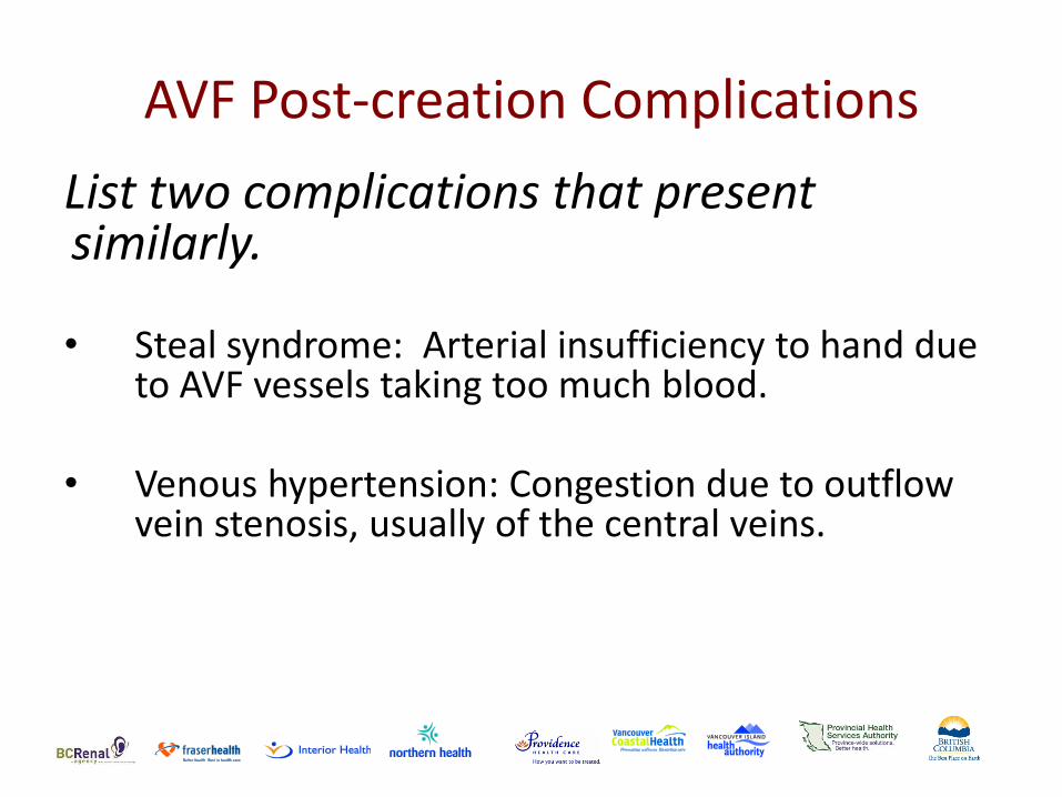
List two complications that present similarly.
• Steal syndrome: Arterial insufficiency to hand due to AVF vessels taking too much blood.
• Venous hypertension: Congestion due to outflow
vein stenosis, usually of the central veins.

AVF Post-creation Complications: Steal Syndrome & Venous Hypertension
Steal Syndrome • Affected hand may be blue (no blood
to fingertips. • No edema is present. • May have wounds.
Venous Hypertension • Affected hand may be red. • Edema is present . • As the hand swells, skin may breakdown.
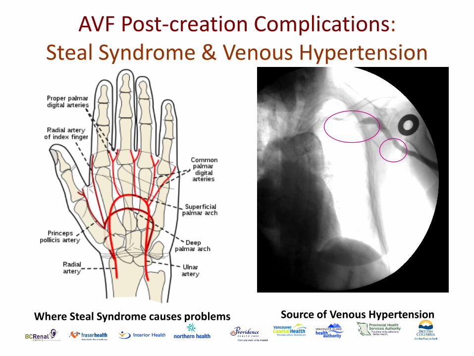
AVF Post-creation Complications: Steal Syndrome & Venous Hypertension
Where Steal Syndrome causes problems Source of Venous Hypertension

AVF Post-creation Complications: Steal Syndrome & Venous Hypertension
Why do they occur?
• Steal Syndrome: – Anastomosis may be too large or poor arteries. – Compromised arterial vessels to the hand.
• Venous Hypertension: – Previous catheter insertions. – Repeated needling causing scar tissue. – Anatomy (approx. 14% of people will develop central
venous stenosis without a previous history of catheter insertions).

AVF Post-creation Complications: Steal Syndrome & Venous Hypertension
What would you expect to notice during your assessment?
•Steal Syndrome:
• Hand cool, painful & cyanotic. May be more pale than frankly cyanotic.
• Delayed capillary refill (compared to other hand); with numbness, tingling & decreased range of motion.
• May have skin breakdown, sores on tips of fingers.

AVF Post-creation Complications: Steal Syndrome or Diabetic Complication?

AVF Post-creation Complications: Steal Syndrome & Venous Hypertension
What would you expect to notice during your assessment? •Venous Hypertension:
• Vein engorgement below stenosis, chest veins dilated, pressure in head (possibly).
• Collateral vessel engorgement on access arm & hand. • On dialysis: High venous pressures, prolonged bleeding. • Hand & fingers may be more ruddy in colour. • Numbness, tingling & decreased range of motion. • May have skin breakdown, sores on tips of fingers. • May have generalized edema to affected limb.
Reference: PVAST

AVF Post-creation Complication: Venous Hypertension

AVF Post-creation Complication: Venous Hypertension
Collateral development
Thinning skin & scar tissue

AVF Post-creation Complications: Steal Syndrome & Venous Hypertension
What do you expect to hear during auscultation?
•Steal Syndrome: – Loud bruit heard on both systole & diastole
•Venous Hypertension: – Water hammer bruit – loud & bounding; may be
high-pitched & sporadic. – Bruit may be heard only on systole.
Reference: PVAST

AVF Post-creation Complications: Steal Syndrome & Venous Hypertension
What do you expect to feel during palpation?
• Steal Syndrome:
– Normal thrill
• Venous Hypertension:
– Thrill: strong, bounding pulse near proximal stenosis
– Decreased quality of pulses
– Small, flat, less pulsatile vein above stenosis

AVF Post-creation Complications: Steal Syndrome & Venous Hypertension
Where will you needle & why?
• Steal Syndrome:
– Doesn’t matter – expect normal arterial & venous pressures
• Venous Hypertension:
– Depends on the location of the stenosis or occlusion and if collateral vessels have developed – you may expect high venous pressures & more positive arterial pressures

AVF Post-creation Complications: Steal Syndrome & Venous Hypertension –
Medical Tests
Steal Syndrome
Allen’s test
Doppler
Finger pressures
Arteriogram
Venous Hypertension
Venogram
Fistulogram

AVF Post-creation Complications: Steal Syndrome & Venous Hypertension –
Medical Interventions
Steal Syndrome Leave
Banding
Digital Revascularization-Interval Ligation (DRIL)
RUDI – Revision using Distal Inflow
Takedown
Venous Hypertension Angioplasty
Bypass graft
Clavicle removal
Takedown

Module #2: Central Venous Catheters (CVCs)
& Complications » Non-cuffed & Tunneled Cuffed CVCs » CVC Patient Teaching » CVC Complications
• Post-insertion bleeding • Pneumothorax • Hemothorax • Air embolus • Cardiac tamponade • Malposition • Kinking
CVCs: Case Study
Returning to the case…..
At five weeks post-AVF creation Mr Kline becomes acutely ill. A decision is made to initiate hemodialysis.
A right IJ tunneled catheter is inserted. It is intended to be used as a bridge until the fistula can be cannulated in 3 weeks.

Non-Cuffed & Cuffed CVCs
What is the difference between non-cuffed & tunneled cuffed catheters?
Non cuffed temporary
• Maximum time recommended is no greater than 2 weeks.
• Chest x-ray needed to confirm placement. • Emergent start. • Needs sutures at all times. • Higher infection rates than cuffed catheters. • Dressing change every run, occlusive preferred.
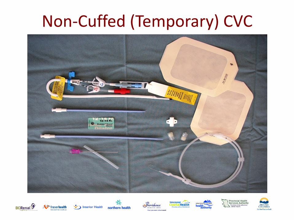
Non-Cuffed (Temporary) CVC
Non-Cuffed & Cuffed CVCs
What is the difference between non-cuffed & tunneled cuffed catheters?
Cuffed • Temporary bridge to an alternate access site. • Permanent use when no other access option exists. • Cuff acts as a barrier to infections. • Connective tissue grows into the cuff & anchors the catheter in
place (fibroses). • After a period of time sutures are not needed to keep catheter
anchored. • Once the tunnel is well healed, a transparent dressing may be used
for up to 7 days at a time. • Softer CVC material creates less tissue stress & better longevity. • Usually inserted under fluoroscopy for exact placement.

Tunneled Cuffed CVC
Source: http://www.cardiointernational.com

Tunneled Cuffed CVC

Catheter Tips
Tunneled Cuffed CVC
What is the most common site for cuffed catheters? Why?
• Right IJ (most common & preferred site) • Left IJ • Right & left external jugulars • Right & left subclavian • Femoral veins
When would we use a Femoral catheter? • ICU / CCU emergency or no more sites left

Tunneled Cuffed CVC
Source: www.vascularphysicians.com/services/tunneled & www.fda.gov/cdrh
• Preferred catheter placement is into the right atrium. • Please DO NOT refer to tunneled cuffed catheters as perm caths.
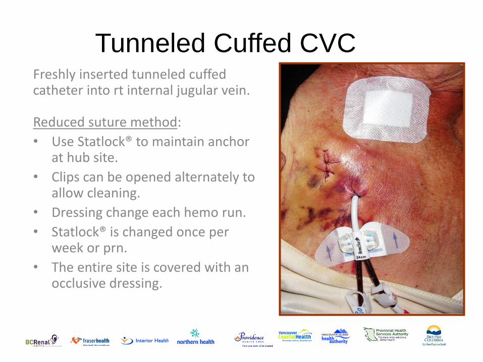
Tunneled Cuffed CVC Freshly inserted tunneled cuffed catheter into rt internal jugular vein.
Reduced suture method:
• Use Statlock® to maintain anchor at hub site.
• Clips can be opened alternately to allow cleaning.
• Dressing change each hemo run.
• Statlock® is changed once per week or prn.
• The entire site is covered with an occlusive dressing.

Tunneled Cuffed CVC
Area between the arrows indicates where cuff is anchored with fibrosed connective tissue.
The cuff acts as a barrier to organisms traveling up the tunnel.
It’s a good idea to note the exposed length of catheter between the exit site & the hub.
This catheter lies over the clavicle.

CVC Patient Teaching
What does Mr. Kline need to know? Insertion: • May be done in Radiology by an Interventional Radiologist (IR). • May be done in the OR by a VA Surgeon. Recovery: • Should be very little discomfort. • Should be very little bleeding. • Tunneled (neck) suture will be required a minimum of 1 week. • Anchoring (exit) suture will be required a minimum of 6 weeks. Routine: • Must keep the exit site dry. • Report any pain or fever with or without chills & rigor. • Never open the catheter. • Keep clamps closed at all times.

CVC Complications
• Post-insertion bleeding
• Pneumothorax
• Hemothorax
• Air embolus
• Cardiac tamponade
• Malposition
• Kinking

CVC Complication:
Post-insertion Bleeding
What to do? • Stop the bleeding
CVC Complication: Pneumothorax
Symptoms: • SOB • Sharp pain in the chest • Sudden shortness of breath • Painful breathing • Tightness in the chest • Dry coughing • Cyanosis
What to do? • Supportive treatment as per shock • Oxygen administration • If physician not already in attendance, call STAT
*Source: http://lungdiseases.about.com

CVC Complication: Hemothorax • Tachypnea • Dyspnea • Cyanosis • Decreased or absent breath sounds on affected side • Tracheal deviation • Dull resonance on percussion • Unequal chest rise • Tachycardia • Hypotension • Pale, cool, clammy skin • Possibly subcutaneous emphysema • Narrowing pulse pressure
Source: http://en.wikipedia.org/wiki/Hemothorax

CVC Complication: Hemothorax
What to do?
• Supportive treatment as per shock
• Oxygen administration
• If physician not already in attendance, call STAT

CVC Complication: Air Embolus
• Chest pain • Dyspnea • Coughing • Cyanosis • Visual problems • Confusion • Hemiparesis • Death
Source: ANNA, Core Curriculum for Nephrology Nursing; 5Th ed. © 2008
CVC Complication: Air Embolus
What to do?
• Turn patient on LEFT side with head DOWN (Trendelenberg)
• Administer 100% oxygen
• If physician not already in attendance, call STAT
Source: ANNA, Core Curriculum for Nephrology Nursing; 5Th ed. © 2008
CVC Complication:
Cardiac Tamponade
• Sharp chest pain, often related to pericarditis, dissipating by the time the more severe cardiac tamponade condition develops
• SOB, sometimes as a result of breathing shallowly on purpose to avoid chest pain but usually, once cardiac tamponade has developed, related to reduced blood flow
• Forward-leaning posture due to pain &/or the need to catch one’s breath
• Weakness &/or fatigue • Bluish tint to skin (cyanosis) • Anxiety • Swelling in the abdomen Source: http://yourtotalhealth.ivillage.com/cardiac-tamponade.html

CVC Complication:
Cardiac Tamponade
What to do?
• Supportive treatment as per shock
• Oxygen administration
• If physician not already in attendance, call STAT
This is a medical emergency likely requiring pericardiocentesis & possibly cardiac window
http://en.wikipedia.org/wiki/Cardiac_tamponade

CVC Complication: Malposition
• Unable to aspirate from either lumen • May or may not be able to flush lumens • High arterial pressures; low venous pressures • Kinked catheter curve
What to do? • Do not use at all if visibly malpositioned • Notify VA Nurse or Nephrologist
• CXR for position • May run lines reversed if able & clearance adequate • May require new catheter

CVC Complication: Kinking
• May present the same as malposition
• Aspiration likely sticky or absent from one or both lumens
• Flushing likely possible
What to do?
• CXR to confirm placement & identify kinking
• Will likely need new catheter
• Notify VA Nurse or Nephrologist

Module #3:
AVF Maturation, Needling & Complications
» AVF Maturation
» AVF Needling
» AVF Complications

AVF Maturation: Case Study
Returning to the case…..
• Mr. Kline is now 8 weeks post-op & still dialyzing with his catheter.
• He should have a mature fistula after 8 - 12 weeks.
• You wonder if you can start needling.

AVF Maturation
AVF maturation = process by which a fistula becomes suitable for cannulation (i.e. develops adequate flow, wall thickness, & diameter).
Reference: “Vascular Anatomy for HD” by Lawrence M. Spergel, MD - BC VAEG Vascular Access Workshop May 2008.

AVF Maturation 1. What are the goals of maturation/needling
assessment?
Rule of “6s”: A mature fistula should be: – >6 weeks old (minimum)
– >6 mm in diameter with discernible margins when tourniquet applied
– <6 mm in depth from the skin surface
– >6 cm long
– >600 mL/min for access blood flow
Any AVF that does not meet the above criteria should be evaluated for non-maturation 4-6 weeks after creation. References: Core Curriculum for Nephrology Nursing “Vascular Anatomy for HD” by Lawrence M. Spergel, MD - BC VAEG Vascular Access Workshop; May 2008

AVF Maturation
2. How do we know if it is OK to start needling?
• Arterialization of vein wall due to increased blood flow » Ideal access flow 800-1200 ml/min
» Vessel should be soft & compressible
» As the vessel becomes more arterialized, the rebound after compression should be stronger.
• Thrill should feel like a vibration or purring
• Bruit should be a continuous whooshing sound (biphasic)
• Vessel depth & size should be appropriate: i.e. >6 mm diameter & <6 mm from skin surface
• No obvious S&S of stenosis; no edema; lift arm to check for collateral vessels

AVF Maturation: Assessory Vessels & Embolization

AVF Maturation
3. What are the tools required for this assessment? – Physical Assessment
• Look
• Feel
• Listen
– Comparison to other limb
– VAN or Advanced Cannulator: Ultrasound to map the fistula for cannulation (site-specific policy)
– VA History: • Documented history of maturation over time
• 2 & 6 week follow up appt entered into PROMIS along with observations
AVF Maturation
True or False?
If the access is not mature by 8 weeks, then Mr. Kline should be seen by the vascular access team. True False True
• May need to have radiological intervention vs. surgery. • Generally, if on dialysis: maturity of 4 – 6 weeks is required before
any radiological intervention is considered
A weak thrill should be palpable at the anastamosis, increasing closer to the venous end.
True False False

AVF Needling: Case Study
Returning to the case…..
• Mr. Kline still has a functioning CVC. He is allergic to chlorhexidine & alcohol swabs.
• Mr. Kline’s access is ready for needling. You are going to needle him today.

AVF Needling
Beautiful development of a radio-cephalic AVF
Arterialized cephalic vein
Healing anastamosis
Plenty of choices for sites

AVF Needling How will you prepare the access arm for needling?
Recommendation: Needle first, then prepare the CVC (more logical & efficient)
•Ask Mr. Kline to wash his hands & AVF arm using soap & water. •Map out the cannulation site for one fistula needle (How would you do this?) •Clean cannulation site with site-specific germicide solution using friction with waffle technique, starting at needle site & moving outward. •For Mr. Kline, use Amuchina because he is allergic to CHG & isopropyl alcohol. •Allow germicidal solution to dry for 1-3 minutes. See recommendations.

AVF Needling
Who should needle this new access? Novice? Skilled? Advanced? Initially, we will use the CVC port for the venous return & the fistula needle for the arterial pull.
The access needle insertion site & needle tip must be 3 – 5 cm away from the anastomosis.
Use a 17g needle for the fistula cannulation.

Is This an Easy Access to Needle?
On inspection it looks easy? But is it?
Brachio-cephalic AVF

Is This an Easy Access to Needle?
Arterial needle
Venous needle
Anastomosis
Vein mapping to identify flow pattern.
NO, it is not easy

Fistulogram Examples
Fistulogram reveals a problem. Can you identify it?
Upper arm Forearm A
B
basilic
cephalic
occluded
Flow returning up the basilic

AVF Needling: Vein Mapping
Result: Moderately complicated.

• “SLEEVE UP” may reveal a developing accessory vessel
• It may also reveal a serious complication.
AVF Needling: Watchful Eyes

AVF Needling: Using a Teflon Needle (a less familiar option)
A
B
C

AVF Needling: Using a Teflon Needle
The cathlon cannot move off the stylet when held in this manner.

AVF Needling: Using a Teflon Needle
Note the flashback in the stylet – this will show first. The blood will then track up the cannula. Once this occurs, the cannula can then be eased off the stylet. Only after the cannula has been advanced into place may the stylet be removed.
Q: What’s wrong in this picture? A: The gloves – there are none! A: Stylet is not prevented from moving.

AVF Needling: Fragile AVF

AVF Needling

AVF Needling: Cannulation Trouble-shooting
• Check your body position
• Check the patient’s position
• Re-angle the depth of the needle
• Re-angle the position of the needle to the access
• Tourniquet
• Check patency
• Rotate wings of needle
What are your tips?

AVF Needling: Cannulation Trouble-shooting
You were unsuccessful in your first needling attempt. What will you do now?
• Remove needle; apply pressure to stop bleeding; then ice to minimize swelling.
• Use CVC only & rest AVF for 1-2 weeks (until swelling & edema have subsided). – Reassess condition of AVF at next treatment.
• Educate patient re ice/ heat application. – Ice x 24 hours, then warmth after that (heating pad on low
or use warm, wrapped hot water bottle).

AVF Complications
• Stenosis, including Juxta-anastomosis stenosis (JAS)
• Infiltration
• Infection
• Aneurysms
• Skin breakdown
AVF Complications: Case Study
Returning to the case…..
Mr. Kline has been dialyzing with his fistula for 6 months with no problems.
Today, he asks if you can get the surseal ready as it has been taking a long time to stop the bleeding from the arterial site the past few runs.
What should you do?

AVF Complications
Chart review reveals:
• Hematoma • Problems with venous cannulation • Occasional arterial supply problems • Frequent clotting of extracorporeal circuit • Prolonged bleeding post dialysis at arterial site • More negative arterial pressures over 3 consecutive runs • A drop in access flow of > 20% from the baseline • Decreasing blood clearance values

Case Study: AVF Complication
The problem is……?
Juxta-anastomosis Stenosis (JAS)
What are the possible causes?
• Trauma during surgery (damage to the vessel intima) • Scarred vessels due to blood draws, IVs, IV drug use • Diseased vessels due to diabetes, PVD, CAD, etc • Sclerotic vessels due to chemo, steroids, mineral
(calcium)
3

AVF Complication: JAS
http://www.nature.com/ki/journal/v64/n4/thumbs/4494044f1ath.gif
RC anastamosis JAS

AVF Complication: JAS
Where would you expect a juxta-anastomosis stenosis to form on the access?
• Within 5 cm distal to the anastomosis
2 year old RC AVF which has never been needled

AVF Complication: JAS
JAS BC AVF JAS

AVF Complication: Stenosis
What is the problem with Mr. Kline’s fistula? • Possible stenosis.
What are the causes of stenosis formation? • Repeated blows/ hematomas. • Scarring due to “one sititis.” • Scarred vessels due to blood draws, IVs, IV drug use. • Diseased vessels – diabetes, PVD, CAD, etc. • Sclerotic vessels – chemo, steroids mineral (calcium). • Repeated Percutaneous Transluminal Angioplasty .
Repeated poking +++ trauma!
6

AVF Complication: Stenosis Why are stenosis’ important to detect? • Ineffective dialysis. • Difficulty cannulating patient discomfort. • Blood loss due to increased venous pressure & bleeding. • Clotting in circuit if inadequate BPS. • Recirculation, PRU, Kt/V, IDY. • Access may thrombose.
Common locations for stenosis formation • Juxta-anastomosis stenosis (reviewed as an early comp’n).
• Anywhere along the outflow tract . • Cannulation sites. • Brachiocephalic arch. • Central venous stenosis – catheter placement.
AVF Complication: Stenosis How will you detect the presence of stenosis on the access arm?
Look for:
•Hard dilated veins, swollen hand/arm. •Lift arm – look for vessel to dilate before the stenosis; easier to see with a R-C AVF. •Aneurysmal areas. •If central stenosis
– look for arm edema – collateral vessels development along the chest wall and torso.

AVF Complication: Stenosis
How will you detect the presence of stenosis on the access arm?
Feel for:
•Dilated veins without compression. •Hardness before the stenosis & may be difficult to palpate after the stenosis. •Narrowing of the vessel. •Thrill which is stronger before & disappears after the stenosis .
– The closer the stenosis is to the anastomosis the more bounding & pulsatile it will feel (water hammer).

AVF Complication: Stenosis
How will you detect the presence of stenosis on the access arm?
Listen for:
• Change in sound of the bruit … – Very loud & bounding (water hammer). – High pitched &/or whistling quality which
disappears distal to the stenosis. – Heard only on systole (severe stenosis).

AVF Complication: Venous Stenosis or Occlusion
Normal Venous Stenosis
Bruit Low pitched
- Continuous
-Diastolic & systolic
-High pitched
-Becomes more monophasic
with severity
Beathard G. Fistula First National Vascular Access Improvement Initiative. A Practitioner’s Resource Guide to Physical Examination of Dialysis Vascular Access. November 2003.
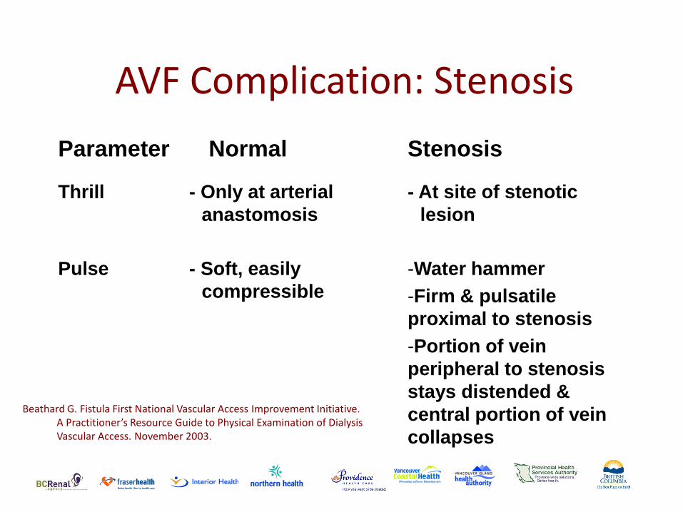
AVF Complication: Stenosis
Parameter Normal Stenosis
Thrill - Only at arterial
anastomosis
- At site of stenotic
lesion
Pulse - Soft, easily
compressible
-Water hammer
-Firm & pulsatile
proximal to stenosis
-Portion of vein
peripheral to stenosis
stays distended &
central portion of vein
collapses
Beathard G. Fistula First National Vascular Access Improvement Initiative. A Practitioner’s Resource Guide to Physical Examination of Dialysis Vascular Access. November 2003.

AVF Complication: Stenosis
Stenosis & occlusion

AVF Complication: ??
To be avoided
collateral development
Same arm: rotated Same arm: 2nd view

AVF Complication: ??
Alternative areas to cannulate
Same arm: 3rd view

AVF Complication: Stenosis
Areas of stenosis developed due to cannulation in same areas
AVF Complication: Diagnosis of Stenosis or Occlusion
Fistulogram
• Puncture fistula with small gauge needle.
• Inject contrast.
• Visualize fistula from arterial anastomosis to central veins.
• Reflux of contrast into artery during injection necessary to examine arterial anastomosis & arterial limb.
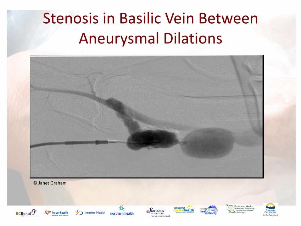
Stenosis in Basilic Vein Between Aneurysmal Dilations
© Janet Graham

Stenosis in the Cephalic Vein of a Radiocephalic AV Fistula
© Janet Graham

AVF Complication: Stenosis in Radiocephalic Fistula (Multiple Areas)
© Janet Graham
Where will you needle & why? What pressures would you expect to see from your choices?
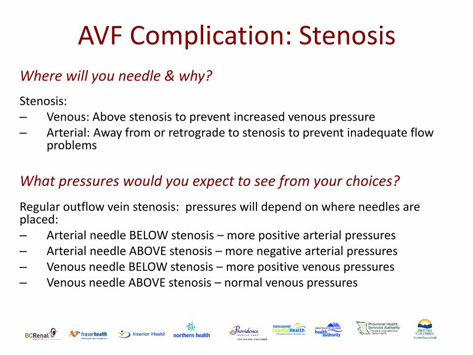
AVF Complication: Stenosis
Where will you needle & why?
Stenosis: – Venous: Above stenosis to prevent increased venous pressure – Arterial: Away from or retrograde to stenosis to prevent inadequate flow
problems
What pressures would you expect to see from your choices?
Regular outflow vein stenosis: pressures will depend on where needles are placed: – Arterial needle BELOW stenosis – more positive arterial pressures – Arterial needle ABOVE stenosis – more negative arterial pressures – Venous needle BELOW stenosis – more positive venous pressures – Venous needle ABOVE stenosis – normal venous pressures

AVF Complication: Stenosis - Management with Angioplasty
• Venous stenosis does not respond as well to angioplasty as arterial stenosis
• Guidelines recommend treatment of stenosis ≥ 50% reduction of normal vessel diameter accompanied by hemodynamic, functional or clinical abnormality
Jindal K, et al. J Am Soc Nephrol 2006;17:S16-23; National Kidney Foundation. KDOQI Clinical Practice Guidelines & Clinical Practice Recommendations 2006 Updates: Vascular Access. Am J Kidney Dis 2006;48(suppl 1):S1-S322.

AVF Complication: Stenosis – Management with Angioplasty
Short vascular sheath inserted
Normal vein proximal to stenosis or distal to post-stenotic dilatation measured
Guidewire advanced into fistula
Angioplasty balloons inflated for ~ 20 – 30 seconds
Balloon removed & another angiogram performed
If residual stenosis > 30%, angioplasty repeated with larger balloon

Fistulogram with Mapping Notes
Recommend potential arterial buttonhole sites. Try to avoid going through scar tissue.
Recommend venous buttonhole site, just below or in front of aneurysm hump.

Fistulogram Thigh AVF

Pre- & Post-angioplasty of Severe Stenosis above Arterial Anastomosis in
Radiocephalic Fistula
Areas of arterial anastomosis © Janet Graham

Endovascular Stent Placement Radiocephalic Fistula
Areas of arterial anastomosis © Janet Graham

AVF Complication: Infiltration
•Edematous upper arm. •Extensive bruising at the ACF where the first needle was attempted. This may have blown: •as the needle entered the vessel (still fragile). •as the needle was flushed with the syringe •as the blood pump was started •mid run due to patient movement or off-centre needle placement •post run with pressure rebound after site was held

AVF Complication: Infiltration

AVF Complication: Infiltration
The problem is…..?
Needle infiltration
What would expect to notice during your assessment?
• Swelling, bruising
• Complaints of pain & tenderness

AVF Complication: Needle Infiltration
Needle Infiltration with immediate bruising & swelling.

AVF Complication: Needle Infiltration What do you do?
• Hold site – including where needle tip was – with two fingers or more until hemostasis achieved.
• If suspect ‘back-blow’ infiltration, use c-clamp digital pressure.
• Apply ice immediately after hemostasis & ask patient to reapply ice at home for 20 minute periods at least once an hour for next 24 hours. Heat or warmth can be applied thereafter.
• Patient may take acetaminophen for discomfort.
• Assess access for degree of hematoma at the time of infiltration & at the next run to determine severity & level of needling skill required.
• Needle away from hematoma site until hematoma resolved.
AVF Complication: Needle Infiltration What would you expect to hear during auscultation? • Presence of bruit – low & continuous. • May be diminished or high pitched if large amount of swelling is
present.
What would you expect to feel during palpation? • Hardness (induration) over bruised area (due to infiltrated blood) -
inappropriate for cannulation.
• Vein less palpable.
What should you look for that would allow cannulation? • Presence of a thrill. • Soft compressible area appropriate for cannulation.
**Check for other nearby vessels that have developed under the influence of overall increased blood flow.**

AVF Complication: Needle Infiltration
Where will you needle & why?
1. “Above bruise” = venous.
2. “Above/ below bruise” = arterial.
Rationale:
• Cannulate away from the infiltrated site to allow for healing.
• If cannulate into or below traumatized area & infiltration occurs, will just further traumatize with possibility of causing compartment syndrome & thrombosis.
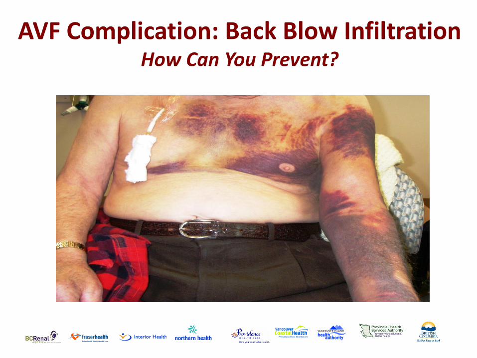
AVF Complication: Back Blow Infiltration How Can You Prevent?

AVF Complication: What is the Problem?

AVF Complication: Infection
What do you notice on assessment? • Redness • Possible edema • Discharge • Pain & tenderness

AVF Complication: Infection
How could it have happened?
• Previous needle sites have not healed
• Ineffective cleaning technique
• Improper cannulation technique
• Scratching/compromised personal hygiene (especially of fingernails)

AVF Complication: Infection
What might you expect to hear during auscultation? • May be no change in bruit if swelling is minimal • Diminished bruit due to swelling (especially if
extensive)
What might you expect to feel during palpation? • Vein may be less palpable • Tissue hardened due to swelling • Tissue warmth due to inflammatory process
AVF Complication: Infection
Where will you needle & why? IT IS NOT ADVISABLE TO NEEDLE INFECTED AVF
Check with MD – may need temporary CVC
• AVF may be salvaged with antibiotics
• a line may need to be inserted
If MD determines AVF can be needled:
• Needle away from infected area

AVF Complication: Infection
What to do? • Observe for drainage & swab for culture
• Note presence of pulses, bruit & thrill
• Document; if possible photograph
• Monitor patient temperature
• Contact doctor before needling
• Avoid infected sites when needling
• Pain management as required
• Coach patient re hygiene & access care

AVF Complication: Buttonhole Infection

AVF Complication: Buttonhole Infection
Due to Repeated Needling of Buttonhole with Sharp Needle

AVF Complications: Case Study
Returning to the case…..
Mr. Kline has been dialyzing with his fistula for 12 months with no problems.
You notice that the dynamic venous pressure at Qb 200ml/min is 200mmHg. You review the documentation on the chart & find problems.

AVF Complications: Case Study Mr. Kline’s chart reveals:
• Needling difficulties • Flashback of “black” blood • Clotted needle • Lack of supply • Hematoma formation • Frequent clotting of extracorporeal circuit • Prolonged bleeding post dialysis • Aneurysm • Trend of elevated dynamic venous & arterial pressures (over 3
consecutive runs) • A drop in access flow of > 20% from the baseline • A drop in KT/V (from 1.3 to 1.0); PRU 72% reduced to 60% • IDY of 130 @ qB of 350 mL/min; previously 180

AVF Complication: Aneurysms

AVF Complication: Aneurysm or Pseudoaneurysm?

AVF Complication: Spontaneous Rupture of an Aneurysm
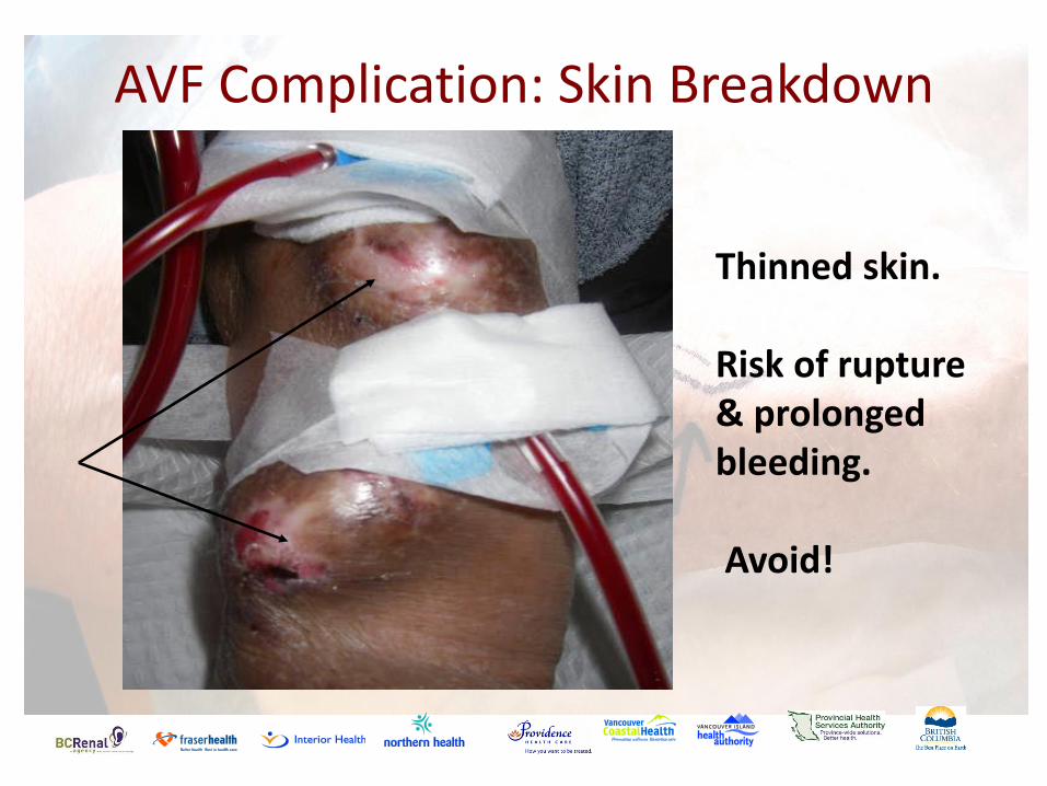
AVF Complication: Skin Breakdown
Thinned skin. Risk of rupture & prolonged bleeding. Avoid!

AVF Complication: Skin Breakdown
Avoid using Tegaderm in this area
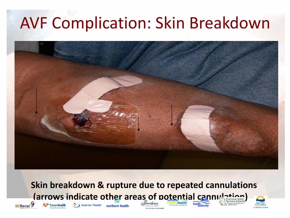
AVF Complication: Skin Breakdown
Skin breakdown & rupture due to repeated cannulations (arrows indicate other areas of potential cannulation)

AVF Complications • Needling difficulties • Flashback of “black” blood • Clotted needle • Lack of arterial supply • Vessel spasm • Hematoma formation • Frequent clotting of extracorporeal circuit • Prolonged bleeding post dialysis • Trend of elevated dynamic venous & arterial pressures (over 3
consecutive runs) • Drop in access flow of > 20% from the baseline • Increase in recirculation to 20% • Drop in values that measure clearance
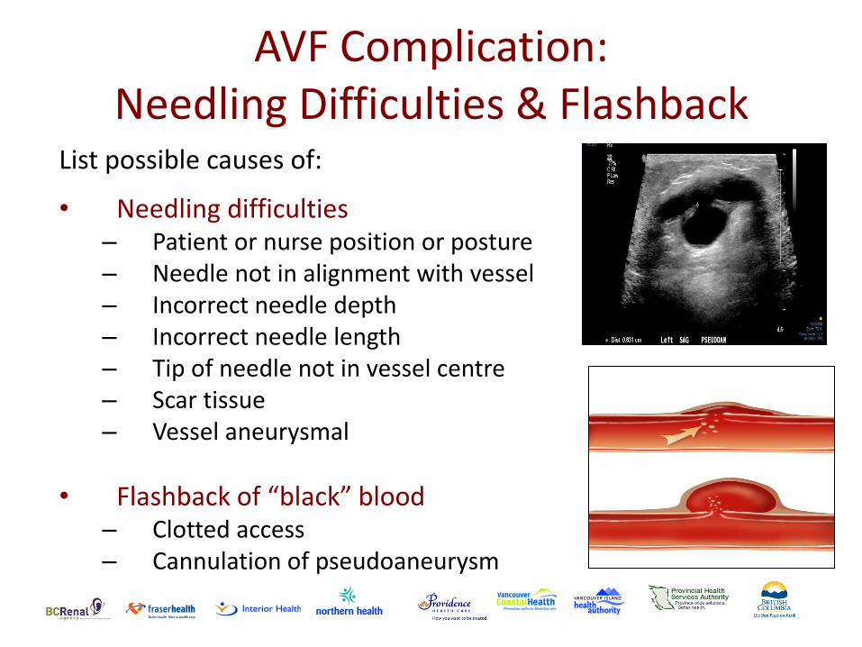
AVF Complication: Needling Difficulties & Flashback
List possible causes of:
• Needling difficulties – Patient or nurse position or posture – Needle not in alignment with vessel – Incorrect needle depth – Incorrect needle length – Tip of needle not in vessel centre – Scar tissue – Vessel aneurysmal
• Flashback of “black” blood – Clotted access – Cannulation of pseudoaneurysm

AVF Complication:
What could of caused this?
We must not forget the possibility of external injury unrelated to the patient’s time in the HD unit.

AVF Complication:
What could of caused this?
Answer: scraping the scab off a BH instead of picking the scab off

Case Study: Access Monitoring State possible causes of (cont’d):
• Clotted needle – Not in vessel – Repeated poking – Took too long to access the access – Cannulated into a pseudoaneurysm that contained clotted blood – Needle placed incorrectly in the vessel (i.e. up against the wall)
• Lack of arterial supply – Arterial stenosis – Cannulated arterial needle above a stenosis – Small vessel needle gauge too big therefore may be sucking up against
vessel wall – Needle placement incorrect – Hypotension, hypovolemia

Case Study: Access Monitoring
State possible causes for (con’t):
• Vessel spasm – No backflow upon aspiration but instills well – Patient may say it’s “vibrating”; nurse may see vibration
or feel it upon needle aspiration – Vessel doesn’t have adequate blood supply so pump is
“sucking” – Venous needle may be difficult to cannulate – Needle alignment off centre – Fragile, new, or immature vessel
• Hematoma formation – Infiltrated vessel – Inadequate hemostasis post needle removal

Case Study: Access Monitoring State possible causes for (con’t):
• Frequent clotting of extracorporeal circuit – High hemoglobin/ platelets; coagulopathy – Heparin inadequate (bolus, running, or early shut-off) – Recirculation – Arterial insufficiency – Inadequate extracorporeal prime
• Prolonged bleeding post dialysis – High INR – Heparin – Stenosis – Inadequate hemostasis – Repeated cannulations same site – Aneurysm (skin becoming very thin over aneurysm)

Module #4:
CVC Complications cont’d » Infection
» Recirculation
» Fibrin sheath
» Degradation of catheter
» Stenosis
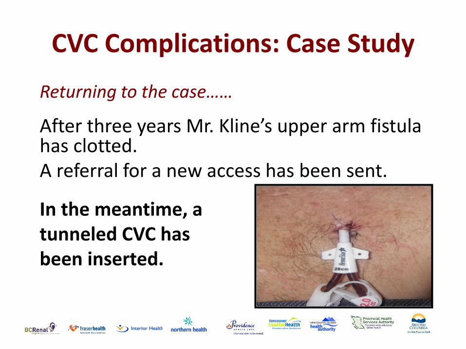
Returning to the case……
After three years Mr. Kline’s upper arm fistula has clotted. A referral for a new access has been sent.
CVC Complications: Case Study
In the meantime, a tunneled CVC has been inserted.

CVC Complications: Case Study
Pre-dialysis, the nurse has problems aspirating from Mr. Kline’s arterial lumen. The venous lumen works well.
What to do? • Reposition patient – side to side, head up - head down,
turn head side to side, pad between shoulders • Increase intrathoracic pressure – coughing or bearing
down • Assess blood pressure – volume status • Flush lines • CXR for line position • tPA – administration route: push/ pause, dwell, infusion,
pack • Review need for a line change

CVC Complication: Infection
– Febrile
– Chills & rigor
– Redness, tenderness & possibly drainage at CVC site
– Elevated WBC
– Positive cultures

CVC Complication: Infection
Infection resulted from a reaction to dressing adhesives. Blisters formed & caused erosion of tissue to the depth of the catheter. Patient has severe diabetes & was prone to related complications.
CVC Complication: Infection
What to do?
• Monitor temperature
• Give Tylenol
• Get blood cultures
• Careful hygiene & catheter care
• Keep patient comfortable – warm blankets
• Review need for catheter removal
CVC Complication: Recirculation – Increase in uremic symptoms
• Loss of appetite
• Malaise
• Weakness
• Itch
• Halitosis
• Increase in lab values
– Drop in PRU, Kt/V, IDY
CVC Complication: Recirculation
What to do?
• Rule out possible reasons:
– Kinked lines
– Thrombus formation: fibrin sheath
– Malposition
– Blood pump too aggressive
• tPA
• Review need for new catheter

CVC Complication: Thrombus
• Intraluminal
• Mural
• Fibrin tail
• Fibrin Sheath
158

CVC Complication: Thrombus (Fibrin
Sheath)
( 1 ) CVC Tip
( 2 ) Fibrin Sheath

CVC Complication: Fibrin Sheath
Dark line shows fibrin sheath
Tip of CVC
Fibrin sheath Beyond CVC

CVC Complication: Thrombus
What to do?
• tPA – push/pause method or 60 minute infusion in 1st hour of HD
• Review possibility of fibrin stripping – invasive procedure with mixed results
• Review need for new catheter

CVC Complication: Catheter Degradation
• Hub cracks
• Clamp failure
• Limb crimping
• Cuff erosion
• Cuff separation
• Cuff extrusion
• Pin holes

CVC Complication: Catheter Degradation
What to do?
Depending on the problem:
• Change clamps
• Change hubs
• Have catheter replaced
• Visually inspect exit site for cuff extrusion
• Ensure correct cleaning solution
• Be alert for unusual blood or air leaks

CVC Complication: Stenosis • Long term placement or where CVC is placed can
influence the development of upstream (central venous) stenosis.
• Stenosis may cause problems with – CVC function – Collateral vessel development – Prolonged bleeding – Venous congestion
• Treatment – angioplasty – removal of CVC if present; – ligation of affected peripheral vascular access.

CVC Complication: Stenotic Sites
SVC = 24%
BCV = 29% SCV =9%
CA-SCV= 38%

Module #5:
AV Grafts (AVGs) » AV Graft Insertion
» AV Graft Complications
» AV Graft Monitoring

AV Graft Insertion: Case Study
Returning to the case…..
• A referral was sent to the VA clinic.
• Physical examination demonstrated very few options for access creation.
• Mr. Kline was sent for vessel mapping & bilateral venograms.
• It was decided to insert a graft in his right forearm with the hopes of an upper arm cephalic vein developing in the future.

AV Graft Insertion
Source: http://www.dialysistips.com
Source: http://www.goremedical.com/images/center/vascular_access.jpg

AVG Monitoring Even if an OR report describes the arterial & venous connection, the nurse should confirm access flow with one of the following tests:
1.Block apex (referred to in literature) – Listen with a stethoscope. – Arterial segment should get louder.
2.Bubble test – Insert needles into graft. Attach male/male
adapter. – Open clamps. – Bubble will travel to venous end.
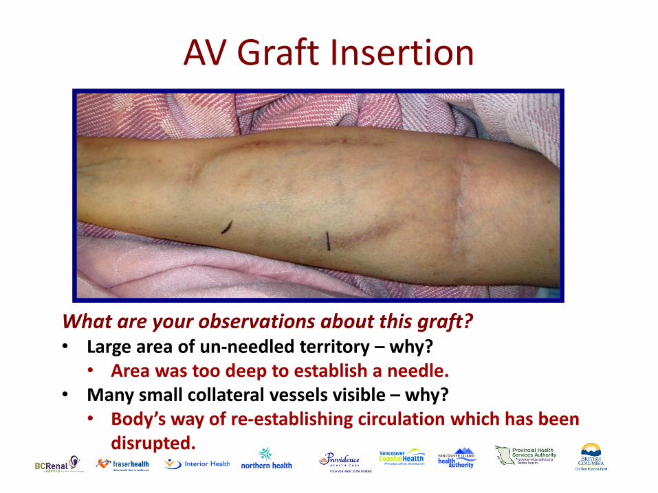
AV Graft Insertion
What are your observations about this graft? • Large area of un-needled territory – why?
• Area was too deep to establish a needle. • Many small collateral vessels visible – why?
• Body’s way of re-establishing circulation which has been disrupted.

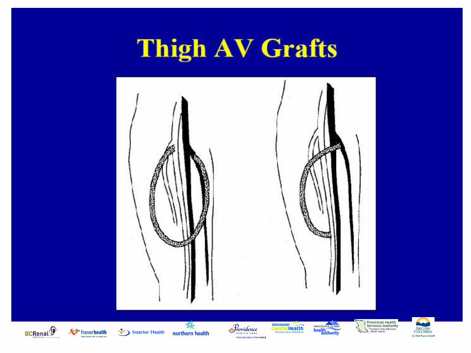
AV Graft Insertion
AV graft placement sites:
– Forearm (preferred)
– Upper arm
– Thigh
– Axillo/femoral bypass
– Axillo/axillo bypass
(necklace graft)

Case Study: AVG
Case Study: AVG

AV Graft Insertion: Bypass Graft - Brachioaxillary
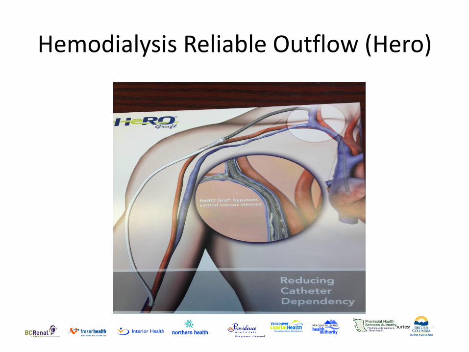
Hemodialysis Reliable Outflow (Hero)
Photo courtesy of CyoLife

Hemodialysis Reliable Outflow (Hero)
• Indications
– Limited AV access
– CVC bound
– Stenotic or occlusive central veins
Hemodialysis Reliable Outflow (Hero)
• Exclusion Criteria
– Severe peripheral vascular disease
– Chronic hypotension
Benefits
Decreased risk of infection
Decreased thrombus formation due to constant flow

Hemodialysis Reliable Outflow (Hero)
• Nursing Implications
– Bruit and thrill as per graft
– Light tourniquet to be used when cannulating

AVG Complications
• Infection (reviewed earlier)
• Stenosis (reviewed earlier)
• Pseudoaneurysm
• Degradation (destruction of graft material)
• Ischemic monomelic neuropathy

AVG Complication: What happened?

AVG Complication: Pseudoaneurysm
• Complication of AVGs. • Rupture and life
threatening hemorrhage are the most common and dangerous consequences of a pseudoaneurysm.
Source: The Internet Journal of Thoracic & Cardiovascular Surgery TM
ISSN: 1524-0274

AVG Complication: Degradation
• Long time in situ & repeated in the same area caused this graft to fail.
• One site-itis is hard to avoid in an old graft. • Eventually graft material whittles away & the fibrotic
tunnel of connective tissue becomes the shunt.

AVG Complication: Ischemic Monomelic Neuropathy
• Sudden diversion of blood supply to the nerves of the forearm & hand. – severe enough to injure nerve fibres but not
severe enough to produce necrosis.
• Condition is painful. – Analgesics are not usually very effective.
• Graft must be removed.

AVG & AVG Monitoring
When should access flows be investigated?
• A drop of >20%
• If drop is >50%, investigations should be within 48 hours
Normal • AVF 500 ml/min
• AVG 650 ml/min
Investigation • Fistulogram +/- angioplasty

AVG & AVG Monitoring
What can affect access flows?
Needling collateral/accessory vessels
Blood pressure
Cannulation attempts
Body position – lying vs sitting
185

AVG & AVG Monitoring High Cardiac Output • Access flows of over 2000ml/min and greater over an
extended period of time can have detrimental affects Patients may experience • SOB • Decrease in activities of daily living • Removal of fluid on dialysis Treatment • Decrease arterial supply • Ligation of access
186

THE END Mr. Kline experienced the full gamut of vascular accesses. He’s
had education & coaching to help him adjust to every new event involved in his journey. He weathered many complications &
challenges common to vascular accesses.
We hope Mr. Kline was eventually back to an AVF since a FISTULA is always the best choice.
This concludes our Vascular Access Workshop presentation.
We hope it has been useful for you.
We welcome all suggestions for further editions. Contact Rick Luscombe.
Related Documents