Variations in ATM Protein Expression During Normal Lymphoid Differentiation and Among B-Cell-Derived Neoplasias Jane Starczynski,* William Simmons § Joanne R. Flavell, § Phillip J. Byrd, ‡ Grant S. Stewart, ‡ Harjit S. Kullar, ‡ Alix Groom, ‡ John Crocker,* Paul A.H. Moss, ‡ Gary M. Reynolds, ¶ Meri Glavina-Durdov, A. Malcolm R. Taylor, ‡ Christopher Fegan, † Tatjana Stankovic, ‡ Paul G. Murray § From the Departments of Histopathology * and Haematology, † Birmingham Heartland’s Hospital, Birmingham, United Kingdom; the Cancer Research UK Institute for Cancer Studies, ‡ Department of Pathology, § Division of Cancer Studies, and Liver Research Laboratories, ¶ University of Birmingham, Birmingham, United Kingdom; and the Department of Pathology, University Hospital Split, Split, Croatia The ataxia telangiectasia mutated (ATM) protein plays a central role in the cellular response to DNA double- strand breaks (DSBs). Developmentally programmed DSBs are restricted to cellular subsets within lym- phoid tissues and we asked whether ATM expression is differentially regulated during lymphoid differenti- ation. We showed that immature B cells in bone mar- row and immature T cells of the thymic cortex were negative or weakly ATM-positive. T cells of thymic medulla and peripheral tissues strongly expressed ATM. High levels of ATM were present in the B lym- phocytes of the mantle zone and in plasma cells, while the majority of germinal center B cells were negative or weakly labeled. Therefore , ATM expres- sion appears to be down-regulated at those stages of lymphoid development where physiological DNA DSBs occur. In B-chronic lymphocytic leukemia and mantle cell lymphoma we observed two categories: ATM-negative tumors , most likely reflecting the pres- ence of ATM mutation, and tumors with abundant ATM expression. Most follicular center-cell lympho- mas and diffuse large B-cell lymphomas , which rarely show inactivation of the ATM gene, were negative or weakly ATM-positive. Tumor cells from most cases of Hodgkin’s disease were ATM-negative. Therefore , un- less ATM inactivation occurs , ATM expression in lym- phoid tumors is likely to reflect their cellular origin. As a result , immunostaining to identify lymphoid neoplasias with ATM inactivation might only be fea- sible for tumors derived from the stages where ATM is constitutively highly expressed. (Am J Pathol 2003, 163:423– 432) Individuals with biallelic inactivation of the ataxia telangi- ectasia mutated ( ATM) gene show a high predisposition to the development of lymphoid tumors of both B- and T-cell origin. While T-cell malignancies in A-T patients show a wide spectrum of phenotypes and include tumors of mature as well as immature T cells, B-cell tumors are derived mostly from the later stages of B-cell differentia- tion. 1 ATM is a 370-kd protein belonging to a family of PI-3 protein kinases with a role in DNA processing, regulation of the cell cycle, and control of telomere length. The principal function of the ATM protein is the integration of cellular responses to DNA double-strand breaks (DSBs). 2–4 In lymphoid tissues DNA DSBs can be cre- ated either during normal lymphoid development by the processes of V(D)J recombination, somatic hypermuta- tion, and isotype switching, 5 or are caused by extrinsic factors such as ionizing radiation (IR). ATM-dependent cellular responses to DNA DSBs include the activation of DNA repair, cell-cycle checkpoints and apoptosis, and involve a complex network of protein-protein interactions that act to prevent propagation of DNA damage and the transmission of DNA errors that could be potentially tu- morigenic for the cell. 4,6 Direct phosphorylation of p53 protein and activation of the p53 pathway is one of the crucial ATM-dependent responses and is important for the ATM-mediated activation of the G1/S checkpoint and the induction of apoptosis. 3 Human cells can process DSBs by either homology- directed or non-homologous repair pathways. Non-ho- mologous end-joining (NHEJ) repair is based on error- prone ligation of broken ends and involves the proteins Ku, DNA-PKCS, Xrcc4, and DNA ligase IV. This pathway is also used during V(D)J recombination of the immune system genes. 5 In contrast, a high-fidelity mechanism of homologous recombination (HR) between sister chroma- Supported by the Leukemia Research Fund, Cancer Research UK and the Kendall (Kay) Leukemia Fund. Accepted for publication April 15, 2003. Address reprint requests to Dr. P.G. Murray, Department of Pathology, Division of Cancer Studies, The Medical School, University of Birmingham, Edgbaston, Birmingham, B15 2TT, UK. E-mail: [email protected]. American Journal of Pathology, Vol. 163, No. 2, August 2003 Copyright © American Society for Investigative Pathology 423

Welcome message from author
This document is posted to help you gain knowledge. Please leave a comment to let me know what you think about it! Share it to your friends and learn new things together.
Transcript
Variations in ATM Protein Expression During NormalLymphoid Differentiation and Among B-Cell-DerivedNeoplasias
Jane Starczynski,* William Simmons§
Joanne R. Flavell,§ Phillip J. Byrd,‡
Grant S. Stewart,‡ Harjit S. Kullar,‡ Alix Groom,‡
John Crocker,* Paul A.H. Moss,‡
Gary M. Reynolds,¶ Meri Glavina-Durdov,�
A. Malcolm R. Taylor,‡ Christopher Fegan,†
Tatjana Stankovic,‡ Paul G. Murray§
From the Departments of Histopathology * and Haematology,†
Birmingham Heartland’s Hospital, Birmingham, United
Kingdom; the Cancer Research UK Institute for Cancer Studies,‡
Department of Pathology,§ Division of Cancer Studies, and Liver
Research Laboratories,¶ University of Birmingham, Birmingham,
United Kingdom; and the Department of Pathology,� University
Hospital Split, Split, Croatia
The ataxia telangiectasia mutated (ATM) protein playsa central role in the cellular response to DNA double-strand breaks (DSBs). Developmentally programmedDSBs are restricted to cellular subsets within lym-phoid tissues and we asked whether ATM expressionis differentially regulated during lymphoid differenti-ation. We showed that immature B cells in bone mar-row and immature T cells of the thymic cortex werenegative or weakly ATM-positive. T cells of thymicmedulla and peripheral tissues strongly expressedATM. High levels of ATM were present in the B lym-phocytes of the mantle zone and in plasma cells,while the majority of germinal center B cells werenegative or weakly labeled. Therefore, ATM expres-sion appears to be down-regulated at those stages oflymphoid development where physiological DNADSBs occur. In B-chronic lymphocytic leukemia andmantle cell lymphoma we observed two categories:ATM-negative tumors, most likely reflecting the pres-ence of ATM mutation, and tumors with abundantATM expression. Most follicular center-cell lympho-mas and diffuse large B-cell lymphomas, which rarelyshow inactivation of the ATM gene, were negative orweakly ATM-positive. Tumor cells from most cases ofHodgkin’s disease were ATM-negative. Therefore, un-less ATM inactivation occurs, ATM expression in lym-phoid tumors is likely to reflect their cellular origin.As a result, immunostaining to identify lymphoidneoplasias with ATM inactivation might only be fea-sible for tumors derived from the stages where ATM is
constitutively highly expressed. (Am J Pathol 2003,163:423–432)
Individuals with biallelic inactivation of the ataxia telangi-ectasia mutated (ATM) gene show a high predispositionto the development of lymphoid tumors of both B- andT-cell origin. While T-cell malignancies in A-T patientsshow a wide spectrum of phenotypes and include tumorsof mature as well as immature T cells, B-cell tumors arederived mostly from the later stages of B-cell differentia-tion.1
ATM is a 370-kd protein belonging to a family of PI-3protein kinases with a role in DNA processing, regulationof the cell cycle, and control of telomere length. Theprincipal function of the ATM protein is the integration ofcellular responses to DNA double-strand breaks(DSBs).2–4 In lymphoid tissues DNA DSBs can be cre-ated either during normal lymphoid development by theprocesses of V(D)J recombination, somatic hypermuta-tion, and isotype switching,5 or are caused by extrinsicfactors such as ionizing radiation (IR). ATM-dependentcellular responses to DNA DSBs include the activation ofDNA repair, cell-cycle checkpoints and apoptosis, andinvolve a complex network of protein-protein interactionsthat act to prevent propagation of DNA damage and thetransmission of DNA errors that could be potentially tu-morigenic for the cell.4,6 Direct phosphorylation of p53protein and activation of the p53 pathway is one of thecrucial ATM-dependent responses and is important forthe ATM-mediated activation of the G1/S checkpoint andthe induction of apoptosis.3
Human cells can process DSBs by either homology-directed or non-homologous repair pathways. Non-ho-mologous end-joining (NHEJ) repair is based on error-prone ligation of broken ends and involves the proteinsKu, DNA-PKCS, Xrcc4, and DNA ligase IV. This pathwayis also used during V(D)J recombination of the immunesystem genes.5 In contrast, a high-fidelity mechanism ofhomologous recombination (HR) between sister chroma-
Supported by the Leukemia Research Fund, Cancer Research UK andthe Kendall (Kay) Leukemia Fund.
Accepted for publication April 15, 2003.
Address reprint requests to Dr. P.G. Murray, Department of Pathology,Division of Cancer Studies, The Medical School, University of Birmingham,Edgbaston, Birmingham, B15 2TT, UK. E-mail: [email protected].
American Journal of Pathology, Vol. 163, No. 2, August 2003
Copyright © American Society for Investigative Pathology
423
tids is conducted by the proteins Rad51, Rad52, Rad54,and BRCA. The Nbs1/hMre11/Rad50 protein complex isimplicated in both HR and NHEJ repair pathways. ATMdirectly phosphorylates repair proteins such as Nbs1 andBRCA1, and was shown to bind directly to the sites ofDNA DSBs7 as well as V(D)J intermediates.8 Further-more, both NHEJ and HEJ repair pathways are found tobe defective in the absence of ATM.9,10 The precisemechanism of DNA repair regulation by ATM, however, isyet to be elucidated.
Earlier reports suggested that ATM expression re-mains constant during cell-cycle progression and alsoafter induction of DNA DSBs by � irradiation.11–13 In thesecircumstances ATM function appears to be determinedby regulation of its kinase activity rather than by proteinexpression.14 In contrast, a rapid change in protein ex-pression has been reported under certain conditions.ATM was found to be up-regulated in the proliferativemyoepithelium of breast ducts compared with the quies-cent myoepithelial cells.15 In addition, the level of ATMprotein dramatically increases in quiescent lymphocytesin response to mitogenic agents.16 Finally, both fibro-blasts and lymphoid cells were found to down-regulateATM in response to epidermal growth factor through amechanism that involves alteration in DNA binding activ-ity of the transcription factor Sp1.17 Accordingly, severalSp1 consensus binding sites were previously identifiedwithin the sequence of the ATM/NPAT/E14 bi-directionalpromoter.18
It has not been reported whether regulation of ATMduring normal lymphoid differentiation involves variationin ATM protein expression. Although variations in expres-sion of ATM transcripts between different tissues havebeen observed, with particularly high-level expression intissues that are frequently exposed to DNA DSBs such asspleen and thymus,19 ATM expression in specific celltypes within lymphoid tissues has not been analyzed. Toaddress this question, we studied a spectrum of humanlymphoid tissues using an antibody directed againstATM. Our findings revealed a clear difference in ATMexpression between different stages of lymphoid devel-opment. ATM was generally absent in both immature Band T cells of the bone marrow and the thymic cortex,respectively. In contrast, T lymphocytes of the thymicmedulla and the peripheral tissues generally expressedhigh levels of ATM. During B-cell differentiation high-levelexpression was observed in pre- and postgerminal cen-ter B cells, but not in germinal center B cells. Thesefindings suggest that down-regulation of ATM expressionmay be important during developmentally programmedgenomic recombinations. Because of the variations inATM expression observed during B-cell differentiation weextended our study to include an analysis of B-cell tu-mors derived from these different stages of B-cell devel-opment. Our results revealed that the majority of tumorsderived from the germinal center stages did not expressATM. In tumors derived from the stages of B-cell differenti-ation where we had previously demonstrated high-levelATM expression we observed two distinct categories: ATM-negative tumors, presumably the result of the presence ofinactivating ATM mutations, and tumors exhibiting strong
ATM expression. Our results have important implications forthe use of protein detection in the identification of tumorsharboring inactivating ATM mutations.
Materials and Methods
Tissues
ATM expression was studied in paraffin-embedded nor-mal lymphoid tissues (lymph node, thymus, spleen, bonemarrow) as well as in frozen thymus and tonsil. Paraffinwax-embedded specimens of a variety of B-lymphoidtumors, including B-cell chronic lymphocytic leukemia(B-CLL), mantle cell lymphoma (MCL), follicular centercell lymphoma (FCCL), diffuse large B-cell lymphoma(DLBCL), and classic Hodgkin’s disease (cHD) were alsoinvestigated. Five-�m paraffin wax sections were cut toVectabond-coated slides and left at 37°C for a minimumof 2 hours before being dewaxed and transferred to PBSbuffer pH 7.4. Frozen sections were cut at 6 �m to coatedslides, fixed in 10% formal saline for 20 minutes andwashed in PBS. Lymphoblastoid cell lines (LCLs) pre-pared from A-T patients and from normal donors wereused to confirm the specificity of the ATM antibody andsubsequently as controls for the ATM staining.
Production of 11G12 ATM Monoclonal Antibody
A 474-bp fragment representing amino acids 992-1144 ofthe ATM cDNA sequence was cloned in frame with thehexa-histidine tag of the vector pQE-32 (Qiagen, Craw-ley, UK) to generate the clone, designated FP8. Bulkexpression in E. coli and purification of the His-taggedATM fusion protein was performed using protocols sug-gested by the manufacturer (Qiagen). Aliquots (50 �g) ofATM fusion protein in Freund’s adjuvant were injectedinto three mice at two-week intervals for a total of eightweeks, with the fourth and final injection in the absence ofFreund’s adjuvant. Sera from the mice were tested byWestern blotting for good antibody responses to the fu-sion protein, before proceeding to monoclonal antibodyproduction. Spleen cells were fused to SP2 mouse my-eloma cells, plated out in 96-well plates and hybridomasselected in medium containing HAT (Sigma Ltd., Poole,Dorset, UK). Supernatants were tested by ELISA toidentify hybridoma clones producing antibodies to ATMfusion protein. Positive clones were expanded andsupernatants tested by Western blotting of protein cellextracts from normal and A-T individuals, to identify thosehybridomas producing monoclonal antibodies specific tothe �370-kd ATM protein (hereafter referred to as 11G12antibody).
Immunohistochemistry
The 11G12 ATM antibody was used at a dilution of 1:10 inimmunohistochemical assays and was detected by thestandard peroxidase-based Avidin Biotin Duet system(catalog number K492; Dako Corp., Ely, UK) or theChemicon IHC Select (Chemicon, Temecula, CA) fol-
424 Starczynski et alAJP August 2003, Vol. 163, No. 2
lowed by the demonstration of peroxidase activity usingthe DAB reaction. Tissue sections were microwaved in0.01 mol/L-citrate buffer (pH 6.0) for a minimum of 45minutes. In the case of the lymphoma specimens tumorswere classed as ATM-negative if fewer than 10% of tumorcells were labeled. Tissues were also analyzed by se-quential double-labeling to demonstrate ATM and arange of hematopoietic differentiation markers, includingCD3 (for T cells), CD20, CD21, and CD22 (for B cells),CD34 (for early hematopoietic cells), CD45 (leukocyteprogenitors in bone marrow), CD42b (for megakaryo-cytes), CD68 (for macrophages), CD79a (for mature Bcells), CD138 (for post-GC B cells, including plasmacells), CD235a (glycophorin A, for cells of the erythroidseries), and myeloperoxidase (for cells of the myeloidseries).
Bound antibodies were detected using Chemicon IHCSelect kit and a combination of DAB (brown) with eitherVector VIP (purple) (Vector Laboratories, Burlingame,CA) or Vector SG (gray/black). DAB was generally but notalways used in the first labeling reaction. The antigenwhich required the shorter retrieval time was labeled inthe first reaction. Controls were used to ensure non-cross-reactivity between first and second labeling stepsand these consisted of a series of consecutive sections inwhich individual antibody steps were omitted. Immuno-phenotyping of FCCL, DLBCL, and cHD was performedusing antibodies to CD10 and CD138. ATM expression inHRS cells of cHD was assessed by dual-labeling with
anti-CD30 antibodies. The presence of DSBs was dem-onstrated using antibodies against phosphorylated his-tone H2AX (�-H2AX).20
Results
ATM Antibody 11G12
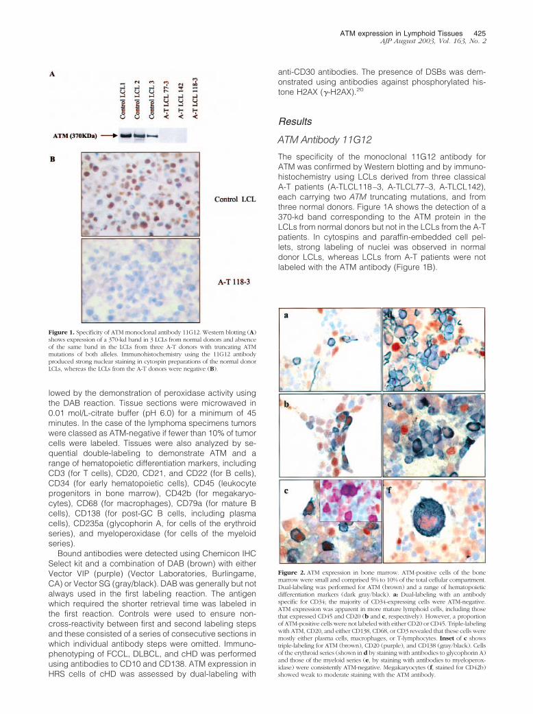
The specificity of the monoclonal 11G12 antibody forATM was confirmed by Western blotting and by immuno-histochemistry using LCLs derived from three classicalA-T patients (A-TLCL118–3, A-TLCL77–3, A-TLCL142),each carrying two ATM truncating mutations, and fromthree normal donors. Figure 1A shows the detection of a370-kd band corresponding to the ATM protein in theLCLs from normal donors but not in the LCLs from the A-Tpatients. In cytospins and paraffin-embedded cell pel-lets, strong labeling of nuclei was observed in normaldonor LCLs, whereas LCLs from A-T patients were notlabeled with the ATM antibody (Figure 1B).
Figure 1. Specificity of ATM monoclonal antibody 11G12. Western blotting (A)shows expression of a 370-kd band in 3 LCLs from normal donors and absenceof the same band in the LCLs from three A-T donors with truncating ATMmutations of both alleles. Immunohistochemistry using the 11G12 antibodyproduced strong nuclear staining in cytospin preparations of the normal donorLCLs, whereas the LCLs from the A-T donors were negative (B).
Figure 2. ATM expression in bone marrow. ATM-positive cells of the bonemarrow were small and comprised 5% to 10% of the total cellular compartment.Dual-labeling was performed for ATM (brown) and a range of hematopoieticdifferentiation markers (dark gray/black). a: Dual-labeling with an antibodyspecific for CD34; the majority of CD34-expressing cells were ATM-negative.ATM expression was apparent in more mature lymphoid cells, including thosethat expressed CD45 and CD20 (b and c, respectively). However, a proportionof ATM-positive cells were not labeled with either CD20 or CD45. Triple-labelingwith ATM, CD20, and either CD138, CD68, or CD3 revealed that these cells weremostly either plasma cells, macrophages, or T-lymphocytes. Inset of c showstriple-labeling for ATM (brown), CD20 (purple), and CD138 (gray/black). Cellsof the erythroid series (shown in d by staining with antibodies to glycophorin A)and those of the myeloid series (e, by staining with antibodies to myeloperox-idase) were consistently ATM-negative. Megakaryocytes (f, stained for CD42b)showed weak to moderate staining with the ATM antibody.
ATM expression in Lymphoid Tissues 425AJP August 2003, Vol. 163, No. 2
ATM Protein Expression in Normal LymphoidTissues
Our studies revealed that ATM-positivity in the bone mar-row was restricted to a minor subpopulation of mostlysmall cells, comprising 5% to 10% of the total cellularcompartment (Figure 2). CD34-positive cells (stem cells/pro-B cells) were mostly ATM-negative, although occa-sional cells were labeled. More mature B cells, as evi-denced by their expression of CD45 (early pro-B cells,mature B cells), CD20, CD21, or CD79a (all mature Bcells), were more frequently ATM-positive (mean num-bers of ATM-positive cells across 3 separate normalbone-marrow specimens that co-expressed B-cell mark-ers were for CD45, 72.8%; CD20, 65.3%; CD21, 35.6%;CD79a, 49.4%). Triple-labeling experiments revealedthat the majority of ATM-positive cells not expressingB-cell markers (CD20, CD21, CD79a) included plasmacells (Figure 2c, inset), macrophages, and T-lympho-cytes. Occasional CD20-, CD79a-, and CD21-expressingcells appeared to lack ATM expression although thesewere generally rare. Remarkably, developing cells of the
erythroid (as detected by expression of CD235a) andmyeloid lineages (by expression of myeloperoxidase)were almost exclusively ATM-negative. Cells of the plate-let series, including megakaryocytes, expressed moder-ate levels of ATM.
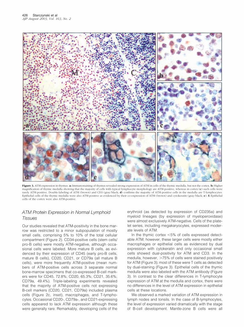
In the thymic cortex �5% of cells expressed detect-able ATM; however, these larger cells were mostly eithermacrophages or epithelial cells as evidenced by dualexpression with cytokeratin and only occasional smallcells showed dual-positivity for ATM and CD3. In themedulla, however, �75% of cells were stained positivelyfor ATM (Figure 3); most of these were T cells as detectedby dual-staining (Figure 3). Epithelial cells of the thymicmedulla were also labeled with the ATM antibody (Figure3). In contrast to the clear differences in T-lymphocyteexpression of ATM at the medulla and cortex, there wereno differences in the level of ATM expression in epithelialcells at these locations.
We observed a marked variation of ATM expression inlymph nodes and tonsils. In the case of B-lymphocytes,the level of expression varied dramatically with the stageof B-cell development. Mantle-zone B cells were all
Figure 3. ATM expression in thymus. a: Immunostaining of thymus revealed strong expression of ATM in cells of the thymic medulla, but not the cortex. b: Highermagnification of thymic medulla showing that the majority of cells with typical lymphocyte morphology are ATM-positive, whereas in cortex (c) such cells wererarely ATM-positive. Double-labeling of ATM (brown) and CD3 (gray/black; d) confirms the majority of ATM-positive cells in the medulla are T-lymphocytes.Epithelial cells of the thymic medulla were also ATM-positive as evidenced by their co-expression of ATM (brown) and cytokeratin (gray/black; e). f: Epithelialcells of the cortex were also ATM-positive.
426 Starczynski et alAJP August 2003, Vol. 163, No. 2
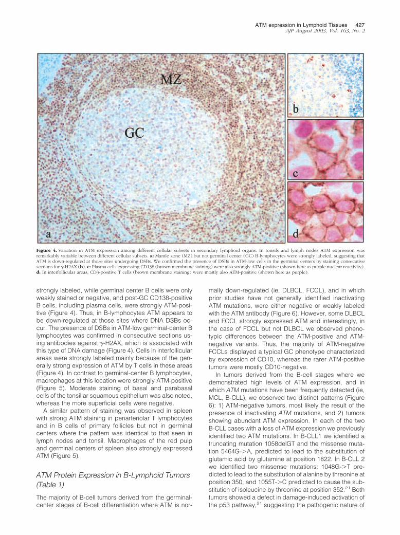
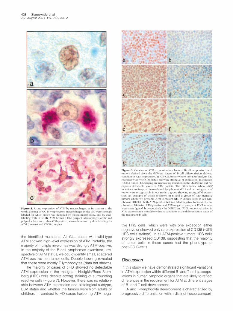
strongly labeled, while germinal center B cells were onlyweakly stained or negative, and post-GC CD138-positiveB cells, including plasma cells, were strongly ATM-posi-tive (Figure 4). Thus, in B-lymphocytes ATM appears tobe down-regulated at those sites where DNA DSBs oc-cur. The presence of DSBs in ATM-low germinal-center Blymphocytes was confirmed in consecutive sections us-ing antibodies against �-H2AX, which is associated withthis type of DNA damage (Figure 4). Cells in interfollicularareas were strongly labeled mainly because of the gen-erally strong expression of ATM by T cells in these areas(Figure 4). In contrast to germinal-center B lymphocytes,macrophages at this location were strongly ATM-positive(Figure 5). Moderate staining of basal and parabasalcells of the tonsillar squamous epithelium was also noted,whereas the more superficial cells were negative.
A similar pattern of staining was observed in spleenwith strong ATM staining in periarteriolar T lymphocytesand in B cells of primary follicles but not in germinalcenters where the pattern was identical to that seen inlymph nodes and tonsil. Macrophages of the red pulpand germinal centers of spleen also strongly expressedATM (Figure 5).
ATM Protein Expression in B-Lymphoid Tumors(Table 1)
The majority of B-cell tumors derived from the germinal-center stages of B-cell differentiation where ATM is nor-
mally down-regulated (ie, DLBCL, FCCL), and in whichprior studies have not generally identified inactivatingATM mutations, were either negative or weakly labeledwith the ATM antibody (Figure 6). However, some DLBCLand FCCL strongly expressed ATM and interestingly, inthe case of FCCL but not DLBCL we observed pheno-typic differences between the ATM-positive and ATM-negative variants. Thus, the majority of ATM-negativeFCCLs displayed a typical GC phenotype characterizedby expression of CD10, whereas the rarer ATM-positivetumors were mostly CD10-negative.
In tumors derived from the B-cell stages where wedemonstrated high levels of ATM expression, and inwhich ATM mutations have been frequently detected (ie,MCL, B-CLL), we observed two distinct patterns (Figure6): 1) ATM-negative tumors, most likely the result of thepresence of inactivating ATM mutations, and 2) tumorsshowing abundant ATM expression. In each of the twoB-CLL cases with a loss of ATM expression we previouslyidentified two ATM mutations. In B-CLL1 we identified atruncating mutation 1058delGT and the missense muta-tion 5464G-�A, predicted to lead to the substitution ofglutamic acid by glutamine at position 1822. In B-CLL 2we identified two missense mutations: 1048G-�T pre-dicted to lead to the substitution of alanine by threonine atposition 350, and 1055T-�C predicted to cause the sub-stitution of isoleucine by threonine at position 352.21 Bothtumors showed a defect in damage-induced activation ofthe p53 pathway,21 suggesting the pathogenic nature of
Figure 4. Variation in ATM expression among different cellular subsets in secondary lymphoid organs. In tonsils and lymph nodes ATM expression wasremarkably variable between different cellular subsets. a: Mantle zone (MZ) but not germinal center (GC) B-lymphocytes were strongly labeled, suggesting thatATM is down-regulated at those sites undergoing DSBs. We confirmed the presence of DSBs in ATM-low cells in the germinal centers by staining consecutivesections for �-H2AX (b). c: Plasma cells expressing CD138 (brown membrane staining) were also strongly ATM-positive (shown here as purple nuclear reactivity).d: In interfollicular areas, CD3-positive T cells (brown membrane staining) were mostly also ATM-positive (shown here as purple).
ATM expression in Lymphoid Tissues 427AJP August 2003, Vol. 163, No. 2
the identified mutations. All CLL cases with wild-typeATM showed high-level expression of ATM. Notably, themajority of multiple myelomas was strongly ATM-positive.In the majority of the B-cell lymphomas examined, irre-spective of ATM status, we could identify small, scatteredATM-positive non-tumor cells. Double-labeling revealedthat these were mostly T lymphocytes (data not shown).
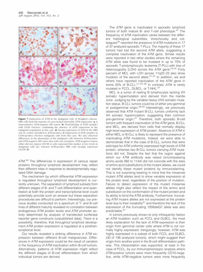
The majority of cases of cHD showed no detectableATM expression in the malignant Hodgkin/Reed-Stern-berg (HRS) cells despite strong staining of surroundingreactive cells (Figure 7). However, there was no relation-ship between ATM expression and histological subtype,EBV status and whether the tumors were from adults orchildren. In contrast to HD cases harboring ATM-nega-
tive HRS cells, which were with one exception eithernegative or showed only rare expression of CD138 (�5%HRS cells stained), in all ATM-positive tumors HRS cellsstrongly expressed CD138, suggesting that the majorityof tumor cells in these cases had the phenotype ofpost-GC B-cells.
Discussion
In this study we have demonstrated significant variationsin ATM expression within different B- and T-cell subpopu-lations in human lymphoid organs that are likely to reflectdifferences in the requirement for ATM at different stagesof B- and T-cell development.
B- and T-lymphocyte development is characterized byprogressive differentiation within distinct tissue compart-
Figure 6. Variation of ATM expression in subsets of B-cell neoplasias. B-celltumors derived from the different stages of B-cell differentiation showedvariation in ATM expression. a: A B-CLL tumor where previous analysis hadrevealed wild-type ATM status, showing strong ATM expression. In contrast,B-CLL1 tumor (b) carrying an inactivating mutation in the ATM gene did notexpress detectable levels of ATM protein. The other tumor where ATMmutations are frequent is mantle cell lymphoma (MCL) and two subgroups oftumor were recognizable in our study; a group showing strong ATM expres-sion, an example of which is shown in c, and a group of ATM-negativetumors where we presume ATM is mutant (d). In diffuse large B-cell lym-phomas (DLBCL) both ATM-positive (e) and ATM-negative tumors (f) wereobserved. Likewise, ATM-positive and ATM-negative groups of FCCL tumorswere seen (g and h, respectively). In DLBCL and FCCL tumors variation inATM expression is most likely due to variations in the differentiation status ofthe malignant B cells.
Figure 5. Strong expression of ATM by macrophages. a: In contrast to theweak labeling of GC B lymphocytes, macrophages in the GC were stronglylabeled for ATM (brown) as identified by typical morphology, and by dual-labeling with CD68 (b; ATM brown, CD68 purple). Macrophages of the redpulp of spleen were also ATM-positive, shown here in c by dual-labeling forATM (brown) and CD68 (purple).
428 Starczynski et alAJP August 2003, Vol. 163, No. 2

ments. In the case of B lymphocytes, early antigen-inde-pendent development occurs in the bone marrow and ischaracterized by stepwise gene rearrangement, involv-ing first the heavy chain D-J segments (pro-B cell stage),followed by heavy chain and then light chain V(D)J re-combination. Mature B cells exported to the peripherythen undergo a second round of proliferation if they en-counter antigen. This occurs in the germinal center andleads to the production of high-affinity antibody produc-ing plasma cells and memory B cells. Class switchingand somatic hypermutation also occur in the activated Bcells of the germinal center. In contrast, T-cell develop-ment occurs in the thymus. Pre-T cells of the thymiccortex rearrange their TCR genes and undergo positiveselection for self-MHC restriction. T cells that survive thisprocess migrate to the medulla from where they travel tothe peripheral tissues and localize to T-cell areas (inter-follicular regions of lymph nodes and periarteriolar cuffsin spleen). Unlike B cells there is no formation of anequivalent to the germinal center during T-cell differenti-ation. Our results show that ATM is expressed at lowlevels at sites where physiological DNA DSBs are re-quired and where cells are undergoing rapid prolifera-tion, ie, in germinal center B cells, in immature B cells andT cells of the bone marrow and thymic cortex, respec-tively. Interestingly, we observed high-level ATM expres-sion at both pre-germinal and postgerminal stages ofB-cell differentiation, as well as in the thymic medulla atthe locations of terminal T-cell differentiation.
One possible explanation for the variations in ATMexpression we have observed in lymphoid cells might bethe requirement for developmentally regulated cellularresponses to DNA DSBs. The alteration in ATM expres-sion might be required to prevent apoptosis in immature
lymphocytes undergoing immune system gene rear-rangements. Indeed, Bhandoola and colleagues22 re-cently reported that immature thymocytes are able totolerate DNA DSBs induced by DNA intercalating agents,whereas mature T cells are signaled to die by an ATM-dependent death mechanism. Here, we demonstrate aclear decrease in ATM expression at the locations ofimmune system gene rearrangements in lymphoid or-gans. We suggest that reduced ATM expression mayrepresent a mechanism for the down-regulation of re-sponses to DNA DSBs. Indeed, we have previouslyshown that decreased levels of ATM expression canresult in significantly reduced ATM function and impairedresponses to DNA DSBs.23 Furthermore, it has been re-ported that during lymphoid development, activation ofthe p53 pathway plays an important role in induction ofapoptosis and elimination of lymphocytes with the accu-mulation of V(D)J intermediates.24 Down-regulation ofATM in immature human B and T lymphocytes observedin this study would therefore be consistent with protectionfrom excessive apoptosis during lymphoid development.In contrast, high-level expression of ATM in pre- as wellas in postgerminal B cells and in mature T lymphocytesmay ensure that cells with disadvantageous V(D)J re-combinations, or potentially tumorigenic DNA damageare eliminated by apoptosis.
During the processing of developmentally pro-grammed DSBs, ATM co-operates with other repair pro-teins. Expression of the DSB repair protein, Nbs1, whichis involved in V(D)J recombination, is also low at the sitesof developmentally programmed DNA DSBs, both in thespleen and thymus.25 However, DNA PKcs and Ku 80show low expression in mantle zone and high expressionin germinal centers, the opposite of that observed for
Table 1. ATM Expression in B-Lymphoid Tumors
Tumor No. ATM-positive cases
Diffuse large B-cell lymphoma 11/47Follicular lymphoma 19/59
CD10-positive 7/41*CD10-negative 11/14
Mantle cell lymphoma 7/15Chronic lymphocytic leukemia† 32/42
Wild-type ATM 15/15Mutant ATM 0/2 B-CLL1 1058delGT; 5464G3A(E1822Q)
B-CLL2 G1048T(A350T); T1055C(I352T)29/31
Hodgkin’s disease (classic) 5/25CD138-positive 5/6CD138-negative 0/19
EBV status‡
(�) 1/10(�) 3/14
SubtypeMC 2/12NS 2/11Lymphocyte-rich classic HD 1/2
Age�15 years 1/615 and over 4/19
*CD10 status not determinable in four cases.†ATM status had previously been determined for these CLL tumors.‡EBV status not available for one ATM-positive case.
ATM expression in Lymphoid Tissues 429AJP August 2003, Vol. 163, No. 2
ATM.26 The differences in expression of various repairproteins throughout lymphoid development may reflecttheir different roles in response to developmentally regu-lated DNA damage.
The mechanism by which differential ATM expressionis regulated throughout lymphoid development is cur-rently unknown. The separation of lymphoid subsets fromdifferent stages of B- and T-cell differentiation and quan-titation at both the protein and transcriptional level couldpotentially provide such an answer but these separationprocedures are difficult to perform. Interestingly, our pre-vious studies conducted on a spectrum of T- and B-celllines of different maturity revealed no correlation betweenendogenous ATM protein levels and ATM promoter ac-tivity determined by analysis of transfected luciferasereporter gene constructs (unpublished data). There is apossibility, therefore, that throughout lymphoid develop-ment ATM protein expression is regulated at a posttran-scriptional level.
Our results revealed a striking difference in ATM ex-pression between different B-cell malignancies. Differ-ences in ATM expression could be the result of variationin the frequency of ATM inactivation within B-cell tumors.Alternatively, patterns of ATM expression might reflectthe different stages of B-cell differentiation from whichindividual tumors are derived.
The ATM gene is inactivated in sporadic lymphoidtumors of both mature B- and T-cell phenotype.27 Thefrequency of ATM inactivation varies between the differ-ent histological subentities. Vorechovsky and col-leagues28 reported the presence of ATM mutations in 17of 37 analyzed sporadic T-PLLs. The majority of these 17tumors had lost the second ATM allele, suggesting acomplete inactivation of the ATM gene. Similar resultswere reported in two other studies where the remainingATM allele was found to be mutated in up to 70% ofsporadic T-prolymphocytic leukemia (T-PLL) with loss ofheterozygosity (LOH) across the ATM gene.29,30 Fortypercent of MCL with LOH across 11q22–23 also showmutation of the second allele.31,32 In addition, we andothers have reported inactivation of the ATM gene insome 20% of B-CLL.21,33–36 In contrast, ATM is rarelymutated in FCCL, DLBCL, or T-NHL.37
MCL is a tumor of resting B lymphocytes lacking VHsomatic hypermutation and derived from the mantlezone. Judging by the variable heavy (VH) somatic muta-tion status, B-CLL tumors could be of either pre-germinalor postgerminal origin.38,39 Interestingly, we previouslyobserved that ATM mutant B-CLL tumors uniformly lackVH somatic hypermutation, suggesting their commonpre-germinal origin.21 Therefore, both sporadic B-celltumors with frequent inactivation of the ATM gene, B-CLLand MCL, are derived from cells that normally displayhigh-level expression of ATM protein. Absence of ATM ineither MCL or B-CLL is likely to represent the presence ofinactivating ATM mutations. Indeed, we were able todemonstrate that in the case of B-CLL, tumors that werewild-type for ATM uniformly expressed high levels of ATMprotein, whereas two B-CLL tumors carrying ATM muta-tions did not. Despite the fact that the region againstwhich our ATM antibody was raised (encompassingamino acids 992 to 1144) did not coincide with the sitesof amino acid substitutions of the mutant alleles, we couldnot identify these mutant proteins by immunostaining.This is not surprising bearing in mind that the missensemutant ATM alleles tend to show variable expression atthe protein level, regardless of the position of mutation.Failure to detect expression of the mutant missensealleles might also reflect the impact of the amino acidsubstitution on the conformation of the mutant protein andits ability to bind the ATM antibody. Furthermore, truncat-ing ATM mutant alleles are not expressed at the proteinlevel due to their instability27 and therefore the lack of theexpression of the truncating 1058delGT allele was alsopredictable.27
In tumors previously shown to only infrequently harboran ATM mutation, such as FCCL and DLBCL, the mostlikely explanation for the lack of ATM expression is theirorigin from germinal center cells where ATM is not nor-mally highly expressed. Intriguingly, however, ATM washighly expressed in a subset of both FCCL and DLBCL(30 of 106 analyzed tumors), which might suggest theirorigin from another point in the B-cell differentiation path-way. This interpretation was supported, at least in thecase of FCCL, by our immunophenotypic analysis whereATM-positive tumors were more frequently CD10-nega-tive, while ATM-negative tumors were more frequently
Figure 7. Expression of ATM in the malignant cells of Hodgkin’s disease.HRS cells from the majority of cases lacked detectable ATM expression. a: Arepresentative ATM-negative HD tumor. b: Dual-labeling of ATM (purple)with CD30 (brown), confirming the absence of ATM expression in themalignant population in this case. d: Strong expression of ATM in the HRScell of a tumor classified as ATM-positive. e: Expression of ATM (purple) inCD30-positive (brown) malignant cells from this case. We also observeddifferences in the phenotype of the malignant cells between ATM-positiveand ATM-negative HD cases; thus with one exception, ATM-negative tumorseither did not express CD138 or only expressed this marker at low levels inmalignant cells (c), whereas ATM-positive HRS cells strongly expressedCD138 (f).
430 Starczynski et alAJP August 2003, Vol. 163, No. 2
CD10-positive, although the significance of CD10 ex-pression by FCCL in relation to cell of origin has yet to beestablished. Surprisingly we found no relationship be-tween ATM and CD10 expression in DLBCL where CD10has been useful in making the distinction between theGC-derived and activated forms of DLBCL. An alternativeexplanation is that DLBCL and FCCL with high ATMexpression might have involvement of genetic factors thatderegulate ATM. We conclude that unless there is evi-dence of ATM inactivation, ATM expression in sporadiclymphoid tumors broadly reflects their cellular origin.
In Hodgkin’s disease, where a role for ATM inactivationis suggested by the increased incidence of this tumor inAT patients, low levels of ATM protein expression mightindicate the presence of inactivating mutations in theATM gene. Alternatively, it might merely reflect the originof HRS cells from a germinal center stage of B-cell dif-ferentiation. Carbone et al40 recently described variationin the extent to which tumor cells from individual cHDcases express a post-GC phenotype as assessed bytheir expression of CD138. We used immunohistochem-istry to examine CD138 expression in tumor cells from ourseries. Our finding that all of the ATM-positive tumorsstrongly expressed CD138, whereas almost all of theATM-negative tumors were either negative or showedonly low CD138 expression favors the stage of differen-tiation as a determinant of ATM expression in HRS cells.However, we did find a single tumor that strongly ex-pressed CD138 but lacked ATM expression; this mightrepresent a post-GC subgroup that carry inactivatingATM mutations. Further studies are required to confirmthis possibility.
The observations from our study have practical impli-cations for the analysis of ATM in different sporadic lym-phoid tumors. We have demonstrated remarkable varia-tions in ATM expression among the lymphoid subsets ofdifferent maturity, indicating that not only the presence ofATM mutations, but also the stage of differentiation, caninfluence ATM expression in lymphoid cells. Conse-quently, it may be difficult to detect a decrease in ATMexpression caused by ATM inactivation in tumors derivedfrom cells with constitutively low levels of ATM expres-sion. In neoplasias derived from stages with high ATMexpression, however, immunocytochemistry could pro-vide an ideal screening procedure for the detection oftumors harboring mutant ATM.
Acknowledgments
We thank Marie Smith and Pamela Edwards for help inthe collection of the pathology samples.
References
1. Taylor AMR, Metcalfe JA, Thick J, Mak Y-F: Leukaemia and lym-phoma in ataxia telangiectasia. Blood 1996, 87:423–438
2. Shiloh Y: Ataxia-telangiectasia and the Nijmegen breakagesyndrome: related disorders but genes apart. Ann Rev Genet 1997,31:635–662
3. Shiloh Y: ATM and ATR: networking cellular responses to DNA dam-age. Curr Opin Genet Dev 2001, 11:71–77
4. Kastan MB, Lin D: The many substrates and functions of ATM. NatRev 2000, 1:179–186
5. Vanasse GJ, Concannon P, Willerford DM: Regulated genomic insta-bility and neoplasia in the lymphoid lineage. Blood 1999, 94:3997–4010
6. Khanna KK, Jackson SP: DNA double strand breaks: signalling,repair and cancer connection. Nat Genet 2001, 27:247–254
7. Suzuki K, Kodama S, Watanabe M: Recruitment of ATM protein todouble strand DNA irradiated with ionizing radiation. J Biol Chem1999, 274:25571–25575
8. Perkins EJ, Nair A, Cowley DO, Van Dyke T, Chang Y, Ramsden DA:Sensing of intermediates in V(D)J recombination by ATM. Genes Dev2002, 16:159–164
9. Luo CM, Tang W, Mekeel KL, DeFrank JS, Anne PR, Powell SN: Highfrequency and error-prone DNA recombination in ataxia telangiecta-sia cell lines. J Biol Chem 1996, 271:4497–4503
10. Morrison C, Sonoda E, Takao N, Shinohara A, Yamamoto K, TakedaS: The controlling role of ATM in homologous recombinational repairof DNA damage. EMBO J 2000, 19:463–471
11. Lakin ND, Weber P, Stankovic T, Rottinghaus ST, Taylor AMR, Jack-son P: Analysis of the ATM protein in wild-type and ataxia telangiec-tasia cells. Oncogene 1996, 13:2707–2716
12. Watters D, Khanna KK, Beamish H, Birrell G, Spring K, Kedar P, GateiM, Stenzel D, Hobson K, Kozlov S, Zhang N, Farrell A, Ramsay J,Gatti R, Lavin M: Cellular localisation of the ataxia-telangiectasia(ATM) gene product and discrimination between mutated and normalforms. Oncogene 1997, 14:1911–1921
13. Brown KD, Ziv Y, Sadanandan SN, Chessa L, Collins FS, Shiloh Y,Tagle DA: The ataxia-telangiectasia gene product, a constitutivelyexpressed nuclear protein that is not up-regulated following genomedamage. Proc Natl Acad Sci USA 1997, 94:1840–1845
14. Pandita TK, Lieberman HB, Lim DS, Dhar S, Zheng W, Taya Y, KastanMB: Ionizing radiation activates the ATM kinase throughout the cellcycle. Oncogene 2000, 19:1386–1391
15. Clarke RA, Kairouz R, Watters D, Lavin MF, Kearsley JH, Lee CS:Upregulation of ATM in sclerosing adenosis of the breast. Mol Pathol1998, 51:224–226
16. Fukao T, Kaneko H, Birrell G, Gatei M, Tashita H, Yoshida T, Cross S,Kedar P, Watters D, Khana KK, Misko I, Kondo N, Lavin MF: ATM isupregulated during the mitogenic response in peripheral bloodmononuclear cells. Blood 1999, 94:1998–2006
17. Gueven N, Keating KE, Chen P, Fukao T, Khanna KK, Watters D,Rodemann PH, Lavin MF: Epidermal growth factor sensitizes cells toionizing radiation by down-regulating protein mutated in ataxia-telan-giectasia. J Biol Chem 2001, 276:8884–8891
18. Byrd PJ, Cooper PR, Stankovic T, Kullar HS, Watts GDJ, Taylor AMR:A gene transcribed from the bidirectional ATM promoter coding for aserine rich protein: amino acid sequence, structure and expressionstudies. Hum Mol Genet 1996, 5:1785–1791
19. Savitsky K, Bar-Shira A, Gilad S, Rotman G, Ziv Y, Vanagaite L, TagleDA, Smith S, Uziel T, Sfez S: A single ataxia telangiectasia gene witha product similar to PI-3 kinase. Science 1995, 268:1749–1753
20. Tomilin NV, Solovjeva LV, Svetlova MP, Pleskach NM, Zalenskaya IA,Yau PM, Bradbury EM: Visualization of focal nuclear sites of DNArepair synthesis induced by bleomycin in human cells. Radiat Res2001, 156:347–354
21. Stankovic T, Stewart G, Fegan C, Biggs P, Last J, Byrd, Moss PAH,Taylor AMR: ATM deficient B-CLL occurs in pre-germinal center cellsand results in defective damage response and unrepaired chromo-some damage. Blood 2002, 99:300–309
22. Bhandoola A, Dolnick B, Fayad N, Nussenzweig A, Singer A: Imma-ture thymocytes undergoing receptor rearrangements are resistant toan ATM-dependent death pathway activated in mature T cells bydouble-stranded DNA breaks. J Exp Med 2000, 192:891–897
23. Stewart GS, Last JIK, Stankovic T, Haites N, Byrd PJ, and Taylor AMR:Presence of residual ATM function in cells derived from A-T patientswith a less severe clinical and cellular phenotype. J Biol Chem 2001,276:30133–30141
24. Guidos CJ, Williams CJ, Grandal I, Knowles G, Huang MT, DanskaJS: V(D)J recombination activates a p53-dependent DNA damagecheckpoint in SCID lymphocyte precursors. Genes Dev 1996, 10:2038–2054
ATM expression in Lymphoid Tissues 431AJP August 2003, Vol. 163, No. 2
25. Wilda M, Demuth I, Concannon P, Sperling K, Hameister H: Expres-sion pattern of the Nijmegen breakage syndrome gene, Nbs1, duringmurine development. Hum Mol Genet 2000, 22:1739–1744
26. Moll U, Lau R, Sypes MA, Gupta MM, Anderson CW: DNA-PK, theDNA-activated protein kinase, is differentially expressed in normaland malignant human tissues. Oncogene 1999, 18:3114–3126
27. Stankovic T, Kidd AM, Sutcliffe A, McGuire GM, Robinson P, Weber P,Bedenham T, Bradwell AR, Easton DF, Lennox GG, Haites N, ByrdPJ, Taylor AMR: ATM mutations and phenotypes in ataxia-telangiec-tasia families in the Br Isles: expression of mutant ATM and the risk ofleukemia, lymphoma, and breast cancer. Am J Hum Genet 19982:334–345
28. Vorechovsky I, Luo L, Dyer MJ, Catovsky D, Amlot PL, Yaxley JC,Foroni L, Hammarstrom L, Webster AD, Yuille MA: Clustering ofmissense mutations in the ataxia telangiectasia gene in a sporadic Tcell leukaemia. Nat Genet 1997, 17:96–99
29. Stilgenbauer S, Schaffner C, Litterst A, Liebisch P, Gilad S, Bar-ShiraA, James MR, Lichter P, Dohner H: Biallelic mutations in the ATMgene in T-prolymphocytic leukaemia. Nat Med 1997, 3:1155–1159
30. Stoppa-Lyonnet D, Soulier J, Lauge A, Dastot H, Garand R, Sigaux F,Stern MH: Inactivation of the ATM gene in T-cell prolymphocyticleukaemia. Blood 1998, 91:3920–3926
31. Schaffner C, Idler I, Stilgenbaue S, Dohner H, Lichter P: Mantle celllymphoma is characterized by inactivation of the ATM gene. Proc NatlAcad Sci USA 2000, 97:2773–2778
32. Camacho E, Hernandez L, Hernadez S, Tort F, Bellosillo B, Bea S,Bosch F, Montserrat E, Cardesa A, Fernandez PL, Campo E: ATMgene inactivation in mantle cell lymphoma mainly occurs by truncat-ing mutations and missense mutations involving the phosphatidylino-sitol-3 kinase domain and is associated with increasing numbers ofchromosomal imbalances. Blood 2002, 99:238–244
33. Stankovic T, Weber P, Stewart G, Bedenham T, Murray J, Byrd PJ,Moss PA, Taylor AM: (1999) Inactivation of ataxia telangiectasiamutated gene in B-cell chronic lymphocytic leukaemia. Lancet 1999,353:26–29
34. Bullrich F, Rasio D, Kitada S, Starostik P, Kipps T, Keating M, AlbitarM, Reed JC, Croce CM: ATM mutations in B-cell chronic lymphocyticleukemia. Cancer Res 1999, 59:24–27
35. Schaffner C, Stilgenbauer S, Rappold GA, Dohner H, Lichter P:Somatic ATM mutations indicate a pathogenic role of ATM in B-cellchronic lymphocytic leukemia. Blood 1999, 94:748–753
36. Pettitt AR, Sherrington PD, Stewart G, Cawley JC, Taylor AM, Stank-ovic T: p53 dysfunction in B-cell chronic lymphocytic leukemia: inac-tivation of ATM as an alternative to TP53 mutation. Blood 2001,98:814–822
37. Gronbaek K, Worm J, Ralfkiaer E, Ahrenkiel V, Hokland P, GuldbergP: ATM mutations are associated with inactivation of the ARF-TP53tumor suppressor pathway in diffuse large B-cell lymphoma. Blood2002, 100:1430–1437
38. Hamblin TJ, Davis Z, Gardiner A, Oscier DG, Stevenson FK: Unmu-tated Ig VH genes are associated with a more aggressive form ofchronic lymphocytic leukaemia. Blood 1999, 94:1848–1854
39. Damle RN, Wasil T, Fais F, Ghiotto F, Valetto A, Allen SL, BuchbinderA, Budman D, Dittmar K, Kolitz J, Lichtman SM, Schulman P, Vin-ciguerra VP, Rai KR, Ferrarini M, Chiorazzi N: Ig V gene mutationstatus and CD38 expression as novel prognostic indicators in chroniclymphocytic leukaemia. Blood 1999, 94:1840–1847
40. Carbone A, Gloghini A, Gaidano G, Franceschi S, Capello D, DrexlerHG, Falini B, Dalla-Favera R: Expression status of BCL-6 and synde-can-1 identifies distinct histogenetic subtypes of Hodgkin’s disease.Blood 1998, 92:2220–2228
432 Starczynski et alAJP August 2003, Vol. 163, No. 2
Related Documents