Variant analysis of COVID-19 genomes T. Koyama, D. Platt & L. Parida IBM TJ Watson Research Center, 1101 Kitchawan RD., Yotktown Heights, NY 10598, USA. Correspondence to Takahiko Koyama (email: [email protected]) (Submitted: 22 February 2020 – Published online: 24 February 2020) DISCLAIMER This paper was submitted to the Bulletin of the World Health Organization and was posted to the COVID-19 open site, according to the protocol for public health emergencies for international concern as described in Vasee Moorthy et al. (http://dx.doi.org/10.2471/BLT.20.251561). The information herein is available for unrestricted use, distribution and reproduction in any medium, provided that the original work is properly cited as indicated by the Creative Commons Attribution 3.0 Intergovernmental Organizations licence (CC BY IGO 3.0). RECOMMENDED CITATION T. Koyama, D. Platt & L. Parida. Variant analysis of COVID-19 genomes. [Preprint]. Bull World Health Organ. E-pub: 24 February 2020. doi: http://dx.doi.org/10.2471/BLT.20.253591

Welcome message from author
This document is posted to help you gain knowledge. Please leave a comment to let me know what you think about it! Share it to your friends and learn new things together.
Transcript

Variant analysis of COVID-19 genomes
T. Koyama, D. Platt & L. Parida
IBM TJ Watson Research Center, 1101 Kitchawan RD., Yotktown Heights, NY 10598, USA.
Correspondence to Takahiko Koyama (email: [email protected])
(Submitted: 22 February 2020 – Published online: 24 February 2020)
DISCLAIMER
This paper was submitted to the Bulletin of the World Health Organization and was posted to the COVID-19 open site, according to the protocol for public health emergencies for international concern as described in Vasee Moorthy et al. (http://dx.doi.org/10.2471/BLT.20.251561). The information herein is available for unrestricted use, distribution and reproduction in any medium, provided that the original work is properly cited as indicated by the Creative Commons Attribution 3.0 Intergovernmental Organizations licence (CC BY IGO 3.0).
RECOMMENDED CITATION
T. Koyama, D. Platt & L. Parida. Variant analysis of COVID-19 genomes. [Preprint]. Bull World Health Organ. E-pub: 24 February 2020. doi: http://dx.doi.org/10.2471/BLT.20.253591

Abstract
In December 2019, a new coronavirus was discovered in Wuhan, China, which is officially named COVID‐
19. Within two months of the discovery of the first patient, it has now spread across China and many areas
globally. Although the fatality rate is estimated to be low, the number of deaths has already exceeded
that of SARS. There have been enormous efforts to contain the virus. Despite the lockdown of the city of
Wuhan, the virus has escaped as many people traveled for Lunar New Year. As more patients are infected
as time goes by, concerns are that the virus will accumulate more variants and that a virulent strain with
stronger toxicity might emerge. Therefore, it is critical to track and characterize them in terms of variants,
patient profiles, geographic locations, symptoms, and treatment responses. In this study, we have
collected 48 publicly available genomes from 2019‐vnCoV infected patients. 80 distinct variants were
identified with 43 missense, 21 synonymous, 3 deletion, 11 non‐coding and 2 non‐coding deletion types.
Most common variants were 28144T>C and synonymous 8782C>T in 13 samples which occur for the same
samples mostly collected outside of Wuhan. In terms of base pair changes, C>T variants occurred most
frequently in 26 distinct variants. All the coding variants were annotated with amino acid information
including cleaved non‐structured proteins within ORF1ab. Within ORF1ab, samples were bearing more
variants in NSP3 domain than other domains. BEAST analysis indicates structured transmission of this
strain, with the possibility of multiple introductions into the population.

Introduction
In late 2019, several patients with severe pneumonia were brought into hospitals across the City of Wuhan.
The virus causing the pneumonia was sequenced and it was found out that it is a strain of beta‐coronavirus
and most similarly related to SARS‐like BAT coronaviruses bat‐SL‐CoVZC45 and bat‐SL‐CoVZXC21 with 88%
similarity, 79.5% homology with SARS, and 50% with MERS(1, 2). Since then, more patients showing the
symptoms were identified in Wuhan. The Chinese Government decided to lock down the city in an effort
to contain the virus. Unfortunately, travels associated with Chinese Lunar New Year aggravated the
situation and the virus has spread across China and to many countries. Based on the US CDC, the
incubation period of the virus can be as long as 14 days. In addition to that, there appears to be many
asymptomatic or mild symptom patients or carriers who might still be transmitting the virus(3).
A study estimated the number of patients to be 75,000 as of January 25, 2020, which far exceeded the
Chinese official released number by factor of 10 at that time(4). As a virus transmitted from person to
person, as time goes by it inevitably mutates and potentially virulent strains might emerge with high
mortality rate. Currently, the mortality rate is estimated lower than that of SARS or MERS(5). Furthermore,
potential treatments and vaccines can accelerate the rate that novel mutations fix in the population with
treatment resistant substrains. Therefore, tracking of detailed demographic and clinical information as
well as substrain information is indispensable to effectively fight against COVID‐19.
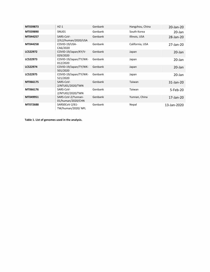
In this study, variants occurred in 48 genomes as shown in Table 1 were annotated with amino acid
changes including non‐structured proteins cleaved from ORF1ab.

Results
129 total variants were found and 80 unique variants as shown in Table 2. Among the 48 genomes we
analyzed, 10 samples did not exhibit any variants except for missing starts and end base pairs. The distinct
variants consist of 43 missense, 21 synonymous, 3 deletion, 11 non‐coding and 2 non‐coding deletion
alleles in Figure 1. Most common variants were 8782C>T(ORF1ab) and 28144T>C (ORF8) in 13 samples
followed by 29095C>T (N) in 5 samples.
The occurrences of 8782C>T and 28144T>C coincide. 29095C>T is found in the subset of them. Both
8782C>T and 29095C>T are synonymous; however, 28144T>C causes amino acid change L84S in ORF8. It
is notable that 12 out of 13 of these variant substrains are found outside of Wuhan. Whereas almost all
no variants substrain are found from samples collected in Wuhan except for MT039873.
For the 43 missense variants, 30 variants are found in ORF1ab, which is the longest ORF occupying 2/3 of
the entire genome. ORF1ab is cleaved into many nonstructural proteins (NSP1‐NSP16). Among NSP’s,
NSP3 has more variants in the analyzed samples.
All three deletions found so far took place in NSP1 of ORF1ab and they are all in‐frame deletions. All non‐
coding deletions are either located in 3’‐UTR or 5’‐UTR and do not seem to affect functions in major way.
In terms of base changes, most frequently observed one is C>T as shown in

Figure 2. Since coronaviruses are single strand RNA, we did not combine C>T with G>A mutations. There
is strong bias in transition versus transversion ratio as expected(6).
Figure 3 shows a consensus tree from BEAST(7). The resulting tree shows a coalescence center with rapid
expansion, except for one side‐branch showing relatively slower population growth, which echoes one of
the clades marked in beige in Figure 1. This group includes the first traveling family cluster showing
transmission without contact with the seafood market (MN938384, MN975262) (8). Further, this cluster
is comprised of samples collected in Japan (LC522973, LC522974, LC522975), the first US case (MN985325),
the Arizona case (MN997409), other US cases (MN994467 ‐ CA, MT044257 ‐‐ IL), a Wuhanese
(NMDC60013002‐04 = WH04 – Wuhan, not patron at seafood market), a Yunnanese (MT049951) and a
Taiwanese patient (MT066175)(1, 9). Another group includes 3 relatively closely related samples
(MN994468 – US CA, MT039890 – Korea from traveler to Wuhan, MT007544 – Australia). A slightly earlier
branch includes (LC522972 from Japan, and GWHABKG00000001 = WIV02 = MN996527 – Wuhan, no
background available). Nearly all of these show coalescence patterns suggestive of more constrained
growth, and almost all of them involve long‐distance travel, suggesting the possibility that the travelers
provided a more constrained growth of effective population size.
Some of the low variant samples identified in Figure 1 appear around the 6‐oclock area of the plot, with
some possibility of differential classification tree construction due to differences in pre‐processing.
Mutation rates estimated for SARS, MERS, and OC43 show a large range, covering a span of 0.27 to 2.38
substitutions ×10‐3 / site / year(10‐16). A value range of 0.5‐1×10‐3 is satisfactory for these preliminary
estimates. With a rate of 1×10‐3 substitutions per site per year, BEAST estimated a median tree height of
1.68 months, 95%CI of 0.92 ‐ 3.64 months, with twice those numbers at the 0.5×10‐3 rate
Methods
China’s National Genomics Data Center (NGDC) has a COVID‐19 dedicated page
(https://bigd.big.ac.cn/ncov), where links to COVID‐19 genomes are available. We have downloaded 52
publicly available genomes from Genbank, the NGDC Genome Warehouse, and the National Microbiology
Data Center (NMDC) as shown in Table 1. Among 52 genomes, GWHABKP00000001, GWHABKW00000001,
NMDC60013002‐02, and NMDC60013002‐05 were not used for the analysis due to unusually high variants
with deletions. Also, MN988713 had ambiguous base and it is converted into appropriate variants.
Likewise, NMDC60013002‐07 has non‐determined bases in 3’‐UTR region, which we ignored in the study.
NC_045512 genome sequence was used for reference and genomic coordinate in this study is based on
this reference genome. Therefore, genomic coordinates must be adjusted to compare with previous
studies, such as Lu et al. It appeared Lu et al. has 25 bp difference prior to the ribosomal slippage site and
24 bp difference after the ribosomal slippage. For instance, 8757C>T in Lu’s paper corresponds to our
8782C>T, whereas after the slippage, 24301A>G is 24325A>G.
Each genome was first aligned to NC_045512 using EMBOSS needle with a default gap penalty of 10 and
extension penalty of 0.5. Then, differences in comparison with NC_045512 were extracted to create
variants table. Based on protein annotations, nucleotide level variants were converted into amino acid
codon variants for alignments when its location within a gene was identified.

For ORF1ab, NC_045512 does not have detailed annotations for non‐structural proteins. SARS related
coronavirus genome NP_828849.2 was used(17). For each non‐structured proteins (NSP1‐NSP16), we
have aligned the protein sequences to NC_045512 ORF1ab protein sequence to create a map of non‐
structured proteins for COVID‐19 ORF1ab. ORF1ab genes has ribosomal slippage site at 13468 near the
beginning of NSP12 and caution must be placed for the map. Utilizing the map, we annotated variants in
ORF1ab region. Similarly, NSP3 was subdivided into domains: Ubl1, AC(HVR), Mac1(X), Mac2(SUD‐N),
Mac3(SUD‐M), DPUP(SUD‐C), Ubl2, PL2pro, betaSM(NAB), G2M, TM1, 3Ecto, TM2, AH1, and Y1+CoV‐Y.
We used the SARS NSP3 AAP33706.1 aligned with our COVID‐19 NSP using EMBOSS needle and
coordinates provided by Lei et al(18).
Multiple alignments were performed in NCBI COBALT after blast searches with queries of COVID‐19
proteins(19). The particular residues where mutations take places were further investigated for cross
species conservations and nature of amino acid changes. The aligned sequence was visualized using
alv(20).
EMBOSS Clustal Omega after application of Muscle was used to create the phylogenetic tree in the Figure
1(21, 22). Since many genomes have different start and end points, we have adjusted the length of
genomes. Although this approach neglects some variants which occur either in start or end such as
NMDC60013002‐04 16C>T, the genomes with no variants are gathered in the bottom of the phylogenetic
tree.
A preliminary analysis using BEAST generated a consensus view among trees of the viral phylogeny, shown
in Figure 3. Sequences were aligned with MAFFT, truncating the first 15 SNPs, and the ragged ends. 1×10⁷
MC samples were collected, assuming an HKY mutation model, with a burn‐in of 1×10⁶ iterations.
Discussion
8782C>T(ORF1ab) and 28144T>C (ORF8) were always found in pair among genomes we analyzed.
Multiple alignments with other coronavirus ORF8 sequences suggests that L84 associated with 28144T>C
(L84S) is not conserved. Interestingly, many of them were from samples collected in USA and Japan. It is
not known whether these substrains have any clinical significance from the information available at this
point.
Annotation in these NSP proteins are important since they can be targets of pharmaceutical agents like
protease inhibitors. NCBI had released new annotation for orf1ab recently. NSP6 is the only difference
and it is considered as a putative protein. Therefore, we retain our NSP annotations, for the time being.
There are 12 distinct variants in NSP3 protein in ORF1ab. NSP3 contains the papain‐like protease and is
deemed important for SARS virus virulence(23). Variants found in samples originated from Wuhan are
located in either TM1 or Y domain which are highly conserved(24). In fact, all the codon I1426, L1417,
G1433, G1716, D1761, and N1890, appeared in in GWHABKF00000001, GWHABKJ00000001,
GWHABKH00000001, GWHABKM00000001, GWHABKO00000001 and NMDC60013002‐01, are
conserved among other coronavirus as shown in

Figure 4. Other four variants M84I in MT039890, P153L in MN988713, V267F in MT039888, A358V in
LC522973, are at not conserved codons. Whereas, I789V in MT027062 and A921T in MT039888 occurred
at highly conserved codons.
Another notable thing about COVID‐19 is ORF10 which does not have any similar proteins in a huge
repository in NCBI. ORF10 is a short protein or peptide of length 38 residues. This unique protein can be
utilized to detect the virus more quickly than PCR based methods. Characterization of ORF10 functions is
strongly desired.
BEAST phylogenies give a tantalizing hint of population structuring in the evolution of COVID‐19 in the
human population. The branches with coalescence patterns most consistent with slow growth are almost
all travelers and individuals with no contact with the seafood market. The rest of the growth occurred
quite rapidly suggesting near exponential effective population growth. Curiously, not only is the slow‐
growth branch dominated by travelers, but the COVID‐19 lineages appear to be phylogenetically related
to each other, suggesting an exposure point for these individuals that is distinct from the rest of the
population.

Conclusion
The rapid increase of cases is providing more genomes that may provide some visibility and evidence of
population structure, particularly of the possibility of multiple introductions of COVID‐19 into the human
population. An understanding of the biological reservoirs carrying these viruses, and how the route to
market has been bringing them into contact with human populations will be important to understand
future risks for novel infections, whether through trade or through recreation and daily work bringing
exposure to wild environments.
This study reveals some structure in how the disease spread depending on whether the subjects were
travelers or not, with effective population size growth more constrained among the travelers. Further,
those travelers all seemed to share lineages not so typical of the rest of the patients which seem to have
experienced much more rapid effective population growth. This suggests the exposure points that the
travelers were infected through were distinct from those that generated the rapid spread through the
Wuhan population, indicating multiple introductions into the population that were differentiated early in
the spread of the disease.
There is, as of this writing, still no sign of slowdown of the COVID‐19 outbreak and number of patients
infected appears to be increasing exponentially. This fight against COVID‐19 will be a long one until we
develop vaccines or effective treatments. It is still an early stage and we have limited knowledge about
the virus. However, we believe that sharing information on variants and clinical information will be
beneficial. We should continue to be vigilant for emergence of new variants or substrains. As more
genomes released in public repositories, the variant analysis will be updated and shared.

List of Abbreviations
COVID‐19 Coronavirus disease 2019 AA Amino Acid EMBOSS The European Molecular Biology Open Software Suite BLAST Basic Local Alignment Search Tool CDC Centers of Disease Control and Prevention MERS Middle East Respiratory Syndrome NCBI National Center for Biotechnology Information NGDC National Genomics Data Center NMDC National Microbiology Data Center NSP Nonstructural Protein ORF Open Reading Frame PCR Polymerase Chain Reaction SARS Severe Acute Respiratory Syndrome
Funding The authors received no specific funding for this work.
Acknowledgements The authors are grateful to those who sequenced genomes in timely manner and deposited in public
domains.

ACCESSION SAMPLE NAME DATA SOURCE LOCATION COLLECTION DATE
GWHABKF00000001 IPBCAMS‐WH‐01 Genome Warehouse Wuhan, China 23‐Dec‐19 GWHABKG00000001 IPBCAMS‐WH‐02 Genome Warehouse Wuhan, China 30‐Dec‐19 GWHABKH00000001 IPBCAMS‐WH‐03 Genome Warehouse Wuhan, China 30‐Dec‐19 GWHABKI00000001 IPBCAMS‐WH‐04 Genome Warehouse Wuhan, China 30‐Dec‐19 GWHABKJ00000001 IPBCAMS‐WH‐05 Genome Warehouse Wuhan, China 1‐Jan‐20 GWHABKK00000001 WIV02/MN996527 Genome Warehouse Wuhan, China 30‐Dec‐19 GWHABKL00000001 WIV04/MN996528 Genome Warehouse Wuhan, China 30‐Dec‐19 GWHABKM00000001 WIV05/MN996529 Genome Warehouse Wuhan, China 30‐Dec‐19 GWHABKN00000001 WIV06/MN996530 Genome Warehouse Wuhan, China 30‐Dec‐19 GWHABKO00000001 WIV07/MN996531 Genome Warehouse Wuhan, China 30‐Dec‐19 GWHABKS00000001 20cov‐1L Genome Warehouse Hangzhou, China 20‐Jan‐20 NMDC60013002‐01 WH01 NMDC Wuhan, China 26‐Dec‐19 NMDC60013002‐03 WH03 NMDC Wuhan, China 1‐Jan‐20 NMDC60013002‐04 WH04 NMDC Wuhan, China 5‐Jan‐20 NMDC60013002‐06 WH19008 NMDC Wuhan, China 30‐Dec‐19 NMDC60013002‐07 YS8011 NMDC Wuhan, China 7‐Jan‐20 NMDC60013002‐08 WH19001 NMDC Wuhan, China 30‐Dec‐19 NMDC60013002‐09 WH19004 NMDC Wuhan, China 1‐Jan‐20 NMDC60013002‐10 WH19005 NMDC Wuhan, China 30‐Dec‐19 MN908947 Wuhan‐Hu‐1 Genbank Wuhan, China 19‐Dec MN938384 COVID‐19_HKU‐SZ‐
002a_2020 Genbank Shenzhen, China 14‐Jan‐20
MN975262 COVID‐19_HKU‐SZ‐005b_2020
Genbank Shenzhen, China 21‐Jan‐20
MN985325 COVID‐19/USA‐WA1/2020
Genbank Snohomish County, Washington, USA
19‐Jan‐20
MN988668 COVID‐19 WHU01 Genbank Wuhan, China 2‐Jan‐20 MN988669 COVID‐19 WHU02 Genbank Wuhan, China 2‐Jan‐20 MN988713 COVID‐19/USA‐IL1/2020 Genbank Chicago, Illinois, USA 21‐Jan‐20 MN994467 COVID‐19/USA‐
CA1/2020 Genbank Los Angels, California,
USA 23‐Jan‐20
MN994468 COVID‐19/USA‐CA2/2020
Genbank Orange County, California, USA
22‐Jan‐20
MN997409 COVID‐19/USA‐AZ1/2020
Genbank Phenix, Arizona, USA 22‐Jan‐20
MT007544 Australia/VIC01/2020 Genbank Clayton, Victoria, Australia
25‐Jan‐20
MT027062 COVID‐19/USA‐CA3/2020
Genbank California, USA 29‐Jan‐20
MT027063 COVID‐19/USA‐CA4/2020
Genbank California, USA 29‐Jan‐20
MT027064 COVID‐19/USA‐CA5/2020
Genbank California, USA 29‐Jan‐20
LC521925 COVID‐19/Japan/AI/I‐004/2020
Genbank Japan 20‐Jan
MT039887 COVID‐19/USA‐WI1/2020
Genbank Wisconsin, USA 31‐Jan‐20
MT039888 COVID‐19/USA‐MA1/2020
Genbank Massachussetts, USA 29‐Jan‐20
MT039873 HZ‐1 Genbank Hangzhou, China 20‐Jan‐20 MT039890 SNU01 Genbank South Korea 20‐Jan MT044257 SARS‐CoV‐
2/IL2/human/2020/USA Genbank Illinois, USA 28‐Jan‐20
MT044258 COVID‐19/USA‐CA6/2020
Genbank California, USA 27‐Jan‐20
LC522972 COVID‐19/Japan/KY/V‐029/2020
Genbank Japan 20‐Jan
LC522973 COVID‐19/Japan/TY/WK‐012/2020
Genbank Japan 20‐Jan
LC522974 COVID‐19/Japan/TY/WK‐501/2020
Genbank Japan 20‐Jan
LC522975 COVID‐19/Japan/TY/WK‐521/2020
Genbank Japan 20‐Jan
MT066175 SARS‐CoV‐2/NTU01/2020/TWN
Genbank Taiwan 31‐Jan‐20
MT066176 SARS‐CoV‐2/NTU02/2020/TWN
Genbank Taiwan 5‐Feb‐20
MT049951 SARS‐CoV‐2/Yunnan‐01/human/2020/CHN
Genbank Yunnan, China 17‐Jan‐20
MT072688 SARS0CoV‐2/61‐TW/human/2020/ NPL
Genbank Nepal 13‐Jan‐2020
Table 1. List of genomes used in the analysis.

ACCESSION GENOME CHANGE
GENE AA_CHANGE TYPE ORF1 PROTEIN
ORF1 AA_CHANGE
GWHABKF00000001 3778A>G ORF1ab
synonymous mutation
GWHABKF00000001 8388A>G ORF1ab N2708S missense nsp3‐pp1a N1890S
GWHABKF00000001 8987T>A ORF1ab F2908I missense nsp4‐pp1a F145I
GWHABKG00000001 104T>A
non‐coding
GWHABKG00000001 111T>C
non‐coding
GWHABKG00000001 112T>G
non‐coding
GWHABKG00000001 119C>G
non‐coding
GWHABKG00000001 120T>C
non‐coding
GWHABKG00000001 124G>A
non‐coding
GWHABKH00000001 6996T>C ORF1ab I2244T missense nsp3‐pp1a I1426T
GWHABKJ00000001 7866G>T ORF1ab G2534V missense nsp3‐pp1a G1716V
GWHABKK00000001 21316G>A ORF1ab D7018N missense nsp16‐pp1ab D220N
GWHABKK00000001 24325A>G S
synonymous mutation
GWHABKM00000001 21137A>G ORF1ab K6958R missense nsp16‐pp1ab K160R
GWHABKM00000001 7016G>A ORF1ab G2251S missense nsp3‐pp1a G1433S
GWHABKO00000001 8001A>C ORF1ab D2579A missense nsp3‐pp1a D1761A
GWHABKO00000001 9534C>T ORF1ab T3090I missense nsp4‐pp1a T327I
LC521925 18512C>T ORF1ab P6083L missense nsp14‐pp1ab P158L
LC521925 1912C>T ORF1ab
synonymous mutation
LC521925 359_382del ORF1ab G32_L39del deletion nsp1‐pp1a G32_L39del
LC522972 11557G>T ORF1ab E3764D missense
LC522972 15324C>T ORF1ab
synonymous mutation
LC522972 25810C>G ORF3a L140V missense
LC522972 29303C>T N P344S missense
LC522973 2662C>T ORF1ab
synonymous mutation
LC522973 28144T>C ORF8 L84S missense
LC522973 29095C>T N
synonymous mutation
LC522973 3792C>T ORF1ab A1176V missense nsp3‐pp1a A358V
LC522973 8782C>T ORF1ab
synonymous mutation
LC522974 2662C>T ORF1ab
synonymous mutation
LC522974 28144T>C ORF8 L84S missense
LC522974 29095C>T N
synonymous mutation
LC522974 8782C>T ORF1ab
synonymous mutation
LC522975 2662C>T ORF1ab
synonymous mutation
LC522975 28144T>C ORF8 L84S missense
LC522975 29095C>T N
synonymous mutation
LC522975 29705G>T
non‐coding
LC522975 8782C>T ORF1ab
synonymous mutation
MN938384 28144T>C ORF8 L84S missense
MN938384 29095C>T N
synonymous mutation
MN938384 8782C>T ORF1ab
synonymous mutation
MN975262 15607T>C ORF1ab
synonymous mutation
MN975262 28144T>C ORF8 L84S missense

MN975262 29095C>T N
synonymous mutation
MN975262 8782C>T ORF1ab
synonymous mutation
MN975262 9561C>T ORF1ab S3099L missense nsp4‐pp1a S336L
MN985325 18060C>T ORF1ab
synonymous mutation
MN985325 28144T>C ORF8 L84S missense
MN985325 8782C>T ORF1ab
synonymous mutation
MN988713 24034C>T S
synonymous mutation
MN988713 26729T>C M
synonymous mutation
MN988713 28077G>C ORF8 V62L missense
MN988713 28144T>C ORF8 L84S missense
MN988713 28854C>T N S194L missense
MN988713 3177C>T ORF1ab P971L missense nsp3‐pp1a P153L
MN988713 490T>A ORF1ab D75E missense nsp1‐pp1a D75E
MN988713 8782C>T ORF1ab
synonymous mutation
MN994467 1548G>A ORF1ab S428N missense nsp2‐pp1a S248N
MN994467 24034C>T S
synonymous mutation
MN994467 26729T>C M
synonymous mutation
MN994467 28077G>C ORF8 V62L missense
MN994467 28144T>C ORF8 L84S missense
MN994467 28792A>T N
synonymous mutation
MN994467 8782C>T ORF1ab
synonymous mutation
MN994468 17000C>T ORF1ab T5579I missense nsp13‐pp1ab T255I
MN994468 26144G>T ORF3a G251V missense
MN997409 11083G>T ORF1ab L3606F missense
MN997409 28144T>C ORF8 L84S missense
MN997409 29095C>T N
synonymous mutation
MN997409 8782C>T ORF1ab
synonymous mutation
MT007544 19065T>C ORF1ab
synonymous mutation
MT007544 22303T>G S S247R missense
MT007544 26144G>T ORF3a G251V missense
MT007544 29750_29758del
non‐coding deletion
MT027062 28854C>T N S194L missense
MT027062 5084A>G ORF1ab I1607V missense nsp3‐pp1a I789V
MT027062 614G>A ORF1ab A117T missense nsp1‐pp1a A117T
MT027063 28854C>T N S194L missense
MT027063 5084A>G ORF1ab I1607V missense nsp3‐pp1a I789V
MT027063 614G>A ORF1ab A117T missense nsp1‐pp1a A117T
MT027064 2091C>T ORF1ab T609I missense nsp2‐pp1a T429I
MT027064 21707C>T S H49Y missense
MT039887 17373C>T ORF1ab
synonymous mutation
MT039888 17423A>G ORF1ab Y5720C missense nsp13‐pp1ab Y396C
MT039888 24034C>T S
synonymous mutation
MT039888 28854C>T N S194L missense
MT039888 3518G>T ORF1ab V1085F missense nsp3‐pp1a V267F

MT039888 5480G>A ORF1ab A1739T missense nsp3‐pp1a A921T
MT039890 12115C>T ORF1ab
synonymous mutation
MT039890 15597T>C ORF1ab
synonymous mutation
MT039890 20936C>T ORF1ab T6891M missense nsp16‐pp1ab T93M
MT039890 22224C>G S S221W missense
MT039890 25775G>T ORF3a W128L missense
MT039890 26144G>T ORF3a G251V missense
MT039890 26354T>A E L37H missense
MT039890 2971G>T ORF1ab M902I missense nsp3‐pp1a M84I
MT039890 6031C>T ORF1ab
synonymous mutation
MT044257 24034C>T S
synonymous mutation
MT044257 26729T>C M
synonymous mutation
MT044257 28077G>C ORF8 V62L missense
MT044257 28144T>C ORF8 L84S missense
MT044257 3177C>T ORF1ab P971L missense nsp3‐pp1a P153L
MT044257 490T>A ORF1ab D75E missense nsp1‐pp1a D75E
MT044257 8782C>T ORF1ab
synonymous mutation
MT044258 508_522del ORF1ab G82_V86del deletion nsp1‐pp1a G82_V86del
MT044258 686_694del ORF1ab K141_F143del deletion nsp1‐pp1a K141_F143del
MT049951 11083G>C ORF1ab L3606F missense
MT049951 21644T>A S Y28N missense
MT049951 28144T>C ORF8 L84S missense
MT049951 75C>A
non‐coding
MT049951 8782C>T ORF1ab
synonymous mutation
MT066175 28144T>C ORF8 L84S missense
MT066175 8782C>T ORF1ab
synonymous mutation
MT066176 9034A>G ORF1ab
synonymous mutation
MT066176 9491C>T ORF1ab H3076Y missense nsp4‐pp1a H313Y
MT072688 24034C>T S
synonymous mutation
NMDC60013002‐01 11764T>A ORF1ab N3833K missense
NMDC60013002‐01 6968C>A ORF1ab L2235I missense nsp3‐pp1a L1417I
NMDC60013002‐04 16C>T
non‐coding
NMDC60013002‐04 28144T>C ORF8 L84S missense
NMDC60013002‐04 29854C>T
non‐coding
NMDC60013002‐04 29856T>A
non‐coding
NMDC60013002‐04 8782C>T ORF1ab
synonymous mutation
NMDC60013002‐06 24325A>G S
synonymous mutation
NMDC60013002‐07 29869del
non‐coding deletion
NMDC60013002‐09 27493C>T ORF7a P34S missense
NMDC60013002‐09 28253C>T ORF8
synonymous mutation
NMDC60013002‐10 20670G>A ORF1ab
synonymous mutation
NMDC60013002‐10 20679G>A ORF1ab
synonymous mutation
Table 2. List of variants found in COVID‐19 genomes.

References
1. Lu R, Zhao X, Li J, Niu P, Yang B, Wu H, et al. Genomic characterisation and epidemiology of 2019 novel coronavirus: implications for virus origins and receptor binding. Lancet. 2020. 2. Wu F, Zhao S, Yu B, Chen Y‐M, Wang W, Hu Y, et al. Complete genome characterisation of a novel coronavirus associated with severe human respiratory disease in Wuhan, China. bioRxiv. 2020:2020.01.24.919183. 3. Rothe C, Schunk M, Sothmann P, Bretzel G, Froeschl G, Wallrauch C, et al. Transmission of 2019‐nCoV Infection from an Asymptomatic Contact in Germany. N Engl J Med. 2020. 4. Wu JT, Leung K, Leung GM. Nowcasting and forecasting the potential domestic and international spread of the 2019‐nCoV outbreak originating in Wuhan, China: a modelling study. The Lancet. 5. Wang C, Horby PW, Hayden FG, Gao GF. A novel coronavirus outbreak of global health concern. Lancet. 2020. 6. Lyons DM, Lauring AS. Evidence for the Selective Basis of Transition‐to‐Transversion Substitution Bias in Two RNA Viruses. Mol Biol Evol. 2017;34(12):3205‐15. 7. Bouckaert R, Vaughan TG, Barido‐Sottani J, Duchêne S, Fourment M, Gavryushkina A, et al. BEAST 2.5: An advanced software platform for Bayesian evolutionary analysis. PLOS Computational Biology. 2019;15(4):e1006650. 8. Chan JF‐W, Yuan S, Kok K‐H, To KK‐W, Chu H, Yang J, et al. A familial cluster of pneumonia associated with the 2019 novel coronavirus indicating person‐to‐person transmission: a study of a family cluster. The Lancet. 9. Holshue ML, DeBolt C, Lindquist S, Lofy KH, Wiesman J, Bruce H, et al. First Case of 2019 Novel Coronavirus in the United States. New England Journal of Medicine. 2020. 10. Vijgen L, Keyaerts E, Moës E, Thoelen I, Wollants E, Lemey P, et al. Complete genomic sequence of human coronavirus OC43: molecular clock analysis suggests a relatively recent zoonotic coronavirus transmission event. Journal of virology. 2005;79(3):1595‐604. 11. Cotten M, Watson SJ, Zumla AI, Makhdoom HQ, Palser AL, Ong SH, et al. Spread, circulation, and evolution of the Middle East respiratory syndrome coronavirus. mBio. 2014;5(1):e01062‐13. 12. Cotten M, Watson SJ, Kellam P, Al‐Rabeeah AA, Makhdoom HQ, Assiri A, et al. Transmission and evolution of the Middle East respiratory syndrome coronavirus in Saudi Arabia: a descriptive genomic study. Lancet (London, England). 2013;382(9909):1993‐2002. 13. Zhao Z, Li H, Wu X, Zhong Y, Zhang K, Zhang Y‐P, et al. Moderate mutation rate in the SARS coronavirus genome and its implications. BMC Evol Biol. 2004;4:21‐. 14. Dudas G, Carvalho LM, Rambaut A, Bedford T. MERS‐CoV spillover at the camel‐human interface. Elife. 2018;7. 15. Rambaut A. Preliminary phylogenetic analysis of 11 nCoV2019 genomes, 2020‐01‐19 2020 [Available from: http://virological.org/t/preliminary‐phylogenetic‐analysis‐of‐11‐ncov2019‐genomes‐2020‐01‐19/329. 16. Rambaut A. Phylogenetic analysis of 23 nCoV‐2019 genomes, 2020‐01‐23 2020 [Available from: http://virological.org/t/phylogenetic‐analysis‐of‐23‐ncov‐2019‐genomes‐2020‐01‐23/335. 17. He R, Dobie F, Ballantine M, Leeson A, Li Y, Bastien N, et al. Analysis of multimerization of the SARS coronavirus nucleocapsid protein. Biochem Biophys Res Commun. 2004;316(2):476‐83. 18. Lei J, Kusov Y, Hilgenfeld R. Nsp3 of coronaviruses: Structures and functions of a large multi‐domain protein. Antiviral Research. 2018;149:58‐74. 19. Papadopoulos JS, Agarwala R. COBALT: constraint‐based alignment tool for multiple protein sequences. Bioinformatics. 2007;23(9):1073‐9.

20. Arvestad L. alv: a console‐based viewer for molecular sequence alignments. Journal of Open Source Software. 2018;3(31):955. 21. Sievers F, Wilm A, Dineen D, Gibson TJ, Karplus K, Li W, et al. Fast, scalable generation of high‐quality protein multiple sequence alignments using Clustal Omega. Mol Syst Biol. 2011;7:539. 22. Edgar RC. MUSCLE: a multiple sequence alignment method with reduced time and space complexity. BMC Bioinformatics. 2004;5(1):113. 23. Niemeyer D, Mosbauer K, Klein EM, Sieberg A, Mettelman RC, Mielech AM, et al. The papain‐like protease determines a virulence trait that varies among members of the SARS‐coronavirus species. PLoS Pathog. 2018;14(9):e1007296. 24. Hurst KR, Koetzner CA, Masters PS. Characterization of a critical interaction between the coronavirus nucleocapsid protein and nonstructural protein 3 of the viral replicase‐transcriptase complex. J Virol. 2013;87(16):9159‐72.

Figure Legends
Figure 1. A graphical representation of variants found in COVID‐19 genomes. Variants are colored depending on the type of mutations (missense, synonymous, non‐coding). Gene structure are displayed in the bottom. Phylogenetic tree is generated using EMBOSS Clustal Omega.
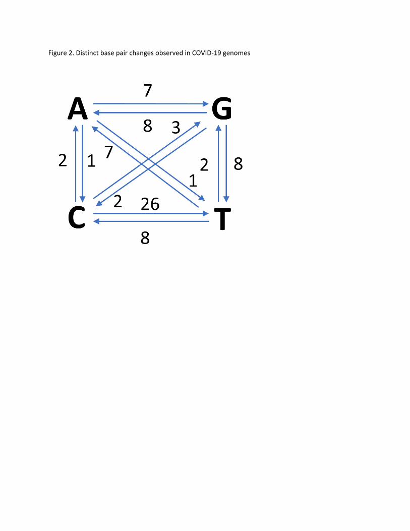
Figure 2. Distinct base pair changes observed in COVID‐19 genomes

Figure 3.BEAST consensus tree for the COVID‐19 genomes.
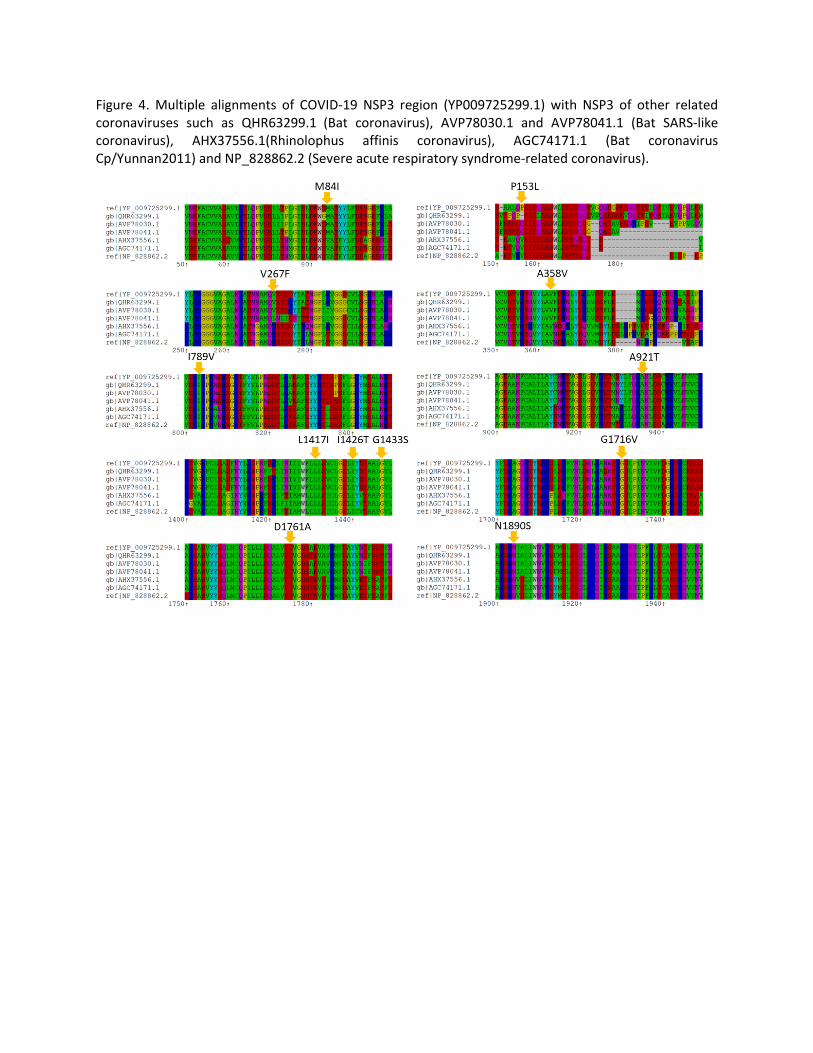
Figure 4. Multiple alignments of COVID‐19 NSP3 region (YP009725299.1) with NSP3 of other related coronaviruses such as QHR63299.1 (Bat coronavirus), AVP78030.1 and AVP78041.1 (Bat SARS‐like coronavirus), AHX37556.1(Rhinolophus affinis coronavirus), AGC74171.1 (Bat coronavirus Cp/Yunnan2011) and NP_828862.2 (Severe acute respiratory syndrome‐related coronavirus).
Related Documents


![FORMER KITCHAWAN INSTITUTE FOR SALE 712 Kitchawan Road ...€¦ · [5] A building used as a private garage for vehicle equipment storage with no more than four overhead doors. [6]](https://static.cupdf.com/doc/110x72/602ae17a61b10556d358a317/former-kitchawan-institute-for-sale-712-kitchawan-road-5-a-building-used-as.jpg)
