Biology Contribution Validation of Heat Shock Protein 70 as a Tumor-Specific Biomarker for Monitoring the Outcome of Radiation Therapy in Tumor Mouse Models Christine Bayer, PhD,* Michael E. Liebhardt, MD,* Thomas E. Schmid, PhD,* Marija Trajkovic-Arsic, PhD, z Kathrin Hube, MD,* Hanno M. Specht, MD,* Daniela Schilling, PhD,* ,y Mathias Gehrmann, PhD,* Stefan Stangl, Dipl Ing,* Jens T. Siveke, MD, z Jan J. Wilkens, DSc,* and Gabriele Multhoff, PhD* ,y *Department of Radiation Oncology, Klinikum rechts der Isar, Technische Universita¨t Mu¨nchen, Munich, Germany; y Clinical Kooperation Group, Innate Immunity in Tumor Biology, HelmholtzZentrum Mu¨nchen, Munich, Germany; and z II Medizinische Klinik, Klinikum rechts der Isar, Technische Universita¨t Mu¨nchen, Munich, Germany Received Sep 24, 2013, and in revised form Nov 4, 2013. Accepted for publication Nov 5, 2013. Summary Soluble heat shock protein 70 (sHsp70) is actively released in a tumor-specific manner. Herein we demon- strated that sHsp70 levels correlate with an increase in tumor volume during growth and monitors tumor control after curative radiation ther- apy in tumor-bearing mice. We propose sHsp70 as a predictive marker with which to monitor the clinical outcome of radiation therapy in patients. Purpose: Tumor cells, in contrast to normal cells, frequently overexpress heat shock protein 70 (Hsp70) in the cytosol, present it on their cell surface, and actively release it. Therefore, soluble Hsp70 (sHsp70) was investigated as a potential tumor biomarker for monitoring the outcome of radiation therapy. Methods and Materials: Plasma from mice bearing membrane Hsp70 (mHsp70)-positive FaDu human squamous cell carcinoma of the head and neck and spontaneous pancreatic ductal adeno- carcinoma (PDAC) was investigated. A cohort of mice with FaDu tumors (0.32 cm 3 ) was irra- diated with 30 Gy, and plasma was collected 24 hours after irradiation, after the tumors had shrunk to 50% of their starting volume and after complete remission. sHsp70 levels in the plasma were quantified by enzyme-linked immunosorbent assay. Results: sHsp70 levels were significantly higher in the blood of tumor-bearing mice than that of control animals. A correlation between increasing sHsp70 plasma levels and tumor volume in the range of 0.01 cm 3 to 0.66 cm 3 was observed. Radiation-induced regression of the tumors was associated with significantly decreased sHsp70 levels, which returned to the level of control animals after complete remission. Conclusion: We propose sHsp70 as an innovative biomarker for detecting tumors and for moni- toring the clinical outcome of radiation therapy in cancer patients. Ó 2014 Elsevier Inc. Reprint requests to: Gabriele Multhoff, PhD, Department of Radiation Oncology, Klinikum rechts der Isar, Technische Universita ¨t Mu ¨nchen, Munich, Germany. Tel: þ49-89-4140-4514; E-mail: Gabriele.multhoff@ lrz.tum.de This work was funded by the DFG cluster of excellence Munich- Centre for Advanced Photonics; the German Federal Ministry of Education and Research (BMBF, 0313909); the m4-Cluster of Excellence: Personalized Medicine (BMBF, 16EX1021C); the SFB824 (DFG); the BMBF Kompetenzverbund Strahlenforschung (03NUK007E); and the Klinikum rechts der Isar, Technische Universita ¨t Mu ¨nchen (KKF:15-06). Conflicts of interest: none. AcknowledgmentdThe authors thank Jessica Pelzel, Constantin A. Maftei, and Olga Zlobinskaya for excellent technical assistance. Int J Radiation Oncol Biol Phys, Vol. 88, No. 3, pp. 694e700, 2014 0360-3016/$ - see front matter Ó 2014 Elsevier Inc. All rights reserved. http://dx.doi.org/10.1016/j.ijrobp.2013.11.008 Radiation Oncology International Journal of biology physics www.redjournal.org

Welcome message from author
This document is posted to help you gain knowledge. Please leave a comment to let me know what you think about it! Share it to your friends and learn new things together.
Transcript
International Journal of
Radiation Oncologybiology physics
www.redjournal.org
Biology Contribution
Validation of Heat Shock Protein 70 as a Tumor-SpecificBiomarker for Monitoring the Outcome of RadiationTherapy in Tumor Mouse ModelsChristine Bayer, PhD,* Michael E. Liebhardt, MD,* Thomas E. Schmid, PhD,*Marija Trajkovic-Arsic, PhD,z Kathrin Hube, MD,* Hanno M. Specht, MD,*Daniela Schilling, PhD,*,y Mathias Gehrmann, PhD,* Stefan Stangl, Dipl Ing,*Jens T. Siveke, MD,z Jan J. Wilkens, DSc,* and Gabriele Multhoff, PhD*,y
*Department of Radiation Oncology, Klinikum rechts der Isar, Technische Universitat Munchen, Munich, Germany;yClinical Kooperation Group, Innate Immunity in Tumor Biology, HelmholtzZentrum Munchen, Munich, Germany; and zIIMedizinische Klinik, Klinikum rechts der Isar, Technische Universitat Munchen, Munich, Germany
Received Sep 24, 2013, and in revised form Nov 4, 2013. Accepted for publication Nov 5, 2013.
Summary
Soluble heat shock protein70 (sHsp70) is activelyreleased in a tumor-specificmanner. Herein we demon-strated that sHsp70 levelscorrelate with an increase intumor volume during growthand monitors tumor controlafter curative radiation ther-apy in tumor-bearing mice.We propose sHsp70 as apredictive marker with whichto monitor the clinicaloutcome of radiation therapyin patients.
Reprint requests to: Gabriele Multhoff, PhD
Oncology, Klinikum rechts der Isar, Technisc
Munich, Germany. Tel: þ49-89-4140-4514; E-
lrz.tum.de
This work was funded by the DFG clust
Centre for Advanced Photonics; the German Fed
and Research (BMBF, 0313909); the m4
Int J Radiation Oncol Biol Phys, Vol. 88, No. 3
0360-3016/$ - see front matter � 2014 Elsevie
http://dx.doi.org/10.1016/j.ijrobp.2013.11.008
Purpose: Tumor cells, in contrast to normal cells, frequently overexpress heat shock protein 70(Hsp70) in the cytosol, present it on their cell surface, and actively release it. Therefore, solubleHsp70 (sHsp70) was investigated as a potential tumor biomarker for monitoring the outcome ofradiation therapy.Methods and Materials: Plasma from mice bearing membrane Hsp70 (mHsp70)-positive FaDuhuman squamous cell carcinoma of the head and neck and spontaneous pancreatic ductal adeno-carcinoma (PDAC) was investigated. A cohort of mice with FaDu tumors (0.32 cm3) was irra-diated with 30 Gy, and plasma was collected 24 hours after irradiation, after the tumors hadshrunk to 50% of their starting volume and after complete remission. sHsp70 levels in theplasma were quantified by enzyme-linked immunosorbent assay.Results: sHsp70 levels were significantly higher in the blood of tumor-bearing mice than that ofcontrol animals. A correlation between increasing sHsp70 plasma levels and tumor volume inthe range of 0.01 cm3 to 0.66 cm3 was observed. Radiation-induced regression of the tumorswas associated with significantly decreased sHsp70 levels, which returned to the level of controlanimals after complete remission.Conclusion: We propose sHsp70 as an innovative biomarker for detecting tumors and for moni-toring the clinical outcome of radiation therapy in cancer patients. � 2014 Elsevier Inc.
, Department of Radiation
he Universitat Munchen,
mail: Gabriele.multhoff@
er of excellence Munich-
eral Ministry of Education
-Cluster of Excellence:
Personalized Medicine (BMBF, 16EX1021C); the SFB824 (DFG); the
BMBF Kompetenzverbund Strahlenforschung (03NUK007E); and the
Klinikum rechts der Isar, Technische Universitat Munchen (KKF:15-06).
Conflicts of interest: none.
AcknowledgmentdThe authors thank Jessica Pelzel, Constantin A.
Maftei, and Olga Zlobinskaya for excellent technical assistance.
, pp. 694e700, 2014
r Inc. All rights reserved.
Volume 88 � Number 3 � 2014 Hsp70 as a tumor-specific biomarker 695
Introduction
Solid tumors continue to respond heterogeneously to ionizingirradiation. There is a great medical need for tumor-specific andminimally invasive tests (1) that can assess the prognosis of apatient and monitor clinical responses. Optimal biomarkers areuseful diagnostic tools to detect tumors i high-risk populations,diagnose cancer in general, and detect specific types of cancer.Presently, approximately 20 different biomarkers are in clinicaluse (1) that reveal various degrees of sensitivity and specificity.
Intracellular heat shock 70 (Hsp70) plays a central role in theprocessing of cytosolic and secretory proteins and is involved incell proliferation, differentiation, and tumorigenesis (2). Manystudies have demonstrated that the highly stress-inducible Hsp70is frequently overexpressed in tumor cells, where it exerts anti-apoptotic functions and serves as a survival protein (4). Hsp70also has been found on the plasma membrane (mHsp70) of tumorbut not normal cells, as determined by the cmHsp70.1 monoclonalantibody (3, 5-7). On the one hand, mHsp70-positive tumors aremore resistant to chemoradiation therapy than their mHsp70-negative counterparts (8, 9). On the other hand, mHsp70 ontumor cells serves as a target structure for activated natural killercells (10, 11). Although not secreted by the canonical secretionpathway, Hsp70 has been found to be actively released from viabletumor cells (12, 13). It appears that Hsp70 is secreted from tumorcells via a nonclassic ER Z endoplasmatic reticulum-Golgiapparatus secretory route that involves lysosomal and exosomaltransport (14). Free Hsp70 in the serum also has been associatedwith cell necrosis, induced by inflammatory diseases (15), but theamount of necrosis-derived Hsp70 was significantly lower thanthat of Hsp70 actively released by viable tumor cells (C. Bayeret al, unpublished observation).
Because mHsp70-positive human and mouse tumor cells areable to secrete Hsp70 (16), we studied the impact of solubleHsp70 (sHsp70) in mice bearing human (xenograft) and murinespontaneous tumors before and after radiation therapy.
Methods and Materials
Cell lines
The established human squamous cell carcinoma of the head andneck (SCCHN) cell line FaDu was obtained from American TypeCulture Collection (Rockville, MD). FaDu cells were cultured instandard Dulbecco modified Eagle medium containing 10% fetalcalf serum (FCS) and supplements (1% nonessential amino acids,1% penicillin-streptomycin-neomycin, and 10 mM HEPES) in ahumidified incubator at 37�C and 5% CO2. Cells were regularlypassaged and tested for the absence of mycoplasma.
Flow cytometry
Single cells from fresh FaDu and pancreatic ductal adenocarci-noma (PDAC) tumors were isolated by mechanical disruption anddigestion with collagenase for 1 hour at 37�C. A sample of2 � 105 cells was washed once with 10% FCS in PBS andincubated with a fluorescein isothiocyanate (FITC)-conjugatedmouse monocolonal antibody specific for membrane-bound Hsp70(cmHsp70.1, immunoglobulin G1 [IgG1]; multimmune GmbH,Germany) or a FITC-labeled isotype-matched IgG1-negative
control antibody (code 345815; BD Biosciences, NJ) on ice inthe dark for 30 minutes. After cells were washed, propidiumiodide-negative viable cells were analyzed on a FACSCalibur flowcytometer (Becton Dickinson, Heidelberg, Germany). The per-centage of cells stained with an isotype-matched control antibodywas subtracted from the percentage of mHsp70-positive cells.
Spheroid assay
FaDu cells were seeded at 2000 cells/well in ultra-low round-bottomed 96-well attachment plates (product no. 7007; CorningLife Sciences, Lowell, MA) and allowed to form spheroids for3 days. The supernatant was then removed from 4 wells/day andfrozen at �80�C. Spheroid volumes were calculated according tothe formula (p/6) � d1 � d2, where d1 is the longest diameter, andd2 is the shortest diameter.
Enzyme-linked immunosorbent assay
Blood from mice was collected from the orbital sinus into lithium-heparin Microvettes (model no. CB 300 LH; Sarstedt, Numbrecht,Germany) after mice were sedated with isoflurane. Plasma wascentrifuged for 15 minutes at 1500 � g at 4�C, divided into ali-quots, and stored at �80�C.
Total sHsp70 levels in supernatants and plasma samples weremeasured using an Hsp70 immunoassay (Duoset, DYC1663; R&DSystems, Minneapolis, MN) according to the manufacturer’s in-structions. The enzyme-linked immunosorbent assay detectsmouse and human Hsp70.
Mice
FaDu xenograft tumorsExperiments were performed using 7- to 10-week-old femaleNaval Medicine Research Institute (nu/nu) mice (Charles River,Sulzfeld, Germany). To suppress residual immune reactions, micewere whole-body irradiated with 4 Gy (200-kV x-rays, 0.5 mm Cufilter, 1 Gy/min), using the Gulmay RS225 A device (GulmayMedical, Camberley, UK). Two to 3 days later, source tumorpieces of z1 mm3 were transplanted into the right hind legs ofanesthetized mice (intraperitoneally [i.p.], 0.50 mg/kg medeto-midin plus 5.0 mg/kg midazolam plus 0.05 mg/kg fentanyl).Immediately after transplantation, the mice were injected intra-muscularly with the respective antagonists (2.5 mg/kg atipame-zole, 0.50 mg/kg flumazenil, and 1.2 mg/kg naloxone).
Irradiation of tumorsFaDu (subcutaneous) tumors were locally irradiated with a singledose of 10, 15, 20, or 30 Gy, using a clinical linear accelerator(Varian Trilogy, CA) at 6-MV photons and a static field of1.5 � 1.5 cm2. Tumor diameters (in 3 dimensions) were measuredtwice per week by ultrasonography (Logiq-5 model; GE Health-care, Solingen, Germany), and volumes were determined by usingthe formula for a rotational ellipsoid (p/6)abc, where a, b, and care the measured diameters of the tumors.
Pancreatic ductal adenocarcinoma mouse modelThe spontaneous PDAC mouse model (Ptf1aþ/Cre;Krasþ/LSL-
G12D;p53LoxP/LoxP) used here has been described previously (17).These mice spontaneously develop rapidly growing PDAC tumors
Bayer et al. International Journal of Radiation Oncology � Biology � Physics696
that usually do not metastasize. Wild-type (wt) animals from thesame strain were used as controls. Wild-type mice were sacrificedat a mean age of 5.4 weeks and Ptf1aþ/Cre;Krasþ/LSL-
G12D;p53LoxP/LoxP mice at a mean age of 6.4 weeks. The presenceof tumor was confirmed after removing the tumor and staining thetumor sections with hematoxylin and eosin (H&E).
The mouse experiments described here were performed ac-cording to German animal welfare regulations.
Histology
The hypoxia marker pimonidazole (Hypoxyprobe, Burlington,MA) was injected i.p. at 100 mg/kg of body weight in a volume of0.1 mL of saline z1.5 hours before tumor excision, and theperfusion marker Hoechst 33,342 (Sigma, Deisenhofen, Germany)was given intravenously at 15 mg/kg of body weight in a volumeof 0.1 mL of saline 1 minute before tumor-bearing mice weresacrificed. Tumor excision followed immediately upon sacrifice.
Cryosections (10 mm) from the middle part of each tumor werefixed in cold (4�C) acetone, air dried, and rehydrated in PBSbefore staining. Pimonidazole was stained with the FITC-labeledantipimonidazole antibody (Hypoxyprobe) diluted 1:50 in primaryantibody diluent (Serotec, Oxford, U.K.) by incubating for 1 hourat 37�C in the dark (18, 19). The pimonidazole hypoxic fraction
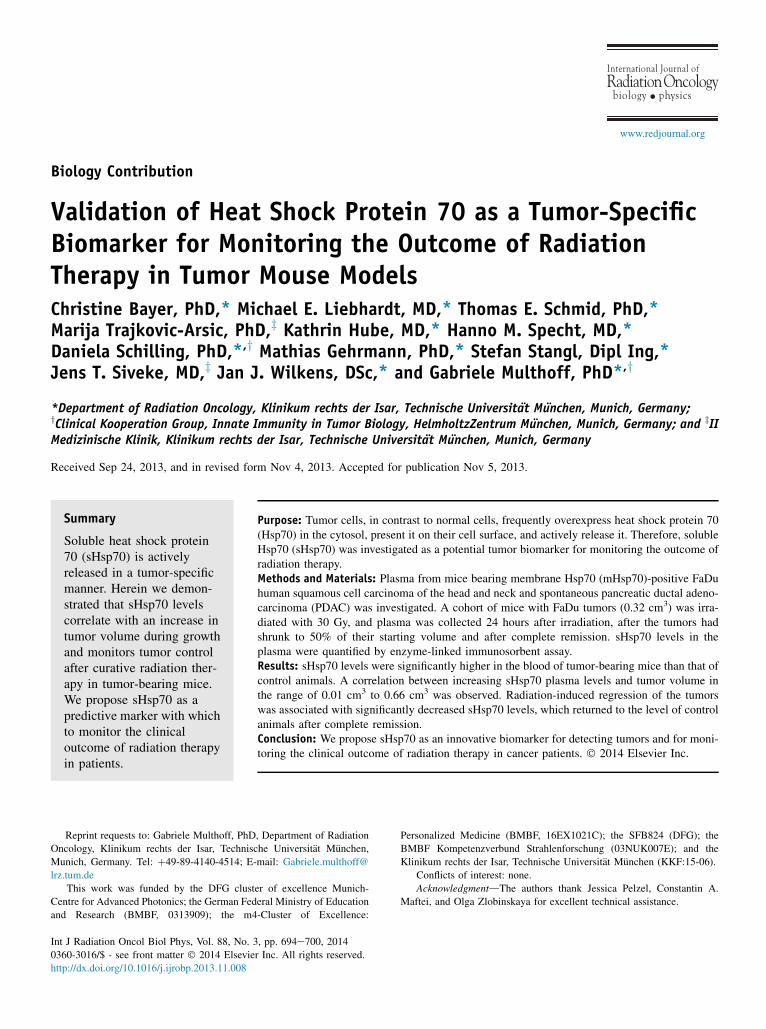
Fig. 1. (A) Representative flow cytometric analysis is shown of mHsp7panel), single-cell suspensions of FaDu xenograft tumors (middle panel)(right panel). The cells stained with the FITC-labeled IgG1 isotype-matcstained with cmHsp70.1monoclonal antibody (16) are shown in thewhiteanalyzed for their Hsp70 secretion by ELISA during growth for 2 weeks.were used to examine the relationship between variables. Abbreviatioimmunosorbent assay; FITC Z fluorescein isothiocyanate; PDAC Z pan
(pHF) was assessed by dividing the hypoxic area by the vitaltumor tissue area after staining with H&E.
Statistical analysis
Statistical analysis was performed using the statistical software forsocial sciences (SPSS, Chicago, IL). Results for the levels ofsHsp70 are means � SEM. The Pearson coefficient of correlation,r, and linear regression analysis were used to determine therelationships between variables. The Student t test was used toevaluate the relevance between tumor-bearing and non-tumor-bearing mice and between irradiated and nonirradiated mice atthe various time points after irradiation. A P value of <.05 wasconsidered statistically significant.
Results
mHsp70 expression, Hsp70 secretion from tumorcell lines, and tumor spheroids
The human FaDu SCCHN cell line shows a mHsp70-positivephenotype on approximately 50% of the cells in vitro (Fig. 1A, leftpanel). FaDu cells have the capacity to secrete Hsp70 into the cell
0 expression in viable FaDu cells from cell culture (FaDu cells, left, and primary cells of cultivated spontaneous PDAC mouse tumorshed control antibody are shown in the gray histogram, and the cellshistogram. (B) Cell culture medium from FaDu tumor spheroids wasLinear regression analysis and Pearson coefficient of correlation (r)ns: cmHsp Z cell membrane Hsp70; ELISA Z enzyme-linkedcreatic ductal adenocarcinoma; mHsp70Z membrane Hsp70.
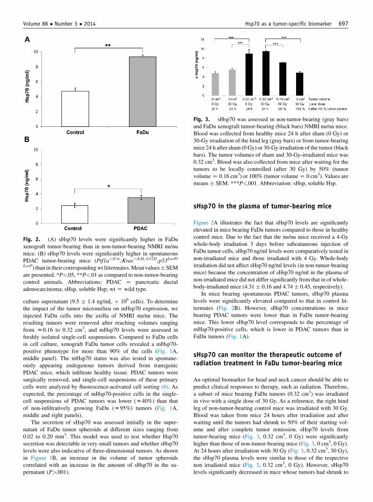
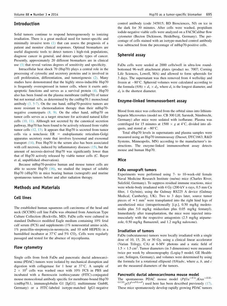
Fig. 2. (A) sHsp70 levels were significantly higher in FaDuxenograft tumor-bearing than in non-tumor-bearing NMRI nu/numice. (B) sHsp70 levels were significantly higher in spontaneousPDAC tumor-bearing mice (Ptf1aþ/Cre;Krasþ/LSL-G12D;p53LoxP/LoxP) than in their correspondingwt littermates.Meanvalues�SEMare presented. *P<.05, **P<.01 as compared to non-tumor-bearingcontrol animals. Abbreviations: PDAC Z pancreatic ductaladenocarcinoma; sHsp, soluble Hsp; wtZ wild type.
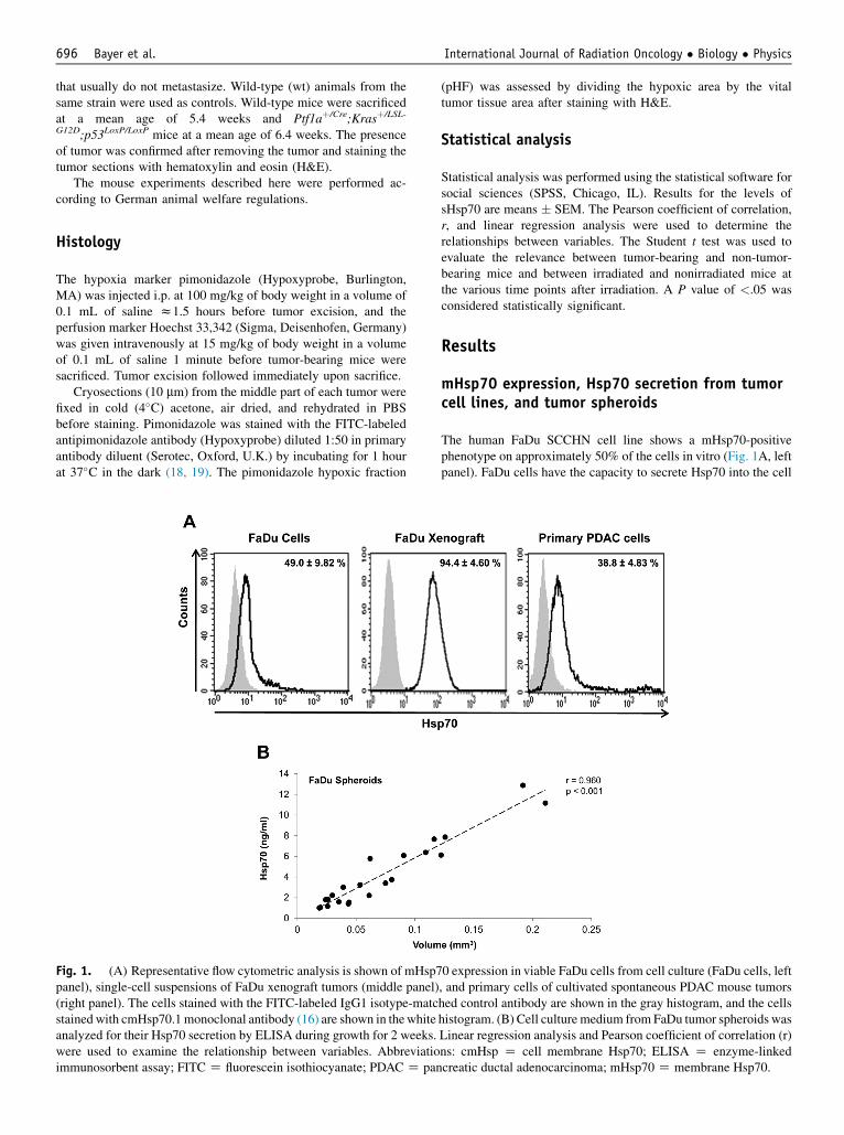
Fig. 3. sHsp70 was assessed in non-tumor-bearing (gray bars)and FaDu xenograft tumor-bearing (black bars) NMRI nu/nu mice.Blood was collected from healthy mice 24 h after sham (0 Gy) or30-Gy irradiation of the hind leg (gray bars) or from tumor-bearingmice 24 h after sham (0Gy) or 30-Gy irradiation of the tumor (blackbars). The tumor volumes of sham and 30-Gy-irradiated mice was0.32 cm3. Blood was also collected from mice after waiting for thetumors to be locally controlled (after 30 Gy) by 50% (tumorvolumeZ 0.16 cm3) or 100% (tumor volumeZ 0 cm3). Values aremeans � SEM. ***P<.001. Abbreviation: sHsp, soluble Hsp.
Volume 88 � Number 3 � 2014 Hsp70 as a tumor-specific biomarker 697
culture supernatant (9.5 � 1.4 ng/mL � 106 cells). To determinethe impact of the tumor micromilieu on mHsp70 expression, weinjected FaDu cells into the axilla of NMRI nu/nu mice. Theresulting tumors were removed after reaching volumes rangingfrom z0.16 to 0.32 cm3, and mHsp70 levels were assessed infreshly isolated single-cell suspensions. Compared to FaDu cellsin cell culture, xenograft FaDu tumor cells revealed a mHsp70-positive phenotype for more than 90% of the cells (Fig. 1A,middle panel). The mHsp70 status was also tested in spontane-ously appearing endogenous tumors derived from transgenicPDAC mice, which infiltrate healthy tissue. PDAC tumors weresurgically removed, and single-cell suspensions of these primarycells were analyzed by fluorescence-activated cell sorting (6). Asexpected, the percentage of mHsp70-positive cells in the single-cell suspensions of PDAC tumors was lower (z40%) than thatof non-infiltratively growing FaDu (z95%) tumors (Fig. 1A,middle and right panels).
The secretion of sHsp70 was assessed initially in the super-natant of FaDu tumor spheroids at different sizes ranging from0.02 to 0.20 mm3. This model was used to test whether Hsp70secretion was detectable in very small tumors and whether sHsp70levels were also indicative of three-dimensional tumors. As shownin Figure 1B, an increase in the volume of tumor spheroidscorrelated with an increase in the amount of sHsp70 in the su-pernatant (P>.001).
sHsp70 in the plasma of tumor-bearing mice
Figure 2A illustrates the fact that sHsp70 levels are significantlyelevated in mice bearing FaDu tumors compared to those in healthycontrol mice. Due to the fact that the nu/nu mice received a 4-Gywhole-body irradiation 3 days before subcutaneous injection ofFaDu tumor cells, sHsp70 ng/ml levels were comparatively tested innon-irradiated mice and those irradiated with 4 Gy. Whole-bodyirradiation did not affect sHsp70 ng/ml levels (in non-tumor-bearingmice) because the concentration of sHsp70 ng/ml in the plasma ofnon-irradiated mice did not differ significantly from that in of whole-body-irradiated mice (4.31 � 0.16 and 4.74 � 0.45, respectively).
In mice bearing spontaneous PDAC tumors, sHsp70 plasmalevels were significantly elevated compared to that in control lit-termates (Fig. 2B). However, sHsp70 concentrations in micebearing PDAC tumors were lower than in FaDu tumor-bearingmice. This lower sHsp70 level corresponds to the percentage ofmHsp70-positive cells, which is lower in PDAC tumors than inFaDu tumors (Fig. 1A).
sHsp70 can monitor the therapeutic outcome ofradiation treatment in FaDu tumor-bearing mice
An optimal biomarker for head and neck cancer should be able topredict clinical responses to therapy, such as radiation. Therefore,a subset of mice bearing FaDu tumors (0.32 cm3) was irradiatedin vivo with a single dose of 30 Gy. As a reference, the right hindleg of non-tumor-bearing control mice was irradiated with 30 Gy.Blood was taken from mice 24 hours after irradiation and afterwaiting until the tumors had shrunk to 50% of their starting vol-ume and after complete tumor remission. sHsp70 levels fromtumor-bearing mice (Fig. 3, 0.32 cm3, 0 Gy) were significantlyhigher than those of non-tumor-bearing mice (Fig. 3, 0 cm3, 0 Gy).At 24 hours after irradiation with 30 Gy (Fig. 3, 0.32 cm3, 30 Gy),the sHsp70 plasma levels were similar to those of the respectivenon irradiated mice (Fig. 3, 0.32 cm3, 0 Gy). However, sHsp70levels significantly decreased in mice whose tumors had shrunk to
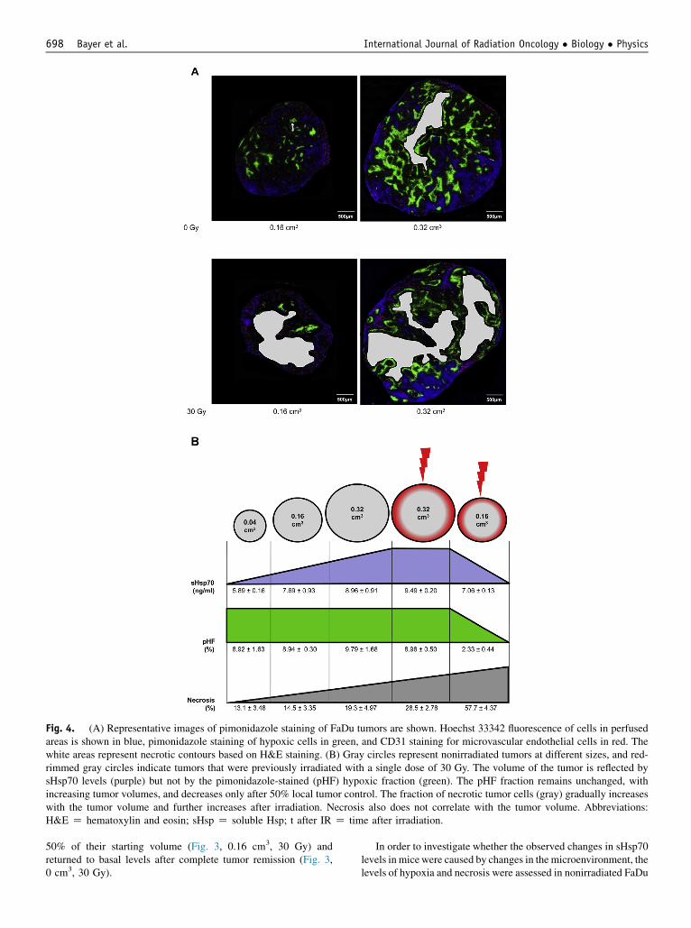
Fig. 4. (A) Representative images of pimonidazole staining of FaDu tumors are shown. Hoechst 33342 fluorescence of cells in perfusedareas is shown in blue, pimonidazole staining of hypoxic cells in green, and CD31 staining for microvascular endothelial cells in red. Thewhite areas represent necrotic contours based on H&E staining. (B) Gray circles represent nonirradiated tumors at different sizes, and red-rimmed gray circles indicate tumors that were previously irradiated with a single dose of 30 Gy. The volume of the tumor is reflected bysHsp70 levels (purple) but not by the pimonidazole-stained (pHF) hypoxic fraction (green). The pHF fraction remains unchanged, withincreasing tumor volumes, and decreases only after 50% local tumor control. The fraction of necrotic tumor cells (gray) gradually increaseswith the tumor volume and further increases after irradiation. Necrosis also does not correlate with the tumor volume. Abbreviations:H&E Z hematoxylin and eosin; sHsp Z soluble Hsp; t after IR Z time after irradiation.
Bayer et al. International Journal of Radiation Oncology � Biology � Physics698
50% of their starting volume (Fig. 3, 0.16 cm3, 30 Gy) andreturned to basal levels after complete tumor remission (Fig. 3,0 cm3, 30 Gy).
In order to investigate whether the observed changes in sHsp70levels in micewere caused by changes in the microenvironment, thelevels of hypoxia and necrosis were assessed in nonirradiated FaDu
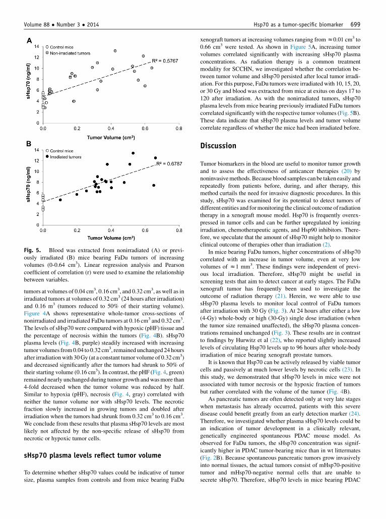
Fig. 5. Blood was extracted from nonirradiated (A) or previ-ously irradiated (B) mice bearing FaDu tumors of increasingvolumes (0-0.64 cm3). Linear regression analysis and Pearsoncoefficient of correlation (r) were used to examine the relationshipbetween variables.
Volume 88 � Number 3 � 2014 Hsp70 as a tumor-specific biomarker 699
tumors at volumes of 0.04 cm3, 0.16 cm3, and 0.32 cm3, as well as inirradiated tumors at volumes of 0.32 cm3 (24 hours after irradiation)and 0.16 m3 (tumors reduced to 50% of their starting volume).Figure 4A shows representative whole-tumor cross-sections ofnonirradiated and irradiated FaDu tumors at 0.16 cm3 and 0.32 cm3.The levels of sHsp70 were compared with hypoxic (pHF) tissue andthe percentage of necrosis within the tumors (Fig. 4B). sHsp70plasma levels (Fig. 4B, purple) steadily increased with increasingtumor volumes from0.04 to 0.32 cm3, remained unchanged 24 hoursafter irradiationwith 30Gy (at a constant tumor volumeof 0.32 cm3)and decreased significantly after the tumors had shrunk to 50% oftheir starting volume (0.16 cm3). In contrast, the pHF (Fig. 4, green)remained nearly unchanged during tumor growth andwasmore than4-fold decreased when the tumor volume was reduced by half.Similar to hypoxia (pHF), necrosis (Fig. 4, gray) correlated withneither the tumor volume nor with sHsp70 levels. The necroticfraction slowly increased in growing tumors and doubled afterirradiation when the tumors had shrunk from 0.32 cm3 to 0.16 cm3.We conclude from these results that plasma sHsp70 levels are mostlikely not affected by the non-specific release of sHsp70 fromnecrotic or hypoxic tumor cells.
sHsp70 plasma levels reflect tumor volume
To determine whether sHsp70 values could be indicative of tumorsize, plasma samples from controls and from mice bearing FaDu
xenograft tumors at increasing volumes ranging fromz0.01 cm3 to0.66 cm3 were tested. As shown in Figure 5A, increasing tumorvolumes correlated significantly with increasing sHsp70 plasmaconcentrations. As radiation therapy is a common treatmentmodality for SCCHN, we investigated whether the correlation be-tween tumor volume and sHsp70 persisted after local tumor irradi-ation. For this purpose, FaDu tumors were irradiated with 10, 15, 20,or 30 Gy and blood was extracted from mice at exitus on days 17 to120 after irradiation. As with the nonirradiated tumors, sHsp70plasma levels from mice bearing previously irradiated FaDu tumorscorrelated significantly with the respective tumor volumes (Fig. 5B).These data indicate that sHsp70 plasma levels and tumor volumecorrelate regardless of whether the mice had been irradiated before.
Discussion
Tumor biomarkers in the blood are useful to monitor tumor growthand to assess the effectiveness of anticancer therapies (20) bynoninvasivemethods.Because blood samples canbe taken easily andrepeatedly from patients before, during, and after therapy, thismethod curtails the need for invasive diagnostic procedures. In thisstudy, sHsp70 was examined for its potential to detect tumors ofdifferent entities and formonitoring the clinical outcome of radiationtherapy in a xenograft mouse model. Hsp70 is frequently overex-pressed in tumor cells and can be further upregulated by ionizingirradiation, chemotherapeutic agents, and Hsp90 inhibitors. There-fore, we speculate that the amount of sHsp70 might help to monitorclinical outcome of therapies other than irradiation (2).
In mice bearing FaDu tumors, higher concentrations of sHsp70correlated with an increase in tumor volume, even at very lowvolumes of z1 mm3. These findings were independent of previ-ous local irradiation. Therefore, sHsp70 might be useful inscreening tests that aim to detect cancer at early stages. The FaDuxenograft tumor has frequently been used to investigate theoutcome of radiation therapy (21). Herein, we were able to usesHsp70 plasma levels to monitor local control of FaDu tumorsafter irradiation with 30 Gy (Fig. 3). At 24 hours after either a low(4-Gy) whole-body or high (30-Gy) single dose irradiation (whenthe tumor size remained unaffected), the sHsp70 plasma concen-trations remained unchanged (Fig. 3). These results are in contrastto findings by Hurwitz et al (22), who reported slightly increasedlevels of circulating Hsp70 levels up to 96 hours after whole-bodyirradiation of mice bearing xenograft prostate tumors.
It is known that Hsp70 can be actively released by viable tumorcells and passively at much lower levels by necrotic cells (23). Inthis study, we demonstrated that sHsp70 levels in mice were notassociated with tumor necrosis or the hypoxic fraction of tumorsbut rather correlated with the volume of the tumor (Fig. 4B).
As pancreatic tumors are often detected only at very late stageswhen metastasis has already occurred, patients with this severedisease could benefit greatly from an early detection marker (24).Therefore, we investigated whether plasma sHsp70 levels could bean indication of tumor development in a clinically relevant,genetically engineered spontaneous PDAC mouse model. Asobserved for FaDu tumors, the sHsp70 concentration was signif-icantly higher in PDAC tumor-bearing mice than in wt littermates(Fig. 2B). Because spontaneous pancreatic tumors grow invasivelyinto normal tissues, the actual tumors consist of mHsp70-positivetumor and mHsp70-negative normal cells that are unable tosecrete sHsp70. Therefore, sHsp70 levels in mice bearing PDAC
Bayer et al. International Journal of Radiation Oncology � Biology � Physics700
tumors were lower than those of FaDu tumor-bearing mice. In linewith this finding, the expression levels of mHsp70 in PDACtumors were lower than those in FaDu tumors (Fig. 1A).
Conclusions
Extensive evidence shows that Hsp70 levels are elevated in tumortissue from a variety of entities and correlate with poor prognosis inbreast, endometrial, uterine cervical, and bladder carcinomas (25). Inparticular, there is strong evidence supporting the diagnostic value ofHsp70 in hepatocellular carcinoma (HCC). Hsp70 can be used todiagnose early HCC and is strongly expressed in advanced HCCtissue (26). However, not much is known about the clinical impact ofmeasuring sHsp70 in plasma or serum. Our data provide solid evi-dence that elevated sHsp70 protein levels are associated with thepresence of primary malignant tumors and are able to reflect tumorsize and can be used to monitor treatment success after radiationtherapy. Whether sHsp70 levels in the blood of cancer patientscorrelate with tumor size and clinical responses needs to be investi-gated. Importantly, clinical trials of Hsp70 as a potential cancerbiomarker will need to take into consideration the fact that Hsp70 inthe blood can be elevated in response to exercise or to some diseasessuch as diabetes, chronic kidney disease, and peripheral vasculardisease (27-29). Nonetheless, a few key studies have indicatedsignificantly higher Hsp70 levels in the blood of non-small-cell lungcancer patients than that in blood of healthy individuals (30, 31). Incml Z chronic myeloic leukemia patients whose circulating Hsp70levels were above the median, the time to progression was shorter(32). Concerning pancreatic cancer, Hsp70 serum levels were foundto be significantly higher in PDAC than in patients with chronicpancreatitis or controls, with high specificity and sensitivity (33).
References
1. Mitchell PS, Parkin RK, Kroh EM, et al. Circulating micrornas as
stable blood-based markers for cancer detection. Proc Natl Acad Sci
U S A 2008;105:10513-10518.
2. Dakappagari N, Neely L, Tangri S, et al. An investigation into the
potential use of serum hsp70 as a novel tumour biomarker for hsp90
inhibitors. Biomarkers 2010;15:31-38.
3. Fujita Y, Nakanishi T,MiyamotoY, et al. Proteomics-based identification
of autoantibody against heat shock protein 70 as a diagnostic marker in
esophageal squamous cell carcinoma. Cancer Lett 2008;263:280-290.
4. Shukla S, Pranay A, D’Cruz AK, et al. Immunoproteomics reveals that
cancer of the tongue and the gingivobuccal complex exhibit differ-
ential autoantibody response. Cancer Biomark 2009;5:127-135.
5. Ferrarini M, Heltai S, Zocchi MR, et al. Unusual expression and
localization of heat-shock proteins in human tumor cells. Int J Cancer
1992;51:613-619.
6. Multhoff G, Botzler C, Wiesnet M, et al. A stress-inducible 72-kda
heat-shock protein (hsp72) is expressed on the surface of human tumor
cells, but not on normal cells. Int J Cancer 1995;61:272-279.
7. Weidle UH, Maisel D, Klostermann S, et al. Intracellular proteins dis-
played on the surface of tumor cells as targets for therapeutic intervention
with antibody-related agents.CancerGenomicsProteomics2011;8:49-63.
8. Gehrmann M, Radons J, Molls M, et al. The therapeutic implications
of clinically applied modifiers of heat shock protein 70 (hsp70)
expression by tumor cells. Cell Stress Chaperones 2008;13:1-10.
9. Schmid TE, Multhoff G. Radiation-induced stress proteins - the role of
heat shock proteins (hsp) in anti- tumor responses. Curr Med Chem
2012;19:1765-1770.
10. Krause SW, Gastpar R, Andreesen R, et al. Treatment of colon and
lung cancer patients with ex vivo heat shock protein 70-peptide-
activated, autologous natural killer cells: A clinical phase I trial. Clin
Cancer Res 2004;10:3699-3707.
11. Multhoff G. Activation of natural killer cells by heat shock protein 70
2002. Int J Hyperthermia 2009;25:169-175.
12. Gastpar R, Gehrmann M, Bausero MA, et al. Heat shock protein
70 surface-positive tumor exosomes stimulate migratory and
cytolytic activity of natural killer cells. Cancer Res 2005;65:5238-
5247.
13. Hightower LE, Guidon PT Jr. Selective release from cultured
mammalian cells of heat-shock (stress) proteins that resemble glia-
axon transfer proteins. J Cell Physiol 1989;138:257-266.
14. Lancaster GI, Febbraio MA. Exosome-dependent trafficking of hsp70:
A novel secretory pathway for cellular stress proteins. J Biol Chem
2005;280:23349-23355.
15. Schmitt E, Gehrmann M, Brunet M, et al. Intracellular and extracel-
lular functions of heat shock proteins: Repercussions in cancer ther-
apy. J Leukoc Biol 2007;81:15-27.
16. Multhoff G, Hightower LE. Distinguishing integral and receptor-
bound heat shock protein 70 (hsp70) on the cell surface by hsp70-
specific antibodies. Cell Stress Chaperones 2011;16:251-255.
17. Ardito CM, Gruner BM, Takeuchi KK, et al. EGF receptor is required for
KRAS-induced pancreatic tumorigenesis.Cancer Cell 2012;22:304-317.
18. Arteel GE, Thurman RG, Yates JM, et al. Evidence that hypoxia
markers detect oxygen gradients in liver: Pimonidazole and retrograde
perfusion of rat liver. Br J Cancer 1995;72:889-895.
19. Ljungkvist AS, Bussink J, Rijken PF, et al. Vascular architecture,
hypoxia, and proliferation in first-generation xenografts of human
head-and-neck squamous cell carcinomas. Int J Radiat Oncol Biol
Phys 2002;54:215-228.
20. Madu CO, Lu Y. Novel diagnostic biomarkers for prostate cancer. J
Cancer 2010;1:150-177.
21. Baumann M, Liertz C, Baisch H, et al. Impact of overall treatment
time of fractionated irradiation on local control of human fadu squa-
mous cell carcinoma in nude mice. Radiother Oncol 1994;32:137-143.
22. Hurwitz MD, Kaur P, Nagaraja GM, et al. Radiation therapy induces
circulating serum hsp72 in patients with prostate cancer. Radiother
Oncol 2010;95:350-358.
23. Mambula SS, Stevenson MA, Ogawa K, et al. Mechanisms for hsp70
secretion: Crossing membranes without a leader. Methods 2007;43:
168-175.
24. MisekDE, Patwa TH, LubmanDM, et al. Early detection and biomarkers
in pancreatic cancer. J Natl Compr Canc Netw 2007;5:1034-1041.
25. Ciocca DR, Calderwood SK. Heat shock proteins in cancer: Diag-
nostic, prognostic, predictive, and treatment implications. Cell Stress
Chaperones 2005;10:86-103.
26. Sakamoto M, Effendi K, Masugi Y. Molecular diagnosis of multistage
hepatocarcinogenesis. Jpn J Clin Oncol 2010;40:891-896.
27. Krepuska M, Szeberin Z, Sotonyi P, et al. Serum level of soluble hsp70
is associated with vascular calcification. Cell Stress Chaperones 2011;
16:257-265.
28. Nakhjavani M, Morteza A, Khajeali L, et al. Increased serum hsp70
levels are associated with the duration of diabetes. Cell Stress Chap-
erones 2010;15:959-964.
29. Wright BH, Corton JM, El-Nahas AM, et al. Elevated levels of
circulating heat shock protein 70 (hsp70) in peripheral and renal
vascular disease. Heart Vessels 2000;15:18-22.
30. Suzuki K, Ito Y, Wakai K, et al. Serum heat shock protein 70 levels
and lung cancer risk: a case-control study nested in A large cohort
study. Cancer Epidemiol Biomarkers Prev 2006;15:1733-1737.
31. Zimmermann M, Nickl S, Lambers C, et al. Discrimination of clinical
stages in non-small cell lung cancer patients by serum hsp27 and
hsp70: A multi-institutional case-control study. Clin Chim Acta 2012;
413:1115-1120.
32. Yeh CH, Tseng R, Zhang Z, et al. Circulating heat shock protein 70
and progression in patients with chronic myeloid leukemia. Leuk Res
2009;33:212-217.
33. Dutta SK, Girotra M, Singla M, et al. Serum hsp70: A novel biomarker
for early detection of pancreatic cancer. Pancreas 2012;41:530-534.
Related Documents