Vaccines to combat the neglected tropical diseases Jeffrey M. Bethony 1 , Rhea N. Cole 2 , Xiaoti Guo 3 , Shaden Kamhawi 4 , Marshall W. Lightowlers 5 , Alex Loukas 6 , William Petri 7 , Steven Reed 2 , Jesus G. Valenzuela 4 , and Peter J. Hotez 1,8 1 Microbiology, Immunology, and Tropical Medicine, George Washington University Medical Center, Washington DC, USA 2 Infectious Diseases Research Institute, Seattle, WA, USA 3 Vaccine Research Center, National Institutes of Health, Bethesda, MD, USA 4 Laboratory of Malaria and Vector Research, National Institute of Allergy and Infectious Diseases, National Institutes of Health, Bethesda, MD, USA 5 University of Melbourne, Melbourne, Australia 6 James Cook University, Cairns, Australia 7 University of Virginia, Charlottesville, VA, USA 8 Sabin Vaccine Institute, Washington DC, USA Summary The neglected tropical diseases (NTDs) represent a group of parasitic and related infectious diseases such as amebiasis, Chagas disease, cysticercosis, echinococcosis, hookworm, leishmaniasis, and schistosomiasis. Together, these conditions are considered the most common infections in low- and middle-income countries, where they produce a level of global disability and human suffering equivalent to better known conditions such as human immunodeficiency virus/acquired immunodeficiency syndrome and malaria. Despite their global public health importance, progress on developing vaccines for NTD pathogens has lagged because of some key technical hurdles and the fact that these infections occur almost exclusively in the world’s poorest people living below the World Bank poverty line. In the absence of financial incentives for new products, the multinational pharmaceutical companies have not embarked on substantive research and development programs for the neglected tropical disease vaccines. Here, we review the current status of scientific and technical progress in the development of new neglected tropical disease vaccines, highlighting the successes that have been achieved (cysticercosis and echinococcosis) and identifying the challenges and opportunities for development of new vaccines for NTDs. Also highlighted are the contributions being made by non-profit product development partnerships that are working to overcome some of the economic challenges in vaccine manufacture, clinical testing, and global access. Keywords neglected tropical diseases; tropical diseases; vaccines; parasitic vaccines © 2010 John Wiley & Sons A/S Correspondence to: Jeffrey M. Bethony, Associate Professor, Immunology & Tropical Medicine, The George Washington University, Ross Hall 727, 2300 Eye Street NW, Washington DC 20037, USA, Tel.: +1 202 994 3535, Fax: +1 202 994 2913, [email protected]. NIH Public Access Author Manuscript Immunol Rev. Author manuscript; available in PMC 2012 September 11. Published in final edited form as: Immunol Rev. 2011 January ; 239(1): 237–270. doi:10.1111/j.1600-065X.2010.00976.x. NIH-PA Author Manuscript NIH-PA Author Manuscript NIH-PA Author Manuscript

Welcome message from author
This document is posted to help you gain knowledge. Please leave a comment to let me know what you think about it! Share it to your friends and learn new things together.
Transcript

Vaccines to combat the neglected tropical diseases
Jeffrey M. Bethony1, Rhea N. Cole2, Xiaoti Guo3, Shaden Kamhawi4, Marshall W.Lightowlers5, Alex Loukas6, William Petri7, Steven Reed2, Jesus G. Valenzuela4, and PeterJ. Hotez1,8
1Microbiology, Immunology, and Tropical Medicine, George Washington University MedicalCenter, Washington DC, USA2Infectious Diseases Research Institute, Seattle, WA, USA3Vaccine Research Center, National Institutes of Health, Bethesda, MD, USA4Laboratory of Malaria and Vector Research, National Institute of Allergy and Infectious Diseases,National Institutes of Health, Bethesda, MD, USA5University of Melbourne, Melbourne, Australia6James Cook University, Cairns, Australia7University of Virginia, Charlottesville, VA, USA8Sabin Vaccine Institute, Washington DC, USA
SummaryThe neglected tropical diseases (NTDs) represent a group of parasitic and related infectiousdiseases such as amebiasis, Chagas disease, cysticercosis, echinococcosis, hookworm,leishmaniasis, and schistosomiasis. Together, these conditions are considered the most commoninfections in low- and middle-income countries, where they produce a level of global disabilityand human suffering equivalent to better known conditions such as human immunodeficiencyvirus/acquired immunodeficiency syndrome and malaria. Despite their global public healthimportance, progress on developing vaccines for NTD pathogens has lagged because of some keytechnical hurdles and the fact that these infections occur almost exclusively in the world’s poorestpeople living below the World Bank poverty line. In the absence of financial incentives for newproducts, the multinational pharmaceutical companies have not embarked on substantive researchand development programs for the neglected tropical disease vaccines. Here, we review thecurrent status of scientific and technical progress in the development of new neglected tropicaldisease vaccines, highlighting the successes that have been achieved (cysticercosis andechinococcosis) and identifying the challenges and opportunities for development of new vaccinesfor NTDs. Also highlighted are the contributions being made by non-profit product developmentpartnerships that are working to overcome some of the economic challenges in vaccinemanufacture, clinical testing, and global access.
Keywordsneglected tropical diseases; tropical diseases; vaccines; parasitic vaccines
© 2010 John Wiley & Sons A/S
Correspondence to: Jeffrey M. Bethony, Associate Professor, Immunology & Tropical Medicine, The George Washington University,Ross Hall 727, 2300 Eye Street NW, Washington DC 20037, USA, Tel.: +1 202 994 3535, Fax: +1 202 994 2913,[email protected].
NIH Public AccessAuthor ManuscriptImmunol Rev. Author manuscript; available in PMC 2012 September 11.
Published in final edited form as:Immunol Rev. 2011 January ; 239(1): 237–270. doi:10.1111/j.1600-065X.2010.00976.x.
NIH
-PA Author Manuscript
NIH
-PA Author Manuscript
NIH
-PA Author Manuscript
IntroductionAs the world’s population soon approaches 7 billion people, approximately 1.4 billionpeople will remain below the World Bank poverty line (1). These individuals, mostly theworld’s subsistence farmers and their families as well as the urban poor, are referred to as‘the bottom billion’ (2). There is geographic dimension to this global poverty, as mostindividuals of the bottom billion live in 58 low- and middle-income countries in Africa,Asia, and Latin America and the Caribbean (2). In 2000 Kofi Annan, the Secretary Generalof the United Nations, began an international effort to lift the world’s poorest people out ofpoverty, which resulted in the drafting of a set of eight Millennium Development Goals(MDGs) for sustainable poverty reduction. Included among these goals was one that wasspecifically devoted to infectious diseases in low-income countries. MDG 6 ‘to combatacquired immunodeficiency syndrome (AIDS), malaria, and other diseases, launched severalinternational initiatives for human immunodeficiency virus (HIV)/AIDS and malaria indeveloping countries, including programs for large-scale interventions that employedavailable drugs and diagnostics. Included among the best-known programs is the UnitedStates (US) President’s Emergency Plan for AIDS Relief (PEPFAR), the US President’sMalaria Initiative (PMI), and the Global Fund to Fight AIDS, Tuberculosis, and Malaria(GFATM). Today these global health initiatives are placing tens of millions in Africa, Asia,and Latin America and the Caribbean on antiretrovirals as well as providing them withantimalarial drugs and insecticide-treated nets (1). In addition, MDG 6 helped to launchproduct development-public private partnerships (PD-PPPs) to develop and test newvaccines for HIV/AIDS and malaria such as the International AIDS Vaccine Initiative(IAVI) and the Malaria Vaccine Initiative of the Program for Appropriate Technology inHealth (PATH-MVI), in addition to large-scale support for these partnerships from the Bill& Melinda Gates Foundation, the US National Institutes of Health, and the Wellcome Trust(1).
Unfortunately research and development (R&D) efforts for vaccines to combat many of the‘other diseases’ outlined in MDG 6 have lagged behind AIDS and malaria vaccine efforts.Here, we outline the current progress in international R&D initiatives to develop newvaccines for one important group of such other diseases, known as the neglected tropicaldiseases (NTDs). This review emphasizes progress in NTD vaccine development since thelaunch of the MDGs a decade ago and since this topic was last reviewed in 2006 (3).Emphasis will be placed on parasitic NTDs as well as vaccines that target the arthropodvectors of some of these infections. Dengue and other viral NTDs, as well as vaccines forcholera and most of the other enteric bacteria are reviewed elsewhere.
Overview of the NTDsThe major clinical and epidemiological features of the NTDs were reviewed previously (1,4–7). Briefly, the NTDs are chronic parasitic and other infections that represent the mostcommon diseases of the world’s poorest people; most of the bottom billion suffers from atleast one NTD (1, 8) (Table 1). The major NTDs are ranked by prevalence in Table 2. Themost common are helminth infections such as hookworm, schistosomiasis, and liver flukeinfections, as well as selected protozoan infections such as leishmaniasis and Chagas diseaseor bacterial infections such as trachoma. Other NTDs such as amebiasis and leptospirosis arealso believed to be extremely common and have a high global prevalence, but there areinsufficient data estimates for these conditions (9, 10).
The NTDs exhibit a number of clinical and epidemiologic features that distinguish themfrom better known infectious diseases. For instance, oftentimes people are infected withNTD pathogens for decades or even their entire lives. Over this period the NTDs produce
Bethony et al. Page 2
Immunol Rev. Author manuscript; available in PMC 2012 September 11.
NIH
-PA Author Manuscript
NIH
-PA Author Manuscript
NIH
-PA Author Manuscript
enormous amounts of disability including chronic anemia and inflammation, malnutrition,disfigurement, and blindness (1, 7). Another important distinguishing feature of NTDs isthat they frequently elicit these chronic morbidities without causing death. The overall lowmortality of the NTDs is considered a key reason why these conditions have been neglectedso long. Without the large numbers of annual deaths, the international policy makers cannotrely on this traditional metric to express the global public health importance of the NTDs.However, using disability adjusted life years (DALYs), i.e. the number of life years lostfrom premature disability or deaths, some estimates indicate that the NTDs may be asimportant as malaria or HIV/AIDS as public health threats (11–13). Moreover, someeconomic analyses indicate that the NTDs not only occur in the setting of poverty but alsocan actually cause poverty (8). The term ‘antipoverty vaccine’ has been applied to new NTDvaccines under development because of the potential of such biologics to improve economicdevelopment as well as health (3).
The poverty-promoting aspects of the NTDs reflect their disproportionate impact on selectedvulnerable populations in developing countries. Growing and developing children forinstance are susceptible to the anemia and malnutrition caused by the most common NTDsworldwide, especially hookworm and other soil-transmitted helminth infections (14) as wellas schistosomiasis (13). As a result, such children experience growth stunting, loss ofintelligence, and cognitive delays (15–17). Presumably through these mechanisms, chronichookworm infection in childhood was determined to reduce future wage earning (18).Adolescent girls, young women, and especially pregnant women also represent a highlysusceptible population (19). Anemia and inflammation from hookworm and schistosomiasisare two important examples that result in increased maternal morbidity and adversepregnancy outcomes (20). In addition, some of the NTDs such as schistosomiasis in thegenital tract and trichomoniasis can result in infertility, and there is even evidence thatfemale genital schistosomiasis increases susceptibility to horizontal transmission from HIV/AIDS (21), while the stigma from disfigurement resulting from lymphatic filariasis,onchocerciasis, and other NTDs also disproportionately affects young women (19).Neurocysticercosis is recognized as a major cause of acquired epilepsy in most low-incomecountries (22). Finally, the NTDs promote poverty because of their documented impact onreducing agricultural worker productivity (8). The disproportionate impact of NTDs onsubsistence farmers means that many are too disabled to go to work or work effectively,with demonstrable economic losses as a result (8). Through the mechanisms outlined above,the NTDs are key but often stealth reasons why the bottom billion cannot escape poverty ordestitution (8). However, exactly how the NTDs exert their public health and economicimpact often varies depending on geographic region (Fig. 1). Therefore, important to theframework for understanding the devastation wrought by the NTDs is to consider themseparately by different regions of the world.
NTDs in sub-Saharan AfricaOf the 800 million people who live in this region, approximately one-half live below theWorld Bank poverty figure. Among these individuals, helminth infections are the mostcommon NTDs accounting for about 85% of the NTD disease burden in the region (10).Overall, the NTD disease burden in sub-Saharan Africa has been estimated to be equivalentto roughly one-half the disease burden resulting from malaria and one-quarter that of HIV/AIDS (10). Hookworm infection (caused predominantly by Necator americanus) andschistosomiasis (Schistosoma haematobium and Schistosoma mansoni) are the mostcommon African helminthiases, with approximately 200 million cases of each infectionoccurring at any given time (23, 24). However, King (12) recently determined that the actualnumber of cases of schistosomiasis in Africa could be two or more times higher. Sub-Saharan Africa accounts for approximately one-third of the world’s hookworm cases and
Bethony et al. Page 3
Immunol Rev. Author manuscript; available in PMC 2012 September 11.
NIH
-PA Author Manuscript
NIH
-PA Author Manuscript
NIH
-PA Author Manuscript
more than 90% of the schistosomiasis cases. As a result of the anemia resulting fromhookworm infection and the anemia as well as chronic inflammation, malnutrition, and end-organ pathology (including bladder cancer) resulting from schistosomiasis, some estimatesindicate that these two helminthiases are also the most important NTDs in Africa terms oftheir overall morbidity and disease burden (10, 12). In addition, sub-Saharan Africa accountsfor one-half of the world’s cases of 120 million cases of lymphatic filariasis occur in sub-Saharan Africa and virtually all of the cases of onchocerciasis (river blindness) and loiasis(Africa eye worm infection) (10). In contrast, guinea worm infection is close to beingeradicated (25). Cysticercosis and/or hydatid disease are endemic in most sub-SaharanAfrican countries, with some regions among the most endemic areas in the world (26, 27).Two important protozoan NTDs are vector-borne kinetoplastid infections. According to anew World Health Organization (WHO) report, the number of cases of human Africantrypanosomiasis (HAT) (28) has dropped below 10 000 for the first time in 50 years, butthere are an unknown number of cases visceral leishmaniasis (VL) (Leishmania donovani)(29). Both HAT and leishmaniasis are found most commonly in conflict and postconflictareas in West and East Africa, respectively (10). Amebiasis (Entamoeba histolytica) is alsobelieved to be extremely common, but there are no prevalence data available (30). Amongthe bacterial NTDs, approximately one-half of the world’s cases of active trachoma(Chlamydia trachomitis) occur in sub-Saharan Africa, especially in the Sahelian countriesand in conflict and postconflict areas of East Africa (10), while most of the world’s cases ofBuruli ulcer (Mycobacterium ulcerans) occur in West Africa. Two tick-borne bacterialNTDs, tick-borne relapsing fever and African tick-bite fever, are common in Africa, as isboth typhoidal and non-typhoidal salmonellosis and yaws; however, no disease burdenestimates are available for these conditions (10).
NTDs in East AsiaDespite the impressive economic growth and urbanization in parts of this region, pockets ofextreme poverty remain. As a result, the soil-transmitted helminth infections are still widelyprevalent. Up to 40% of the world’s cases of ascariasis and trichuriasis and one third of thehookworm cases occur in Southeast Asia and China, with the largest number in Indonesia,Philippines, Myanmar, and the Southwestern provinces of China (31). In many of thesesame areas, lymphatic filariasis is still endemic. Food-borne trematode infections are alsohighly endemic to this region, including high rates of liver fluke infection caused byOpisthorchis viverrini (especially in northern Thailand and Lao PDR) and Clonorchissinensis (China and North Korea). More than 20 million people are infected with liver flukesin these areas, which have been identified as carcinogens causing bile duct cancer (32, 33).About 1 million cases of an Asian form of schistosomiasis (Schistosoma japonicum) with animportant water buffalo animal reservoir occur primarily along the tributaries and drainagebasins of the Yangtze River in China and in the Philippines and one focal area of Indonesia(34). The west and Tibetan highland regions of China include areas where echinococcosispresents a major threat to health (35). Data on enteric protozoan infections are largely non-existent, while for the bacterial NTDs, almost one-half of the global trachoma cases werefound to occur in China, Indonesia, and Cambodia, as does about 10% of the leprosy(Mycobacterium leprae) cases (31). East Timor has not achieved its leprosy eliminationtarget of one case per 10 000 (36). Melioidosis (Burkholderia pesudomallei) is anotherimportant bacterial infection associated with sepsis and high mortality in northern Thailand,Malaysia, and Singapore (31).
NTDs in South AsiaHookworm and other soil-transmitted helminth infections are extremely common in themost populous South Asian countries of India, Bangladesh, and Nepal, with an overallprevalence equivalent to that found in Southeast Asia and China (37). In addition, about
Bethony et al. Page 4
Immunol Rev. Author manuscript; available in PMC 2012 September 11.
NIH
-PA Author Manuscript
NIH
-PA Author Manuscript
NIH
-PA Author Manuscript

50% of the global disease burden of lymphatic filariasis occurs in South Asia. There is also ahuge socioeconomic burden resulting from lymphatic filariasis because of diminished abilityto work in both rural and urban pursuits (38). By some estimates, India loses close to $1billion annually from lymphatic filariasis (8). VL is endemic to India (especially BiharState), Nepal, and Bangladesh, where it is an opportunistic infection of HIV/AIDS. By someestimates more than 4 million cases occur, with another 200 million people at risk forinfection (39). Amebiasis is also widespread, with seroprevalence estimates ranging between2% and 55% in the 1990s, although there is minimal surveillance conducted for thisinfection (40). Among the bacterial NTDs, India annually reports the greatest number ofnew cases of leprosy annually, and three states in India have not yet achieved eliminationtargets of less than one per 10 000 cases (36). Along with East Timor and Brazil, Nepal isone of three countries worldwide not to have achieved this elimination target (36).Leptospirosis (Leptospira spp.) is also an important infection in South Asia.
NTDs in Latin America and the Caribbean (LAC)The NTD burden and geographic distribution of the NTDs in LAC have been reviewedpreviously (41). Most of the NTDs in the Americas were imported from West Africa duringthe 500 years of the Middle Passage of the Atlantic slave trade (42). Among the ‘bottom 100million’, referring to the people who live on less than US$2 per day, the most commonNTDs include the soil-transmitted helminth infections, with the greatest number of casesoccurring in Brazil, Mexico, and Guatemala. Approximately 65% of LAC’s 50 million casesof hookworm infection and more than 80% of the 2–7 million cases of intestinalschistosomiasis (S. mansoni) occur in Brazil (43). Indeed, Brazil accounts for more than50% of all of the NTDs in the Americas (41). Almost 1 million cases of lymphatic filariasisstill occur in four countries in the LAC region, led by Haiti with 80% of the cases followedby Brazil, Dominican Republic, and Guyana (43). Onchocerciasis is near elimination in theAmericas through the Onchocerciasis Elimination Programme for the Americas (OEPA),and cysticercosis, fascioloiasis, and echinococcosis are important zoonotic helminthiases infocal areas. Chagas disease is the most common NTD in Latin America following thehelminthic NTDs. Approximately 8–9 million cases occur in the LAC region, including tensof thousands of new cases annually (41, 44). Most of these cases occur in areas of extremepoverty, especially in Bolivia, where the quality of dwellings is sufficiently poor to facilitatethe ecological habitats of the assassin bug intermediate host vectors. The disease isresponsible for millions of cases of cardiomyopathy and possibly hundreds of thousands ofcases of megaesophagous and megacolon (44) making it one of the highest disease burdenconditions in LAC (41). Both forms of leishmaniasis are common in Latin America, and ithas been suggested that guerilla activities and drug trafficking in the region may contributeto the emergence of these sandfly transmitted conditions (45). An estimated 1 million casesof trachoma occur mostly in the Amazon region of Brazil and neighboring countries, whileleprosy has still not been eliminated in the nation of Brazil (36). Leptospirosis is animportant zoonotic bacterial infection from rats living in the favelas of Brazilian cities (46),and bartonellosis (Bartonella spp.) is an important vector-borne transmitted bacterialinfection in the Andes region, which like leishmaniasis is transmitted by sandflies. In theUS, Chagas disease has now emerged as an important NTD in the states bordering withMexico (47). However, neglected infections of poverty in the US are not exclusively relatedto immigration, as large numbers of African Americans living in poverty are affected by avariety of neglected infections including toxocariasis and the protozoan infectionstrichomoniasis and toxoplasmosis (47).
History and rationale of NTD vaccinesThe history of large-scale control of the NTDs began with Jamot and his colleagues (1, 48)working in West Africa during the first part of the 20th century. Using mobilized teams in a
Bethony et al. Page 5
Immunol Rev. Author manuscript; available in PMC 2012 September 11.
NIH
-PA Author Manuscript
NIH
-PA Author Manuscript
NIH
-PA Author Manuscript
military-style campaign, the prevalence and incidence of human African trypanosomiasiswas greatly reduced through widespread case detection and treatment of individuals with T.br. gambiense in their blood or spinal fluid (1, 48). Later in the middle part of the 20thcentury, the drug diethylcarbamazine citrate (DEC) was shown to be effective in clearingmicrofilariae from the blood in patients with lymphatic filariasis (1), leading to the practiceof treating large populations simultaneously with DEC through a program of mass drugadministration (MDA) to effect a reduction in NTD prevalence and in some cases actuallyeliminate the infection as a public health problem (49). During the last decade of the 20thcentury, the People’s Republic of China expanded MDA to become the first large country toeliminate lymphatic filariasis through this practice (1). Today, highly cost effective MDAprograms are in place for the control or elimination of lymphatic filariasis, onchocerciasis,leprosy, trachoma, and other NTDs using either extremely low-cost generic drugs or drugsdonated free-of-charge by several different multinational pharmaceutical companies (49). Asa result, these diseases have been eliminated in several countries and there is optimism thatincreased drug coverage could extend the list of nations that have eliminated some of theirNTDs as major public health problems (49). The observation of extensive geographicoverlap among many of the NTDs (Fig. 1), along with high rates of co-endemicity, has alsoled to stepped-up global efforts and financing to simultaneously administer several drugs oreven combine them into a low-cost and highly cost-effective package in order to controlseveral NTDs in parallel (6, 11, 31, 49, 50). Through support from the United States Agencyfor International Development (USAID) and the British Department for InternationalDevelopment (DFID), national control and elimination programs for NTDs based primarilyon MDA are now in place for at least 14 countries, mostly in sub-Saharan African (5, 51).
For many NTDs, however, MDA is either not possible or efficient for purposes of control orelimination (51). For these diseases, there is an urgent need for new control tools, includingvaccines (3, 9, 51). The major NTDs requiring vaccines include some of the high prevalencehelminth and protozoan infections, i.e. hookworm infection, schistosomiasis, and amebiasis,and other enteric protozoan infections because of high rates of drug failure and/or rapidpost-treatment re-infection, which have so far thwarted effective control through MDA (52,53). In addition, there is an equal need for vaccines to combat the zoonotic and vector-borneNTDs associated with severe morbidity such as leptospirosis, leishmaniasis, and Chagasdisease, anti-cancer vaccines to prevent neoplasms that result from chronic neglectedinfections caused by liver flukes and schistosomes, and therapeutic vaccines for atypicalintracellular bacterial infections, including Buruli ulcer and possibly leprosy (3, 51). SomeNTDs offer opportunities for the development of transmission-blocking vaccines, includingcysticercosis, echinococcosis, Asian schistosomiasis, and some forms of leishmaniasis.Through this strategy, NTDs would be controlled indirectly by decreasing or removing thesource of human infections via the pathogen’s animal reservoirs. Indeed, this strategy isshowing great promise with new, effective recombinant vaccines against cysticercosis andechinococcosis beginning to be implemented (54, 55).
Because the NTDs almost exclusively affect the world’s poorest people, there is notraditional commercial market for new vaccines. As a result, R&D efforts for antipovertyvaccines have greatly lagged behind more traditional vaccines for childhood infections andother diseases. In addition, there are formidable scientific hurdles, which have thwartedNTD vaccine development, including complex genomes (especially for the eukaryoticpathogens), the absence of in vitro systems to maintain the NTD pathogens in the laboratory,suitable animal models of disease, and adequate correlates of protection.
The first generation of NTD vaccines developed in the 20th century was comprised of wholeorganisms, which were either attenuated (typically with radiation) or killed with heat orformalin (reviewed in 3). For instance, it was shown during the 1960s that living helminth
Bethony et al. Page 6
Immunol Rev. Author manuscript; available in PMC 2012 September 11.
NIH
-PA Author Manuscript
NIH
-PA Author Manuscript
NIH
-PA Author Manuscript
larvae could be attenuated by X-ray or γ-irradiation; such vaccines were developed, with thehookworm and Dictyocaulus viviparous vaccine marketed as veterinary products (56, 57). Inaddition, whole cell vaccines (both killed and living vaccines) derived from eggs were alsodeveloped for Chlamydia infections, but in some cases these vaccines actually worsened thecourse of the disease, while heat-killed and formalinized whole cell vaccine from leptospiralcultures were developed in Japan (58). Leishmanization, which is the practice of injectingliving Leishmania parasites from active lesions into human hosts, was developed in ancienttimes even before vaccination and subsequently used during the Iran–Iraq war during the1980s (3, 59). For the most part, these vaccines were expensive to produce and, when livingorganisms were required, expensive to maintain in their laboratory. However, in the lastdecade, the availability of genomes and proteomes for NTD pathogens, access to newadjuvants, and partial financial support from the Bill & Melinda Gates Foundation and othersources, both public and private, has made it possible to expand R&D efforts for antipovertyvaccines. These initiatives are leading to new vaccines now entering clinical testing.
Technical challenges for NTD vaccinesThe challenges of NTD vaccine development are not limited to the discovery of antigens,adjuvants or delivery methods, but also to product and clinical development of thesevaccines (Table 3). Product development is the technological foundation that underlies themanufacture of new vaccines and is central for it to successfully reach the people for whomthe vaccine is intended. Clinical development is the testing in humans from phase 1 to 4 ofthe safety, immunogenicity and efficacy of a vaccine. Herein, we discuss several of thetechnical challenges that are unique to the discovery and product and clinical developmentof NTD vaccines.
Technical challenge 1: antigen discoveryDespite the recent availability genomic and bioinformatic data from completed genomeprojects for a number of NTD pathogens including schistosomes, filariae, and most of theprotozoan and bacterial pathogens, efforts to develop vaccines against these organisms hasbeen slow. Whereas so-called reverse vaccinology approaches based on the availability ofpathogen genomes have led to recent successes in developing vaccines against a serogroupB meningococcus and a Group B streptococcus for instance (60), it has been difficult toapply similar successful paradigms to NTD pathogens (3). Outlined below are several othermajor challenges that confront the successful development of antipoverty vaccines.
While in silico approaches have helped to launch discovery programs for new vaccinestargeting some viral and bacterial pathogens, the far more complicated genomes ofeukaryotic parasites require the evaluation of considerably more gene targets. In some cases,innovative approaches using signal traps and other technologies have been used tospecifically identify secreted and surface exposed eukaryotic proteins (61), but so far nouniversal approach to mining eukaryotic genomes and antigen selection has emerged.
Technical challenge 2: process developmentEffective recombinant vaccine antigens which protect against infections with taeniid cestodeparasites, such as those causing cysticercosis and echinococcosis, have been successfullyproduced using ‘simple’ bacterial (Escherichia coli) expression (62). However, for manyeukaryotic antigens, similar expression systems do not produce recombinant proteins thatfold properly and resemble native proteins. This observation has been made for a number ofeukaryotic parasite proteins (63), including helminth antigens. To date, however, highthroughput reverse vaccinology approaches have required bacterial expression systems (60),so that there remains an urgent need to adapt this approach for eukaryotic expression.
Bethony et al. Page 7
Immunol Rev. Author manuscript; available in PMC 2012 September 11.
NIH
-PA Author Manuscript
NIH
-PA Author Manuscript
NIH
-PA Author Manuscript
Moreover, there is an additional constraint that most of the NTD vaccines must be made atextremely low cost. To ensure that antigens are expressed at lowest cost and maximal yield,either great care must be taken to ensure that parasite proteins can be expressed inprokaryotic systems in a manner which conform to the native antigen or high throughputexpression systems must be developed using low cost yeast expression systems, such asPichia pastoris or Saccharomyces cervisiae. Recently, a tobacco-based expression systemhas also emerged as a viable alternative (64), but it is unlikely this approach would beamenable to high throughput approaches.
Technical challenge 3: preclinical developmentThe life cycles of many viral and bacterial pathogens are relatively straightforward tomaintain in vitro, and permissive animal models are available for the target pathogens,making the testing of vaccines for efficacy, immunogenicity, and potency straightforward.However, many of the NTDs are eukaryotic pathogens that are difficult to maintain in vitroor as laboratory strains; in some cases, only a single stage of the life cycle can beconsistently maintained in vitro. A concomitant limitation is the paucity of laboratory animalmodels permissive to these pathogens. For example, in efforts to access material for thedevelopment of a vaccine against the food-borne trematode O. viverrini, the intermediatehost is a cyprinoid fish, which must be harvested from local water sources and the encystedmetacercarial stage of the pathogen removed and transported to laboratories in the US (32,33). In other cases, uncommon small animals (e.g. jirds for Onchocerca volvulus) are theonly permissive animal models, with limitations on immunological reagents and housing.Finally, some NTD pathogens require large and expensive animals models, many of whichare considered ‘sensitive’ species (e.g. canines for hookworm or non-human primates forSchistosoma spp). In many cases, there is considerable scientific debate as to whether theanimal models reproduce the natural history of the NTDs as they occur in the human host.Therefore, careful consideration must be given to determine how preclinical testing inlaboratory animals can be used on the critical path for NTD vaccine development.
These limitations are most apparent in the potency testing stage of vaccine pre-clinicaldevelopment (65). Potency testing is used to ensure the quality and consistency of vaccinemanufacture, and usually performed for the ‘release’ of the drug product (vaccineformulation after cGMP manufacture) and then continually (usually in 6 month or yearlyintervals) to ensure the ongoing stability of the vaccine formulation. The InternationalConference on Harmonization (ICH) defines potency testing as: ‘The measure of biologicalactivity using a suitably quantitative biological assay (also called a potency assay orbioassay), based on the attribute of the product which is linked to the relevant biologicalproperties,’ [ICH, Section Q6B (66)].
Traditionally, the term ‘potency’ has been reserved for bioassays that involve the lethalchallenge of an animal immunized with a specific dose of the vaccine and then challengedwith the target pathogen (reviewed in 67). If the vaccine formulation is potent, the animalwill elicit an immune response that parallels protection in the human host (67). This modelis used for a number of well-established vaccines, e.g. pertussis, tetanus, diphtheria, rabies,leptospira, and clostridial vaccines (reviewed in 67). In many cases, the potency of thevaccine is quantified as the Protective Dose 50 (PD50): the specific dose of the vaccineformulation that protects 50% of the animals in a dose group against the lethal challengefrom the target pathogen (67, 68). As outlined discussed extensively in Jariwala et al (65),the ‘immunization and lethal challenge’ model for potency requires the following: (i) alethal dose of the pathogen (ii) lethality by the target pathogen that can be induced by asimilar mechanism as that induced lethality in the human host (not just toxicity), and (iii) acorrelate of protection using the vaccine in humans. Many of the NTD pathogens fail tomeet these requirements for the following reasons: (i) a pathogenesis that is often chronic
Bethony et al. Page 8
Immunol Rev. Author manuscript; available in PMC 2012 September 11.
NIH
-PA Author Manuscript
NIH
-PA Author Manuscript
NIH
-PA Author Manuscript
and not lethal, (ii) clinical outcomes take years or even decades to manifest, (iii) vaccineendpoints that are nearly impossible to measure in laboratory animal models, or (iv) nonaturally acquired immunity in humans against the NTD pathogend (e.g. hookworms). Assuch, traditional potency testing is seldom an option for NTD vaccine development. Aspointed out by Arciniega (69), potency testing need not be the only tool to ensure theconsistent quality in the manufacturing process of a vaccine. Many regulatory bodies nowaccept that a potency assay for an NTD vaccine may not need to directly measure theprotective immune mechanism of a vaccine formulation and instead could measure someaspect of consistent manufacture, e.g. a consistent level of antibody in an animal model inresponse to a defined dose of the drug product. Table 4 is an example of how a potency testwas developed for a recombinant NTD vaccine (65).
Technical challenge 4a: clinical trials in resource-poor settingsClinical testing of NTD vaccines is affected by the economic and geographicalcharacteristics of NTDs, which often occur among the bottom billion (2), i.e. thoseindividuals typically resident in the low and middle income countries in the tropics whereNTDs are endemic. These resource poor settings pose numerous challenges (Table 5) for theclinical development of NTD vaccines, including little or no infrastructure for early vaccineclinical development and few trained research personnel (75). This is most critical duringthe early stages of clinical development: phase 1 or first-in-human testing. In acute shortageare the clinical laboratories necessary for the accurate and certified clinical chemistryevaluations; in many case, the clinical trial infrastructure (e.g. clinics, research pharmacies,certified clinical laboratories, personnel trained in Good Clinical Practices, etc.) areimplemented by the sponsor.
Another important barrier is that the nature of the patient population enrolled into phase 1through 3 testing. Many of the bottom billion fall into the category of a ‘vulnerablepopulations’ from the perspective of ethical committees due to their socioeconomic andeducational conditions, which often include illiteracy. The obligation of researchers toensure that potential volunteers understand the risks and benefits of clinical trialparticipation is especially challenging with such populations (76, 77). Traditionally,‘informing’ potential research subjects and obtaining their voluntary permission toparticipate has been accomplished by means of reading and signing an informed consentdocument. By signing the informed consent, it is assumed that the clinical trial volunteer hasfreely exercised his or her will in deciding to participate and that this was decision wasformed an independent evaluation of the proposed research; that is, the participant made atruly informed decision about participating in the proposed research. However, researchindicates that despite the use of thorough informed consent documents, the comprehensionof the proposed research and an the understanding of the potential risks and benefits ofparticipating in a clinical trial are less than ideal among population resident in resource poorsettings (76, 77). Hence, much effort often goes into educating and informing thesepopulations not only of the nature of the current clinical trials but the basic distinctionbetween medical ‘research’ and medical ‘care’ (78–80). At times, even the basiccomponents of the disease itself must be explained to participants in order for them to decideon the risk and benefits proposed by participating in a clinical trial.
Most problematic is that many of populations in which NTD vaccines will undergo earlyclinical testing are often underserved by the local medical infrastructure and are unfamiliarwith the distinction between standard of care medical practice and clinical research. Thelatter poses problems of enrolling truly informed and consenting participants into clinicaltrials. The daunting complexity of the technical and scientific information presented duringthe informed consent process can prove especially challenging to volunteers with limitededucation (78–80). Often even the most simply written informed consent document contains
Bethony et al. Page 9
Immunol Rev. Author manuscript; available in PMC 2012 September 11.
NIH
-PA Author Manuscript
NIH
-PA Author Manuscript
NIH
-PA Author Manuscript

extensive and complex information that may not satisfactorily convey an understanding ofthe study procedures to be undertaken or of the potential risks and benefits of participationto individuals in such settings. In an effort to adequately inform volunteers, investigatorsconducting early phase clinical research on NTD vaccines in resource-poor areas haveincreased the amount of information in informed consent documents as well as developedseveral strategies involved in community preparation (81).
Technical challenge 4b: the immune response to NTD infection and the ‘IgE trap’As noted above, many of the NTDs are endemic to the same geographic area (co-endemic):a single individual can often have several of NTD infections at once. This is most apparentin helminth infection, where it is common for individuals (especially children) to haveseveral of these infections simultaneously. Many of the NTD pathogens, especially thehelminths, are associated with a systemic downmodulation of the immune response, withmeasurable attenuation of responses to bystander antigens and routine vaccine vaccination(82–84); for example, it is well accepted that T-helper 2 (Th2) responses are elicited duringnatural helminth infections, e.g. schistosomiasis, onchocerciasis, and filariasis (82–84). Aspart of this Th2 response, individuals develop elevated levels of total and parasite-specificimmunoglobulin E (IgE), as well as increased levels of interleukin-4 (IL-4), IL-5 and IL-13,with concomitant increases in eosinophils and mast cells (83). The Th2 response duringhelminth infection is induced against a background of potent, parasite-inducedimmunoregulation, referred to as a ‘modified’ Th2 response. This modified Th2 responsecan consist of alternatively activated macrophages, Foxp3+ CD4 regulatory T (Treg) cells,and CD4+ Tr1-IL-10-producing T cells (82, 83). The effect of this response is to create animmune environment so extensively downregulated that it should protect the host not onlyfrom the strong inflammatory effects of helminth infections but also against the effects ofother IgE-mediated disorders such as atopy, asthma, and anaphylaxis (85). Reduced allergicresponses have been shown in studies of infection of mice with various helminth infections(reviewed in 86). Moreover, epidemiological evidence suggests that hookworm infection isassociated with reduced skin reactivity to common allergens and a lowered risk of extrinsicasthma (87).
However, this response can also pose other problems for helminth vaccines. Recombinant N.americanus Ancylostoma Secreted Protein-2 (Na-ASP-2) is a 21.3 kDa protein secreted byinfective hookworm larvae upon entry into the human host (88–90). Immune responses toadministered Na-ASP-2 showed significant protection in laboratory animal models (91). In aPhase 1 study conducted in hookworm-naive adults living in the US, Na-ASP-2 adjuvantedwith Alhydrogel was well-tolerated and immunogenic (92). However, in a parallel Phase 1trial of this vaccine in adults living in a hookworm endemic area of Brazil, vaccination witha single dose of Na-ASP-2 (10 μg) resulted in generalized urticarial reactions in severalvolunteers. Subsequent analysis showed that the urticarial reactions were associated withelevated levels of IgE antibodies specific for Na-ASP-2, present before receivingimmunization from their previous hookworm infection. A survey of adults and children fromthe same hookworm-endemic area revealed that a significant proportion had elevated levelsof IgE to Na-ASP-2. Hence, vaccinating with Na-ASP-2 posed risks for the population ingeneral. To date, the only feasible alternatives has been to either re-engineer the Na-ASP-2antigen to remove or mutate epitopes recognized by IgE or identify new vaccine antigensthat are protective but not recognized by IgE antibodies induced by natural infection(discussed below). Currently, screening for pre-existing levels of antigen-specific IgE isused as a critical step in our selection of potential vaccine antigens.
Bethony et al. Page 10
Immunol Rev. Author manuscript; available in PMC 2012 September 11.
NIH
-PA Author Manuscript
NIH
-PA Author Manuscript
NIH
-PA Author Manuscript
Vaccines for soil-transmitted helminthsHookworm vaccines
The soil-transmitted helminth (STH) infections are among the most common afflictions ofhumankind, especially the three most common STH infections, i.e. ascariasis, trichuriasis,and hookworm (37). They are also among the most significant NTDs in terms of diseaseburden with some estimates indicating that the three major STH infections result in 39.1million DALYs lost annually (93), a value roughly equivalent to malaria or tuberculosis(94). The current approach to the control of these major STH infections in developingcountries is the annual or twice-yearly administration of a single dose of either albendazole(400 mg) or mebendazole (500 mg) (37). This strategy is sometimes referred to as‘deworming’ and is currently practiced extensively in low- and middle-income countriesespecially in schools to reduce the worm burdens of children (49), with resultantimprovements in child growth and cognition (37). In a recent meta-analysis, it wasdetermined that single dose albendazole or mebendazole is most effective for producingcures or reducing the worm burdens of the STH infection ascariasis but much less so fortrichuriasis and hookworm infection (95). Of particular concern are the findings that singledose mebendazole produces only 15% cure rates for hookworm infection (95), and theefficacy of mebendazole can diminish with frequent and periodic use (96), leading tosuggestion that anthelminthic drug resistance may be developing against hookworm,particularly N. americanus. Moreover, high rates of post-treatment re-infection are commonfor hookworm as they are other STH infections (97). Therefore, while anthelminthicchemotherapy approaches remain the mainstay of control for ascariasis and trichuriasis, forhookworm new controls tools are considered necessary such as a vaccine (56, 98).
Recent developments in hookworm vaccinesThe prospects for developing a vaccine against human hookworm infection, particularly forN. americanus infection, which is responsible for almost 90% of the human hookworm casesworldwide has been reviewed (56, 98, 99) and is briefly summarized here. As mentionedabove (Technical challenges for NTD vaccines), the initial lead candidate antigen of theHHVI was a 21 kDa recombinant protein known as Ancylostoma secreted protein 2 (ASP-2)(56, 88, 91). During phase 1 testing in a hookworm endemic area of Brazil, pre-existinglevels of Na-ASP-2-specific IgE among adults resulted in generalized urticaria responseafter a single vaccination (unpublished observation). Based on the outcome of this phase 1study, the HHVI is no longer pursing larval antigens (such as ASP-2) as candidates forvaccine development (100).
The HHVI is now focused on candidate antigens from the adult hookworm, especiallyantigens involved in parasite blood feeding (reviewed in 100) (Fig. 2). Hookworms ingestblood, and approximately 25–30 adult hookworms can cause the blood loss ofapproximately 1 ml daily, which contains an amount of iron roughly equivalent to a child’sdaily iron intake (101). Much of the pathology associated with human hookworm infectionis associated with the blood loss from feeding adults, which can lead to iron deficiency andanemia and protein malnutrition (14). Interfering with hookworm blood ingestion throughvaccination represents a viable and alternative strategy to larval vaccination (56).
Two lead antigens have emerged as promising candidates for a human hookworm vaccinebased on this strategy (100). One of these is a 45 kDa aspartic protease, known as Na-APR-1(102–104). Na-APR-1 is a hemoglobin-digesting protease found in the hookwormalimentary canal (105). The enzyme is critical for parasite hemoglobin digestion. Theimmunization of canines with recombinant Ac-APR-1 induced antibody and cellularresponses that resulted in significantly reduced worm burdens and fecal egg counts in
Bethony et al. Page 11
Immunol Rev. Author manuscript; available in PMC 2012 September 11.
NIH
-PA Author Manuscript
NIH
-PA Author Manuscript
NIH
-PA Author Manuscript
vaccinated dogs compared with control dogs after challenge with infective larvae ofAncylostoma caninum. More importantly, vaccinated dogs were protected against blood lossand did not develop anemia compared with control canines. In addition, the IgG fromvaccinated canines decreased the catalytic activity of the recombinant enzyme in vitro, andthe antibody bound in situ to the intestines of worms recovered from vaccinated dogs,implying that the vaccine interferes with the parasite’s ability to digest blood (102). Becauseit is not practical to immunize humans with an enzymatically active protease, Na-APR-1cloned from N. americanus was inactivated by site directed mutagenesis (two aspartic acidresidues to alanines). When expressed as a recombinant protein, the mutagenized geneelicited neutralizing antibodies and host protection (103). Na-APR-1 is currently undergoingprocess development.
A second adult-stage hookworm antigen, Na-GST-1, is also involved in parasite bloodfeeding. This 24 kDa glutathione S-transferase (GST) from N. americanus (or A. caninum)reduced host worm burdens immunized in hamsters (106–108). The mechanism of action ofvaccines containing Na-GST-1 also appears to be antibody mediated. It was shown thathookworm GST-1 molecules belong to a unique Nu class of enzymes, which are involved inheme binding (107, 109). From the X-ray crystal structure of Na-GST-1 (109), it has beenhypothesized that the molecule forms homodimers large enough to accommodate heme,hematin, and related molecules. Hence, Na-GST-1 may function to detoxify heme (107–109). Na-GST-1 expressed in the yeast P. pastoris has completed both process developmentand cGMP manufacture and is expected to undergo a regulatory submission and possiblyPhase 1 clinical testing soon. Ultimately, Na-GST-1 and Na-APR-1 would be used togethera bivalent vaccine (100).
Much of the product and clinical development of the human hookworm vaccine will beconducted in Brazil (100). With 32 million cases, Brazil has the largest number of cases ofhookworm in the western hemisphere. Moreover, it has a sophisticated biotechnologyinfrastructure through both its Oswaldo Cruz Foundation (FIOCRUZ) and InstitutoButantan, which create ideal partners for the HHVI. The HHVI will work with both the USFDA and ANVISA, the national regulatory authority in Brazil to advance regulatory filingsin both countries and downstream consider product licensure in that country.
Vaccines for blood flukesSchistosomiasis vaccines
Schistosomiasis is caused by blood flukes of the genus Schistosoma and is arguably themost important human helminth infection in terms of global mortality. Recently, King et al.(13) increased their assessment of the public health impact of schistosomiasis by includingnot only gross organ pathology in the calculation of DALYs, but also the anemia, pain,diarrhea, exercise intolerance, and under-nutrition that results from chronic infection.However, recent progress in the control of schistosomiasis has led some to suggest that itmay be ‘consigned to history’ by 2015 – the target stated in the MDGs (110). Since the1990s, the major approach to schistosomiasis control has been periodic treatment withpraziquantel (PZQ), with the most recent version of schistosomiasis control consisting of theintegration of PZQ into control programs for other neglected tropical diseases (11, 111).However, the sustainability of PZQ treatment for the long-term control of schistosomiasisremains a concern. Indeed, the justification for developing vaccines against schistosomiasishave not changed for over a generation, i.e. high disease burden, high rates of post-treatmentreinfection, the inability of mass chemotherapy to interrupt transmission and controlmorbidity. Most remarkable is the exclusive reliance on praziquantel for control, even in theface of significant concerns about drug resistance and an absence of new drugs in thedevelopment pipeline (112, 113).
Bethony et al. Page 12
Immunol Rev. Author manuscript; available in PMC 2012 September 11.
NIH
-PA Author Manuscript
NIH
-PA Author Manuscript
NIH
-PA Author Manuscript

A central assumption in schistosomiasis control programs is that the repeated use of PZQleads to a regression of the end organ pathology related to infection. Indeed, treatment withPZQ has been shown to reverse both liver and urinary tract pathology due to S. mansoni andS. haematobium, respectively (reviewed in 114). However, there is no evidence that PZQacts directly on the liver and urinary tract to reduce granulomata and fibrosis – in fact, this isunlikely since PZQ has no direct effect on the schistosome eggs that are the cause of thepathology (114). The benefit of PZQ in this regard is probably due to a temporary reductionin the number of egg-laying adult worms in the host, thereby slowing the progression to anadvanced disease state and even allowing for regression of existing lesions. However, post-therapy reversal of both peri-portal liver fibrosis and urinary tract pathology is variable andtemporary, with lesions usually recurring from 12 to 18 months after treatment, at least inthe case of S. haematobium (115, 116). Therefore, use of PZQ to prevent and treat organpathology would require sustained chemotherapy efforts, applied systematically andperiodically on a mass scale for an indefinite period of time, which does not appear to be asustainable proposition.
While the development and spread of PZQ resistance is uncertain, the possibility ofresistance reinforces the need for alternatives to single drug treatment. The reduced efficacyof PZQ treatment has already been reported in both Egypt and Senegal (117–119), and PZQ-resistant schistosomes can be selected for in the laboratory (reviewed in 120). It is not anunreasonable supposition, given the experience with Plasmodium falciparum andgastrointestinal nematodes of livestock, that the selection of drug-resistant schistosomes isinevitable. For these reasons, the window of opportunity provided by PZQ should beconsidered transitory, and the time afforded should be used to develop a vaccine, which canbe used once PZQ is no longer effective, or even before then, to prevent or limit resistance.Additionally, an effective drug discovery program should be strongly encouraged tosufficiently arm the chemotherapeutic arsenal against schistosomiasis (121).
Human immune responseAs with other helminth infections, there is very little evidence to conclude that protectiveimmunity develops in response to chronic schistosome infection or can be induced byrepeated treatment with PZQ. The best evidence for the acquisition of immunity toschistosome infection comes from studies of populations living in endemic areas, wheredeclining levels of infection are seen with increasing age. This age – intensity relationshiphas been observed for all three of the major schistosomes (S. mansoni, S. haematobium, andS. japonicum) and is commonly referred to as the ‘convex age infection curve’, in which themean intensity of infection (usually measured as fecal egg counts) rises throughoutchildhood, peaks in late adolescence, and then declines rapidly in adults (122). Severalhypotheses have been proposed to explain this curve, including the slow acquisition ofimmunity triggered by antigens released by dying worms in the host (123), hormonal andphysiological changes of adolescence that alter the ability of schistosomes to penetrate theskin, or behavioral changes that result in reduced environmental exposure (124). Evidencethat the curve is due to acquired immunity comes from the observation of a ‘peak shift’ inwhich maximum infection intensity occurs at younger ages in areas of higher transmission(125), presumably because more intense exposure to infection results in earlier acquisitionof immunity, similar to what is observed with P. falciparum malaria.
Over a decade ago, groups of individuals were identified as ‘Putatively Resistant’ (PR) byremaining egg-negative despite constant exposure to S. mansoni transmission (126, 127).More specifically, PR individuals were defined as (1) negative over 5 years for S. mansoniinfection based on fecal egg counts; (2) never treated with anthelmintic drugs; (3)continually exposed to infection; and (4) maintaining a vigorous cellular and humoralimmune response to crude schistosome antigen preparations (126–128). A role for immunity
Bethony et al. Page 13
Immunol Rev. Author manuscript; available in PMC 2012 September 11.
NIH
-PA Author Manuscript
NIH
-PA Author Manuscript
NIH
-PA Author Manuscript

in protecting these individuals is inferred from their vigorous, but very different immuneresponse to the crude S. mansoni antigen extracts [i.e. schistosomula tegument extract(STEG) and soluble adult worm antigen preparation (SWAP)] than individuals who arechronically infected.
Proof of conceptThe ‘gold standard’ against which Schistosoma spp vaccines are judged is the attenuation ofinvasive cercariae with ionizing radiation (gamma, X-rays, or UV) (reviewed in 129). Basedupon the development of successful viral and bacterial vaccines in the early 20th century,this attenuation strategy was developed, optimized and standardized in laboratory modelsduring the late 1970s (130–132). The attenuation of infective cercariae has traditionally beenachieved using a gamma source of radiation (131), although X-rays have also been used(133). In this model, protection is measured by enumerating the adult worms recovered byperfusion of the portal vasculature from vaccinated mice compared with control(unvaccinated or vaccinated with adjuvant) mice (129). A single exposure to 500 optimallyradiation-attenuated cercariae can achieve protection of 60–70% (129). While nearly allstudies of the radiation attenuated cercariae vaccine have been performed in C57Bl/6 mice(which are considered to be a high responder strain), protective immunity in other strains hasbeen achieved, with levels depending upon various genetic factors, including host MHC(129). The radiation-attenuated vaccine has also been used in many different host speciesand against S. mansoni, S. haematobium, Schistosoma bovis, and S. japonicum (134–137).The radiation-attenuated vaccine for S. mansoni has been shown to protect in a variety ofhost species such as rats (138) and non-human primates, including baboons andchimpanzees (139, 140). Radiation-attenuated larvae of S. haematobium induce protection inbaboons (141). Over the past 25 years, a substantial inventory of data has accrued whichreveals many features of the radiation-attenuated larvae vaccine that are critical to ourunderstanding of how to induce protective immunity and are well reviewed in Hewitson etal. (129).
Context for the development of schistosomiasis vaccinesThe radiation attenuated vaccine model raised hopes for the development of molecularvaccines against schistosomes. However, no single antigen has consistently induced thesesame levels of protection, particularly in recombinant form. Nearly 15 years ago, the WHOinitiated an independent trial of the six most promising vaccine candidates of S. mansoniorigin. This was a reflection of the advances made in molecular biology during the 1980sthat enabled the selection and purification of recombinant schistosome molecules, whichcould be tested in laboratory animal models (mice). As reported by Bergquist and Colley(142), these trials failed to identify a candidate antigen protective above the 40% thresholdset by the WHO. Moreover, studies of the human immune response to these candidates alsofailed to identify one with outstanding potential (143, 144).
Ongoing development of schistosomiasis vaccinesOnly one schistosome antigen has entered into clinical trials. The Institut Pasteur togetherwith the French Institut National de la Santé et de la Recherche Médicale have taken arecombinant 28 kDa GST cloned from S. haematobium through both phase 1 and 2 clinicaltesting in Europe and West Africa (Senegal and Niger). Sh28-GST (Bilhvax) is arecombinant protein formulated with an aluminum hydroxide adjuvant (145, 146). Bilhvaxappears to be immunogenic and well-tolerated in healthy adults from non-endemic (France)and S. haematobium endemic areas in African (reviewed in 145, 146). A number of otherantigens have shown promise in preclinical studies (reviewed in 57, 112). Of note is a 14kDa fatty acid binding protein known as Sm14 (147), which in experimental animals (miceand rabbits) elicits protection against S. mansoni as well as Fasciola hepatica, a trematode
Bethony et al. Page 14
Immunol Rev. Author manuscript; available in PMC 2012 September 11.
NIH
-PA Author Manuscript
NIH
-PA Author Manuscript
NIH
-PA Author Manuscript

fluke responsible for human and veterinary fascioliasis (148). Recombinant Sm14 is beingdeveloped as an anthelminthic vaccine for use against both fascioliasis of livestock andhuman schistosomiasis due to S. mansoni. Previous problems with dimerization have beensolved. Sm 14 now appears to be a viable and stable vaccine candidate for clinical testing(149). Sm-p80 is another S. mansoni antigen at an advanced stage of pre-clinicaldevelopment. This antigen encodes the large subunit of a calcium-dependent neutralprotease (150–152), and has been tested as DNA vaccine ina DNA prime and protein-boostschedule as well as with a more conventional recombinant protein schedule. In all cases,Smp80 has shown excellent protection in a variety of animal models, including a non-humanprimates (150–152).
Recent developments in schistosomiasis vaccinesOver the past few years several major advances in schistosome molecular biology haveoccurred: the transcriptome (153), the genome (154, 155), and much of the tegumentproteome of S. mansoni (156–159) have either been completed or mostly characterized. Thisupsurge in molecular information (particular the marriage of nucleotide and proteinsequence data to rapidly link proteins to mRNAs) is now bearing fruit in terms of a wholenew suite of promising vaccine antigens. These proteomic and transcriptomic analyses havealso reminded us that the most important target of the schistosome is the tegument. Indeed,there is some consensus that previous failures to develop an efficacious schistosome vaccinewere due to the complex immunoevasive strategies employed by the parasite to avoidelimination from its intravascular environment (160), with much of this immune evasionattributed to the dynamic nature of the tegument. Mammalian stage schistosomes have ahost-interactive outer surface tegument consisting of a single, contiguous, double-bilayer(heptalaminate) membrane that covers the entire worm. The tegument is thought to beinvolved in several key physiologic processes: parasite nutrition, osmoregulation, and theevasion of host immunity (reviewed in 161). For many microbial pathogens, the host-exposed capsular surface is the target of the most protective vaccines and includessuccessful examples of metazoan parasite vaccines, such as the cattle tick Boophilusmicroplus (162, 163), the gastrointestinal nematode Haemonchus contortus (164), andseveral species of cestode parasites (62). Based on this knowledge, the schistosometegument is now the target of intensive development of a vaccine (167).
Substantial recent proteomic analyses have been utilized to identify the proteins present inthe tegument and exposed to the host (157–159). Despite the abundance of proteins foundwithin this structure (157), few tegument proteins are found in the outer tegument of liveworms, where they are likely to be exposed to the host immune system (158). To identifyproteins that contain membrane-targeting signals and are putatively expressed in the outertegument, we used signal sequence trapping to identify two S. mansoni cDNAs of particularinterest – Sm-tsp-1 and Sm-tsp-2 (168). These mRNAs encoded novel tetraspanins, i.e. four-transmembrane domain proteins homologous to surface receptors on B and T cells.Tetraspanins have two extracellular (EC) domains – the small loop (EC-1) and the largeloop (EC-2). In recent descriptions of the S. mansoni adult worm tegument (157, 159),TSP-2 was one of relatively few integral membrane proteins to be consistently found in thetegument, and not in underlying tissues. Sm-TSP-2 is thought to play a critical role integument development and maturation (169). The ultrastructural morphology of adult wormsand schistosomula treated in vitro with Sm-tsp-2 double-stranded RNA (dsRNA) displays adistinctly vacuolated and thinner tegument compared with controls, suggestive of impairedclosure of tegumentary invaginations (169). A marked and significant reduction (83%) ofadult parasites were recovered from mice injected with schistosomulae pre-treated with Sm-tsp-2 dsRNA than control mice injected with untreated schistosomulae (169). These datasuggest that tetraspanins are important role in maintaining the integrity of the tegument,
Bethony et al. Page 15
Immunol Rev. Author manuscript; available in PMC 2012 September 11.
NIH
-PA Author Manuscript
NIH
-PA Author Manuscript
NIH
-PA Author Manuscript
including its structure and development. We have identified and are evaluating othertetraspanins in experimental animal models such as Sm-tsp-2, which is the most highlyupregulated mRNA in maturing schistosomulae (61). Finally, addition, there is someprecedent for the evaluation tetraspanins as vaccine candidates: Sj23 is a tegumenttetraspanin used in DNA vaccine for water buffaloes, an important reservoir for S.japonicum in China (170).
Because the TSPs are putatively exposed to the host immune system, we screened the seraof individuals who are putatively resistant to S. mansoni infection from Brazil for antibodiesagainst recombinant versions of these proteins. These putatively resistant individuals hadelevated levels of the cytophilic antibodies IgG1 and IgG3 compared with age, sex, andwater contact matched individuals chronically infected with S. mansoni from the sameendemic area (61). Previous studies in Brazil (144) and Egypt (143) assessed the immuneresponses of resistant and susceptible individuals to a panel of S. mansoni vaccine antigens,mostly those tested by the WHO, with no single antigen uniquely recognized by putativelyresistant individuals. However, unlike Sm-TSP-2, none of these proteins tested by WHOwere apical membrane proteins exposed to the host in the outer tegument membrane (158).Of note in our studies in Brazil was the absence of IgE to Sm-TSP-2 in both putativelyresistant and chronically infected individuals, enabling us to avoid one of the more recentlyidentified technical challenges for helminth vaccines – the IgE trap (see above).
The second EC domain fragment of a schistosome tetraspanin known as Sm-TSP-2 has beenselected by the HHVI for development as a human vaccine antigen. When the 9 kDa ECdomain was expressed in either P. pastoris or E. coli and formulated with either Freund’scomplete adjuvant (61), aluminum hydroxide, or aluminum hydroxide together with CpGs,it provided high levels of protection in mice vaccinated with the antigen followed bychallenge with S. mansoni cercariae. The Sm-TSP-2 recombinant schistosomiasis vaccinewould be intended primarily for school-aged children living in the S. mansoni endemicregions of sub-Saharan Africa and Brazil. This population was selected because they areconsidered at greatest risk for acquiring the largest number of schistosomes and becausethey suffer the greatest morbidity compared to any other age-group. The vaccine would beadministered as an injectable product and ideally would prevent the reacquisition ofschistosomes in the blood stream following initial treatment with PZQ (vaccine-linkedchemotherapy) (reviewed in 112). The ‘proof of concept’ for the efficacy of the vaccinewould be obtained in a phase 2b study that follows safety studies (phase 1) and would bebased on reductions in schistosome egg counts in school-aged children compared with age-matched controls.
The absence of a commercial market for a schistosomiasis vaccine linked with PZQchemotherapy requires that the vaccine be developed through a PD-PPP mechanism (Globalaccess of NTD vaccines, below). It also requires that a schistosomiasis vaccine be producedat extremely low cost; our economic studies indicate that helminth vaccines require costingbelow US$1–2 per dose. Such economic requirements likely prevent expensive vaccinebiotechnologies, including mammalian cell culture, insect expression vectors, and prime-boost strategies using adenovirus vectors or DNA vaccines. Therefore, we are focusingexpressing this protein in extremely low-cost bacteria and yeast expression vectors.
Veterinary (transmission-blocking) vaccinesCysticercosis and echinococcosis vaccines
Cysticercosis and echinococcosis (hydatid disease) are caused by infection with larval stagesof the taeniid tapeworm parasites Taenia solium and Echinococcus granulosus, respectively.These are zoonotic diseases and livestock animals are involved in their transmission.
Bethony et al. Page 16
Immunol Rev. Author manuscript; available in PMC 2012 September 11.
NIH
-PA Author Manuscript
NIH
-PA Author Manuscript
NIH
-PA Author Manuscript
Vaccination of humans would provide the most direct means to prevent cysticercosis andechinococcosis; however, an alternative option would be to utilize vaccines in the normalanimal hosts of the parasites, indirectly achieving a reduction in human incidence bydecreasing or removing the source of infective material for humans. The latter strategywould be considerably less expensive to develop and implement.
Two different mammalian hosts are involved in the life cycle of taeniid cestode parasites, ina prey-predator cycle. The adult tapeworm lives in the small intestine of a carnivore(definitive host) while the larval stages encyst in the body tissues of an omnivore orherbivore (intermediate host). The life cycle is completed when tissues infected with thelarval stages are eaten by a suitable definitive host species. For T. solium, humans act as theobligate definitive host and pigs act as the animal intermediate host. Dogs act as definitivehosts for E. granulosus, and while numerous herbivorous species may be intermediate hosts,sheep and goats are most commonly associated with transmission of the parasite leading toinfections in humans. Humans may act as intermediate hosts for both T. solium(cysticercosis) and E. granulosus (echinococcosis/hydatid disease), and it is these infectionsof the body organs with the parasites’ metacestode stages that causes substantial humanmorbidity and mortality globally.
Potentially both the definitive and intermediate hosts of these species could be targeted fordevelopment of transmission blocking vaccines. Notwithstanding some recent encouragingdata (171, 172), there is little convincing evidence in favor of the existence ofimmunologically mediated resistance to infection with taeniid cestodes in their definitivehosts (173). This contrasts with the situation in the parasites’ intermediate hosts whereunequivocal evidence exists for immunologically mediated resistance to infection. This facthas favored the successful development of transmission blocking vaccines and the followingdiscussion focuses on vaccination against infection in the parasites’ intermediate hosts.
Acquired immunityTaeniid cestodes are unusual eukaryotic parasites because acquired immunity can be readilydemonstrated. Indeed, the first convincing proof that it was possible to achieve immunityagainst a metazoan parasite with obtained for infection with Taenia taeniaeformis, a naturaltaeniid cestode parasite of rodents, when it was discovered that infected animals wereimmune to a subsequent re-exposure to the parasite (174, reviewed in 175). Subsequently itwas shown that acquired immunity could be demonstrated for many, if not all, species oftaeniid cestode in their intermediate hosts (reviewed in 176).
Correlates of protectionEarly investigations into acquired immunity to Taenia and Echinococcus species found thatimmunity could be transferred with colostrum from an infected dam or to a naive recipientwith passively transferred serum or purified IgG from an infected donor (174, 177–179).The protective efficacy of specific antibody against T. taeniaeformis in both in rats (180)and mice (181) was found to be abrogated entirely by cobra venom factor, implicatingcomplement in the mechanism by which host protective immunity was manifest. Passiveprotection was found to be effective only if the antibodies were transferred within the firstfew days of an infection (180–182), indicating that the susceptible phase in the parasite’sdevelopment was the invasive or early developing parasite and that mature parasites wererelatively insusceptible to host immune attack. While all of this information is not availablefor taeniid species other than T. taeniaeformis, the available evidence suggests that thesegeneral features are common to many or all taeniid cestode infections in their intermediatehosts (173, 175, 179).
Bethony et al. Page 17
Immunol Rev. Author manuscript; available in PMC 2012 September 11.
NIH
-PA Author Manuscript
NIH
-PA Author Manuscript
NIH
-PA Author Manuscript
Impact on development of a vaccineShortly after Miller (174) established that immunity to re-infection with T. taeniaeformisoccurred in rats, immunization studies showed that immunity could also be stimulated byimmunization with parasite extracts (183). Subsequently, it has been found that protectioncould be afforded against other taeniid species by immunization of their hosts with non-living parasite extracts (176). Rajasekariah and colleagues (184) discovered that the richestsource of host protective antigens was the infective form of the parasite known as theoncosphere.
Proof of concept: animal modelsResearch towards the development of transmission blocking vaccines for cysticercosis andechinococcosis affecting humans took a major step forward with the successful developmentof a recombinant vaccine against cysticercosis in sheep caused by Taenia ovis (165, 185).This was the first highly successful recombinant vaccine against any eukaryotic parasite andhas been recognized as a milestone in the history of parasitology (186). Not only did the T.ovis vaccine development program provide a blueprint for how an effective vaccine could bedeveloped, it also provided cDNA probes, which could be used as tools for identification ofpotential antigen-encoding genes in other taeniid species.
Proof of concept: in vitro modelsAntibody is the principal, if not the only, specific host protective immune mechanism whichprotects the intermediate hosts of taeniid cestodes against a challenge infection with eggs.This is the case both for immunity stimulated by prior infection as well as immunitystimulated by vaccination with oncosphere antigens. The presence of protective antibody inserum can be demonstrated through their capacity to kill oncospheres or early developingparasites in in vitro culture. This phenomenon was first demonstrated for the parasite Taeniasaginata by Silverman (187) and been utilized for investigations into protective antibodiesagainst several taeniid species (188–190).
Successful development of effective vaccines against cysticercosis and echinococcosisFollowing the development of the recombinant vaccine against T. ovis in 1989 (165), theknowledge and tools developed with that parasite were utilized to assist with the productionof effective recombinant vaccines against infection with several other taeniid cestode species(reviewed in 62). Vaccine trials in Australia, New Zealand, Argentina, and China confirmedthe efficacy of the EG95 recombinant antigen against E. granulosus infection in sheep andother host species (54, 166, 191). Vaccine trials in pigs against cysticercosis caused by T.solium confirmed the effectiveness of recombinant oncosphere antigens to protect againstthis species also. Independent vaccine trials carried out in pigs with the TSOL18 antigen inMexico, Peru, Honduras, and Cameroon have all achieved 99–100% protection against anexperimental challenge infection with T. solium (62, 192, 193). The effectiveness of thesevaccines in experimental challenge trials in the parasites’ natural host species is highlightedin Table 6.
Field trials of the EG95 vaccine against echinococcosis are currently underway in thePatagonian region of Argentina. Recently, results were published of the first field trial of theTSOL18 vaccine, which was carried out in far north Cameroon. The vaccine completelyeliminated the transmission of T. solium by the pigs involved in the trial (194). Thisrepresents an extraordinary level of success for an anti-parasite vaccine and augers well forthe implementation of programs to eradicate T. solium (195, 196).
Bethony et al. Page 18
Immunol Rev. Author manuscript; available in PMC 2012 September 11.
NIH
-PA Author Manuscript
NIH
-PA Author Manuscript
NIH
-PA Author Manuscript
Challenges for further development and implementationThe most significant challenge facing the TSOL18 and EG95 vaccines against cysticercosisand echinococcosis relate to the lack of incentive for livestock owners to vaccinate theiranimals against the parasites causing these diseases. There is relatively little direct economicimpact due to T. solium or E. granulosus infections in livestock. The overwhelmingimportance of the parasites lies in their effects on human morbidity and mortality. There arenot compelling economic reasons why livestock owners would choose to spend time ormoney to prevent these infections being transmitted. For this reason, control of cysticercosisand echinococcosis is likely to depend on disease control investments made by governmentsand philanthropic agencies.
Delivery systemsAt this time, the TSOL18 and EG95 vaccines both require at least two immunizations tostimulate a high level of immunity. The necessity to treat each animal individually on morethan one occasion is not ideal in the often poor and remote environments in which thesevaccines are needed most. Alteration to the vaccine delivery technology such that only asingle exposure of the animals was required to stimulate long lasting immunity wouldrepresent a major improvement. Currently available technologies suggest this could possiblybe achieved through the adoption of live recombinant vaccine delivery technologies.
To date the TSOL18 and EG95 vaccines have been utilized as stand-alone vaccines. Analternative approach to the vaccines’ delivery could provide incentives for livestock ownersto use the vaccines. The development of products that combined the cestode antigens withvaccines against economically important pathogens of livestock would be likely to enhancethe acceptability and application of these vaccines. For example, combination of TSOL18with one of the existing commercial vaccines against classical swine fever or a futurevaccine against African swine fever. For echinococcosis, the very commonly used clostridialvaccines provide obvious candidates for development of combination vaccines with EG95for use in sheep, goats and cattle.
FormulationFormulation of vaccines for application in livestock animals does not pose the same level ofrestrictions as are imposed in the development of vaccines for use in humans. The adjuvantthat has been utilized successfully in experimental trials of the TSOL18 and EG95 vaccinesto date, Quil A, and its less pure parent saponin, are already licensed for use in a variety ofveterinary vaccines.
Progress on clinical development and commercial-scale productionThe EG95 hydatid vaccine has undergone substantial clinical development as well as theestablishment of Good Manufacturing Practice production protocols. The vaccine waslicensed for application in China in June 2007 and is currently being assessed forregistration in Argentina. Field trials are underway in Argentina in the Tehuelchecommunities of Chubut province and the Mapuche communities of Rio Negro province.Clinical trials are underway in Turkey with a clostridial/EG95 combination vaccine forsheep. Clinical development and scale up of the TSOL18 cysticercosis vaccine for pigs arein their infancy; substantial progress is anticipated in these areas over the next few years.
Enteric protozoa vaccinesGiardia lamblia (9.5 cases/100 000 USA population) is the most common parasite identifiedin stool samples of individuals in the US, present in about 4% of stool specimens submittedto clinical laboratories for O&P testing. It is also the most common cause of diarrhea in
Bethony et al. Page 19
Immunol Rev. Author manuscript; available in PMC 2012 September 11.
NIH
-PA Author Manuscript
NIH
-PA Author Manuscript
NIH
-PA Author Manuscript
returning travelers (197). There are two major genotypes of G. lamblia that may differ intheir propensity to cause diarrhea (198, 199). Water and food-borne transmission are themost frequent mechanisms of spread, with person-to-person spread important in day caresettings and among sexually active homosexual males. Giardiasis is highly infectious,ingestion of as few as 10–25 cysts produces disease in human volunteers. Giardiasis occursin all parts of the world and is a common cause of water-borne outbreaks of diarrhea in theUnited States. Giardia exists in the small intestine, where EC trophozoites remain in intimatecontact with the intestinal epithelium. Vaccination against giardiasis is potentiallycomplicated by the fact that the parasite can undergo antigenic variation. Cyst wall proteinsare candidate antigens for a giardia vaccine (200, 201). Recently disruption of antigenicvariation was demonstrated to be an approach to vaccination (202).
Cryptosporidium spp. infection (1.4 cases/100 000 USA population) results from ingestionof fecally contaminated water or food containing the infectious oocyst form.Cryptosporidium parvum and Cryptosporidium hominis are the predominant causes ofcryptosporidiosis in humans (203). In one study of diarrheal disease in Dhaka Bangladeshcryptosporidiosis was responsible for 2.8% of patients with diarrhea severe enough towarrant a hospital visit (198). Sporozoites are released from the oocyst in the small intestineand attach to the epithelial cell surface. Upon invasion of the epithelial cells the parasiteundergoes both the sexual and asexual stages of the life cycle. Infection withCryptosporidium spp. in a normal host leads to days to several weeks of non-bloodydiarrhea. In patients with AIDS and diminished CD4+ T cells, infection may be persistentand require therapy. Individuals with diminished mannose-binding lectin levels (204) andwith certain HLA haplotypes (205) are also more susceptible. The most effective therapy forcryptosporidiosis in AIDS is antiretroviral treatment (ART) of HIV, which has led to a 90%decrease in the incidence of cryptosporidiosis in the US. In patients with severe AIDS whoare not responding to or are intolerant of ART, neither paromomycin nor nitazoxanide areeffective treatment (203, 206). Vaccination against C. parvum is focused onimmunodominant antigens expressed on the surface of sporozoites (207–210). Immune seraagainst these antigens are able to partially block sporozoite invasion. Additional sporoziteantigens, to which invasion-neutralizing antibody responses are directed, included CpMuc4and CpMuc5 which are mucin-like glycoproteins (209). Because the entire parasite life cycletakes place in the intestinal epithelium, a mucosal immune response is considered critical,and recent approaches have used salmonella as a vaccine delivery vehicle (207).
E. histolytica infection is foremost a problem for children in the developing world (211).The WHO estimates that approximately 50 million people worldwide suffer from invasiveamebic infection each year, resulting in 40 to 100 thousand deaths annually (212). Carefullyconducted serologic studies in Mexico, where amebiasis is endemic, demonstrated antibodyto E. histolytica in 8.4 % of the population (213). In the urban slum of Fortaleza, Brazil,25% of the people tested carried antibody to E. histolytica; the prevalence of anti-amebicantibodies in children aged 6 to 14 years was 40% (214). Our prospective study of preschoolchildren in a slum of Dhaka Bangladesh demonstrated new E. histolytica infection in 39% ofchildren over a 1-year period of observation, with 10% of the children having an E.histolytica infection associated with diarrhea and 3% with dysentery (215). Amebiasis isalso the second most common cause of diarrhea in returning travelers (197). Amebiasis isalso a Category B biodefense agent. There are several unfortunate properties of E.histolytica that could be exploited for misuse in an act of bioterror or war. The organism hasa low infectious dose (<10 cysts) and is resistant to chlorination. Infection can be acquiredfrom fecal contamination of municipal water supplies, as most recently seen in Tblissi in theRepublic of Georgia (216).
Bethony et al. Page 20
Immunol Rev. Author manuscript; available in PMC 2012 September 11.
NIH
-PA Author Manuscript
NIH
-PA Author Manuscript
NIH
-PA Author Manuscript
Vaccination against amebiasis in our laboratory is focusing on the parasite Gal/GalNAclectin. It is an attractive vaccine antigen, as it has essential roles in parasite adherence,killing and phagocytosis, and is antigenically conserved between strains. Trophozoitesadhere to the colonic mucus and epithelial cells through interaction of a galactose and N-acetyl-D-galactosamine (Gal/GalNAc) specific lectin with host Gal/GalNAc-containingglycoconjugates (217). The trophozoite kills host epithelial and immune cells at points ofinvasion in a process that requires the activity of the Gal/GalNAc lectin. Finally, E.histolytica ingests the corpse of the dead cell, in part via the lectin (218). A critical recentadvance by our team of investigators has been the discovery of acquired immunity inchildren to intestinal amebiasis (219) (Fig. 3). Immunity is associated with a mucosal IgAresponse against the carbohydrate recognition domain (CRD) of the Gal/GalNAc lectin:children with this response had 86% fewer new infections over 1 year of prospectiveobservation, with an overall median duration of protection of approximately 600 days (220).Immunizations with native Gal/GalNAc lectin, native Igl, and with recombinant proteinscontaining parts of the cysteine-rich EC portion of Hgl, have been protective in severaldifferent investigators labs with the gerbil model of amebic liver abscess, and in our lab forthe murine model of amebic colitis (reviewed in 222). LecA absorbed in alum is an effectivevaccine in the murine model of amebic colitis. The LecA fragment contains all of theneutralizing antibody epitopes of the complete Gal/GalNAc lectin. Vaccination with thealum-absorbed LecA fragment of the parasite Gal/GalNAc lectin provided 68% protectionfrom amebic colitis in the mouse model (221). Work is underway to develop LecA absorbedin alum as a vaccine product for the protection of children in the developing world, as wellas international travelers.
Vaccines for leishmaniasisLeishmaniasis causes human suffering on a global scale, threatening approximately 350million people in endemic areas, with an estimated 12 million current cases and 2 millionadditional cases annually (222). Several species of protozoan parasites of the genusLeishmania are transmitted by the bite of infected sand flies and cause human infectionsranging from disfiguring cutaneous lesions to potentially fatal VL. Availablechemotherapeutics are largely effective, though often toxic, and drug-resistance is an issue(223). Even if ideal drugs were available, elimination of leishmaniasis can only be achievedthrough vaccination as humans are the main reservoir for many Leishmania spp., andelimination of insect vectors is not an alternative. For these reasons, we at the InfectiousDiseases Research Institute (IDRI) have been focused on vaccine development.
Leishmania parasites reside mainly within macrophages, and therefore vaccines thatstimulate cellular immune responses are required for control of intracellular replication.Appropriate CD4+ T-cell responses correlate with protection against leishmaniasis inhumans and in animal models (224). The discovery of the Th1/Th2 separation of CD4response based on cytokine production was aided largely by studies using resistant andsusceptible inbred mouse strains (225–227). Using crude or defined antigens withappropriate adjuvants, protection against visceral and cutaneous disease has been achievedin mice, hamsters, dogs, and non-human primates (59, 228–235). Protection studies,particularly in mice, have corroborated the Th1-dependence of effective immunity againstLeishmania. Thus, understanding how to induce protective immune responses againstLeishmania has broad relevance to the development of T-cell vaccines and vaccines againstintracellular organisms.
Partial clinical efficacy has been obtained using first generation vaccines, primarily forcutaneous leishmaniasis (CL) (236–238), though results have been inconsistent. Thesestudies involved the use of crude preparations that cannot be standardized or be optimally
Bethony et al. Page 21
Immunol Rev. Author manuscript; available in PMC 2012 September 11.
NIH
-PA Author Manuscript
NIH
-PA Author Manuscript
NIH
-PA Author Manuscript
formulated to induce desired immune responses while avoiding undesirable immuneresponses. Defined antigens delivered as plasmid DNA, vectored DNA, or as recombinantprotein have advantages in this regard, and have proven to be effective in animal models. Ofthese platform technologies, only recombinant proteins have advanced to licensure in humanvaccines, while both protein- and DNA-based vaccines have advanced as veterinaryproducts. While recombinant proteins provide a versatile, scalable, and cost-effectiveapproach for vaccine development, they generally induce only weak T-cell responses.However, this can be overcome with the inclusion of adjuvants. We have optimizedadjuvants for vaccine targets requiring potent CD4+ T-cell responses, includingleishmaniasis. For our human vaccine development we are emphasizing recombinantprotein/adjuvant, while our canine vaccine program includes evaluation of nucleic acid aswell as protein vaccine constructs.
The development of a safe, effective, and practical vaccine against leishmaniasis involves:(i) identification of effective antigens and (ii) delivering antigens in formulations that induceeffective T-cell responses. Although partial efficacy has been demonstrated with crude firstgeneration vaccines, attempts to turn such preparations into sustainable products have beenunsuccessful. To date, there has been no licensure of effective T-cell vaccines, althoughseveral are in development (e.g. tuberculosis, malaria, HIV, leishmaniasis), as well asimmune-therapeutics for cancer. Our approach has been to identify protein antigens, createpolyprotein fusions, optimize the proteins for maximum immunogenicity, and developantigen delivery platforms, including adjuvant formulations that promote appropriate T-cellresponses and protection in animal models. Criteria for antigen selection have also includedconservation among Leishmania species, potentially allowing the development of a vaccineeffective against both VL and CL.
Turning antigens into effective immunogens requires understanding of the nature of thedesired immune response and selection of delivery platforms capable of inducing such aresponse. Our first defined vaccine against leishmaniasis consisted of a combination of fourrecombinant antigens formulated with GM-CSF (used before the availability of otheradjuvants). This vaccine was successfully used to treat drug refractory mucosalleishmaniasis (ML) caused by Leishmania braziliensis (239, 240) and was the first exampleof a defined vaccine being successfully used for immunochemotherapy for this disease,providing proof-of-concept for this approach.
A major breakthrough in the development of vaccine candidates against leishmaniasis, aswell as other diseases requiring potent and directed T-cell responses, occurred with theidentification of adjuvants capable of inducing Th1 responses. The discovery that properlyformulated Toll-like receptor (TLR) agonists can stimulate Th1 immune responses hasprofoundly impacted vaccine development against intracellular pathogens such asLeishmania. In particular, the extensive experience with monophosphoryl lipid A (MPL), aTLR4 agonist obtained from the cell wall of Salmonella, and MPL’s approval in vaccinesfor hepatitis B and human papilloma virus have demonstrated the safety and efficacy ofengaging TLR4. MPL is the only TLR agonist in approved vaccines and thus has anextensive history of safety and efficacy. We have used MPL in preclinical models ofleishmaniasis and have demonstrated efficacy in several species. The next generationvaccine candidate consisted of a poly-protein, designed and produced to be more costeffective than came with the use of recombinant fusion protein Leish-111f (L111f) togetherwith MPL formulated in an oil-in-water emulsion (MPL-SE) (59, 241). This vaccine antigen(Fig. 4) was shown to protect mice, hamsters, and rhesus macaques, when formulated withan effective adjuvant or as DNA (230, 241–244), and has subsequently been used inmultiple clinical trials. L111f was shown to be safe and immunogenic as well as to havetherapeutic efficacy in humans (240, 245, 246) and in dogs (233, 244). The antigens were
Bethony et al. Page 22
Immunol Rev. Author manuscript; available in PMC 2012 September 11.
NIH
-PA Author Manuscript
NIH
-PA Author Manuscript
NIH
-PA Author Manuscript
chosen based on their ability to protect mice or, in the case of LeIF, to act as an adjuvantthrough the stimulation of IL-12, as well as on their conservation among Leishmania species(247).
The primary patient (and reservoir) populations for leishmaniasis are humans and dogs.Dogs are a natural host for VL and represent both a disease protection model as well as atarget for epidemiological intervention of disease transmission. In endemic regions of theMediterranean and Latin America, dogs are the most important reservoir of Leishmaniainfantum. Humans are the VL reservoir in the Indian subcontinent and parts of Africa.Relatively little is known with regard to the generation of protective T-cell responses indogs, although we (233, 244) and others (234, 235) have demonstrated partial efficacy inboth prophylactic and therapeutic vaccine approaches in canine leishmaniasis. Thus, canineVL studies have provided important proof of concept for the use of vaccines to effectivelytreat fatal VL. Clinical trials to evaluate this concept in human VL will begin next year.
We have performed several clinical trials using recombinant Leishmania antigensformulated in granulocyte macrophage colony stimulating factor (GM-CSF) ormonophosphoryl lipid A-squalene (MPL-SE). Several interesting observations haveemerged from our clinical studies. Individuals with active CL or ML have strong anti-Leishmania immune responses, including high antibody levels and significant T-cellresponses. Thus, treating these individuals with vaccine may seem counterintuitive.However, we have found that infected individuals responded poorly to vaccine antigensprior to immunization, but responded to the vaccine antigens with both specific antibody(Fig. 5) and T cells (Fig. 6A) following immunization. Thus, infected individuals can beimmunized with properly formulated antigens, which often times are not well recognized bythe infected individual, resulting in the generation of vaccine antigen-specific T cells whichseems to correlate with disease resolution (Fig. 6B).
In a recent study in CL patients in Brazil, a significantly faster cure rate was observed inpatients who received vaccine in addition to chemotherapy, as opposed to chemotherapyalone (Fig. 7). Cumulatively, our clinical results have demonstrated safety and partialefficacy of therapeutic vaccination and point to the possibility of using this approach inpatients who fail chemotherapy, as well as potentially devising protocols involving reduceddoses of drug in combination with vaccination. Further innovation has been in the area ofadjuvant development. Because of the effectiveness of MPL-based adjuvants in animalmodels of leishmaniasis, as well as in humans, we have focused on improving MPL.Although we have a license to MPL from GSK, there are several reasons for emphasizingthe development of synthetic molecules based on MPL. For one thing, continued costeffective access to the molecule cannot be guaranteed. In addition, we developed structureswith increased potency over MPL, allowing the use of comparatively smaller doses. Usinginformation from the crystal structure of the human TLR4, we selected one molecule forfurther development, based on the ability of this molecule to fit in the human MD2 structure.We have developed formulations of this novel synthetic TLR4 agonist glucopyranosyl lipidA (GLA), which is more potent than MPL in in vitro studies with human cells, and haveshown that GLA is an effective adjuvant in models of CL and VL. Furthermore, GLA can besynthesized in large amounts (we currently have nearly 1 million human doses in inventory)and is independent from control by pharmaceutical companies. In addition to being aneffective adjuvant molecule, GLA can synergize with ligands of other TLRs.
It is evident that solid protection can be achieved in experimental models using recombinantproteins properly formulated in safe and effective adjuvants. The fact that certain protectiveantigens are highly shared between Leishmania species, that protection can be achieved withan adjuvant approved in vaccines in over 100 countries, that VL vaccine development can be
Bethony et al. Page 23
Immunol Rev. Author manuscript; available in PMC 2012 September 11.
NIH
-PA Author Manuscript
NIH
-PA Author Manuscript
NIH
-PA Author Manuscript
pursued in both dogs and humans, and that vaccine products can be pursued for bothprophylactic and therapeutic applications are all advantages for targeting Leishmania forvaccine development. IDRI has completed or has ongoing clinical trials in several countries,including USA, Brazil, Peru, Colombia, Venezuela, India, and Sudan. It is hoped andexpected that information from these trials will lead to one or more safe and effectivevaccines for human and canine leishmaniasis.
Vector saliva: a neglected component of neglected diseasesSome 50% of the neglected tropical diseases listed by the WHO are vector-borne(http://www.who.int/neglected_diseases). These include leishmaniasis transmitted byphlebotomine sand flies, American trypanosomiasis (Chagas disease) by triatomine bugs,African trypanosomiasis (human sleeping sickness) by tsetse flies, dengue by Aedesmosquitoes, lymphatic filariasis by mosquitoes, onchocerciasis (river blindness) by blackflies, and Buruli ulcer by aquatic insects. Over the past two decades, there has been a steadyand mounting evidence of the immunogenicity and immunomodulatory properties of thesalivary proteins of these diverse vectors and their significant influence over vector-transmitted diseases (248–252). This is not surprising, since salivary molecules of diseasevectors are composed of a myriad of potent and pharmacologically active molecules evolvedto assist with blood meal acquisition (248, 253).
Exacerbation of disease by vector salivaSeveral reports have demonstrated that saliva of certain vectors enhances disease bypromoting survival of the pathogen they transmit. Saliva of the sand flies Lutzomyialongipalpis (254) and Phlebotomus papatsi (255), vectors of visceral and cutaneousleishmaniasis, respectively, enhanced infection with Leishmania major in mice. Morerecently, hyaluronidase, an enzyme present in both Phlebotomus and Lutzomyia species,was proposed a one of the factors responsible for disease exacerbation by sand fly saliva(256). As for triatomines, saliva of Rhodnius prolixus attracted inflammatory cells to the bitesite and blocked nitric oxide production by Trypanosoma cruzi-exposed macrophagesresulting in disease enhancement in mice (257). Similarly, Caljon et al. (258, 259) proposedthat saliva of the tsetse fly, the vector of African trypanosomiasis, diminishes the hostinflammatory response at the site of infection biasing host immunity towards a Th2 responseand putatively enhancing Trypanosoma brucei infection in mice (259). Additionally, salivaof the mosquito Aedes aegypti, the primary vector of dengue virus, also promotes a Th2immune response in the host upregulating anti-inflammatory cytokines and downregulatingthe pro-inflammatory cytokine interferon-γ (IFN-γ) (260, 261). Subsequently, Boppana etal. (262) identified a salivary protein from Ae. aegypti (SAAG-4) that modulated CD4+ Thimmune responses inducing Th2 responsiveness and IL-4 production while reducingexpression of the antiviral Th1 cytokine IFN-γ. This modulation of the host immuneresponse by mosquito saliva was put forward as a possible mechanism for potentiation ofviral infections (251, 261). Saliva of the black fly is also suspected to contribute to efficienttransmission of O. volvulus, the etiological agent of human onchocerciasis. Recently, salivaof black fly Simulium vittatum was shown to contain an immunosuppressive protein thatinhibits proliferation of both CD4+ and CD8+ T cells and induces their apoptosis (263).Understanding the mechanism of exacerbation and revealing the identity of the salivarymolecules responsible for it will permit the reversal/neutralization of their effect and aconsequent reduction of disease burden. Maxadilan is a good example of such an approach.A potent vasodilator and immunomodulator in Lu. longipalpis saliva, maxadilan was shownto exacerbate infection with L. major, and immunization against it neutralized its diseaseenhancing effect resulting in protection (264). The emergence of powerful tools such as highthroughput genomics, bioinformatics, functional genomics tools, and sensitive multiplex
Bethony et al. Page 24
Immunol Rev. Author manuscript; available in PMC 2012 September 11.
NIH
-PA Author Manuscript
NIH
-PA Author Manuscript
NIH
-PA Author Manuscript
technologies should accelerate the discovery of the identity, putative function, andimmunomodulatory properties of other salivary molecules from these vectors (265–267).
Adaptive immunity to saliva and diseaseThe disease exacerbative properties of saliva, often resulting from the bioactive property ofone or more of its molecules, should not be confounded with antigenic molecules in salivathat induce an adaptive immune response in the host. This acquired immunity can be eitherprotective or exacerbative depending of the nature and dominance of the salivarycomponents of a vector species. Exposure to uninfected bites of the sand fly Phlebotomuspapatasi induces a strong delayed-type hypersensitivity response and IFN-γ production atthe bite site that confers protection in mice challenged by L. major-infected flies (274); incontrast, acquired immunity to Lutzomyia intermedia saliva results in disease exacerbationnot protection (268). Moreover, P. papatasi saliva, despite its overall protective property,was shown to contain molecules that alone induce a protective (PpSP15) or exacerbative(PpSP44) immune response in the host (269, 270). It is likely that Lu. intermedia saliva alsocontains molecules with similar profiles despite the overall exacerbative effect of totalsaliva. Interestingly, the protective nature of salivary molecules was also reported foraquatic insects, where pre-exposure to members of the family Naucoridae that transmit M.ulcerans protected mice from Buruli ulcers (271), demonstrating the broadness of thisphenomenon. This emphasizes the importance of vector saliva as an untapped source ofantigens for vaccines against vector-borne neglected diseases. Unfortunately, for themajority of neglected diseases, the adaptive immune response generated by vector saliva orits components remains unknown.
Correlates of protectionPathogens of vector-borne neglected diseases are diverse including viruses, bacteria, andprotozoan and eukaryotic parasites. It is unclear for many how immunity to vector saliva canimpact transmitted pathogens, often attributing it to a similarity of antigens between the two.Though this is not impossible, for most vector-borne diseases, the reason immunity to vectorsaliva has such an impact on pathogens revolves around the fact that pathogens are co-deposited into the host skin together with saliva. The immune response generated againstsalivary molecules will therefore impact the pathogens residing in the vicinity. Knowing thecorrelates of protection from neglected diseases and generating them using a salivary proteinis the rationale for development of vector-based vaccines.
As an example, it is established that immunity to leishmaniasis is a cell-mediated responsecharacterized by the presence of a CD4+ Th1 effector response; conversely, a Th2 responsesignals susceptibility to infection (272, 273). In most experimental models studied,immunity to sand fly saliva in general (274–276) and to certain sand fly salivary proteinsspecifically (269, 270, 277) reproduced the correlates of protection from Leishmania,driving a strong adaptive immune response dominated by IFN-γ-producing CD4+ Th1 cells;this saliva-induced immune response resulted in robust protection against both CL and VL.Of note, protection by vector saliva against visceral disease (277) underlines its importancein initiating a faster and stronger Leishmania-specific immune response and therefore ofhaving a long-lasting effect on host immunity. We hypothesize that inherent properties ofprotective sand fly salivary proteins govern early events involving antigen processing, cellrecruitment, and cytokine induction to produce a distinctly rapid and vigorous immuneresponse that in the presence of Leishmania drives an accelerated and improved parasite-specific Th1 response.
Bethony et al. Page 25
Immunol Rev. Author manuscript; available in PMC 2012 September 11.
NIH
-PA Author Manuscript
NIH
-PA Author Manuscript
NIH
-PA Author Manuscript

Animal models of vector salivary vaccines: sand flies as an exampleHaving a defined antigen brings the process of vaccine development a step closer towards aneffective vaccine. The use of modern transcriptomic analysis combined with functionalgenomics produced a number of potential sand fly salivary vaccines (270, 277–279). Inrodent models of infection, vaccination with a DNA plasmid encoding a salivary proteininduced a strong immune response resulting in protection from both CL and VL in theabsence of adjuvant. The salivary protein PpSP15 from P. papatasi conferred powerfulprotection against L. major infection (270). Furthermore, this protection was observed in B-cell-deficient C57BL/6 mice establishing the cell-mediated basis of this protection (269). Ina hamster model of VL, vaccination with LJM19, a salivary protein from Lu. longipalpis,protected animals against the fatal outcome of infection by L. infantum chagasi (277). Theprotective effect was associated with an early saliva-specific Th1 cellular immune responsein the skin and correlated with a later Leishmania-specific Th1 protective immune responsein the liver and spleen (277). Promisingly, the Th1 anti-saliva immunity observed in rodentswas also observed in dogs, a key reservoir of leishmaniasis (279).
Immunization of dogs with the salivary proteins LJM17 and LJL143 from Lu. Longipalpisusing a combination of DNA plasmid, recombinant protein, and canarypox virus resulted ina strong and long lasting Th1 cellular and humoral immunity. Importantly, salivary gland-stimulated peripheral blood mononuclear cells from vaccinated dogs killed L. infantumwithin macrophages upon the addition of autologous T cells (279). Importantly, in a similarassay L. infantum chagasi was killed by sand fly saliva-stimulated peripheral blood mononu-clear cells from human volunteers exposed to uninfected sand fly bites (276) suggesting thatprotective anti-saliva immune responses may be relevant in humans.
Challenges for vector salivary vaccine development: sand flies as an exampleThe concept of vector saliva-based vaccines is relatively recent. Thus far, defined salivaryvaccine candidates for vectors of neglected diseases have only been identified for sand flies.At this point a major challenge is to move these potential candidates towards large-scaleproduction and clinical trials. In general, properties of salivary molecules should make themamenable for large-scale production: they are secreted proteins increasing the probability ofsoluble recombinant protein expression, the majority shares no sequence homology toproteins in humans or other mammals, and they are immunogenic. Another importantconsideration is the heavy financial burden of such an undertaking. Thus, the selectionprocess of a vaccine candidate must be stringent. Emphasis should be placed on the use ofmaterials from animals targeted by the vaccine (instead of mouse models) to establish areliable prediction of the type, quality, and magnitude of the immunity induced by thesalivary vaccine candidates. The combination of recent technological advances in genomicsand immunology are opening the path for this transition. The rapid evolution of highthroughput transcriptomics linked with high throughput DNA plasmid construction,nucleofection technology and multiparameter flow cytometry now allow the rapid screeningof complete repertoires of salivary molecules and the identification of inducers of thedesired immune response. These salivary proteins can then be moved forward fordevelopment.
Impact on vaccine developmentSalivary molecules of vectors of neglected diseases offer several advantages as vaccines orcomponents of vaccines against the pathogens they transmit: they are mostly foreignmolecules that have no homologs in humans; they are mostly well-tolerated as demonstratedby repeatedly exposed humans in endemic areas; and most importantly, they provide aunique opportunity to fight neglected diseases on two fronts, driving a better pathogen-specific immune response while maintaining an independent saliva-specific immune
Bethony et al. Page 26
Immunol Rev. Author manuscript; available in PMC 2012 September 11.
NIH
-PA Author Manuscript
NIH
-PA Author Manuscript
NIH
-PA Author Manuscript

response that contributes to and ameliorates protection. Another tantalizing aspect ofvaccinating with a salivary molecule is that subjects will be naturally boosted by bites of thetargeted vector, which will promote the maintenance of memory cells and prolong theefficacy of a salivary vaccine.
Despite the promising properties of a salivary vaccine, they also offer some challenges.Ideally, a vector salivary vaccine should be conserved within and between different vectorspecies/populations. Mejia et al. (253) reflected on the possibility of a ‘pan arthropodvaccine’. This would indeed be the perfect vaccine; however, it is difficult to envision auniversal antigen considering the diversity of vectors and the divergent evolution of theirsalivary proteins. More likely, we foresee situations where development of a vector salivaryvaccine is warranted, for instance, having a primary vector largely responsible for pathogentransmission within a sizeable geographical region or targeting vectors in areas of highmortality and morbidity.
For most of the neglected vector-borne diseases mentioned here, potentiation andenhancement of infection by vector saliva has been reported. Additionally, as shown forleishmaniasis, distinct salivary proteins induce a potent immune response in the host withprofound effects on disease outcome. It is therefore clear that vector saliva represents avaluable yet neglected component in vaccine development for neglected vector-bornediseases. Investing in research on vector salivary vaccine candidates offers much to gain andnothing to lose.
Overcoming economic challenges: PD-PPP and the pharmaceuticalindustry
In addition to the technological hurdles, the economic challenges have until very recentlydiscouraged the multinational pharmaceutical companies from embarking on NTD vaccineR&D. Instead, leading the way for the production of a new generation of vaccines to combatNTDs are the PD-PPPs (Table 7). The PD-PPPs are non-profit organizations that useindustry practices or partner with industry for purposes of developing, manufacturing, andclinically testing vaccines (Table 6). Because of their non-profit status, they attract privateand public donor support (3, 9). Among the first PD-PPPs was the International VaccineInstitute (IVI) (http://www.ivi.org). Established in the 1990s through initial support of theUnited Nations Development Program (UNDP) and the Korean government and located inSeoul, Korea, IVI is advancing the clinical testing of enteric and arboviral vaccines as wellas taking leadership on developing new vaccines for dengue. Following scale-up funding ofthe Gates Foundation in this new century, additional support became available for malariavaccines through a PATH-MVI (http://www.malariavaccine.org/) and tuberculosis vaccinesthrough Sequella, later renamed Aeras Global TB Vaccine Foundation(http://www.aeras.org/home/home.php). Subsequently, funds became available to establishthe following PD-PPPs: the IDRI (http://www.idri.org/) in Seattle to develop leishmaniasisvaccines and to expand the availability of adjuvants for other antipoverty vaccines; the SabinVaccine Institute (http://www.sabin.org) linked to George Washington University inWashington DC, for hookworm, schistosomiasis, and other helminth vaccines; FraunhoferCenter for Molecular Biology in Newark, Delaware for human African trypanosomiasisvaccines; and other organizations. However, recently both Novartis and Merck & Co. havemade major commitments to developing new NTD vaccines, establishing spin-offenterprises in Siena, Italy and Delhi, India, respectively. There is great excitement that themajor multinational pharmaceutical companies have cautiously begun a concerted effort toenter into the NTD space, with the possibility that additional companies will follow.
Bethony et al. Page 27
Immunol Rev. Author manuscript; available in PMC 2012 September 11.
NIH
-PA Author Manuscript
NIH
-PA Author Manuscript
NIH
-PA Author Manuscript
Several PD-PPPs have developed strategic partnerships with vaccine manufacturers inmiddle income countries, most of which belong to an umbrella organization known as theDeveloping Country Vaccine Manufacturers Network. For example, IDRI and the SabinVaccine Institute work closely with the public sector Institute Butantan in Sao Paulo Brazil,and to FIOCRUZ, the Oswaldo Cruz Foundation. Additional opportunities are underexploration with both public and private vaccine manufacturers in China, India, andIndonesia, which have joined in a Developing Country Vaccine Manufacturers Network(http://www.dcvmn.com). It is anticipated that such middle income country vaccinemanufacturers may be especially tolerant of the low-profit margins anticipated for NTDvaccines or in some cases zero profit margins, in the expectation that final products wouldbe purchased by governments in the endemic countries or by the Global Alliance forVaccines and Immunization (GAVI). Each of the PD-PPPs has written global accessroadmaps to ensure their vaccines are produced and developed at affordable prices.
Additional partnerships may be anticipated. The observation that almost one-half of theNTDs occurs in the world’s Islamic countries, including Asian nations such as Indonesiaand Bangladesh, as well as the African countries of Sudan, Niger, Mali, Chad, and others,adds an additional geopolitical dimension to the urgent need to control these conditions(280). Discussions are in progress by some of the PD-PPPs to partner with some of thewealthier countries in the Middle East as well as with Malaysia and Singapore to advanceNTD vaccines to address the diseases of the most impoverished Islamic nations. Theobservation that polio vaccines were developed jointly by the US and the Soviet Union atthe height of the Cold War has led to calls to embark on similar ‘vaccine diplomacy’ effortsin the Middle East and elsewhere (281).
Global access for NTD vaccinesWhile the technical hurdles to develop new NTD vaccines as outlined in the previoussection are substantial, equally formidable are the barriers that prevent the global uptake andaccess to these new vaccines. Even for commercially successful products such as the yeast-derived recombinant hepatitis B vaccine, almost 30 years following proof of the vaccine’sefficacy were required before high levels of coverage began to be achieved in developingcountries (282); similarly, coverage for the Haemophilus influenzae type B (Hib) vaccine isstill extremely low almost 20 years since the Hib vaccine was licensed (282).
In the cases of the hepatitis B and Hib vaccines, a key barrier to their global update andaccess has been cost. Both vaccines required a high degree of technological sophistication toproduce as well as years of labor-intensive and expensive R&D activities. The vaccinemanufacturers, in turn, charged consumers living in the US, Europe, Japan, and elsewherecosts sufficient to ensure that they recouped their financial investment in R&D. Ultimately,only as the decades passed would the costs of these vaccines trickle down to a point wherethey might become affordable in low- and middle-income countries in Asia, Africa, and theAmericas. Efforts to greatly shorten the time frame for trickle down and to ensure otherfinancing and global access strategies are now actively under development through theactivities of the Global Alliance for Vaccines and Immunization (GAVI)(http://www.gavial-liance.org), a 10-year-old global partnership established to accelerate theaccess and use of vaccines newly developed by the major pharmaceutical manufacturers.
The trickle down model outlined above will not apply to most of the NTD vaccines. Becausethe NTDs occur only among the world’s subsistence farmers and their families or the urbanslum dwellers, for most NTD vaccines there is no significant US or European market.Further, in contrast to vaccines to combat malaria or tuberculosis, there is no real market forinternational travelers or the military. Indeed up until just a few years ago, the major vaccinemanufacturers had largely avoided embarking on R&D efforts to develop NTD vaccines.
Bethony et al. Page 28
Immunol Rev. Author manuscript; available in PMC 2012 September 11.
NIH
-PA Author Manuscript
NIH
-PA Author Manuscript
NIH
-PA Author Manuscript

This situation has started to change with the pharmaceutical giants Novartis and Merck &Co. recently establishing global health vaccine ventures, the Novartis Vaccines Institute forGlobal Health (Siena, Italy)(http://www.novartis.com/research/corporate-research/nvgh.shtml), and the MSD-Well-come Hilleman Laboratories (New Delhi, India) (http://www.hillemanlaboratories.in/),respectively. However, today most of the NTD vaccines are being developed through PDPs,i.e. non-profit organizations that use industry business practices (283). While many of thePDPs are committed to small molecule drug development, several NTD vaccine PDPs arenow in place including Sabin Vaccine Development, the PDP of the Sabin Vaccine Institutein Washington DC, which is developing vaccines for hookworm, schistosomiasis, and liverfluke infection, the IDRI in Seattle, WA, for leishmaniasis and leprosy vaccines, and the IVIin Seoul, Korea, for dengue and enteric bacterial pathogen vaccines.
Among the elements of global access strategies for these organizations are those to ensurethat their NTD vaccines are produced at the lowest possible costs and with a technologyplatform, which can be readily integrated into health systems found in low-incomecountries. Thus, in the area of vaccine design, low cost expression vectors and column resinsare preferred, while for vaccine development the NTD vaccine PDPs frequently partner withdeveloping country manufacturers and clinical trials sites located in innovative developingcountries (IDCs), such as Brazil, India, and China (284). For instance, both Sabin VaccineDevelopment and IDRI develop, manufacture, and tests new NTD vaccines in closecollaboration with Brazilian institutions such as the Instituto Butantan and FIOCRUZ(Oswaldo Cruz Foundation).
Another key aspect of global access is vaccine introduction, which includes efforts tointegrate new NTD vaccines into existing health systems. Because many of the NTDs areimportant causes of morbidity among older children, adolescents, and adults, the vaccinesfor these conditions may not need to be administered to infants and as part of the ExpandedProgramme on Immunization (EPI), which was first launched in Ethiopia in 1980 in order toexpand access to vaccines for diphtheria, pertussis, tetanus, polio, measles, and tuberculosisduring infancy (http://www.who.int/countries/eth/areas/immunization/en/). For example,recombinant vaccines for hookworm, schistosomiasis, and possibly other NTDs will likelybe administered to school-aged or preschool children either in schools or through childhealth days. Therefore, considerable advocacy and consensus building with the WHO andother international agencies will be required to ensure global access, including possibleefforts to link vaccine campaigns for the NTDs with those being put forward for otherschool-based childhood vaccines including the human papillomavirus vaccine (285).
Another important aspect of global access will be identifying mechanisms to finance thesenew NTD vaccines, particularly since the people who need them most will not be able toafford them. Based on past successes with the revolving fund of the Pan American HealthOrganization, it may one day be possible to distribute some NTD vaccines free of charge inthis part of the world (286), while governments such as Brazil with its large-scalemanufacturing capabilities may be able to purchase and distribute vaccines nationally andfor all of Latin America. For the poorest countries in Africa and Asia, however, there willneed to be stepped-up efforts similar to those being advanced for other childhood vaccinescurrently procured through GAVI. Recently, an innovative sustainable immunizationfinancing mechanism was created jointly between GAVI, the Sabin Vaccine Institute, andthe health ministries of several disease-endemic countries(http://www.sabin.org/advocacy-eduation/sustainable-immunization-financing). Finally,patents and other intellectual property will need to be made available to vaccinemanufacturers in IDCs, while in parallel knowledge is publicly disseminated through thepeer-reviewed literature, including open access journals (http://www.plosntds.org).
Bethony et al. Page 29
Immunol Rev. Author manuscript; available in PMC 2012 September 11.
NIH
-PA Author Manuscript
NIH
-PA Author Manuscript
NIH
-PA Author Manuscript
AcknowledgmentsJ. M. B. and P. J. H. are funded by the Bill and Melinda Gates Foundation through the Sabin Vaccine Institute.Research funding (M. W. L) from the Wellcome Trust grant 075818 and the Australian National Health andMedical Research Council grants 350279, 400109, 628320.
References1. Hotez, P. Forgotten People, Forgotten Diseases: The Neglected Tropical Diseases and their Impact
on Global Health and Development. Washington, DC: American Society for Microbiology Press;2008.
2. Collier, P. The Bottom Billion: Why the Poorest Countries Are Failing and What Can Be DoneAbout It. Oxford: Oxford University Press; 2007.
3. Hotez PJ, Ferris MT. The antipoverty vaccines. Vaccine. 2006; 24:5787–5799. [PubMed:16759763]
4. Hotez P. Combating parasitic diseases in innovative developing countries by reverse vaccinology.Parasite Immunol. 2006; 28:242–243.
5. Hotez PJ. A plan to defeat neglected tropical diseases. Sci Am. 2010; 302:90–94. [PubMed:20063641]
6. Hotez PJ, et al. Control of neglected tropical diseases. N Engl J Med. 2007; 357:1018–1027.[PubMed: 17804846]
7. Musgrove P, Hotez PJ. Turning neglected tropical diseases into forgotten maladies. Health Aff(Millwood). 2009; 28:1691–1706. [PubMed: 19887410]
8. Hotez PJ, Fenwick A, Savioli L, Molyneux DH. Rescuing the bottom billion through control ofneglected tropical diseases. Lancet. 2009; 373:1570–1575. [PubMed: 19410718]
9. Hotez PJ, Brown AS. Neglected tropical disease vaccines. Biologicals. 2009; 37:160–164.[PubMed: 19278869]
10. Hotez PJ, Kamath A. Neglected tropical diseases in sub-saharan Africa: review of their prevalence,distribution, and disease burden. PLoS Negl Trop Dis. 2009; 3:e412. [PubMed: 19707588]
11. Hotez PJ, Molyneux DH, Fenwick A, Ottesen E, Ehrlich Sachs S, Sachs JD. Incorporating a rapid-impact package for neglected tropical diseases with programs for HIV/AIDS, tuberculosis, andmalaria. PLoS Med. 2006; 3:e102. [PubMed: 16435908]
12. King CH. Parasites and poverty: the case of schistosomiasis. Acta Trop. 2010; 113:95–104.[PubMed: 19962954]
13. King CH, Dickman K, Tisch DJ. Reassessment of the cost of chronic helmintic infection: a meta-analysis of disability-related outcomes in endemic schistosomiasis. Lancet. 2005; 365:1561–1569.[PubMed: 15866310]
14. Hotez PJ, Brooker S, Bethony JM, Bottazzi ME, Loukas A, Xiao S. Hookworm infection. N Engl JMed. 2004; 351:799–807. [PubMed: 15317893]
15. Sakti H, et al. Evidence for an association between hookworm infection and cognitive function inIndonesian school children. Trop Med Int Health. 1999; 43:322–334. [PubMed: 10402967]
16. Jukes MC, et al. Heavy schistosomiasis associated with poor short-term memory and slowerreaction times in Tanzanian schoolchildren. Trop Med Int Health. 2002; 7:104–117. [PubMed:11841700]
17. Miguel E, Kremer M. Worms: identifying impacts on education and health in the presence ooftreatment externalities. Econometrica. 2004; 72:159–217.
18. Bleakley H. Disease and development: evidence from hookworm eradication in the AmericanSouth. Q J Economics. 2007; 122:73–117.
19. Hotez PJ. Empowering women and improving female reproductive health through control ofneglected tropical diseases. PLoS Negl Trop Dis. 2009; 3:e559. [PubMed: 19936248]
20. Brooker S, Hotez PJ, Bundy DA. Hookworm-related anaemia among pregnant women: asystematic review. PLoS Negl Trop Dis. 2008; 2:e291. [PubMed: 18820740]
21. Kjetland EF, et al. Association between genital schistosomiasis and HIV in rural Zimbabweanwomen. AIDS. 2006; 20:593–600. [PubMed: 16470124]
Bethony et al. Page 30
Immunol Rev. Author manuscript; available in PMC 2012 September 11.
NIH
-PA Author Manuscript
NIH
-PA Author Manuscript
NIH
-PA Author Manuscript

22. Garcia HH, Del Brutto OH. Neurocysticercosis: updated concepts about an old disease. LancetNeurol. 2005; 4:653–661. [PubMed: 16168934]
23. de Silva NR, Brooker S, Hotez PJ, Montresor A, Engels D, Savioli L. Soil-transmitted helminthinfections: updating the global picture. Trends Parasitol. 2003; 19:547–551. [PubMed: 14642761]
24. Steinmann P, Keiser J, Bos R, Tanner M, Utzinger J. Schistosomiasis and water resourcesdevelopment: systematic review, meta-analysis, and estimates of people at risk. Lancet Infect Dis.2006; 6:411–425. [PubMed: 16790382]
25. WHO. Dracunculiasis eradication. Wkly Epidemiol Rec. 2008; 83:159–167. [PubMed: 18453066]
26. Eckert, J.; Gemmell, MA.; Meslin, F-X.; Pawlowski, ZS. WHO/OIE Manual on Echinococcosis inHumans and Animals: A Public Health Problem of Global Concern. Paris: WHO/-FAO; 2001.
27. Murrell, KDE. WHO/FAO/OIE Guidelines for the Surveillance, Prevention and Control ofTaeniasis/Cysticercosis. Paris: OIE/WHO/FAO; 2005.
28. WHO. Human African trypanosomiasis (sleeping sickness). Wkly Epidemiol Rec. 1999; 74:245–246. [PubMed: 10446530]
29. Reithinger R, Brooker S, Kolaczinski JH. Visceral leishmaniasis in eastern Africa –current status.Trans R Soc Trop Med Hyg. 2007; 101:1169–1170. [PubMed: 17632193]
30. Stauffer W, Abd-Alla M, Ravdin JI. Prevalence and incidence of Entamoeba histolytica infectionin South Africa and Egypt. Arch Med Res. 2006; 37:266–269. [PubMed: 16380329]
31. Hotez PJ, Ehrenberg JP. Escalating the global fight against neglected tropical diseases throughinterventions in the Asia Pacific region. Adv Parasitol. 2010; 72:31–53. [PubMed: 20624527]
32. Sripa B, et al. Opisthorchiasis and Opisthorchis-associated cholangiocarcinoma in Thailand andLaos. Acta Trop. 201010.1016/j.actatropica.2010.07.006
33. Sripa B, et al. Liver fluke induces cholangiocarcinoma. PLoS Med. 2007; 4:e201. [PubMed:17622191]
34. Wang LD, et al. A strategy to control transmission of Schistosoma japonicum in China. N Engl JMed. 2009; 360:121–128. [PubMed: 19129526]
35. Craig PS, Liu D, Ding Z. Hydatid disease in China. Parasitol Today. 1991; 7:46–50. [PubMed:15463420]
36. Gautam VP. Treatment of leprosy in India. J Postgrad Med. 2009; 55:220–224. [PubMed:19884754]
37. Bethony J, et al. Soil-transmitted helminth infections: ascariasis, trichuriasis, and hookworm.Lancet. 2006; 367:1521–1532. [PubMed: 16679166]
38. Perera M, Whitehead M, Molyneux D, Weerasooriya M, Gunatilleke G. Neglected patients with aneglected disease? A qualitative study of lymphatic filariasis. PLoS Negl Trop Dis. 2007; 1:e128.[PubMed: 18060080]
39. Joshi A, et al. Can visceral leishmaniasis be eliminated from Asia? J Vector Borne Dis. 2008;45:105–111. [PubMed: 18592839]
40. Bansal D, Malla N, Mahajan RC. Drug resistance in amoebiasis. Indian J Med Res. 2006;123:115–118. [PubMed: 16575108]
41. Hotez PJ, Bottazzi ME, Franco-Paredes C, Ault SK, Periago MR. The neglected tropical diseasesof Latin America and the Caribbean: a review of disease burden and distribution and a roadmapfor control and elimination. PLoS Negl Trop Dis. 2008; 2:e300. [PubMed: 18820747]
42. Hotez P. Neglected diseases amid wealth in the United States and Europe. Health Aff (Millwood).2009; 28:1720–1725. [PubMed: 19887412]
43. Hotez PJ. The giant anteater in the room: Brazil’s neglected tropical diseases problem. PLoS NeglTrop Dis. 2008; 2:e177. [PubMed: 18327292]
44. Franco-Paredes C, et al. Chagas disease: an impediment in achieving the Millennium DevelopmentGoals in Latin America. BMC Int Health Hum Rights. 2007; 7:7. [PubMed: 17725836]
45. Beyrer C, Villar JC, Suwanvanichkij V, Singh S, Baral SD, Mills EJ. Neglected diseases, civilconflicts, and the right to health. Lancet. 2007; 370:619–627. [PubMed: 17707757]
46. Ko AI, Goarant C, Picardeau M. Leptospira: the dawn of the molecular genetics era for anemerging zoonotic pathogen. Nat Rev Microbiol. 2009; 7:736–747. [PubMed: 19756012]
Bethony et al. Page 31
Immunol Rev. Author manuscript; available in PMC 2012 September 11.
NIH
-PA Author Manuscript
NIH
-PA Author Manuscript
NIH
-PA Author Manuscript
47. Hotez PJ. Neglected diseases and poverty in “The Other America”: the greatest health disparity inthe United States? PLoS Negl Trop Dis. 2007; 1:e149. [PubMed: 18160982]
48. Jannin J, Louis FJ, Lucas P, Simarro PP. [Control of human African trypanosomiasis: back tosquare one]. Med Trop (Mars). 2001; 61:437–440. [PubMed: 11803838]
49. Hotez PJ. Mass drug administration and integrated control for the world’s high-prevalenceneglected tropical diseases. Clin Pharmacol Ther. 2009; 85:659–664. [PubMed: 19322166]
50. Molyneux DH, Hotez PJ, Fenwick A. “Rapid-impact interventions”: how a policy of integratedcontrol for Africa’s neglected tropical diseases could benefit the poor. PLoS Med. 2005; 2:e336.[PubMed: 16212468]
51. Hotez PJ, Pecoul B. “Manifesto” for advancing the control and elimination of neglected tropicaldiseases. PLoS Negl Trop Dis. 2010; 4:e718. [PubMed: 20520793]
52. Hotez PJ, Bethony JM, Oliveira SC, Brindley PJ, Loukas A. Multivalent anthelminthic vaccine toprevent hookworm and schistosomiasis. Expert Rev Vaccines. 2008; 7:745–752. [PubMed:18665774]
53. Hotez PJ, Brindley PJ, Bethony JM, King CH, Pearce EJ, Jacobson J. Helminth infections: thegreat neglected tropical diseases. J Clin Invest. 2008; 118:1311–1321. [PubMed: 18382743]
54. Heath DD, Jensen O, Lightowlers MW. Progress in control of hydatidosis using vaccination – areview of formulation and delivery of the vaccine and recommendations for practical use incontrol programmes. Acta Trop. 2003; 85:133–143. [PubMed: 12606090]
55. Lightowlers MW. Eradication of Taenia solium cysticercosis: a role for vaccination of pigs. Int JParasitol. 2010; 40:1183–1192. [PubMed: 20470777]
56. Loukas A, Bethony J, Brooker S, Hotez P. Hookworm vaccines: past, present, and future. LancetInfect Dis. 2006; 6:733–741. [PubMed: 17067922]
57. McManus DP, Loukas A. Current status of vaccines for schistosomiasis. Clin Microbiol Rev.2008; 21:225–242. [PubMed: 18202444]
58. Koizumi N, Watanabe H. Leptospirosis vaccines: past, present, and future. J Postgrad Med. 2005;51:210–214. [PubMed: 16333195]
59. Coler RN, Reed SG. Second-generation vaccines against leishmaniasis. Trends Parasitol. 2005;21:244–249. [PubMed: 15837614]
60. Mora M, Veggi D, Santini L, Pizza M, Rappuoli R. Reverse vaccinology. Drug Discov Today.2003; 8:459–464. [PubMed: 12801798]
61. Tran MH, et al. Tetraspanins on the surface of Schistosoma mansoni are protective antigens againstschistosomiasis. Nat Med. 2006; 12:835–840. [PubMed: 16783371]
62. Lightowlers MW. Cestode vaccines: origins, current status and future prospects. Parasitology.2006; 133(Suppl):S27–S42. [PubMed: 17274847]
63. Monsalve RI, Lu G, King TP. Expression of yellow jacket and wasp venom Ag5 allergens inbacteria and in yeast. Arb Paul Ehrlich Inst Bundesamt Sera Impfstoffe Frankf A M. 1999; (93):181–188. [PubMed: 11487873]
64. Mett V, Farrance CE, Green BJ, Yusibov V. Plants as biofactories. Biologicals. 2008; 36:354–358.[PubMed: 18938088]
65. Jariwala AR, et al. Potency testing for the experimental Na-GST-1 hookworm vaccine. Expert RevVaccines. 2010; 9:1219–1230. [PubMed: 20923271]
66. International Conference on Harmonization. Q6B specifications test procedures and acceptancecriteria for biotechnological/biological products. 1999.
67. Hendriksen CF. Replacement, reduction and refinement alternatives to animal use in vaccinepotency measurement. Expert Rev Vaccines. 2009; 8:313–322. [PubMed: 19249973]
68. Giersing BK, Dubovsky F, Saul A, Denamur F, Minor P, Meade B. Potency assay design foradjuvanted recombinant proteins as malaria vaccines. Vaccine. 2006; 24:4264–4270. [PubMed:16767804]
69. Arciniega J. What’s in a name? The meaning of the term “potency” as applied to immunobiologicsfor human use and its implications for alternatives and validation. Dev Animal Vet Sci. 2000;31:979–986.
Bethony et al. Page 32
Immunol Rev. Author manuscript; available in PMC 2012 September 11.
NIH
-PA Author Manuscript
NIH
-PA Author Manuscript
NIH
-PA Author Manuscript

70. Fujiwara RT, Geiger SM, Bethony J, Mendez S. Comparative immunology of human and animalmodels of hookworm infection. Parasite Immunol. 2006; 28:285–293. [PubMed: 16842265]
71. Miller TA. Vaccination against the canine hookworm diseases. Adv Parasitol. 1971; 9:153–183.[PubMed: 4932829]
72. Miller TA. The diagnosis of hookworm disease. Mod Vet Pract. 1974; 55:706–709. [PubMed:4418868]
73. Miller TA. Industrial development and field use of the canine hookworm vaccine. Adv Parasitol.1978; 16:333–342. [PubMed: 364958]
74. Xue J, et al. Necator americanus: optimization of the golden hamster model for testing anthelminticdrugs. Exp Parasitol. 2005; 111:219–223. [PubMed: 16198341]
75. Cutts FT, Enwere G, Zaman SM, Yallop FG. Operational challenges in large clinical trials:examples and lessons learned from the Gambia pneumococcal vaccine trial. PLoS Clin Trials.2006; 1:e16. [PubMed: 16871317]
76. Krosin MT, Klitzman R, Levin B, Cheng J, Ranney ML. Problems in comprehension of informedconsent in rural and peri-urban Mali, West Africa. Clin Trials. 2006; 3:306–313. [PubMed:16895047]
77. Sudore RL, Landefeld CS, Williams BA, Barnes DE, Lindquist K, Schillinger D. Use of amodified informed consent process among vulnerable patients: a descriptive study. J Gen InternMed. 2006; 21:867–873. [PubMed: 16881949]
78. Chen X, et al. Differences in perception of dysentery and enteric fever and willingness to receivevaccines among rural residents in China. Vaccine. 2006; 24:561–571. [PubMed: 16159688]
79. Pack R, et al. Willingness to be vaccinated against shigella and other forms of dysentery: acomparison of three regions in Asia. Vaccine. 2006; 24:485–494. [PubMed: 16137802]
80. Kaljee LM, Pack R, Pach A, Nyamete A, Stanton BF. Sociobehavioural research methods for theintroduction of vaccines in the Diseases of the Most Impoverished Programme. J Health PopulNutr. 2004; 22:293–303. [PubMed: 15609782]
81. Gazzinelli MF, et al. Health education through analogies: preparation of a community for clinicaltrials of a vaccine against hookworm in an endemic area of Brazil. PLoS Negl Trop Dis. 2010;4:e749. [PubMed: 20651933]
82. Maizels RM. Infections and allergy – helminths, hygiene and host immune regulation. Curr OpinImmunol. 2005; 17:656–661. [PubMed: 16202576]
83. Maizels RM, Balic A, Gomez-Escobar N, Nair M, Taylor MD, Allen JE. Helminth parasites –masters of regulation. Immunol Rev. 2004; 201:89–116. [PubMed: 15361235]
84. Maizels RM, Holland MJ, Falcone FH, Zang XX, Yazdanbakhsh M. Vaccination against helminthparasites – the ultimate challenge for vaccinologists? Immunol Rev. 1999; 171:125–147.[PubMed: 10582168]
85. Yazdanbakhsh M, Kremsner PG, van Ree R. Allergy, parasites, and the hygiene hypothesis.Science. 2002; 296:490–494. [PubMed: 11964470]
86. Erb KJ. Helminths, allergic disorders and IgE-mediated immune responses: where do we stand?Eur J Immunol. 2007; 37:1170–1173. [PubMed: 17447233]
87. Leonardi-Bee J, Pritchard D, Britton J. Asthma and current intestinal parasite infection: systematicreview and meta-analysis. Am J Respir Crit Care Med. 2006; 174:514–523. [PubMed: 16778161]
88. Goud GN, et al. Expression of the Necator americanus hookworm larval antigen Na-ASP-2 inPichia pastoris and purification of the recombinant protein for use in human clinical trials.Vaccine. 2005; 23:4754–4764. [PubMed: 16054275]
89. Goud GN, et al. Cloning, yeast expression, isolation, and vaccine testing of recombinantAncylostoma-secreted protein (ASP)-1 and ASP-2 from Ancylostoma ceylanicum. J Infect Dis.2004; 189:919–929. [PubMed: 14976610]
90. Hawdon JM, Narasimhan S, Hotez PJ. Ancylostoma secreted protein 2: cloning andcharacterization of a second member of a family of nematode secreted proteins from Ancylostomacaninum. Mol Biochem Parasitol. 1999; 99:149–165. [PubMed: 10340481]
91. Bethony J, et al. Antibodies against a secreted protein from hookworm larvae reduce the intensityof hookworm infection in humans and vaccinated laboratory animals. FASEB J. 2005; 19:1743–1745. [PubMed: 16037096]
Bethony et al. Page 33
Immunol Rev. Author manuscript; available in PMC 2012 September 11.
NIH
-PA Author Manuscript
NIH
-PA Author Manuscript
NIH
-PA Author Manuscript
92. Bethony JM, et al. Randomized, placebo-controlled, double-blind trial of the Na-ASP-2 hookwormvaccine in unexposed adults. Vaccine. 2008; 26:2408–2417. [PubMed: 18396361]
93. Chan MS. The global burden of intestinal nematode infections – fifty years on. Parasitol Today.1997; 13:438–443. [PubMed: 15275146]
94. Hotez PJ, Bethony J, Bottazzi ME, Brooker S, Diemert D, Loukas A. New technologies for thecontrol of human hookworm infection. Trends Parasitol. 2006; 22:327–331. [PubMed: 16709466]
95. Keiser J, Utzinger J. Efficacy of current drugs against soil-transmitted helminth infections:systematic review and meta-analysis. JAMA. 2008; 299:1937–1948. [PubMed: 18430913]
96. Albonico M, Bickle Q, Ramsan M, Montresor A, Savioli L, Taylor M. Efficacy of mebendazoleand levamisole alone or in combination against intestinal nematode infections after repeatedtargeted mebendazole treatment in Zanzibar. Bull World Health Organ. 2003; 81:343–352.[PubMed: 12856052]
97. Albonico M, et al. Rate of reinfection with intestinal nematodes after treatment of children withmebendazole or albendazole in a highly endemic area. Trans R Soc Trop Med Hyg. 1995; 89:538–541. [PubMed: 8560535]
98. Bethony JM, Loukas A, Hotez PJ, Knox DP. Vaccines against blood-feeding nematodes of humansand livestock. Parasitology. 2006; 133(Suppl):S63–S79. [PubMed: 17274849]
99. Diemert DJ, Bethony JM, Hotez PJ. Hookworm vaccines. Clin Infect Dis. 2008; 46:282–288.[PubMed: 18171264]
100. Hotez PJ, Bethony JM, Diemert DJ, Pearson M, Loukas A. Developing vaccines to combathookworm infection and intestinal schistosomiasis. Nat Rev Microbiol. 2010; 8:814–826.[PubMed: 20948553]
101. Crompton DW. The public health importance of hookworm disease. Parasitology. 2000;121(Suppl):S39–S50. [PubMed: 11386690]
102. Loukas A, et al. Vaccination with recombinant aspartic hemoglobinase reduces parasite load andblood loss after hookworm infection in dogs. PLoS Med. 2005; 2:e295. [PubMed: 16231975]
103. Pearson MS, et al. An enzymatically inactivated hemoglobinase from Necator americanus inducesneutralizing antibodies against multiple hookworm species and protects dogs againstheterologous hookworm infection. FASEB J. 2009; 23:3007–3019. [PubMed: 19380510]
104. Pearson MS, et al. Neutralizing antibodies to the hookworm hemoglobinase Na-APR-1:implications for a multivalent vaccine against hookworm infection and schistosomiasis. J InfectDis. 2010; 201:1561–1569. [PubMed: 20367477]
105. Ranjit N, et al. Proteolytic degradation of hemoglobin in the intestine of the human hookwormNecator americanus. J Infect Dis. 2009; 199:904–912. [PubMed: 19434933]
106. Xiao S, et al. The evaluation of recombinant hookworm antigens as vaccines in hamsters(Mesocricetus auratus) challenged with human hookworm, Necator americanus. Exp Parasitol.2008; 118:32–40. [PubMed: 17645877]
107. Zhan B, et al. Biochemical characterization and vaccine potential of a heme-binding glutathionetransferase from the adult hookworm Ancylostoma caninum. Infect Immun. 2005; 73:6903–6911. [PubMed: 16177370]
108. Zhan B, et al. Molecular cloning, biochemical characterization, and partial protective immunity ofthe heme-binding glutathione S-transferases from the human hookworm Necator americanus.Infect Immun. 2010; 78:1552–1563. [PubMed: 20145100]
109. Asojo OA, et al. X-ray structures of Na-GST-1 and Na-GST-2 two glutathione S-transferase fromthe human hookworm Necator americanus. BMC Struct Biol. 2007; 7:42. [PubMed: 17594497]
110. Fenwick A. Waterborne infectious diseases – could they be consigned to history? Science. 2006;313:1077–1081. [PubMed: 16931751]
111. Hotez P, Ottesen E, Fenwick A, Molyneux D. The neglected tropical diseases: the ancientafflictions of stigma and poverty and the prospects for their control and elimination. Adv ExpMed Biol. 2006; 582:23–33. [PubMed: 16802616]
112. Bergquist NR, Leonardo LR, Mitchell GF. Vaccine-linked chemotherapy: can schistosomiasiscontrol benefit from an integrated approach? Trends Parasitol. 2005; 21:112–117. [PubMed:15734657]
Bethony et al. Page 34
Immunol Rev. Author manuscript; available in PMC 2012 September 11.
NIH
-PA Author Manuscript
NIH
-PA Author Manuscript
NIH
-PA Author Manuscript
113. Bethony JM, Diemert DJ, Oliveira SC, Loukas A. Can schistosomiasis really be consigned tohistory without a vaccine? Vaccine. 2008; 26:3373–3376. [PubMed: 18513839]
114. Richter J. The impact of chemotherapy on morbidity due to schistosomiasis. Acta Trop. 2003;86:161–183. [PubMed: 12745135]
115. Campagne G, Garba A, Barkire H, Vera C, Sidiki A, Chippaux JP. [Continued ultrasonic follow-up of children infected with Schistosoma haematobium after treatment with praziquantel]. TropMed Int Health. 2001; 6:24–30. [PubMed: 11263461]
116. Hatz CF, et al. Evolution of Schistosoma haematobium-related pathology over 24 months aftertreatment with praziquantel among school children in southeastern Tanzania. Am J Trop MedHyg. 1998; 59:775–781. [PubMed: 9840596]
117. Botros S, Sayed H, Amer N, El-Ghannam M, Bennett JL, Day TA. Current status of sensitivity topraziquantel in a focus of potential drug resistance in Egypt. Int J Parasitol. 2005; 35:787–791.[PubMed: 15925597]
118. Doenhoff MJ, Kusel JR, Coles GC, Cioli D. Resistance of Schistosoma mansoni to praziquantel:is there a problem? Trans R Soc Trop Med Hyg. 2002; 96:465–469. [PubMed: 12474468]
119. Ismail M, et al. Resistance to praziquantel: direct evidence from Schistosoma mansoni isolatedfrom Egyptian villagers. Am J Trop Med Hyg. 1999; 60:932–935. [PubMed: 10403323]
120. Hagan P, Appleton CC, Coles GC, Kusel JR, Tchuem-Tchuente LA. Schistosomiasis control:keep taking the tablets. Trends Parasitol. 2004; 20:92–97. [PubMed: 14747023]
121. Utzinger J, Xiao SH, Tanner M, Keiser J. Artemisinins for schistosomiasis and beyond. CurrOpin Investig Drugs. 2007; 8:105–116.
122. Fulford AJ, et al. Water contact observations in Kenyan communities endemic forschistosomiasis: methodology and patterns of behaviour. Parasitology. 1996; 113:223–241.[PubMed: 8811848]
123. Woolhouse ME, Hagan P. Seeking the ghost of worms past. Nat Med. 1999; 5:1225–1227.[PubMed: 10545976]
124. Fulford AJ, Webster M, Ouma JH, Kimani G, Dunne DW. Puberty and age-related changes insusceptibility to schistosome infection. Parasitol Today. 1998; 14:23–26. [PubMed: 17040685]
125. Woolhouse ME. Patterns in parasite epidemiology: the peak shift. Parasitol Today. 1998; 14:428–434. [PubMed: 17040835]
126. Caldas IR, et al. Susceptibility and resistance to Schistosoma mansoni reinfection: parallelcellular and isotypic immunologic assessment. Am J Trop Med Hyg. 2000; 62:57–64. [PubMed:10761724]
127. Correa-Oliveira R, Caldas IR, Gazzinelli G. Natural versus drug-induced resistance inSchistosoma mansoni infection. Parasitol Today. 2000; 16:397–399. [PubMed: 10951600]
128. Brito CF, Caldas IR, Coura Filho P, Correa-Oliveira R, Oliveira SC. CD4+ T cells ofschistosomiasis naturally resistant individuals living in an endemic area produce interferon-gamma and tumour necrosis factor-alpha in response to the recombinant 14 kDa Schistosomamansoni fatty acid-binding protein. Scand J Immunol. 2000; 51:595–601. [PubMed: 10849370]
129. Hewitson JP, Hamblin PA, Mountford AP. Immunity induced by the radiation-attenuatedschistosome vaccine. Parasite Immunol. 2005; 27:271–280. [PubMed: 16138848]
130. Bickle QD, James ER. Resistance against Schistosoma mansoni induced by immunization of micewith cryopreserved schistosomula. Trans R Soc Trop Med Hyg. 1978; 72:677–678. [PubMed:734736]
131. Minard P, Dean DA, Jacobson RH, Vannier WE, Murrell KD. Immunization of mice withcobalt-60 irradiated Schistosoma mansoni cercariae. Am J Trop Med Hyg. 1978; 27:76–86.[PubMed: 626284]
132. Murrell KD, Clark S, Dean DA, Vannier WE. Influence of mouse strain on induction ofresistance with irradiated Schistosoma mansoni cercariae. J Parasitol. 1979; 65:829–831.[PubMed: 512780]
133. Hsu SY, Hsu HF, Burmeister LF. Schistosoma mansoni: vaccination of mice with highly x-irradiated cercariae. Exp Parasitol. 1981; 52:91–104. [PubMed: 7238729]
Bethony et al. Page 35
Immunol Rev. Author manuscript; available in PMC 2012 September 11.
NIH
-PA Author Manuscript
NIH
-PA Author Manuscript
NIH
-PA Author Manuscript
134. Agnew AM, Murare HM, Doenhoff MJ. Specific cross-protection between Schistosoma bovisand S. haematobium induced by highly irradiated infections in mice. Parasite Immunol. 1989;11:341–349. [PubMed: 2506508]
135. Anderson S, Shires VL, Wilson RA, Mountford AP. In the absence of IL-12, the induction ofTh1-mediated protective immunity by the attenuated schistosome vaccine is impaired, revealingan alternative pathway with Th2-type characteristics. Eur J Immunol. 1998; 28:2827–2838.[PubMed: 9754570]
136. Dean DA, Mangold BL, Harrison RA, Ricciardone MD. Homologous and heterologous protectiveimmunity to Egyptian strains of Schistosoma mansoni and S. haematobium induced byultraviolet-irradiated cercariae. Parasite Immunol. 1996; 18:403–410. [PubMed: 9229394]
137. Ruppel A, Shi YE, Moloney NA. Schistosoma mansoni and S. japonicum: comparison of levelsof ultraviolet irradiation for vaccination of mice with cercariae. Parasitology. 1990; 101:23–26.[PubMed: 2122396]
138. Ford MJ, Bickle QD, Taylor MG. Immunization of rats against Schistosoma mansoni usingirradiated cercariae, lung schistosomula and liver-stage worms. Parasitology. 1984; 89:327–344.[PubMed: 6504562]
139. Soisson LA, Reid GD, Farah IO, Nyindo M, Strand M. Protective immunity in baboonsvaccinated with a recombinant antigen or radiation-attenuated cercariae of Schistosoma mansoniis antibody-dependent. J Immunol. 1993; 151:4782–4789. [PubMed: 8409436]
140. Yole DS, Pemberton R, Reid GD, Wilson RA. Protective immunity to Schistosoma mansoniinduced in the olive baboon Papio anubis by the irradiated cercaria vaccine. Parasitology. 1996;112:37–46. [PubMed: 8587800]
141. Harrison RA, et al. Immunization of baboons with attenuated schistosomula of Schistosomahaematobium: levels of protection induced by immunization with larvae irradiated with 20 and60 krad. Trans R Soc Trop Med Hyg. 1990; 84:89–99. [PubMed: 2111950]
142. Bergquist NR, Colley DG. Schistosomiasis vaccine: research to development. Parasitol Today.1998; 14:99–104. [PubMed: 17040715]
143. Al-Sherbiny M, Osman A, Barakat R, El Morshedy H, Bergquist R, Olds R. In vitro cellular andhumoral responses to Schistosoma mansoni vaccine candidate antigens. Acta Trop. 2003;88:117–130. [PubMed: 14516923]
144. Ribeiro de Jesus A, et al. Human immune responses to Schistosoma mansoni vaccine candidateantigens. Infect Immun. 2000; 68:2797–2803. [PubMed: 10768975]
145. Capron A, Capron M, Dombrowicz D, Riveau G. Vaccine strategies against schistosomiasis: fromconcepts to clinical trials. Int Arch Allergy Immunol. 2001; 124:9–15. [PubMed: 11306914]
146. Capron A, Riveau G, Capron M, Trottein F. Schistosomes: the road from host–parasiteinteractions to vaccines in clinical trials. Trends Parasitol. 2005; 21:143–149. [PubMed:15734662]
147. Moser D, Tendler M, Griffiths G, Klinkert MQ. A 14-kDa Schistosoma mansoni polypeptide ishomologous to a gene family of fatty acid binding proteins. J Biol Chem. 1991; 266:8447–8454.[PubMed: 2022660]
148. Tendler M, Simpson AJ. The biotechnology-value chain: development of Sm14 as aschistosomiasis vaccine. Acta Trop. 2008; 108:263–266. [PubMed: 18834847]
149. Ramos CR, et al. Stability improvement of the fatty acid binding protein Sm14 from S. mansoniby Cys replacement: structural and functional characterization of a vaccine candidate. BiochimBiophys Acta. 2009; 1794:655–662. [PubMed: 19150418]
150. Ahmad G, Torben W, Zhang W, Wyatt M, Siddiqui AA. Sm-p80-based DNA vaccineformulation induces potent protective immunity against Schistosoma mansoni. Parasite Immunol.2009; 31:156–161. [PubMed: 19222788]
151. Ahmad G, et al. Protective and antifecundity effects of Sm-p80-based DNA vaccine formulationagainst Schistosoma mansoni in a non-human primate model. Vaccine. 2009; 27:2830–2837.[PubMed: 19366570]
152. Ahmad G, et al. Prime-boost and recombinant protein vaccination strategies using Sm-p80protects against Schistosoma mansoni infection in the mouse model to levels previously
Bethony et al. Page 36
Immunol Rev. Author manuscript; available in PMC 2012 September 11.
NIH
-PA Author Manuscript
NIH
-PA Author Manuscript
NIH
-PA Author Manuscript

attainable only by the irradiated cercarial vaccine. Parasitol Res. 2009; 105:1767–1777.[PubMed: 19809833]
153. Verjovski-Almeida S, et al. Transcriptome analysis of the acoelomate human parasiteSchistosoma mansoni. Nat Genet. 2003; 35:148–157. [PubMed: 12973350]
154. Berriman M, et al. The genome of the blood fluke Schistosoma mansoni. Nature. 2009; 460:352–358. [PubMed: 19606141]
155. Schistosoma japonicum Genome Sequencing and Functional Analysis Consortium. TheSchistosoma japonicum genome reveals features of host-parasite interplay. Nature. 2009;460:345–351. [PubMed: 19606140]
156. Knudsen GM, Medzihradszky KF, Lim KC, Hansell E, McKerrow JH. Proteomic analysis ofSchistosoma mansoni cercarial secretions. Mol Cell Proteomics. 2005; 4:1862–1875. [PubMed:16112986]
157. van Balkom BW, et al. Mass spectrometric analysis of the Schistosoma mansoni tegumental sub-proteome. J Proteome Res. 2005; 4:958–966. [PubMed: 15952743]
158. Braschi S, Wilson RA. Proteins exposed at the adult schistosome surface revealed bybiotinylation. Mol Cell Proteomics. 2006; 5:347–356. [PubMed: 16269422]
159. Braschi S, Curwen RS, Ashton PD, Verjovski-Almeida S, Wilson A. The tegument surfacemembranes of the human blood parasite Schistosoma mansoni: a proteomic analysis afterdifferential extraction. Proteomics. 2006; 6:1471–1482. [PubMed: 16447162]
160. Pearce EJ, MacDonald AS. The immunobiology of schistosomiasis. Nat Rev Immunol. 2002;2:499–511. [PubMed: 12094224]
161. Loukas A, Tran M, Pearson MS. Schistosome membrane proteins as vaccines. Int J Parasitol.2007; 37:257–263. [PubMed: 17222846]
162. Willadsen P, et al. Immunologic control of a parasitic arthropod identification of a protectiveantigen from Boophilus microplus. J Immunol. 1989; 143:1346–1351. [PubMed: 2745982]
163. Willadsen P. The molecular revolution in the development of vaccines against ectoparasites. VetParasitol. 2001; 101:353–368. [PubMed: 11707306]
164. Smith TS, Munn EA, Graham M, Tavernor AS, Greenwood CA. Purification and evaluation ofthe integral membrane protein H11 as a protective antigen against Haemonchus contortus. Int JParasitol. 1993; 23:271–280. [PubMed: 8496010]
165. Johnson KS, et al. Vaccination against ovine cysticercosis using a defined recombinant antigen.Nature. 1989; 338:585–587. [PubMed: 2648160]
166. Lightowlers MW, et al. Vaccination against hydatidosis using a defined recombinant antigen.Parasite Immunol. 1996; 18:457–462. [PubMed: 9226681]
167. Jazwinski SM, Chen JB, Sun J. A single gene change can extend yeast life span: the role of Ras incellular senescence. Adv Exp Med Biol. 1993; 330:45–53. [PubMed: 8368142]
168. Smyth D, McManus DP, Smout MJ, Laha T, Zhang W, Loukas A. Isolation of cDNAs encodingsecreted and transmembrane proteins from Schistosoma mansoni by a signal sequence trapmethod. Infect Immun. 2003; 71:2548–2554. [PubMed: 12704127]
169. Tran MH, et al. Suppression of mRNAs encoding tegument tetraspanins from Schistosomamansoni results in impaired tegument turnover. PLoS Pathog. 2010; 6:e1000840. [PubMed:20419145]
170. Da’dara AA, et al. DNA-based vaccines protect against zoonotic schistosomiasis in water buffalo.Vaccine. 2008; 26:3617–3625. [PubMed: 18524429]
171. Sciutto E, et al. Renewed hope for a vaccine against the intestinal adult Taenia solium. J Parasitol.2007; 93:824–831. [PubMed: 17918361]
172. Zhang W, McManus DP. Vaccination of dogs against Echinococcus granulosus: a means tocontrol hydatid disease? Trends Parasitol. 2008; 24:419–424. [PubMed: 18678528]
173. Lightowlers, MW.; Mitchell, GF.; Rickard, MD. Cestodes. In: Warren, KS.; Agabian, N., editors.Immunology and Molecular Biology of Parasitic Infections. Cambridge: Blackwell Scientific;1993. p. 438-472.
174. Miller HM Jr, Gardiner ML. Passive immunity to infection with a metazoan parasite, Cysticercusfasciolaris, in the albino rat. J Prev Med. 1932; 6:479–496.
Bethony et al. Page 37
Immunol Rev. Author manuscript; available in PMC 2012 September 11.
NIH
-PA Author Manuscript
NIH
-PA Author Manuscript
NIH
-PA Author Manuscript

175. Lightowlers MW. Fact or hypothesis: concomitant immunity in taeniid cestode infections.Parasite Immunol. 2010; 32:582–589. [PubMed: 20626813]
176. Rickard MD, Williams JF. Hydatidosis/cysticercosis: immune mechanisms and immunizationagainst infection. Adv Parasitol. 1982; 21:229–296. [PubMed: 6187188]
177. Mitchell GF, Rajasekariah GR, Rickard MD. A mechanism to account for mouse strain variationin resistance to the larval cestode, Taenia taeniaeformis. Immunology. 1980; 39:481–489.[PubMed: 7380476]
178. Musoke AJ, Williams JF. Immunoglobulins associated with passive transfer of resistance toTaenia taeniaeformis in the mouse. Immunology. 1975; 28:97–101. [PubMed: 803917]
179. Lightowlers, MW.; Gemmell, MA.; Harrison, GBL.; Heath, DD.; Rickard, MD.; Roberts, MG.Control of tissue parasites. II. Cestodes. In: Yong, WK., editor. Animal Parasite Control UtilizingBiotechnology. Boca Raton: CRC Press,Inc; 1992. p. 171-198.
180. Musoke AJ, Williams JF. The immunological response of the rat to infection with Taeniataeniaeformis V. Sequence of appearance of protective immunoglobulins and the mechanism ofaction of 7Sgamma2a antibodies. Immunology. 1975; 29:855–866. [PubMed: 1201860]
181. Mitchell GF, Goding JW, Rickard MD. Studies on immune responses to larval cestodes in mice.Increased susceptibility of certain mouse strains and hypothymic mice to Taenia taeniaeformisand analysis of passive transfer of resistance with serum. Aust J Exp Biol Med Sci. 1977;55:165–186. [PubMed: 332137]
182. Miller HM Jr. Specific immune serums as inhibitors of infections of a metazoan parasite(Cysticercus fasciolaris). Am J Hyg. 1934; 19:270–277.
183. Miller HM. The production of artificial immunity in the albino rat to a metazoan parasite. J PrevMed. 1931; 5:429–452.
184. Rajasekariah GR, Mitchell GF, Rickard MD. Taenia taeniaeformis in mice protectiveimmunization with oncospheres and their products. Int J Parasitol. 1980; 10:155–160. [PubMed:7372398]
185. Harrison GB, et al. Identification and cDNA cloning of two novel low molecular weight host-protective antigens from Taenia ovis oncospheres. Int J Parasitol. 1996; 26:195–204. [PubMed:8690544]
186. Cox FEG. Milestones in parasitology. Parasitol Today. 1993; 9:347–348.
187. Silverman PH. A technique for studying the in vitro effect of serum on activated taeniidhexacanth embryos. Nature. 1955; 176:598–599. [PubMed: 13265786]
188. Heath DD, Smyth JD. In vitro cultivation of Echinococcus granulosus, Taenia hydatigena, T.ovis, T. pisiformis and T. serialis from oncosphere to cystic larva. Parasitology. 1970; 61:329–343. [PubMed: 4101140]
189. Heath DD, Lawrence SB. Echinococcus granulosus cysts: early development in vitro in thepresence of serum from infected sheep. Int J Parasitol. 1981; 11:261–266. [PubMed: 7287291]
190. Kyngdon CT, et al. In vitro oncosphere-killing assays to determine immunity to the larvae ofTaenia pisiformis, Taenia ovis, Taenia saginata, and Taenia solium. J Parasitol. 2006; 92:273–281. [PubMed: 16729683]
191. Lightowlers MW, et al. Vaccination trials in Australia and Argentina confirm the effectiveness ofthe EG95 hydatid vaccine in sheep. Int J Parasitol. 1999; 29:531–534. [PubMed: 10428628]
192. Gonzalez AE, et al. Vaccination of pigs to control human neurocysticercosis. Am J Trop MedHyg. 2005; 72:837–839. [PubMed: 15964973]
193. Flisser A, et al. Induction of protection against porcine cysticercosis by vaccination withrecombinant oncosphere antigens. Infect Immun. 2004; 72:5292–5297. [PubMed: 15322025]
194. Assana E, et al. Elimination of Taenia solium transmission to pigs in a field trial of the TSOL18vaccine in Cameroon. Int J Parasitol. 2010; 40:515–519. [PubMed: 20138046]
195. Lightowlers MW. Eradication of Taenia solium cysticercosis: a role for vaccination of pigs. Int JParasitol. 2010; 40:1183–1192. [PubMed: 20470777]
196. Schantz PM, Cruz M, Sarti E, Pawlowski Z. Potential eradicability of taeniasis and cysticercosis.Bull Pan Am Health Organ. 1993; 27:397–403. [PubMed: 8312963]
Bethony et al. Page 38
Immunol Rev. Author manuscript; available in PMC 2012 September 11.
NIH
-PA Author Manuscript
NIH
-PA Author Manuscript
NIH
-PA Author Manuscript
197. Freedman DO, et al. Spectrum of disease and relation to place of exposure among ill returnedtravelers. N Engl J Med. 2006; 354:119–130. [PubMed: 16407507]
198. Haque R, et al. Prospective case-control study of the association between common entericprotozoal parasites and diarrhea in Bangladesh. Clin Infect Dis. 2009; 48:1191–1197. [PubMed:19323634]
199. Ng CT, Gilchrist CA, Lane A, Roy S, Haque R, Houpt ER. Multiplex real-time PCR assay usingScorpion probes and DNA capture for genotype-specific detection of Giardia lamblia on fecalsamples. J Clin Microbiol. 2005; 43:1256–1260. [PubMed: 15750093]
200. Davids BJ, et al. A new family of giardial cysteine-rich non-VSP protein genes and a novel cystprotein. PLoS One. 2006; 1:e44. [PubMed: 17183673]
201. Lee P, Abdul-Wahid A, Faubert GM. Comparison of the local immune response against Giardialamblia cyst wall protein 2 induced by recombinant Lactococcus lactis and Streptococcusgordonii. Microbes Infect. 2009; 11:20–28. [PubMed: 18992359]
202. Rivero FD, Saura A, Prucca CG, Carranza PG, Torri A, Lujan HD. Disruption of antigenicvariation is crucial for effective parasite vaccine. Nat Med. 2010; 16:551–557. [PubMed:20418884]
203. Chen XM, Keithly JS, Paya CV, LaRusso NF. Cryptosporidiosis. N Engl J Med. 2002; 346:1723–1731. [PubMed: 12037153]
204. Carmolli M, et al. Deficient serum mannose-binding lectin levels and MBL2 polymorphismsincrease the risk of single and recurrent Cryptosporidium infections in young children. J InfectDis. 2009; 200:1540–1547. [PubMed: 19827946]
205. Kirkpatrick BD, et al. Association between Cryptosporidium infection and human leukocyteantigen class I and class II alleles. J Infect Dis. 2008; 197:474–478. [PubMed: 18248305]
206. White AC Jr. Nitazoxanide: an important advance in anti-parasitic therapy. Am J Trop Med Hyg.2003; 68:382–383. [PubMed: 12875283]
207. Benitez AJ, McNair N, Mead JR. Oral immunization with attenuated Salmonella enterica serovarTyphimurium encoding Cryptosporidium parvum Cp23 and Cp40 antigens induces a specificimmune response in mice. Clin Vaccine Immunol. 2009; 16:1272–1278. [PubMed: 19605593]
208. Jenkins MC. Present and future control of cryptosporidiosis in humans and animals. Expert RevVaccines. 2004; 3:669–671. [PubMed: 15606351]
209. O’Connor RM, et al. Polymorphic mucin antigens CpMuc4 and CpMuc5 are integral toCryptosporidium parvum infection in vitro. Eukaryot Cell. 2009; 8:461–469. [PubMed:19168754]
210. O’Connor RM, Wanyiri JW, Cevallos AM, Priest JW, Ward HD. Cryptosporidium parvumglycoprotein gp40 localizes to the sporozoite surface by association with gp15. Mol BiochemParasitol. 2007; 156:80–83. [PubMed: 17719100]
211. Haque R, Huston CD, Hughes M, Houpt E, Petri WA Jr. Amebiasis. N Engl J Med. 2003;348:1565–1573. [PubMed: 12700377]
212. WHO. Amoebiasis. Wkly Epidemiol Rec. 1997; 72:97–99. [PubMed: 9100475]
213. Caballero-Salcedo A, et al. Seroepidemiology of amebiasis in Mexico. Am J Trop Med Hyg.1994; 50:412–419. [PubMed: 8166347]
214. Braga LL, et al. Seroepidemiology of Entamoeba histolytica in a slum in northeastern Brazil. AmJ Trop Med Hyg. 1996; 55:693–697. [PubMed: 9025700]
215. Haque R, et al. Entamoeba histolytica infection in children and protection from subsequentamebiasis. Infect Immun. 2006; 74:904–909. [PubMed: 16428733]
216. Barwick RS, Mohammed HO, McDonough PL, White ME. Epidemiologic features of equineLeptospira interrogans of human significance. Prev Vet Med. 1998; 36:153–165. [PubMed:9762736]
217. Petri WA Jr, Haque R, Mann BJ. The bittersweet interface of parasite and host: lectin–carbohydrate interactions during human invasion by the parasite Entamoeba histolytica. AnnuRev Microbiol. 2002; 56:39–64. [PubMed: 12142490]
218. Boettner DR, et al. Entamoeba histolytica phagocytosis of human erythrocytes involves PATMK,a member of the transmembrane kinase family. PLoS Pathog. 2008; 4:e8. [PubMed: 18208324]
Bethony et al. Page 39
Immunol Rev. Author manuscript; available in PMC 2012 September 11.
NIH
-PA Author Manuscript
NIH
-PA Author Manuscript
NIH
-PA Author Manuscript
219. Haque R, Ali IM, Sack RB, Farr BM, Ramakrishnan G, Petri WA Jr. Amebiasis and mucosal IgAantibody against the Entamoeba histolytica adherence lectin in Bangladeshi children. J Infect Dis.2001; 183:1787–1793. [PubMed: 11372032]
220. Chaudhry OA, Petri WA Jr. Vaccine prospects for amebiasis. Expert Rev Vaccines. 2005; 4:657–668. [PubMed: 16221067]
221. Guo X, et al. Protection against intestinal amebiasis by a recombinant vaccine is transferable by Tcells and mediated by gamma interferon. Infect Immun. 2009; 77:3909–3918. [PubMed:19564375]
222. Scientific Working Group on Leishmaniasis. Meeting report; 2–4 February 2004; Geneva,Switzerland. Available fromhttp://appswhoint/tdr/publications/tdr-research-publications/swg-report-leishmaniasis/pdf/swg_leishpdf
223. Murray HW. Clinical and experimental advances in treatment of visceral leishmaniasis.Antimicrob Agents Chemother. 2001; 45:2185–2197. [PubMed: 11451673]
224. Reed SG, Scott P. T-cell and cytokine responses in leishmaniasis. Curr Opin Immunol. 1993;5:524–531. [PubMed: 8216928]
225. Scott P, Pearce E, Cheever AW, Coffman RL, Sher A. Role of cytokines and CD4+ T-cell subsetsin the regulation of parasite immunity and disease. Immunol Rev. 1989; 112:161–182. [PubMed:2575073]
226. Sypek JP, et al. Resolution of cutaneous leishmaniasis: interleukin 12 initiates a protective Thelper Type 1 immune response. J Exp Med. 1993; 177:1797–1802. [PubMed: 8098733]
227. Heinzel FP, Schoenhaut DS, Rerko RM, Rosser LE, Gately MK. Recombinant interleukin 12cures mice infected with Leishmania major. J Exp Med. 1993; 177:1505–1509. [PubMed:8097524]
228. Sacks DL, Melby PC. Animal models for the analysis of immune responses to leishmaniasis. CurrProtoc Immunol. 2001; Chapter 19(Unit 19):2. [PubMed: 18432753]
229. Coler RN, Goto Y, Bogatzki L, Raman V, Reed SG. Leish-111f, a recombinant poly-proteinvaccine that protects against visceral leishmaniasis by elicitation of CD4+ T cells. Infect Immun.2007; 75:4648–4654. [PubMed: 17606603]
230. Coler RN, et al. Immunization with a poly-protein vaccine consisting of the T-Cell antigens thiol-specific antioxidant, Leishmania major stress-inducible protein 1, and Leishmania elongationinitiation factor protects against leishmaniasis. Infect Immun. 2002; 70:4215–4225. [PubMed:12117930]
231. Goto Y, et al. Leishmania infantum sterol 24-c-methyltransferase formulated with MPL-SEinduces cross-protection against L. major infection. Vaccine. 2009; 27:2884–2890. [PubMed:19428898]
232. Goto Y, Bogatzki LY, Bertholet S, Coler RN, Reed SG. Protective immunization against visceralleishmaniasis using leishmania sterol 24-c-methyltransferase formulated in adjuvant. Vaccine.2007; 25:7450–7458. [PubMed: 17804125]
233. Trigo J, et al. Treatment of canine visceral leishmaniasis by the vaccine Leish-111f + MPL-SE.Vaccine. 2010; 28:3333–3340. [PubMed: 20206667]
234. Borja-Cabrera GP, et al. Long lasting protection against canine kalaazar using the FML-QuilAsaponin vaccine in an endemic area of Brazil (Sao Goncalo do Amarante, RN). Vaccine. 2002;20:3277–3284. [PubMed: 12213397]
235. Borja-Cabrera GP, et al. Effective immunotherapy against canine visceral leishmaniasis with theFML-vaccine. Vaccine. 2004; 22:2234–2243. [PubMed: 15149782]
236. Greenblatt CL. The present and future of vaccination for cutaneous leishmaniasis. Prog Clin BiolRes. 1980; 47:259–285. [PubMed: 7010374]
237. Handman E. Leishmaniasis: current status of vaccine development. Clin Microbiol Rev. 2001;14:229–243. [PubMed: 11292637]
238. Palatnik-de-Sousa CB. Vaccines for leishmaniasis in the fore coming 25 years. Vaccine. 2008;26:1709–1724. [PubMed: 18295939]
239. Badaro R, et al. Immunotherapy for drug-refractory mucosal leishmaniasis. J Infect Dis. 2006;194:1151–1159. [PubMed: 16991091]
Bethony et al. Page 40
Immunol Rev. Author manuscript; available in PMC 2012 September 11.
NIH
-PA Author Manuscript
NIH
-PA Author Manuscript
NIH
-PA Author Manuscript
240. Badaro R, et al. Successful use of a defined antigen/GM-CSF adjuvant vaccine to treat mucosalleishmaniasis refractory to antimony: a case report. Braz J Infect Dis. 2001; 5:223–232.[PubMed: 11712967]
241. Reed SG, Coler RN, Campos-Neto A. Development of a leishmaniasis vaccine: the importance ofMPL. Expert rev vaccines. 2003; 2:239–252. [PubMed: 12899575]
242. Campos-Neto A, et al. Protection against cutaneous leishmaniasis induced by recombinantantigens in murine and nonhuman primate models of the human disease. Infect Immun. 2001;69:4103–4108. [PubMed: 11349082]
243. Coler RN, Goto Y, Bogatzki L, Raman V, Reed SG. Leish-111f, a Recombinant Poly-proteinVaccine that protects against visceral Leishmaniasis by the elicitation of CD4+ T cells. InfectImmun. 2007; 75:4648–454. [PubMed: 17606603]
244. Miret J, et al. Evaluation of an immunochemotherapeutic protocol constituted of N-methylmeglumine antimoniate (Glucantime) and the recombinant Leish-110f + MPL-SE vaccine to treatcanine visceral leishmaniasis. Vaccine. 2008; 26:1585–1594. [PubMed: 18328956]
245. Nascimento E, et al. A clinical trial to evaluate the safety and immunogenicity of the LEISH-F1 +MPL-SE vaccine when used in combination with meglumine antimoniate for the treatment ofcutaneous leishmaniasis. Vaccine. 2010; 28:6581–6587. [PubMed: 20688040]
246. Velez ID, et al. Safety and immunogenicity of a defined vaccine for the prevention of cutaneousleishmaniasis. Vaccine. 2009; 28:329–337. [PubMed: 19879995]
247. Skeiky YA, et al. A recombinant Leishmania antigen that stimulates human peripheral bloodmononuclear cells to express a Th1-type cytokine profile and to produce Interleukin 12. J ExpMed. 1995; 181:1527–1537. [PubMed: 7699334]
248. Ribeiro JM, Francischetti IM. Role of arthropod saliva in blood feeding: sialome and post-sialome perspectives. Annu Rev Entomol. 2003; 48:73–88. [PubMed: 12194906]
249. Titus RG, Bishop JV, Mejia JS. The immunomodulatory factors of arthropod saliva and thepotential for these factors to serve as vaccine targets to prevent pathogen transmission. ParasiteImmunol. 2006; 28:131–141. [PubMed: 16542315]
250. Andrade BB, de Oliveira CI, Brodskyn CI, Barral A, Barral-Netto M. Role of sand fly saliva inhuman and experimental leishmaniasis: current insights. Scand J Immunol. 2007; 66:122–127.[PubMed: 17635789]
251. Schneider BS, Higgs S. The enhancement of arbovirus transmission and disease by mosquitosaliva is associated with modulation of the host immune response. Trans R Soc Trop Med Hyg.2008; 102:400–408. [PubMed: 18342898]
252. Oliveira F, Jochim RC, Valenzuela JG, Kamhawi S. Sand flies, Leishmania, and transcriptome-borne solutions. Parasitol Int. 2009; 58:1–5. [PubMed: 18768167]
253. Mejia JS, Bishop JV, Titus RG. Is it possible to develop panarthropod vaccines? Trends Parasitol.2006; 22:367–370. [PubMed: 16784890]
254. Titus RG, Ribeiro JM. Salivary gland lysates from the sand fly Lutzomyia longipalpis enhanceLeishmania infectivity. Science. 1988; 239:1306–1308. [PubMed: 3344436]
255. Belkaid Y, et al. Development of a natural model of cutaneous leishmaniasis: powerful effects ofvector saliva and saliva preexposure on the long-term outcome of Leishmania major infection inthe mouse ear dermis. J Exp Med. 1998; 188:1941–1953. [PubMed: 9815271]
256. Volfova V, Hostomska J, Cerny M, Votypka J, Volf P. Hyaluronidase of bloodsucking insectsand its enhancing effect on leishmania infection in mice. PLoS Negl Trop Dis. 2008; 2:e294.[PubMed: 18820742]
257. Mesquita RD, et al. Trypanosoma cruzi infection is enhanced by vector saliva throughimmunosuppressant mechanisms mediated by lysophosphatidylcholine. Infect Immun. 2008;76:5543–5552. [PubMed: 18794282]
258. Caljon G, Van Den Abbeele J, Sternberg JM, Coosemans M, De Baetselier P, Magez S. Tsetse flysaliva biases the immune response to Th2 and induces anti-vector antibodies that are a useful toolfor exposure assessment. Int J Parasitol. 2006; 36:1025–1035. [PubMed: 16777113]
259. Caljon G, Van Den Abbeele J, Stijlemans B, Coosemans M, De Baetselier P, Magez S. Tsetse flysaliva accelerates the onset of Trypanosoma brucei infection in a mouse model associated with areduced host inflammatory response. Infect Immun. 2006; 74:6324–6330. [PubMed: 16954393]
Bethony et al. Page 41
Immunol Rev. Author manuscript; available in PMC 2012 September 11.
NIH
-PA Author Manuscript
NIH
-PA Author Manuscript
NIH
-PA Author Manuscript
260. Zeidner NS, Higgs S, Happ CM, Beaty BJ, Miller BR. Mosquito feeding modulates Th1 and Th2cytokines in flavivirus susceptible mice: an effect mimicked by injection of sialokinins, but notdemonstrated in flavivirus resistant mice. Parasite Immunol. 1999; 21:35–44. [PubMed:10081770]
261. Schneider BS, Soong L, Zeidner NS, Higgs S. Aedes aegypti salivary gland extracts modulateanti-viral and TH1/TH2 cytokine responses to sindbis virus infection. Viral Immunol. 2004;17:565–573. [PubMed: 15671753]
262. Boppana VD, Thangamani S, Adler AJ, Wikel SK. SAAG-4 is a novel mosquito salivary proteinthat programmes host CD4 T cells to express IL-4. Parasite Immunol. 2009; 31:287–295.[PubMed: 19493208]
263. Tsujimoto H, Gray EW, Champagne DE. Black fly salivary gland extract inhibits proliferationand induces apoptosis in murine splenocytes. Parasite Immunol. 2010; 32:275–284. [PubMed:20398228]
264. Morris RV, Shoemaker CB, David JR, Lanzaro GC, Titus RG. Sandfly maxadilan exacerbatesinfection with Leishmania major and vaccinating against it protects against L. major infection. JImmunol. 2001; 167:5226–5230. [PubMed: 11673536]
265. Ribeiro JM, et al. An annotated catalogue of salivary gland transcripts in the adult femalemosquito, Aedes aegypti. BMC Genomics. 2007; 8:6. [PubMed: 17204158]
266. Andersen JF, Pham VM, Meng Z, Champagne DE, Ribeiro JM. Insight into the sialome of theBlack Fly, Simulium vittatum. J Proteome Res. 2009; 8:1474–1488. [PubMed: 19166301]
267. Alves-Silva J, et al. An insight into the sialome of Glossina morsitans morsitans. BMC Genomics.2010; 11:213. [PubMed: 20353571]
268. de Moura TR, et al. Enhanced Leishmania braziliensis infection following pre-exposure tosandfly saliva. PLoS Negl Trop Dis. 2007; 1:e84. [PubMed: 18060088]
269. Valenzuela JG, et al. Toward a defined anti-Leishmania vaccine targeting vector antigens:characterization of a protective salivary protein. J Exp Med. 2001; 194:331–342. [PubMed:11489952]
270. Oliveira F, Lawyer PG, Kamhawi S, Valenzuela JG. Immunity to distinct sand fly salivaryproteins primes the anti-Leishmania immune response towards protection or exacerbation ofdisease. PLoS Negl Trop Dis. 2008; 2:e226. [PubMed: 18414648]
271. Marsollier L, et al. Protection against Mycobacterium ulcerans lesion development by exposure toaquatic insect saliva. PLoS Med. 2007; 4:e64. [PubMed: 17326707]
272. Sacks D, Noben-Trauth N. The immunology of susceptibility and resistance to Leishmania majorin mice. Nat Rev Immunol. 2002; 2:845–858. [PubMed: 12415308]
273. Okwor I, Uzonna J. Persistent parasites and immunologic memory in cutaneous leishmaniasis:implications for vaccine designs and vaccination strategies. Immunol Res. 2008; 41:123–136.[PubMed: 18389179]
274. Kamhawi S, Belkaid Y, Modi G, Rowton E, Sacks D. Protection against cutaneous leishmaniasisresulting from bites of uninfected sand flies. Science. 2000; 290:1351–1354. [PubMed:11082061]
275. Thiakaki M, Rohousova I, Volfova V, Volf P, Chang KP, Soteriadou K. Sand fly specificity ofsaliva-mediated protective immunity in Leishmania amazonensis-BALB/c mouse model.Microbes Infect. 2005; 7:760–766. [PubMed: 15866511]
276. Vinhas V, et al. Human anti-saliva immune response following experimental exposure to thevisceral leishmaniasis vector, Lutzomyia longipalpis. Eur J Immunol. 2007; 37:3111–3121.[PubMed: 17935072]
277. Gomes R, et al. Immunity to a salivary protein of a sand fly vector protects against the fataloutcome of visceral leishmaniasis in a hamster model. Proc Natl Acad Sci U S A. 2008;105:7845–7850. [PubMed: 18509051]
278. Oliveira F, et al. From transcriptome to immunome: identification of DTH inducing proteins froma Phlebotomus ariasi salivary gland cDNA library. Vaccine. 2006; 24:374–390. [PubMed:16154670]
Bethony et al. Page 42
Immunol Rev. Author manuscript; available in PMC 2012 September 11.
NIH
-PA Author Manuscript
NIH
-PA Author Manuscript
NIH
-PA Author Manuscript
279. Collin N, et al. Sand fly salivary proteins induce strong cellular immunity in a natural reservoir ofvisceral leishmaniasis with adverse consequences for Leishmania. PLoS Pathog. 2009;5:e1000441. [PubMed: 19461875]
280. Hotez PJ. The neglected tropical diseases and their devastating health and economic impact onthe member nations of the Organisation of the Islamic Conference. PLoS Negl Trop Dis. 2009;3:e539. [PubMed: 19859530]
281. Hotez PJ. Peace through vaccine diplomacy. Science. 2010; 327:1301. [PubMed: 20223952]
282. Mahoney RT, Maynard JE. The introduction of new vaccines into developing countries. Vaccine.1999; 17:646–652. [PubMed: 10067669]
283. Moran M, Guzman J, Ropars AL, Illmer A. The role of product development partnerships inresearch and development for negelcted diseases. Int Health. 2010; 2:114–122.
284. Morel CM, et al. Health innovation networks to help developing countries address neglecteddiseases. Science. 2005; 309:401–404. [PubMed: 16020723]
285. Watson M, Shaw D, Molchanoff L, McInnes C. Challenges, lessons learned and results followingthe implementation of a human papilloma virus school vaccination program in South Australia.Aust N Z J Public Health. 2009; 33:365–370. [PubMed: 19689598]
286. Andrus JK, de Quadros C, Matus CR, Luciani S, Hotez P. New vaccines for developing countries:will it be feast or famine? Am J Law Med. 2009; 35:311–322. [PubMed: 19697751]
287. Molyneux DH, Hotez PJ, Fenwick A. “Rapid-impact interventions”: how a policy of integratedcontrol for Africa’s neglected tropical diseases could benefit the poor. PLoS Med. 2005; 2:e336.[PubMed: 16212468]
Bethony et al. Page 43
Immunol Rev. Author manuscript; available in PMC 2012 September 11.
NIH
-PA Author Manuscript
NIH
-PA Author Manuscript
NIH
-PA Author Manuscript
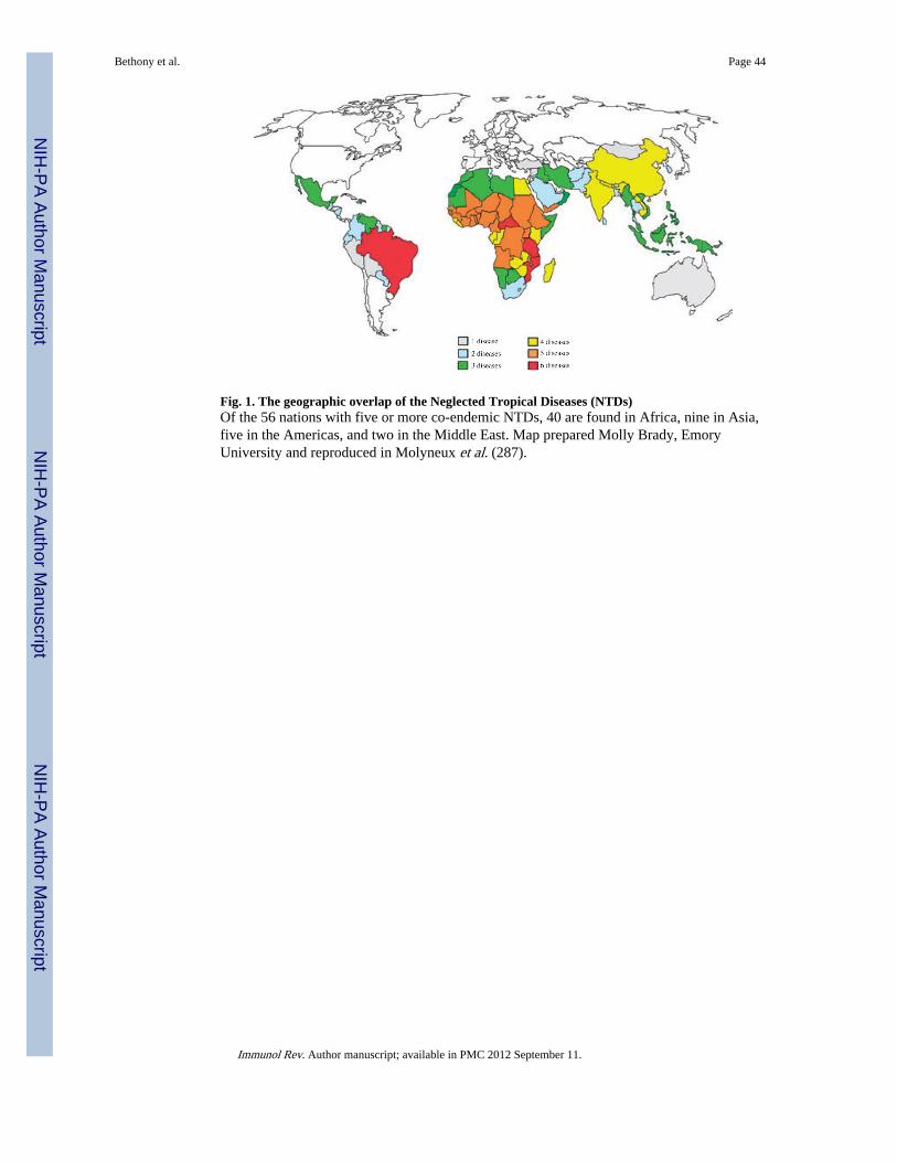
Fig. 1. The geographic overlap of the Neglected Tropical Diseases (NTDs)Of the 56 nations with five or more co-endemic NTDs, 40 are found in Africa, nine in Asia,five in the Americas, and two in the Middle East. Map prepared Molly Brady, EmoryUniversity and reproduced in Molyneux et al. (287).
Bethony et al. Page 44
Immunol Rev. Author manuscript; available in PMC 2012 September 11.
NIH
-PA Author Manuscript
NIH
-PA Author Manuscript
NIH
-PA Author Manuscript
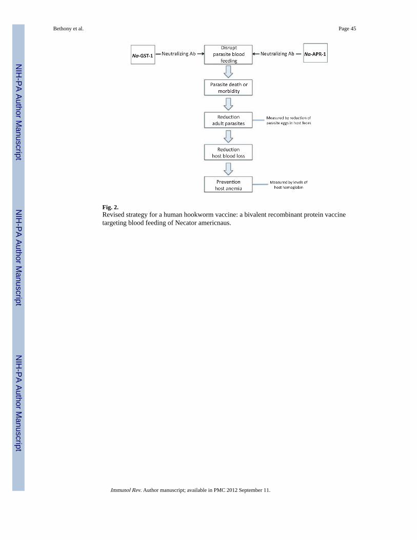
Fig. 2.Revised strategy for a human hookworm vaccine: a bivalent recombinant protein vaccinetargeting blood feeding of Necator americnaus.
Bethony et al. Page 45
Immunol Rev. Author manuscript; available in PMC 2012 September 11.
NIH
-PA Author Manuscript
NIH
-PA Author Manuscript
NIH
-PA Author Manuscript
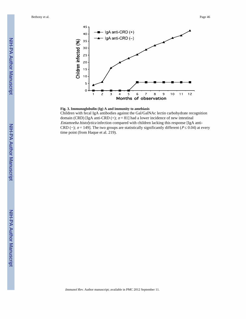
Fig. 3. Immunoglobulin (Ig) A and immunity to amebiasisChildren with fecal IgA antibodies against the Gal/GalNAc lectin carbohydrate recognitiondomain (CRD) [IgA anti-CRD (+); n = 81] had a lower incidence of new intestinalEntamoeba histolytica infection compared with children lacking this response [IgA anti-CRD (−); n = 149]. The two groups are statistically significantly different (P ≤ 0.04) at everytime point (from Haque et al. 219).
Bethony et al. Page 46
Immunol Rev. Author manuscript; available in PMC 2012 September 11.
NIH
-PA Author Manuscript
NIH
-PA Author Manuscript
NIH
-PA Author Manuscript

Fig. 4. Leish-111f (L111f): a tandemly linked protein of three subunitsEach component of L111f, the first recombinant Leishmania vaccine candidate to enterclinical trials, was cloned from a Leishmania major expression library. LeIF, (ribosomalinitiation factor identified by serological screening with human sera from healthy infectedindividuals); LmSTI1 (temperature inducible protein, identified by screening with sera fromBALB/c mice infected with L. major); thiol-specific antioxidant (TSA, identified throughscreening with sera from BALB/c mice immunized with protective Leishmania antigens.
Bethony et al. Page 47
Immunol Rev. Author manuscript; available in PMC 2012 September 11.
NIH
-PA Author Manuscript
NIH
-PA Author Manuscript
NIH
-PA Author Manuscript
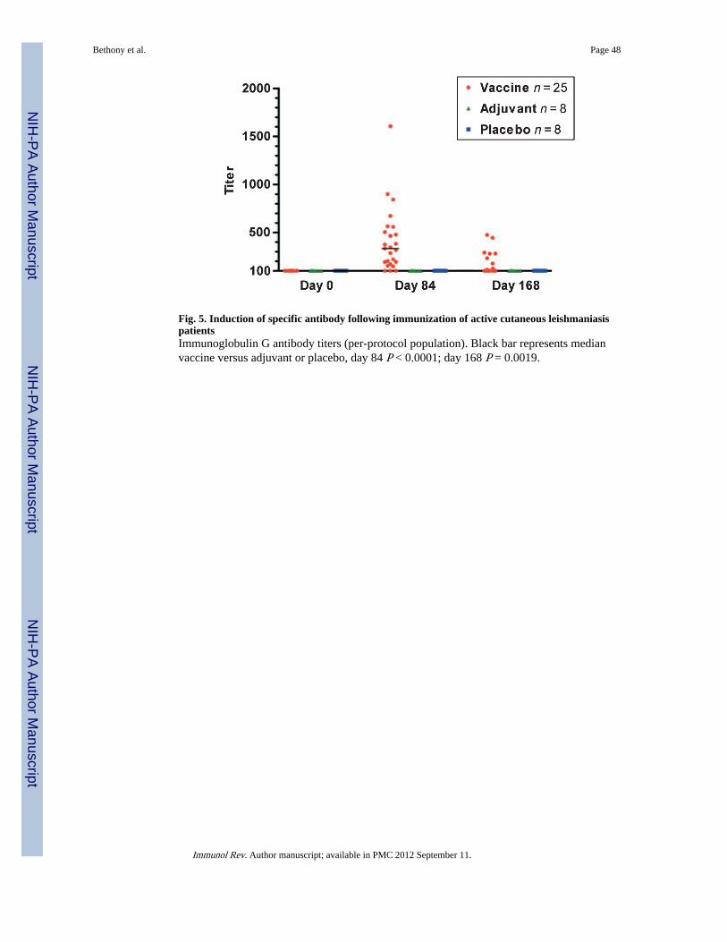
Fig. 5. Induction of specific antibody following immunization of active cutaneous leishmaniasispatientsImmunoglobulin G antibody titers (per-protocol population). Black bar represents medianvaccine versus adjuvant or placebo, day 84 P < 0.0001; day 168 P = 0.0019.
Bethony et al. Page 48
Immunol Rev. Author manuscript; available in PMC 2012 September 11.
NIH
-PA Author Manuscript
NIH
-PA Author Manuscript
NIH
-PA Author Manuscript

Fig. 6. (A). Induction of specific cellular immune responses following immunization of activecutaneous leishmaniasis patients using Fluorescence Activated Cell Sorting (FACS)FACS analysis of un-vaccinated mucosal leishmaniasis (ML) patient subject’s cellscollected before (day 0) and after (day 84) three immunizations with Leish-111f (L111f) inMPL-SE which does not cure (chemotherapy only). Cells were analyzed following in vitrostimulation with vaccine antigen. (B). FACS analysis of ML patient subject’s cells collectedbefore (day 0) and after (day 84) three immunizations with L111f in MPL-SE incombination with standard antimony therapy (immunochemotherapy). Cells were analyzedfollowing in vitro stimulation with vaccine antigen.
Bethony et al. Page 49
Immunol Rev. Author manuscript; available in PMC 2012 September 11.
NIH
-PA Author Manuscript
NIH
-PA Author Manuscript
NIH
-PA Author Manuscript
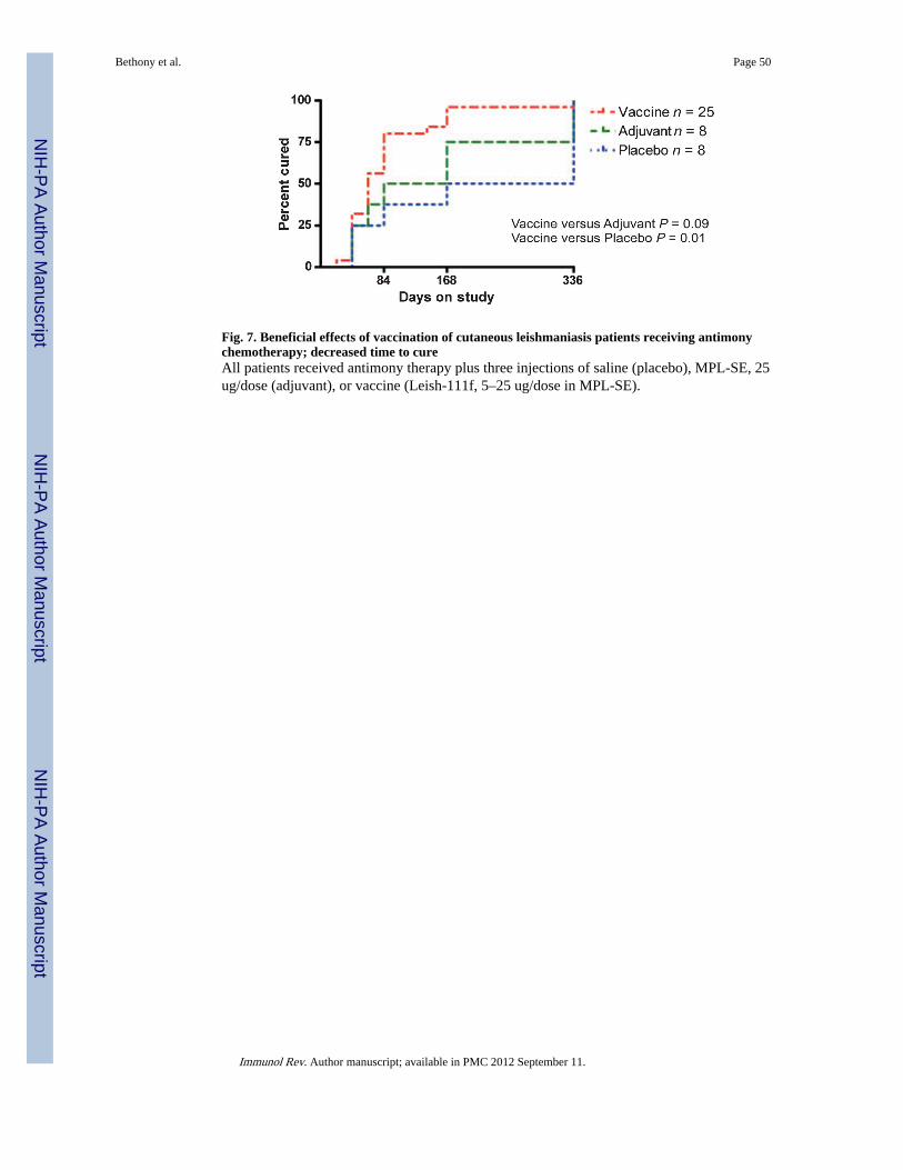
Fig. 7. Beneficial effects of vaccination of cutaneous leishmaniasis patients receiving antimonychemotherapy; decreased time to cureAll patients received antimony therapy plus three injections of saline (placebo), MPL-SE, 25ug/dose (adjuvant), or vaccine (Leish-111f, 5–25 ug/dose in MPL-SE).
Bethony et al. Page 50
Immunol Rev. Author manuscript; available in PMC 2012 September 11.
NIH
-PA Author Manuscript
NIH
-PA Author Manuscript
NIH
-PA Author Manuscript
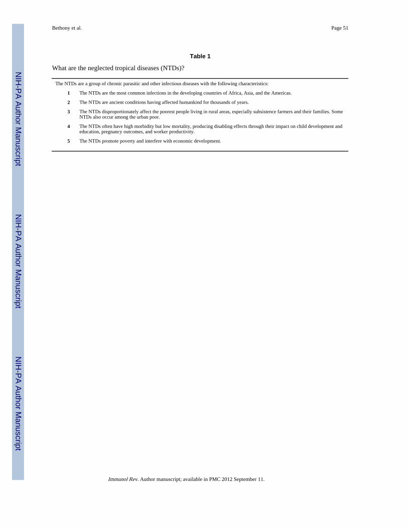
NIH
-PA Author Manuscript
NIH
-PA Author Manuscript
NIH
-PA Author Manuscript
Bethony et al. Page 51
Table 1
What are the neglected tropical diseases (NTDs)?
The NTDs are a group of chronic parasitic and other infectious diseases with the following characteristics:
1 The NTDs are the most common infections in the developing countries of Africa, Asia, and the Americas.
2 The NTDs are ancient conditions having affected humankind for thousands of years.
3 The NTDs disproportionately affect the poorest people living in rural areas, especially subsistence farmers and their families. SomeNTDs also occur among the urban poor.
4 The NTDs often have high morbidity but low mortality, producing disabling effects through their impact on child development andeducation, pregnancy outcomes, and worker productivity.
5 The NTDs promote poverty and interfere with economic development.
Immunol Rev. Author manuscript; available in PMC 2012 September 11.
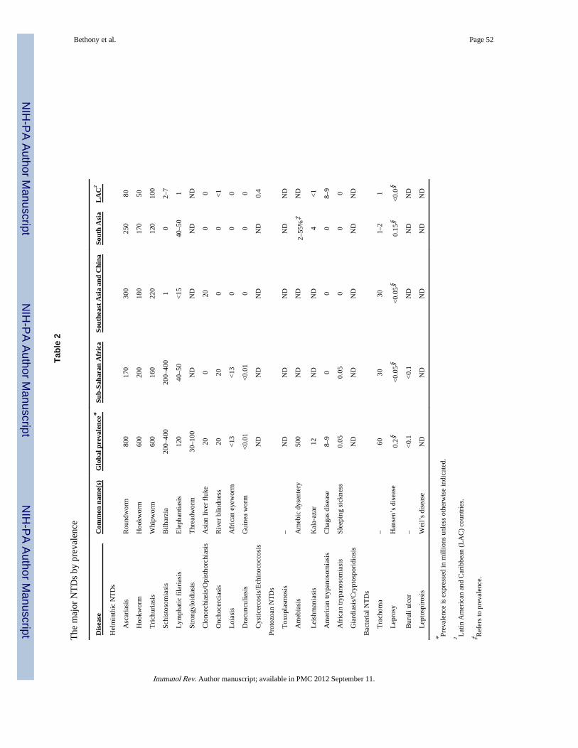
NIH
-PA Author Manuscript
NIH
-PA Author Manuscript
NIH
-PA Author Manuscript
Bethony et al. Page 52
Tabl
e 2
The
maj
or N
TD
s by
pre
vale
nce
Dis
ease
Com
mon
nam
e(s)
Glo
bal p
reva
lenc
e*Su
b-Sa
hara
n A
fric
aSo
uthe
ast
Asi
a an
d C
hina
Sout
h A
sia
LA
C†
Hel
min
thic
NT
Ds
A
scar
iasi
sR
ound
wor
m80
017
030
025
080
H
ookw
orm
Hoo
kwor
m60
020
018
017
050
T
rich
uria
sis
Whi
pwor
m60
016
022
012
010
0
Sc
hist
osom
iasi
sB
ilhar
zia
200–
400
200–
400
10
2–7
L
ymph
atic
fila
rias
isE
leph
antia
sis
120
40–5
0<
1540
–50
1
St
rong
yloi
dias
isT
hrea
dwor
m30
–100
ND
ND
ND
ND
C
lono
rchi
asis
/Opi
stho
rchi
asis
Asi
an li
ver
fluk
e20
020
00
O
ncho
cerc
iasi
sR
iver
blin
dnes
s20
200
0<
1
L
oias
isA
fric
an e
yew
orm
<13
<13
00
0
D
racu
ncul
iasi
sG
uine
a w
orm
<0.
01<
0.01
00
0
C
ystic
erco
sis/
Ech
inoc
occo
sis
ND
ND
ND
ND
0.4
Prot
ozoa
n N
TD
s
T
oxop
lasm
osis
–N
DN
DN
DN
DN
D
A
meb
iasi
sA
meb
ic d
ysen
tery
500
ND
ND
2–55
%‡
ND
L
eish
man
iasi
sK
ala-
azar
12N
DN
D4
<1
A
mer
ican
tryp
anos
omia
sis
Cha
gas
dise
ase
8–9
00
08–
9
A
fric
an tr
ypan
osom
iasi
sSl
eepi
ng s
ickn
ess
0.05
0.05
00
0
G
iard
iasi
s/C
rypt
ospo
ridi
osis
ND
ND
ND
ND
ND
Bac
teri
al N
TD
s
T
rach
oma
–60
3030
1–2
1
L
epro
syH
anse
n’s
dise
ase
0.2§
<0.
05§
<0.
05§
0.15
§<
0.0§
B
urul
i ulc
er–
<0.
1<
0.1
ND
ND
ND
L
epto
spir
osis
Wei
l’s
dise
ase
ND
ND
ND
ND
ND
* Prev
alen
ce is
exp
ress
ed in
mill
ions
unl
ess
othe
rwis
e in
dica
ted.
† Lat
in A
mer
ican
and
Car
ibbe
an (
LA
C)
coun
trie
s.
‡ Ref
ers
to p
reva
lenc
e.
Immunol Rev. Author manuscript; available in PMC 2012 September 11.
NIH
-PA Author Manuscript
NIH
-PA Author Manuscript
NIH
-PA Author Manuscript
Bethony et al. Page 53§ R
efer
s to
new
cas
es a
nnua
lly.
NT
Ds,
neg
lect
ed tr
opic
al d
isea
ses;
ND
, not
det
erm
ined
.
Immunol Rev. Author manuscript; available in PMC 2012 September 11.
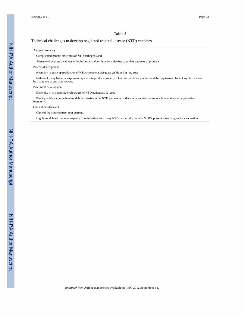
NIH
-PA Author Manuscript
NIH
-PA Author Manuscript
NIH
-PA Author Manuscript
Bethony et al. Page 54
Table 3
Technical challenges to develop neglected tropical disease (NTD) vaccines
Antigen discovery
Complicated genetic structures of NTD pathogens and
Absence of genome databases or bioinformatic algorithms for selecting candidate antigens of promise.
Process development
Necessity to scale up production of NTDs vaccine at adequate yields and at low cost.
Failure of many bacterial expression systems to produce properly folded recombinant proteins and the requirement for eukaryotic or otherless common expression vectors.
Preclinical development
Difficulty in maintaining cycle stages of NTD pathogens in vitro.
Paucity of laboratory animal models permissive to the NTD pathogens or that can accurately reproduce human disease or protectiveimmunity.
Clinical development
Clinical trials in resource-poor settings.
Highly modulated immune response from infection with many NTDs, especially helmith NTDS, present some dangers for vaccination.
Immunol Rev. Author manuscript; available in PMC 2012 September 11.
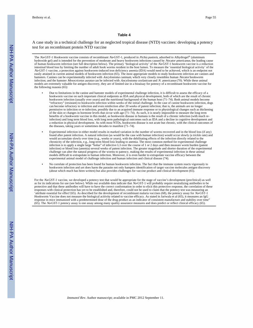
NIH
-PA Author Manuscript
NIH
-PA Author Manuscript
NIH
-PA Author Manuscript
Bethony et al. Page 55
Table 4
A case study in a technical challenge for an neglected tropical disease (NTD) vaccines: developing a potencytest for an recombinant protein NTD vaccine
The Na-GST-1 Hookworm vaccine consists of recombinant Na-GST-1, produced in Pichia pastoris, adsorbed to Alhydrogel® (aluminumhydroxide gel) and is intended for the prevention of moderate and heavy hookworm infections caused by Necator americanus, the leading causeof human hookworm infection (see full description below). The primary ‘biological activity’ of the Na-GST-1 hookworm vaccine is a reductionintestinal blood loss by limiting the number of adult hook worms resident in the host lumen. To measure the ‘essential biological activity’ of theNa-GST-1 vaccine, a protection against hookworm-induced iron deficiency anemia (IDA) would need to be achieved, which is an endpoint noteasily attained in current animal models of hookworm infection (65). The most appropriate models to study hookworm infection are canines andhamsters. Canines can be experimentally infected with Ancylostoma caninum, which very closely resembles human Necator hookworminfection; and the hamster Mesocricetus auratus can be infected with Ancylostoma ceylanicum and N. americanus (70). While these animalmodels are extremely valuable for antigen discovery, they are of limited use in a bioassay for potency of a recombinant hookworm vaccine forthe following reasons (65):
• Due to limitations in the canine and hamster models of experimental challenge infection, it is difficult to assess the efficacy of ahookworm vaccine on such important clinical endpoints as IDA and physical development, both of which are the result of chronichookworm infection (usually over years) and the nutritional background of the human host (71–74). Both animal models become“refractory” (resistant) to hookworm infection within weeks of the initial challenge. In the case of canine hookworm infection, dogscan become refractory to infection and even reinfection after 20 weeks of patent infection; that is, the animals are no longerpermissive to infection or re-infection, possibly due to an acquired immune response or to physiological changes such as thickeningof the skin or changes in hormone levels that occur with age (71–74). As such, it is nearly impossible to measure the long termbenefits of a hookworm vaccine in this model, as hookworm disease in humans is the result of a chronic infection (with much re-infection) and long term blood loss, with long term pathological outcomes such as IDA and a decline in cognitive development anda reduction in physical development. As with most NTDs, hookworm disease is not acute but chronic, with the clinical outcomes ofthe diseases, taking years or sometimes decades to manifest (71–74).
• Experimental infection in either model results in marked variation in the number of worms recovered and in the blood loss (if any)found after patent infection. A natural infection (as would be the case with human infection) would occur slowly (a trickle rate) andwould accumulate slowly over time (e.g., weeks or years), with the debilitating effects of the infection directly related to thechronicity of the infection, e.g., long-term blood loss leading to anemia. The most common method for experimental challengeinfection is to apply a single large “bolus” of infective L3 over the course of 1 or 2 days and then measure worm burden (patentinfection) or blood loss (anemia) several weeks of patent infection. The greater magnitude and shorter duration of the experimentalchallenge can alter the natural progress of the worms to patency, making the results of experimental infection in these animalmodels difficult to extrapolate to human infection. Moreover, it is even harder to extrapolate vaccine efficacy between theexperimental animal model of challenge infection and human infection and clinical disease (74).
• No correlate of protection has been found for human hookworm infection. The fact that the immune system reacts vigorously tohookworm infection and yet does harm the parasite not only hampers identification of target vaccine molecules antigen discovery(about which much has been written) but also provides challenges for vaccine product and clinical development (65).
For the Na-GST-1 vaccine, we developed a potency test that would be appropriate for the stage of vaccine’s development (preclinical) as wellas for its indications for use (see below). While our available data indicate that Na-GST-1 will probably require neutralizing antibodies to beprotective and that these antibodies will have to have the correct conformation in order to elicit this protective response, the correlation of theseresponses with clinical protection has yet to be established and, therefore, could not be used to claim that the potency test was measuring an‘attribute essential for effect’(65). As described for the development of recombinant malaria vaccines (68), the potency assay for Na-GST-1Hookworm Vaccine does not measure the biological acitivity related to vaccine efficacy. As stated in Jariwala et al (65), it measures an IgGresponse in mice immunized with a predetermined dose of the drug product as an indicator of consistent manufacture and stability over time”(65). The Na-GST-1 potency assay is one assay among many quality assurance measures and does predict or reflect clinical efficacy (65).
Immunol Rev. Author manuscript; available in PMC 2012 September 11.
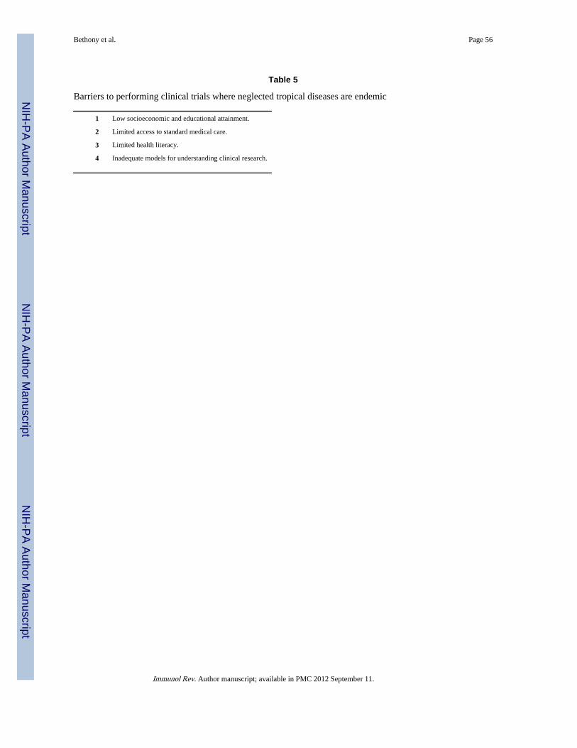
NIH
-PA Author Manuscript
NIH
-PA Author Manuscript
NIH
-PA Author Manuscript
Bethony et al. Page 56
Table 5
Barriers to performing clinical trials where neglected tropical diseases are endemic
1 Low socioeconomic and educational attainment.
2 Limited access to standard medical care.
3 Limited health literacy.
4 Inadequate models for understanding clinical research.
Immunol Rev. Author manuscript; available in PMC 2012 September 11.
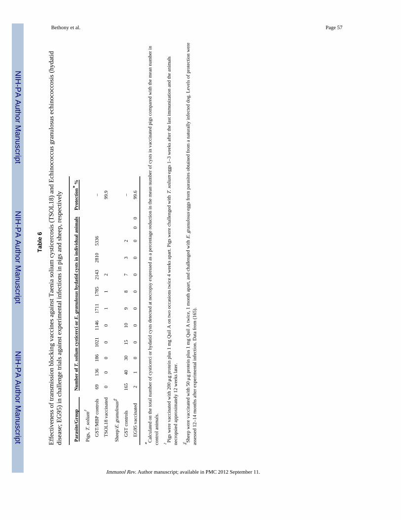
NIH
-PA Author Manuscript
NIH
-PA Author Manuscript
NIH
-PA Author Manuscript
Bethony et al. Page 57
Tabl
e 6
Eff
ectiv
enes
s of
tran
smis
sion
blo
ckin
g va
ccin
es a
gain
st T
aeni
a so
lium
cys
ticer
cosi
s (T
SOL
18)
and
Ech
inoc
occu
s gr
anul
osus
ech
inoc
occo
sis
(hyd
atid
dise
ase;
EG
95)
in c
halle
nge
tria
ls a
gain
st e
xper
imen
tal i
nfec
tions
in p
igs
and
shee
p, r
espe
ctiv
ely
Par
asit
e/G
roup
Num
ber
of T
. sol
ium
cys
tice
rci o
r E
. gra
nulo
sus
hyda
tid
cyst
s in
indi
vidu
al a
nim
als
Pro
tect
ion*
%
Pigs
, T. s
oliu
m†
G
ST/M
BP
cont
rols
6913
618
610
2111
4617
1117
8521
4328
1053
36–
T
SOL
18 v
acci
nate
d0
00
00
11
299
.9
Shee
p/E
. gra
nulo
sus‡
G
ST c
ontr
ols
165
4030
1510
98
73
2–
E
G95
vac
cina
ted
21
00
00
00
00
00
99.6
* Cal
cula
ted
on th
e to
tal n
umbe
r of
cys
ticer
ci o
r hy
datid
cys
ts d
etec
ted
at n
ecro
psy
expr
esse
d as
a p
erce
ntag
e re
duct
ion
in th
e m
ean
num
ber
of c
ysts
in v
acci
nate
d pi
gs c
ompa
red
with
the
mea
n nu
mbe
r in
cont
rol a
nim
als.
† Pigs
wer
e va
ccin
ated
with
200
μg
prot
ein
plus
1 m
g Q
uil A
on
two
occa
sion
s tw
ice
4 w
eeks
apa
rt. P
igs
wer
e ch
alle
nged
with
T. s
oliu
m e
ggs
1–3
wee
ks a
fter
the
last
imm
uniz
atio
n an
d th
e an
imal
sne
crop
sied
app
roxi
mat
ely
12 w
eeks
late
r.
‡ Shee
p w
ere
vacc
inat
ed w
ith 5
0 μ
g pr
otei
n pl
us 1
mg
Qui
l A tw
ice,
1 m
onth
apa
rt, a
nd c
halle
nged
with
E. g
ranu
losu
s eg
gs f
rom
par
asite
s ob
tain
ed f
rom
a n
atur
ally
infe
cted
dog
. Lev
els
of p
rote
ctio
n w
ere
asse
ssed
12–
14 m
onth
s af
ter
expe
rim
enta
l inf
ectio
n. D
ata
from
(16
5).
Immunol Rev. Author manuscript; available in PMC 2012 September 11.
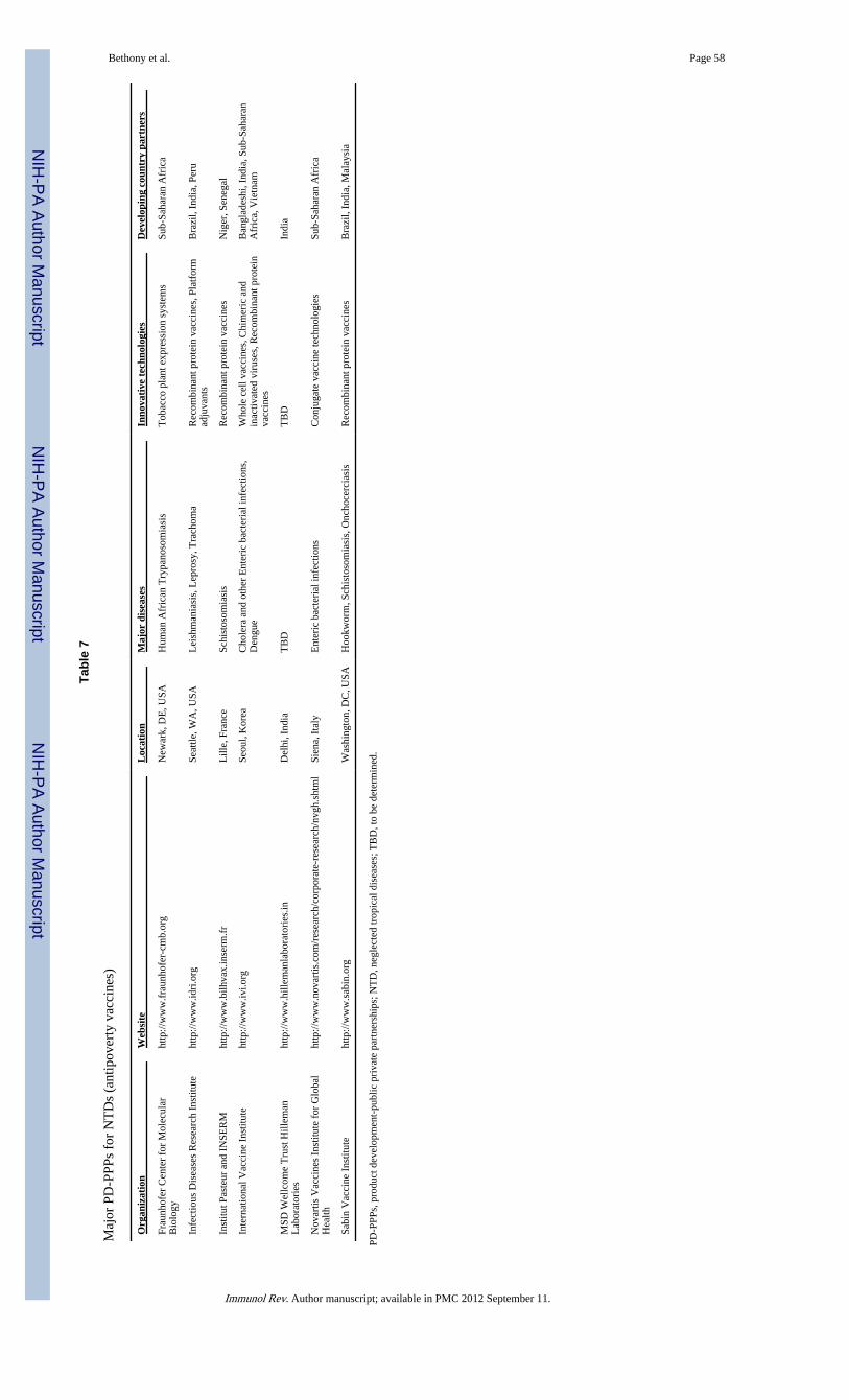
NIH
-PA Author Manuscript
NIH
-PA Author Manuscript
NIH
-PA Author Manuscript
Bethony et al. Page 58
Tabl
e 7
Maj
or P
D-P
PPs
for
NT
Ds
(ant
ipov
erty
vac
cine
s)
Org
aniz
atio
nW
ebsi
teL
ocat
ion
Maj
or d
isea
ses
Inno
vati
ve t
echn
olog
ies
Dev
elop
ing
coun
try
part
ners
Frau
nhof
er C
ente
r fo
r M
olec
ular
Bio
logy
http
://w
ww
.fra
unho
fer-
cmb.
org
New
ark,
DE
, USA
Hum
an A
fric
an T
rypa
noso
mia
sis
Tob
acco
pla
nt e
xpre
ssio
n sy
stem
sSu
b-Sa
hara
n A
fric
a
Infe
ctio
us D
isea
ses
Res
earc
h In
stitu
teht
tp://
ww
w.id
ri.o
rgSe
attle
, WA
, USA
Lei
shm
ania
sis,
Lep
rosy
, Tra
chom
aR
ecom
bina
nt p
rote
in v
acci
nes,
Pla
tfor
mad
juva
nts
Bra
zil,
Indi
a, P
eru
Inst
itut P
aste
ur a
nd I
NSE
RM
http
://w
ww
.bilh
vax.
inse
rm.f
rL
ille,
Fra
nce
Schi
stos
omia
sis
Rec
ombi
nant
pro
tein
vac
cine
sN
iger
, Sen
egal
Inte
rnat
iona
l Vac
cine
Ins
titut
eht
tp://
ww
w.iv
i.org
Seou
l, K
orea
Cho
lera
and
oth
er E
nter
ic b
acte
rial
infe
ctio
ns,
Den
gue
Who
le c
ell v
acci
nes,
Chi
mer
ic a
ndin
activ
ated
vir
uses
, Rec
ombi
nant
pro
tein
vacc
ines
Ban
glad
eshi
, Ind
ia, S
ub-S
ahar
anA
fric
a, V
ietn
am
MSD
Wel
lcom
e T
rust
Hill
eman
Lab
orat
orie
sht
tp://
ww
w.h
illem
anla
bora
tori
es.in
Del
hi, I
ndia
TB
DT
BD
Indi
a
Nov
artis
Vac
cine
s In
stitu
te f
or G
loba
lH
ealth
http
://w
ww
.nov
artis
.com
/res
earc
h/co
rpor
ate-
rese
arch
/nvg
h.sh
tml
Sien
a, I
taly
Ent
eric
bac
teri
al in
fect
ions
Con
juga
te v
acci
ne te
chno
logi
esSu
b-Sa
hara
n A
fric
a
Sabi
n V
acci
ne I
nstit
ute
http
://w
ww
.sab
in.o
rgW
ashi
ngto
n, D
C, U
SAH
ookw
orm
, Sch
isto
som
iasi
s, O
ncho
cerc
iasi
sR
ecom
bina
nt p
rote
in v
acci
nes
Bra
zil,
Indi
a, M
alay
sia
PD-P
PPs,
pro
duct
dev
elop
men
t-pu
blic
pri
vate
par
tner
ship
s; N
TD
, neg
lect
ed tr
opic
al d
isea
ses;
TB
D, t
o be
det
erm
ined
.
Immunol Rev. Author manuscript; available in PMC 2012 September 11.
Related Documents