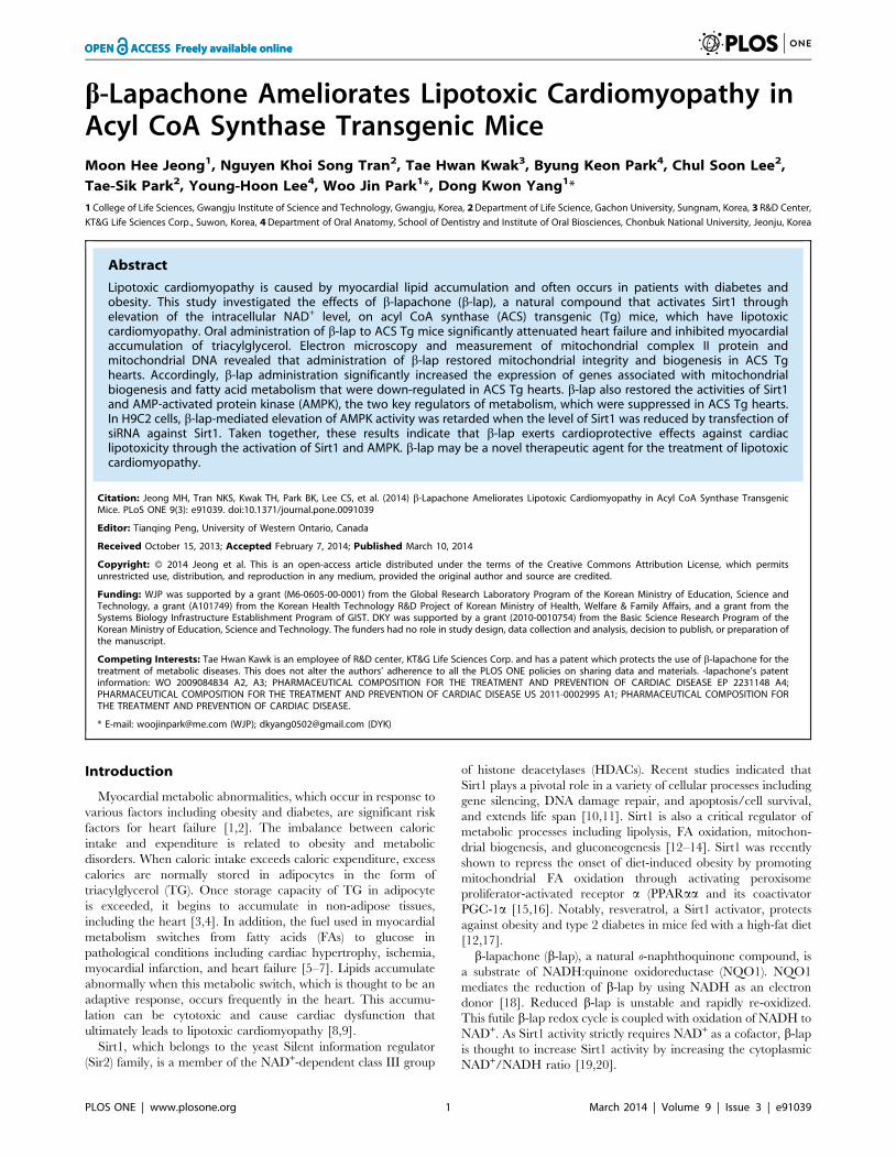
b-Lapachone Ameliorates Lipotoxic Cardiomyopathy in Acyl CoA Synthase Transgenic Mice Moon Hee Jeong 1 , Nguyen Khoi Song Tran 2 , Tae Hwan Kwak 3 , Byung Keon Park 4 , Chul Soon Lee 2 , Tae-Sik Park 2 , Young-Hoon Lee 4 , Woo Jin Park 1 *, Dong Kwon Yang 1 * 1 College of Life Sciences, Gwangju Institute of Science and Technology, Gwangju, Korea, 2 Department of Life Science, Gachon University, Sungnam, Korea, 3 R&D Center, KT&G Life Sciences Corp., Suwon, Korea, 4 Department of Oral Anatomy, School of Dentistry and Institute of Oral Biosciences, Chonbuk National University, Jeonju, Korea Abstract Lipotoxic cardiomyopathy is caused by myocardial lipid accumulation and often occurs in patients with diabetes and obesity. This study investigated the effects of b-lapachone (b-lap), a natural compound that activates Sirt1 through elevation of the intracellular NAD + level, on acyl CoA synthase (ACS) transgenic (Tg) mice, which have lipotoxic cardiomyopathy. Oral administration of b-lap to ACS Tg mice significantly attenuated heart failure and inhibited myocardial accumulation of triacylglycerol. Electron microscopy and measurement of mitochondrial complex II protein and mitochondrial DNA revealed that administration of b-lap restored mitochondrial integrity and biogenesis in ACS Tg hearts. Accordingly, b-lap administration significantly increased the expression of genes associated with mitochondrial biogenesis and fatty acid metabolism that were down-regulated in ACS Tg hearts. b-lap also restored the activities of Sirt1 and AMP-activated protein kinase (AMPK), the two key regulators of metabolism, which were suppressed in ACS Tg hearts. In H9C2 cells, b-lap-mediated elevation of AMPK activity was retarded when the level of Sirt1 was reduced by transfection of siRNA against Sirt1. Taken together, these results indicate that b-lap exerts cardioprotective effects against cardiac lipotoxicity through the activation of Sirt1 and AMPK. b-lap may be a novel therapeutic agent for the treatment of lipotoxic cardiomyopathy. Citation: Jeong MH, Tran NKS, Kwak TH, Park BK, Lee CS, et al. (2014) b-Lapachone Ameliorates Lipotoxic Cardiomyopathy in Acyl CoA Synthase Transgenic Mice. PLoS ONE 9(3): e91039. doi:10.1371/journal.pone.0091039 Editor: Tianqing Peng, University of Western Ontario, Canada Received October 15, 2013; Accepted February 7, 2014; Published March 10, 2014 Copyright: ß 2014 Jeong et al. This is an open-access article distributed under the terms of the Creative Commons Attribution License, which permits unrestricted use, distribution, and reproduction in any medium, provided the original author and source are credited. Funding: WJP was supported by a grant (M6-0605-00-0001) from the Global Research Laboratory Program of the Korean Ministry of Education, Science and Technology, a grant (A101749) from the Korean Health Technology R&D Project of Korean Ministry of Health, Welfare & Family Affairs, and a grant from the Systems Biology Infrastructure Establishment Program of GIST. DKY was supported by a grant (2010-0010754) from the Basic Science Research Program of the Korean Ministry of Education, Science and Technology. The funders had no role in study design, data collection and analysis, decision to publish, or preparation of the manuscript. Competing Interests: Tae Hwan Kawk is an employee of R&D center, KT&G Life Sciences Corp. and has a patent which protects the use of b-lapachone for the treatment of metabolic diseases. This does not alter the authors’ adherence to all the PLOS ONE policies on sharing data and materials. -lapachone’s patent information: WO 2009084834 A2, A3; PHARMACEUTICAL COMPOSITION FOR THE TREATMENT AND PREVENTION OF CARDIAC DISEASE EP 2231148 A4; PHARMACEUTICAL COMPOSITION FOR THE TREATMENT AND PREVENTION OF CARDIAC DISEASE US 2011-0002995 A1; PHARMACEUTICAL COMPOSITION FOR THE TREATMENT AND PREVENTION OF CARDIAC DISEASE. * E-mail: [email protected] (WJP); [email protected] (DYK) Introduction Myocardial metabolic abnormalities, which occur in response to various factors including obesity and diabetes, are significant risk factors for heart failure [1,2]. The imbalance between caloric intake and expenditure is related to obesity and metabolic disorders. When caloric intake exceeds caloric expenditure, excess calories are normally stored in adipocytes in the form of triacylglycerol (TG). Once storage capacity of TG in adipocyte is exceeded, it begins to accumulate in non-adipose tissues, including the heart [3,4]. In addition, the fuel used in myocardial metabolism switches from fatty acids (FAs) to glucose in pathological conditions including cardiac hypertrophy, ischemia, myocardial infarction, and heart failure [5–7]. Lipids accumulate abnormally when this metabolic switch, which is thought to be an adaptive response, occurs frequently in the heart. This accumu- lation can be cytotoxic and cause cardiac dysfunction that ultimately leads to lipotoxic cardiomyopathy [8,9]. Sirt1, which belongs to the yeast Silent information regulator (Sir2) family, is a member of the NAD + -dependent class III group of histone deacetylases (HDACs). Recent studies indicated that Sirt1 plays a pivotal role in a variety of cellular processes including gene silencing, DNA damage repair, and apoptosis/cell survival, and extends life span [10,11]. Sirt1 is also a critical regulator of metabolic processes including lipolysis, FA oxidation, mitochon- drial biogenesis, and gluconeogenesis [12–14]. Sirt1 was recently shown to repress the onset of diet-induced obesity by promoting mitochondrial FA oxidation through activating peroxisome proliferator-activated receptor a (PPARaa and its coactivator PGC-1a [15,16]. Notably, resveratrol, a Sirt1 activator, protects against obesity and type 2 diabetes in mice fed with a high-fat diet [12,17]. b-lapachone (b-lap), a natural o-naphthoquinone compound, is a substrate of NADH:quinone oxidoreductase (NQO1). NQO1 mediates the reduction of b-lap by using NADH as an electron donor [18]. Reduced b-lap is unstable and rapidly re-oxidized. This futile b-lap redox cycle is coupled with oxidation of NADH to NAD + . As Sirt1 activity strictly requires NAD + as a cofactor, b-lap is thought to increase Sirt1 activity by increasing the cytoplasmic NAD + /NADH ratio [19,20]. PLOS ONE | www.plosone.org 1 March 2014 | Volume 9 | Issue 3 | e91039

Welcome message from author
This document is posted to help you gain knowledge. Please leave a comment to let me know what you think about it! Share it to your friends and learn new things together.
Transcript
b-Lapachone Ameliorates Lipotoxic Cardiomyopathy inAcyl CoA Synthase Transgenic MiceMoon Hee Jeong1, Nguyen Khoi Song Tran2, Tae Hwan Kwak3, Byung Keon Park4, Chul Soon Lee2,
Tae-Sik Park2, Young-Hoon Lee4, Woo Jin Park1*, Dong Kwon Yang1*
1 College of Life Sciences, Gwangju Institute of Science and Technology, Gwangju, Korea, 2 Department of Life Science, Gachon University, Sungnam, Korea, 3 R&D Center,
KT&G Life Sciences Corp., Suwon, Korea, 4 Department of Oral Anatomy, School of Dentistry and Institute of Oral Biosciences, Chonbuk National University, Jeonju, Korea
Abstract
Lipotoxic cardiomyopathy is caused by myocardial lipid accumulation and often occurs in patients with diabetes andobesity. This study investigated the effects of b-lapachone (b-lap), a natural compound that activates Sirt1 throughelevation of the intracellular NAD+ level, on acyl CoA synthase (ACS) transgenic (Tg) mice, which have lipotoxiccardiomyopathy. Oral administration of b-lap to ACS Tg mice significantly attenuated heart failure and inhibited myocardialaccumulation of triacylglycerol. Electron microscopy and measurement of mitochondrial complex II protein andmitochondrial DNA revealed that administration of b-lap restored mitochondrial integrity and biogenesis in ACS Tghearts. Accordingly, b-lap administration significantly increased the expression of genes associated with mitochondrialbiogenesis and fatty acid metabolism that were down-regulated in ACS Tg hearts. b-lap also restored the activities of Sirt1and AMP-activated protein kinase (AMPK), the two key regulators of metabolism, which were suppressed in ACS Tg hearts.In H9C2 cells, b-lap-mediated elevation of AMPK activity was retarded when the level of Sirt1 was reduced by transfection ofsiRNA against Sirt1. Taken together, these results indicate that b-lap exerts cardioprotective effects against cardiaclipotoxicity through the activation of Sirt1 and AMPK. b-lap may be a novel therapeutic agent for the treatment of lipotoxiccardiomyopathy.
Citation: Jeong MH, Tran NKS, Kwak TH, Park BK, Lee CS, et al. (2014) b-Lapachone Ameliorates Lipotoxic Cardiomyopathy in Acyl CoA Synthase TransgenicMice. PLoS ONE 9(3): e91039. doi:10.1371/journal.pone.0091039
Editor: Tianqing Peng, University of Western Ontario, Canada
Received October 15, 2013; Accepted February 7, 2014; Published March 10, 2014
Copyright: � 2014 Jeong et al. This is an open-access article distributed under the terms of the Creative Commons Attribution License, which permitsunrestricted use, distribution, and reproduction in any medium, provided the original author and source are credited.
Funding: WJP was supported by a grant (M6-0605-00-0001) from the Global Research Laboratory Program of the Korean Ministry of Education, Science andTechnology, a grant (A101749) from the Korean Health Technology R&D Project of Korean Ministry of Health, Welfare & Family Affairs, and a grant from theSystems Biology Infrastructure Establishment Program of GIST. DKY was supported by a grant (2010-0010754) from the Basic Science Research Program of theKorean Ministry of Education, Science and Technology. The funders had no role in study design, data collection and analysis, decision to publish, or preparation ofthe manuscript.
Competing Interests: Tae Hwan Kawk is an employee of R&D center, KT&G Life Sciences Corp. and has a patent which protects the use of b-lapachone for thetreatment of metabolic diseases. This does not alter the authors’ adherence to all the PLOS ONE policies on sharing data and materials. -lapachone’s patentinformation: WO 2009084834 A2, A3; PHARMACEUTICAL COMPOSITION FOR THE TREATMENT AND PREVENTION OF CARDIAC DISEASE EP 2231148 A4;PHARMACEUTICAL COMPOSITION FOR THE TREATMENT AND PREVENTION OF CARDIAC DISEASE US 2011-0002995 A1; PHARMACEUTICAL COMPOSITION FORTHE TREATMENT AND PREVENTION OF CARDIAC DISEASE.
* E-mail: [email protected] (WJP); [email protected] (DYK)
Introduction
Myocardial metabolic abnormalities, which occur in response to
various factors including obesity and diabetes, are significant risk
factors for heart failure [1,2]. The imbalance between caloric
intake and expenditure is related to obesity and metabolic
disorders. When caloric intake exceeds caloric expenditure, excess
calories are normally stored in adipocytes in the form of
triacylglycerol (TG). Once storage capacity of TG in adipocyte
is exceeded, it begins to accumulate in non-adipose tissues,
including the heart [3,4]. In addition, the fuel used in myocardial
metabolism switches from fatty acids (FAs) to glucose in
pathological conditions including cardiac hypertrophy, ischemia,
myocardial infarction, and heart failure [5–7]. Lipids accumulate
abnormally when this metabolic switch, which is thought to be an
adaptive response, occurs frequently in the heart. This accumu-
lation can be cytotoxic and cause cardiac dysfunction that
ultimately leads to lipotoxic cardiomyopathy [8,9].
Sirt1, which belongs to the yeast Silent information regulator
(Sir2) family, is a member of the NAD+-dependent class III group
of histone deacetylases (HDACs). Recent studies indicated that
Sirt1 plays a pivotal role in a variety of cellular processes including
gene silencing, DNA damage repair, and apoptosis/cell survival,
and extends life span [10,11]. Sirt1 is also a critical regulator of
metabolic processes including lipolysis, FA oxidation, mitochon-
drial biogenesis, and gluconeogenesis [12–14]. Sirt1 was recently
shown to repress the onset of diet-induced obesity by promoting
mitochondrial FA oxidation through activating peroxisome
proliferator-activated receptor a (PPARaa and its coactivator
PGC-1a [15,16]. Notably, resveratrol, a Sirt1 activator, protects
against obesity and type 2 diabetes in mice fed with a high-fat diet
[12,17].
b-lapachone (b-lap), a natural o-naphthoquinone compound, is
a substrate of NADH:quinone oxidoreductase (NQO1). NQO1
mediates the reduction of b-lap by using NADH as an electron
donor [18]. Reduced b-lap is unstable and rapidly re-oxidized.
This futile b-lap redox cycle is coupled with oxidation of NADH to
NAD+. As Sirt1 activity strictly requires NAD+ as a cofactor, b-lap
is thought to increase Sirt1 activity by increasing the cytoplasmic
NAD+/NADH ratio [19,20].
PLOS ONE | www.plosone.org 1 March 2014 | Volume 9 | Issue 3 | e91039
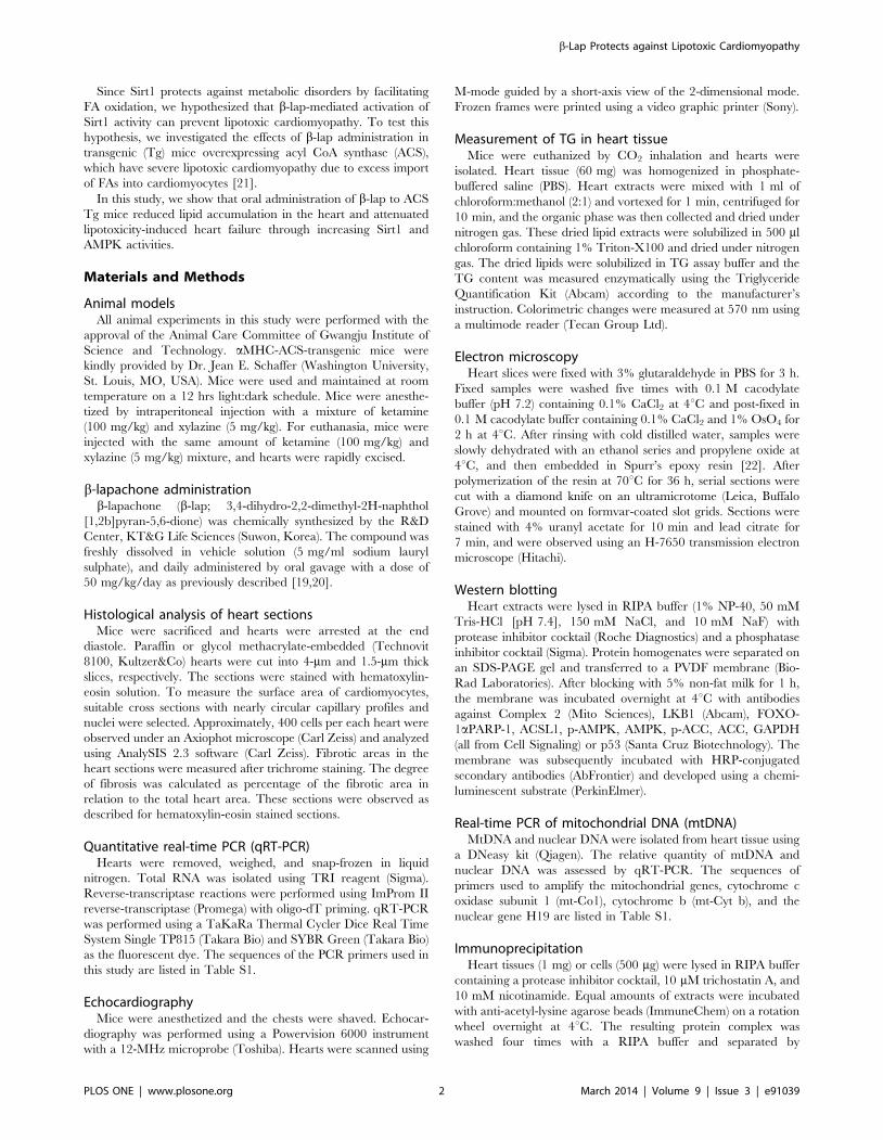
Since Sirt1 protects against metabolic disorders by facilitating
FA oxidation, we hypothesized that b-lap-mediated activation of
Sirt1 activity can prevent lipotoxic cardiomyopathy. To test this
hypothesis, we investigated the effects of b-lap administration in
transgenic (Tg) mice overexpressing acyl CoA synthase (ACS),
which have severe lipotoxic cardiomyopathy due to excess import
of FAs into cardiomyocytes [21].
In this study, we show that oral administration of b-lap to ACS
Tg mice reduced lipid accumulation in the heart and attenuated
lipotoxicity-induced heart failure through increasing Sirt1 and
AMPK activities.
Materials and Methods
Animal modelsAll animal experiments in this study were performed with the
approval of the Animal Care Committee of Gwangju Institute of
Science and Technology. aMHC-ACS-transgenic mice were
kindly provided by Dr. Jean E. Schaffer (Washington University,
St. Louis, MO, USA). Mice were used and maintained at room
temperature on a 12 hrs light:dark schedule. Mice were anesthe-
tized by intraperitoneal injection with a mixture of ketamine
(100 mg/kg) and xylazine (5 mg/kg). For euthanasia, mice were
injected with the same amount of ketamine (100 mg/kg) and
xylazine (5 mg/kg) mixture, and hearts were rapidly excised.
b-lapachone administrationb-lapachone (b-lap; 3,4-dihydro-2,2-dimethyl-2H-naphthol
[1,2b]pyran-5,6-dione) was chemically synthesized by the R&D
Center, KT&G Life Sciences (Suwon, Korea). The compound was
freshly dissolved in vehicle solution (5 mg/ml sodium lauryl
sulphate), and daily administered by oral gavage with a dose of
50 mg/kg/day as previously described [19,20].
Histological analysis of heart sectionsMice were sacrificed and hearts were arrested at the end
diastole. Paraffin or glycol methacrylate-embedded (Technovit
8100, Kultzer&Co) hearts were cut into 4-mm and 1.5-mm thick
slices, respectively. The sections were stained with hematoxylin-
eosin solution. To measure the surface area of cardiomyocytes,
suitable cross sections with nearly circular capillary profiles and
nuclei were selected. Approximately, 400 cells per each heart were
observed under an Axiophot microscope (Carl Zeiss) and analyzed
using AnalySIS 2.3 software (Carl Zeiss). Fibrotic areas in the
heart sections were measured after trichrome staining. The degree
of fibrosis was calculated as percentage of the fibrotic area in
relation to the total heart area. These sections were observed as
described for hematoxylin-eosin stained sections.
Quantitative real-time PCR (qRT-PCR)Hearts were removed, weighed, and snap-frozen in liquid
nitrogen. Total RNA was isolated using TRI reagent (Sigma).
Reverse-transcriptase reactions were performed using ImProm II
reverse-transcriptase (Promega) with oligo-dT priming. qRT-PCR
was performed using a TaKaRa Thermal Cycler Dice Real Time
System Single TP815 (Takara Bio) and SYBR Green (Takara Bio)
as the fluorescent dye. The sequences of the PCR primers used in
this study are listed in Table S1.
EchocardiographyMice were anesthetized and the chests were shaved. Echocar-
diography was performed using a Powervision 6000 instrument
with a 12-MHz microprobe (Toshiba). Hearts were scanned using
M-mode guided by a short-axis view of the 2-dimensional mode.
Frozen frames were printed using a video graphic printer (Sony).
Measurement of TG in heart tissueMice were euthanized by CO2 inhalation and hearts were
isolated. Heart tissue (60 mg) was homogenized in phosphate-
buffered saline (PBS). Heart extracts were mixed with 1 ml of
chloroform:methanol (2:1) and vortexed for 1 min, centrifuged for
10 min, and the organic phase was then collected and dried under
nitrogen gas. These dried lipid extracts were solubilized in 500 ml
chloroform containing 1% Triton-X100 and dried under nitrogen
gas. The dried lipids were solubilized in TG assay buffer and the
TG content was measured enzymatically using the Triglyceride
Quantification Kit (Abcam) according to the manufacturer’s
instruction. Colorimetric changes were measured at 570 nm using
a multimode reader (Tecan Group Ltd).
Electron microscopyHeart slices were fixed with 3% glutaraldehyde in PBS for 3 h.
Fixed samples were washed five times with 0.1 M cacodylate
buffer (pH 7.2) containing 0.1% CaCl2 at 4uC and post-fixed in
0.1 M cacodylate buffer containing 0.1% CaCl2 and 1% OsO4 for
2 h at 4uC. After rinsing with cold distilled water, samples were
slowly dehydrated with an ethanol series and propylene oxide at
4uC, and then embedded in Spurr’s epoxy resin [22]. After
polymerization of the resin at 70uC for 36 h, serial sections were
cut with a diamond knife on an ultramicrotome (Leica, Buffalo
Grove) and mounted on formvar-coated slot grids. Sections were
stained with 4% uranyl acetate for 10 min and lead citrate for
7 min, and were observed using an H-7650 transmission electron
microscope (Hitachi).
Western blottingHeart extracts were lysed in RIPA buffer (1% NP-40, 50 mM
Tris-HCl [pH 7.4], 150 mM NaCl, and 10 mM NaF) with
protease inhibitor cocktail (Roche Diagnostics) and a phosphatase
inhibitor cocktail (Sigma). Protein homogenates were separated on
an SDS-PAGE gel and transferred to a PVDF membrane (Bio-
Rad Laboratories). After blocking with 5% non-fat milk for 1 h,
the membrane was incubated overnight at 4uC with antibodies
against Complex 2 (Mito Sciences), LKB1 (Abcam), FOXO-
1aPARP-1, ACSL1, p-AMPK, AMPK, p-ACC, ACC, GAPDH
(all from Cell Signaling) or p53 (Santa Cruz Biotechnology). The
membrane was subsequently incubated with HRP-conjugated
secondary antibodies (AbFrontier) and developed using a chemi-
luminescent substrate (PerkinElmer).
Real-time PCR of mitochondrial DNA (mtDNA)MtDNA and nuclear DNA were isolated from heart tissue using
a DNeasy kit (Qiagen). The relative quantity of mtDNA and
nuclear DNA was assessed by qRT-PCR. The sequences of
primers used to amplify the mitochondrial genes, cytochrome c
oxidase subunit 1 (mt-Co1), cytochrome b (mt-Cyt b), and the
nuclear gene H19 are listed in Table S1.
ImmunoprecipitationHeart tissues (1 mg) or cells (500 mg) were lysed in RIPA buffer
containing a protease inhibitor cocktail, 10 mM trichostatin A, and
10 mM nicotinamide. Equal amounts of extracts were incubated
with anti-acetyl-lysine agarose beads (ImmuneChem) on a rotation
wheel overnight at 4uC. The resulting protein complex was
washed four times with a RIPA buffer and separated by
b-Lap Protects against Lipotoxic Cardiomyopathy
PLOS ONE | www.plosone.org 2 March 2014 | Volume 9 | Issue 3 | e91039
SDS-PAGE, followed by immunoblotting with specific primary
antibodies.
Cell culture and shRNA transfectionH9C2 cells were obtained from the Korean Cell Line Bank, and
were grown in Dulbecco’s modified Eagle’s medium (Hyclone)
supplemented with 10% fetal bovine serum (Hyclone) and
antibiotics (Invitrogen). Sirt1-shRNA and scramble-shRNA were
synthesized by GeneScript and subcloned into the RNAi-Ready
pSIREN-DNR-DsRed-Express vector (Clontech). The resulting
vectors were transfected into H9C2 cells using Lipofectamin 2000
(Invitrogen) according to the manufacturer’s instructions. After
incubation for 24 h, H9C2 cells were treated with 5 mM b-lap for
30 min as previously described [19].
StatisticsAll data are reported as mean 6 SD. Statistical significance was
analyzed using the Student’s t test or two-way ANOVA for
multiple comparisions (Statview 5.0, SAS). p,.05, or p,0.001 was
considered statistically significant.
Results
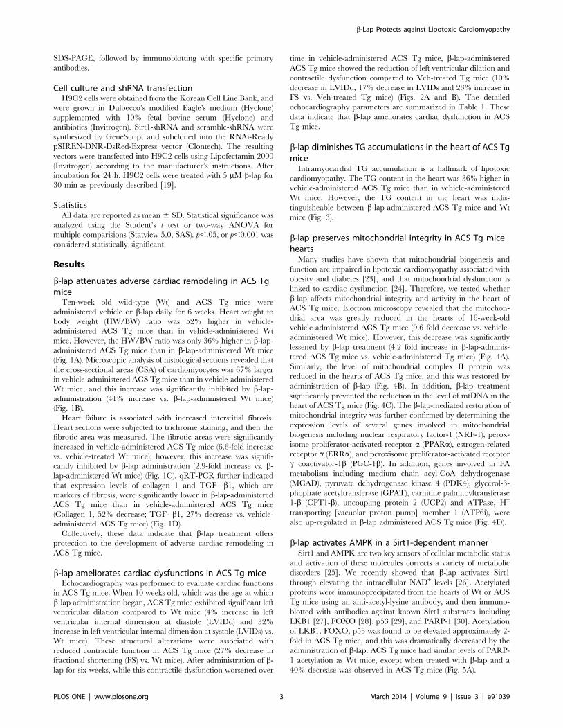
b-lap attenuates adverse cardiac remodeling in ACS Tgmice
Ten-week old wild-type (Wt) and ACS Tg mice were
administered vehicle or b-lap daily for 6 weeks. Heart weight to
body weight (HW/BW) ratio was 52% higher in vehicle-
administered ACS Tg mice than in vehicle-administered Wt
mice. However, the HW/BW ratio was only 36% higher in b-lap-
administered ACS Tg mice than in b-lap-administered Wt mice
(Fig. 1A). Microscopic analysis of histological sections revealed that
the cross-sectional areas (CSA) of cardiomyocytes was 67% larger
in vehicle-administered ACS Tg mice than in vehicle-administered
Wt mice, and this increase was significantly inhibited by b-lap-
administration (41% increase vs. b-lap-administered Wt mice)
(Fig. 1B).
Heart failure is associated with increased interstitial fibrosis.
Heart sections were subjected to trichrome staining, and then the
fibrotic area was measured. The fibrotic areas were significantly
increased in vehicle-administered ACS Tg mice (6.6-fold increase
vs. vehicle-treated Wt mice); however, this increase was signifi-
cantly inhibited by b-lap administration (2.9-fold increase vs. b-
lap-administered Wt mice) (Fig. 1C). qRT-PCR further indicated
that expression levels of collagen 1 and TGF- b1, which are
markers of fibrosis, were significantly lower in b-lap-administered
ACS Tg mice than in vehicle-administered ACS Tg mice
(Collagen 1, 52% decrease; TGF- b1, 27% decrease vs. vehicle-
administered ACS Tg mice) (Fig. 1D).
Collectively, these data indicate that b-lap treatment offers
protection to the development of adverse cardiac remodeling in
ACS Tg mice.
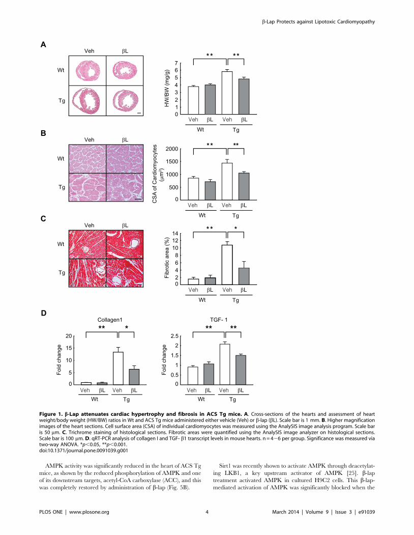
b-lap ameliorates cardiac dysfunctions in ACS Tg miceEchocardiography was performed to evaluate cardiac functions
in ACS Tg mice. When 10 weeks old, which was the age at which
b-lap administration began, ACS Tg mice exhibited significant left
ventricular dilation compared to Wt mice (4% increase in left
ventricular internal dimension at diastole (LVIDd) and 32%
increase in left ventricular internal dimension at systole (LVIDs) vs.
Wt mice). These structural alterations were associated with
reduced contractile function in ACS Tg mice (27% decrease in
fractional shortening (FS) vs. Wt mice). After administration of b-
lap for six weeks, while this contractile dysfunction worsened over
time in vehicle-administered ACS Tg mice, b-lap-administered
ACS Tg mice showed the reduction of left ventricular dilation and
contractile dysfunction compared to Veh-treated Tg mice (10%
decrease in LVIDd, 17% decrease in LVIDs and 23% increase in
FS vs. Veh-treated Tg mice) (Figs. 2A and B). The detailed
echocardiography parameters are summarized in Table 1. These
data indicate that b-lap ameliorates cardiac dysfunction in ACS
Tg mice.
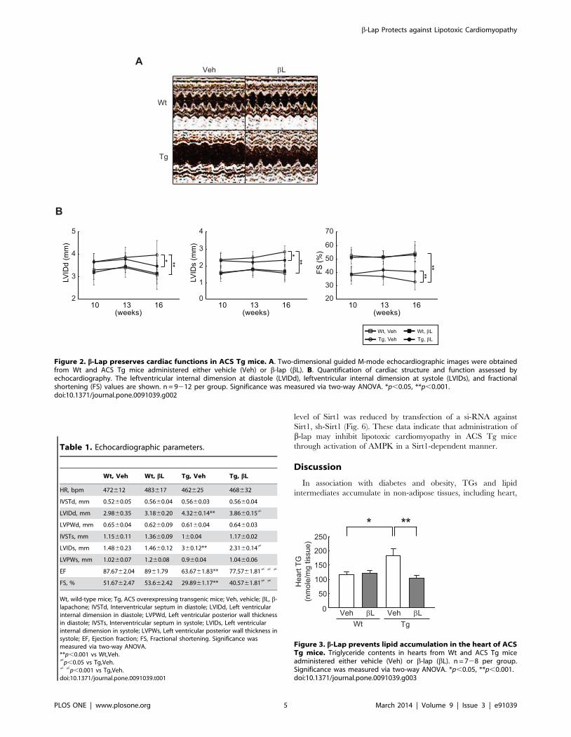
b-lap diminishes TG accumulations in the heart of ACS Tgmice
Intramyocardial TG accumulation is a hallmark of lipotoxic
cardiomyopathy. The TG content in the heart was 36% higher in
vehicle-administered ACS Tg mice than in vehicle-administered
Wt mice. However, the TG content in the heart was indis-
tinguisheable between b-lap-administered ACS Tg mice and Wt
mice (Fig. 3).
b-lap preserves mitochondrial integrity in ACS Tg micehearts
Many studies have shown that mitochondrial biogenesis and
function are impaired in lipotoxic cardiomyopathy associated with
obesity and diabetes [23], and that mitochondrial dysfunction is
linked to cardiac dysfunction [24]. Therefore, we tested whether
b-lap affects mitochondrial integrity and activity in the heart of
ACS Tg mice. Electron microscopy revealed that the mitochon-
drial area was greatly reduced in the hearts of 16-week-old
vehicle-administered ACS Tg mice (9.6 fold decrease vs. vehicle-
administered Wt mice). However, this decrease was significantly
lessened by b-lap treatment (4.2 fold increase in b-lap-adminis-
tered ACS Tg mice vs. vehicle-administered Tg mice) (Fig. 4A).
Similarly, the level of mitochondrial complex II protein was
reduced in the hearts of ACS Tg mice, and this was restored by
administration of b-lap (Fig. 4B). In addition, b-lap treatment
significantly prevented the reduction in the level of mtDNA in the
heart of ACS Tg mice (Fig. 4C). The b-lap-mediated restoration of
mitochondrial integrity was further confirmed by determining the
expression levels of several genes involved in mitochondrial
biogenesis including nuclear respiratory factor-1 (NRF-1), perox-
isome proliferator-activated receptor a (PPARa), estrogen-related
receptor a (ERRa), and peroxisome proliferator-activated receptor
c coactivator-1b (PGC-1b). In addition, genes involved in FA
metabolism including medium chain acyl-CoA dehydrogenase
(MCAD), pyruvate dehydrogenase kinase 4 (PDK4), glycerol-3-
phophate acetyltransferase (GPAT), carnitine palmitoyltransferase
1-b (CPT1-b), uncoupling protein 2 (UCP2) and ATPase, H+
transporting [vacuolar proton pump] member 1 (ATP6i), were
also up-regulated in b-lap administered ACS Tg mice (Fig. 4D).
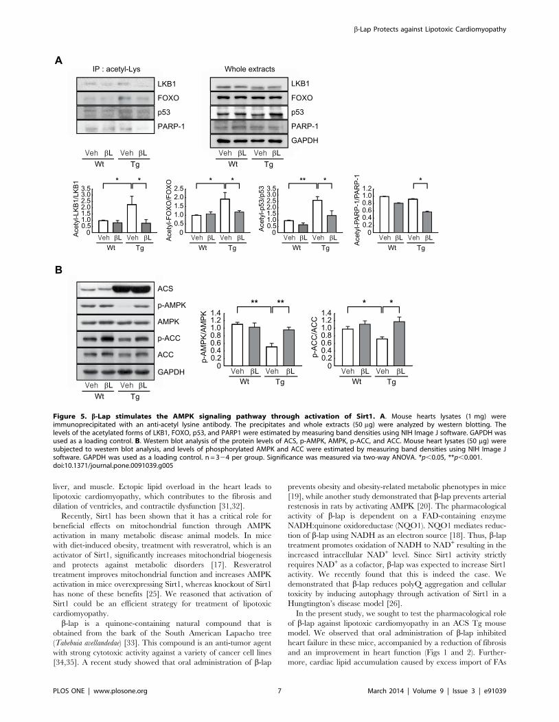
b-lap activates AMPK in a Sirt1-dependent mannerSirt1 and AMPK are two key sensors of cellular metabolic status
and activation of these molecules corrects a variety of metabolic
disorders [25]. We recently showed that b-lap activates Sirt1
through elevating the intracellular NAD+ levels [26]. Acetylated
proteins were immunoprecipitated from the hearts of Wt or ACS
Tg mice using an anti-acetyl-lysine antibody, and then immuno-
blotted with antibodies against known Sirt1 substrates including
LKB1 [27], FOXO [28], p53 [29], and PARP-1 [30]. Acetylation
of LKB1, FOXO, p53 was found to be elevated approximately 2-
fold in ACS Tg mice, and this was dramatically decreased by the
administration of b-lap. ACS Tg mice had similar levels of PARP-
1 acetylation as Wt mice, except when treated with b-lap and a
40% decrease was observed in ACS Tg mice (Fig. 5A).
b-Lap Protects against Lipotoxic Cardiomyopathy
PLOS ONE | www.plosone.org 3 March 2014 | Volume 9 | Issue 3 | e91039
AMPK activity was significantly reduced in the heart of ACS Tg
mice, as shown by the reduced phosphorylation of AMPK and one
of its downstream targets, acetyl-CoA carboxylase (ACC), and this
was completely restored by administration of b-lap (Fig. 5B).
Sirt1 was recently shown to activate AMPK through deacetylat-
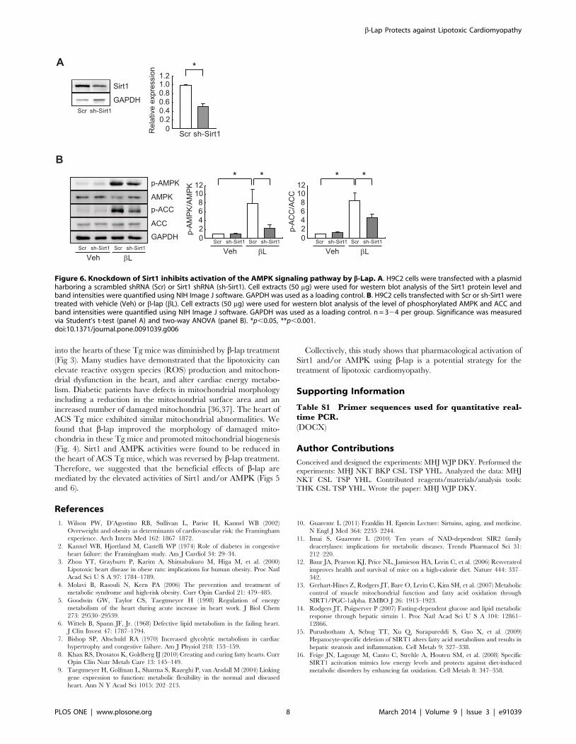
ing LKB1, a key upstream activator of AMPK [25]. b-lap
treatment activated AMPK in cultured H9C2 cells. This b-lap-
mediated activation of AMPK was significantly blocked when the
Figure 1. b-Lap attenuates cardiac hypertrophy and fibrosis in ACS Tg mice. A. Cross-sections of the hearts and assessment of heartweight/body weight (HW/BW) ratios in Wt and ACS Tg mice administered either vehicle (Veh) or b-lap (bL). Scale bar is 1 mm. B. Higher magnificationimages of the heart sections. Cell surface area (CSA) of individual cardiomyocytes was measured using the AnalySIS image analysis program. Scale baris 50 mm. C. Trichrome staining of histological sections. Fibrotic areas were quantified using the AnalySIS image analyzer on histological sections.Scale bar is 100 mm. D. qRT-PCR analysis of collagen I and TGF- b1 transcript levels in mouse hearts. n = 426 per group. Significance was measured viatwo-way ANOVA. *p,0.05, **p,0.001.doi:10.1371/journal.pone.0091039.g001
b-Lap Protects against Lipotoxic Cardiomyopathy
PLOS ONE | www.plosone.org 4 March 2014 | Volume 9 | Issue 3 | e91039
level of Sirt1 was reduced by transfection of a si-RNA against
Sirt1, sh-Sirt1 (Fig. 6). These data indicate that administration of
b-lap may inhibit lipotoxic cardiomyopathy in ACS Tg mice
through activation of AMPK in a Sirt1-dependent manner.
Discussion
In association with diabetes and obesity, TGs and lipid
intermediates accumulate in non-adipose tissues, including heart,
Figure 2. b-Lap preserves cardiac functions in ACS Tg mice. A. Two-dimensional guided M-mode echocardiographic images were obtainedfrom Wt and ACS Tg mice administered either vehicle (Veh) or b-lap (bL). B. Quantification of cardiac structure and function assessed byechocardiography. The leftventricular internal dimension at diastole (LVIDd), leftventricular internal dimension at systole (LVIDs), and fractionalshortening (FS) values are shown. n = 9212 per group. Significance was measured via two-way ANOVA. *p,0.05, **p,0.001.doi:10.1371/journal.pone.0091039.g002
Table 1. Echocardiographic parameters.
Wt, Veh Wt, bL Tg, Veh Tg, bL
HR, bpm 472612 483617 462625 468632
IVSTd, mm 0.5260.05 0.5660.04 0.5660.03 0.5660.04
LVIDd, mm 2.9860.35 3.1860.20 4.3260.14** 3.8660.15?
LVPWd, mm 0.6560.04 0.6260.09 0.6160.04 0.6460.03
IVSTs, mm 1.1560.11 1.3660.09 160.04 1.1760.02
LVIDs, mm 1.4860.23 1.4660.12 360.12** 2.3160.14?
LVPWs, mm 1.0260.07 1.260.08 0.960.04 1.0460.06
EF 87.6762.04 8961.79 63.6761.83** 77.5761.81? ? ?
FS, % 51.6762.47 53.662.42 29.8961.17** 40.5761.81? ?
Wt, wild-type mice; Tg, ACS overexpressing transgenic mice; Veh, vehicle; bL, b-lapachone; IVSTd, Interventricular septum in diastole; LVIDd, Left ventricularinternal dimension in diastole; LVPWd, Left ventricular posterior wall thicknessin diastole; IVSTs, Interventricular septum in systole; LVIDs, Left ventricularinternal dimension in systole; LVPWs, Left ventricular posterior wall thickness insystole; EF, Ejection fraction; FS, Fractional shortening. Significance wasmeasured via two-way ANOVA.**p,0.001 vs Wt,Veh.?p,0.05 vs Tg,Veh.? ?p,0.001 vs Tg,Veh.doi:10.1371/journal.pone.0091039.t001
Figure 3. b-Lap prevents lipid accumulation in the heart of ACSTg mice. Triglyceride contents in hearts from Wt and ACS Tg miceadministered either vehicle (Veh) or b-lap (bL). n = 728 per group.Significance was measured via two-way ANOVA. *p,0.05, **p,0.001.doi:10.1371/journal.pone.0091039.g003
b-Lap Protects against Lipotoxic Cardiomyopathy
PLOS ONE | www.plosone.org 5 March 2014 | Volume 9 | Issue 3 | e91039
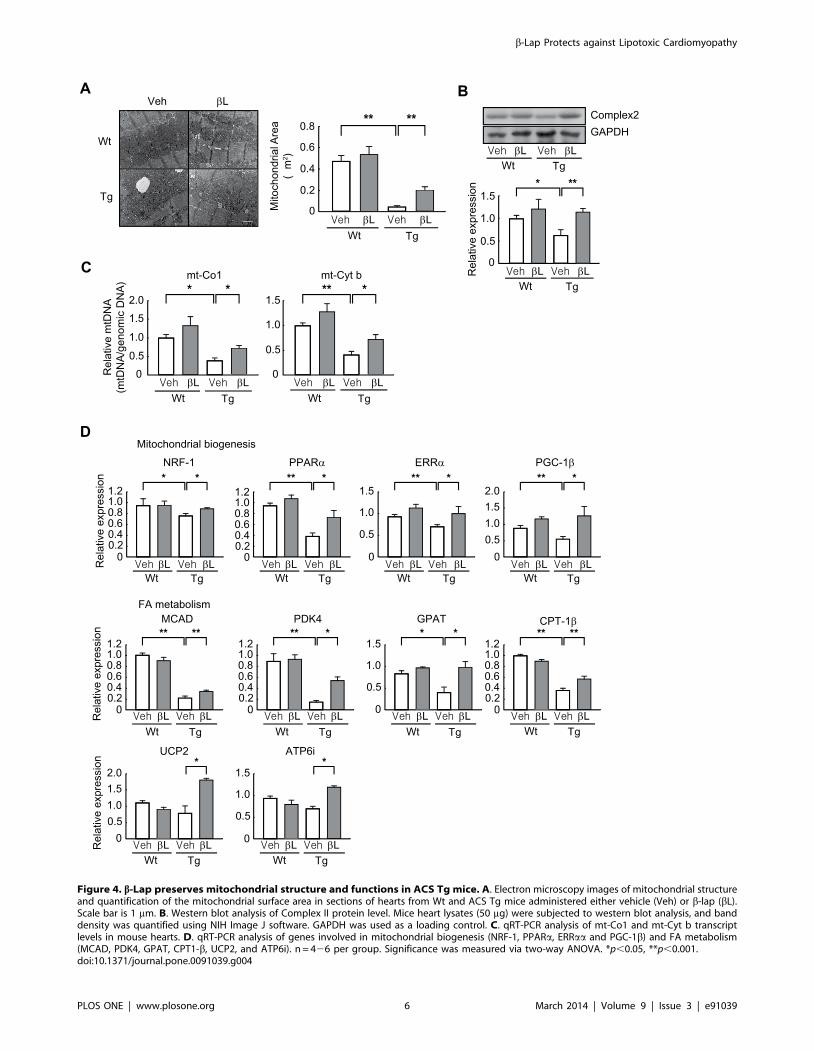
Figure 4. b-Lap preserves mitochondrial structure and functions in ACS Tg mice. A. Electron microscopy images of mitochondrial structureand quantification of the mitochondrial surface area in sections of hearts from Wt and ACS Tg mice administered either vehicle (Veh) or b-lap (bL).Scale bar is 1 mm. B. Western blot analysis of Complex II protein level. Mice heart lysates (50 mg) were subjected to western blot analysis, and banddensity was quantified using NIH Image J software. GAPDH was used as a loading control. C. qRT-PCR analysis of mt-Co1 and mt-Cyt b transcriptlevels in mouse hearts. D. qRT-PCR analysis of genes involved in mitochondrial biogenesis (NRF-1, PPARa, ERRaa and PGC-1b) and FA metabolism(MCAD, PDK4, GPAT, CPT1-b, UCP2, and ATP6i). n = 426 per group. Significance was measured via two-way ANOVA. *p,0.05, **p,0.001.doi:10.1371/journal.pone.0091039.g004
b-Lap Protects against Lipotoxic Cardiomyopathy
PLOS ONE | www.plosone.org 6 March 2014 | Volume 9 | Issue 3 | e91039
liver, and muscle. Ectopic lipid overload in the heart leads to
lipotoxic cardiomyopathy, which contributes to the fibrosis and
dilation of ventricles, and contractile dysfunction [31,32].
Recently, Sirt1 has been shown that it has a critical role for
beneficial effects on mitochondrial function through AMPK
activation in many metabolic disease animal models. In mice
with diet-induced obesity, treatment with resveratrol, which is an
activator of Sirt1, significantly increases mitochondrial biogenesis
and protects against metabolic disorders [17]. Resveratrol
treatment improves mitochondrial function and increases AMPK
activation in mice overexpressing Sirt1, whereas knockout of Sirt1
has none of these benefits [25]. We reasoned that activation of
Sirt1 could be an efficient strategy for treatment of lipotoxic
cardiomyopathy.
b-lap is a quinone-containing natural compound that is
obtained from the bark of the South American Lapacho tree
(Tabebuia avellandedae) [33]. This compound is an anti-tumor agent
with strong cytotoxic activity against a variety of cancer cell lines
[34,35]. A recent study showed that oral administration of b-lap
prevents obesity and obesity-related metabolic phenotypes in mice
[19], while another study demonstrated that b-lap prevents arterial
restenosis in rats by activating AMPK [20]. The pharmacological
activity of b-lap is dependent on a FAD-containing enzyme
NADH:quinone oxidoreductase (NQO1). NQO1 mediates reduc-
tion of b-lap using NADH as an electron source [18]. Thus, b-lap
treatment promotes oxidation of NADH to NAD+ resulting in the
increased intracellular NAD+ level. Since Sirt1 activity strictly
requires NAD+ as a cofactor, b-lap was expected to increase Sirt1
activity. We recently found that this is indeed the case. We
demonstrated that b-lap reduces polyQ aggregation and cellular
toxicity by inducing autophagy through activation of Sirt1 in a
Hungtington’s disease model [26].
In the present study, we sought to test the pharmacological role
of b-lap against lipotoxic cardiomyopathy in an ACS Tg mouse
model. We observed that oral administration of b-lap inhibited
heart failure in these mice, accompanied by a reduction of fibrosis
and an improvement in heart function (Figs 1 and 2). Further-
more, cardiac lipid accumulation caused by excess import of FAs
Figure 5. b-Lap stimulates the AMPK signaling pathway through activation of Sirt1. A. Mouse hearts lysates (1 mg) wereimmunoprecipitated with an anti-acetyl lysine antibody. The precipitates and whole extracts (50 mg) were analyzed by western blotting. Thelevels of the acetylated forms of LKB1, FOXO, p53, and PARP1 were estimated by measuring band densities using NIH Image J software. GAPDH wasused as a loading control. B. Western blot analysis of the protein levels of ACS, p-AMPK, AMPK, p-ACC, and ACC. Mouse heart lysates (50 mg) weresubjected to western blot analysis, and levels of phosphorylated AMPK and ACC were estimated by measuring band densities using NIH Image Jsoftware. GAPDH was used as a loading control. n = 324 per group. Significance was measured via two-way ANOVA. *p,0.05, **p,0.001.doi:10.1371/journal.pone.0091039.g005
b-Lap Protects against Lipotoxic Cardiomyopathy
PLOS ONE | www.plosone.org 7 March 2014 | Volume 9 | Issue 3 | e91039
into the hearts of these Tg mice was diminished by b-lap treatment
(Fig 3). Many studies have demonstrated that the lipotoxicity can
elevate reactive oxygen species (ROS) production and mitochon-
drial dysfunction in the heart, and alter cardiac energy metabo-
lism. Diabetic patients have defects in mitochondrial morphology
including a reduction in the mitochondrial surface area and an
increased number of damaged mitochondria [36,37]. The heart of
ACS Tg mice exhibited similar mitochondrial abnormalities. We
found that b-lap improved the morphology of damaged mito-
chondria in these Tg mice and promoted mitochondrial biogenesis
(Fig. 4). Sirt1 and AMPK activities were found to be reduced in
the heart of ACS Tg mice, which was reversed by b-lap treatment.
Therefore, we suggested that the beneficial effects of b-lap are
mediated by the elevated activities of Sirt1 and/or AMPK (Figs 5
and 6).
Collectively, this study shows that pharmacological activation of
Sirt1 and/or AMPK using b-lap is a potential strategy for the
treatment of lipotoxic cardiomyopathy.
Supporting Information
Table S1 Primer sequences used for quantitative real-time PCR.
(DOCX)
Author Contributions
Conceived and designed the experiments: MHJ WJP DKY. Performed the
experiments: MHJ NKT BKP CSL TSP YHL. Analyzed the data: MHJ
NKT CSL TSP YHL. Contributed reagents/materials/analysis tools:
THK CSL TSP YHL. Wrote the paper: MHJ WJP DKY.
References
1. Wilson PW, D’Agostino RB, Sullivan L, Parise H, Kannel WB (2002)
Overweight and obesity as determinants of cardiovascular risk: the Framingham
experience. Arch Intern Med 162: 1867–1872.
2. Kannel WB, Hjortland M, Castelli WP (1974) Role of diabetes in congestive
heart failure: the Framingham study. Am J Cardiol 34: 29–34.
3. Zhou YT, Grayburn P, Karim A, Shimabukuro M, Higa M, et al. (2000)
Lipotoxic heart disease in obese rats: implications for human obesity. Proc Natl
Acad Sci U S A 97: 1784–1789.
4. Molavi B, Rasouli N, Kern PA (2006) The prevention and treatment of
metabolic syndrome and high-risk obesity. Curr Opin Cardiol 21: 479–485.
5. Goodwin GW, Taylor CS, Taegtmeyer H (1998) Regulation of energy
metabolism of the heart during acute increase in heart work. J Biol Chem
273: 29530–29539.
6. Wittels B, Spann JF, Jr. (1968) Defective lipid metabolism in the failing heart.
J Clin Invest 47: 1787–1794.
7. Bishop SP, Altschuld RA (1970) Increased glycolytic metabolism in cardiac
hypertrophy and congestive failure. Am J Physiol 218: 153–159.
8. Khan RS, Drosatos K, Goldberg IJ (2010) Creating and curing fatty hearts. Curr
Opin Clin Nutr Metab Care 13: 145–149.
9. Taegtmeyer H, Golfman L, Sharma S, Razeghi P, van Arsdall M (2004) Linking
gene expression to function: metabolic flexibility in the normal and diseased
heart. Ann N Y Acad Sci 1015: 202–213.
10. Guarente L (2011) Franklin H. Epstein Lecture: Sirtuins, aging, and medicine.
N Engl J Med 364: 2235–2244.
11. Imai S, Guarente L (2010) Ten years of NAD-dependent SIR2 family
deacetylases: implications for metabolic diseases. Trends Pharmacol Sci 31:
212–220.
12. Baur JA, Pearson KJ, Price NL, Jamieson HA, Lerin C, et al. (2006) Resveratrol
improves health and survival of mice on a high-calorie diet. Nature 444: 337–
342.
13. Gerhart-Hines Z, Rodgers JT, Bare O, Lerin C, Kim SH, et al. (2007) Metabolic
control of muscle mitochondrial function and fatty acid oxidation through
SIRT1/PGC-1alpha. EMBO J 26: 1913–1923.
14. Rodgers JT, Puigserver P (2007) Fasting-dependent glucose and lipid metabolic
response through hepatic sirtuin 1. Proc Natl Acad Sci U S A 104: 12861–
12866.
15. Purushotham A, Schug TT, Xu Q, Surapureddi S, Guo X, et al. (2009)
Hepatocyte-specific deletion of SIRT1 alters fatty acid metabolism and results in
hepatic steatosis and inflammation. Cell Metab 9: 327–338.
16. Feige JN, Lagouge M, Canto C, Strehle A, Houten SM, et al. (2008) Specific
SIRT1 activation mimics low energy levels and protects against diet-induced
metabolic disorders by enhancing fat oxidation. Cell Metab 8: 347–358.
Figure 6. Knockdown of Sirt1 inhibits activation of the AMPK signaling pathway by b-Lap. A. H9C2 cells were transfected with a plasmidharboring a scrambled shRNA (Scr) or Sirt1 shRNA (sh-Sirt1). Cell extracts (50 mg) were used for western blot analysis of the Sirt1 protein level andband intensities were quantified using NIH Image J software. GAPDH was used as a loading control. B. H9C2 cells transfected with Scr or sh-Sirt1 weretreated with vehicle (Veh) or b-lap (bL). Cell extracts (50 mg) were used for western blot analysis of the level of phosphorylated AMPK and ACC andband intensities were quantified using NIH Image J software. GAPDH was used as a loading control. n = 324 per group. Significance was measuredvia Student’s t-test (panel A) and two-way ANOVA (panel B). *p,0.05, **p,0.001.doi:10.1371/journal.pone.0091039.g006
b-Lap Protects against Lipotoxic Cardiomyopathy
PLOS ONE | www.plosone.org 8 March 2014 | Volume 9 | Issue 3 | e91039
17. Lagouge M, Argmann C, Gerhart-Hines Z, Meziane H, Lerin C, et al. (2006)
Resveratrol improves mitochondrial function and protects against metabolicdisease by activating SIRT1 and PGC-1alpha. Cell 127: 1109–1122.
18. Jaiswal AK (2000) Regulation of genes encoding NAD(P)H:quinone oxidore-
ductases. Free Radic Biol Med 29: 254–262.19. Hwang JH, Kim DW, Jo EJ, Kim YK, Jo YS, et al. (2009) Pharmacological
stimulation of NADH oxidation ameliorates obesity and related phenotypes inmice. Diabetes 58: 965–974.
20. Kim SY, Jeoung NH, Oh CJ, Choi YK, Lee HJ, et al. (2009) Activation of
NAD(P)H:quinone oxidoreductase 1 prevents arterial restenosis by suppressingvascular smooth muscle cell proliferation. Circ Res 104: 842–850.
21. Chiu HC, Kovacs A, Ford DA, Hsu FF, Garcia R, et al. (2001) A novel mousemodel of lipotoxic cardiomyopathy. J Clin Invest 107: 813–822.
22. Spurr AR (1969) A low-viscosity epoxy resin embedding medium for electronmicroscopy. J Ultrastruct Res 26: 31–43.
23. Boudina S, Sena S, Theobald H, Sheng X, Wright JJ, et al. (2007)
Mitochondrial energetics in the heart in obesity-related diabetes: direct evidencefor increased uncoupled respiration and activation of uncoupling proteins.
Diabetes 56: 2457–2466.24. Tsutsui H, Kinugawa S, Matsushima S (2009) Mitochondrial oxidative stress
and dysfunction in myocardial remodelling. Cardiovasc Res 81: 449–456.
25. Price NL, Gomes AP, Ling AJ, Duarte FV, Martin-Montalvo A, et al. (2012)SIRT1 is required for AMPK activation and the beneficial effects of resveratrol
on mitochondrial function. Cell Metab 15: 675–690.26. Shin BH, Lim Y, Oh HJ, Park SM, Lee SK, et al. (2013) Pharmacological
activation of Sirt1 ameliorates polyglutamine-induced toxicity through theregulation of autophagy. PLoS One 8: e64953.
27. Lan F, Cacicedo JM, Ruderman N, Ido Y (2008) SIRT1 modulation of the
acetylation status, cytosolic localization, and activity of LKB1. Possible role inAMP-activated protein kinase activation. J Biol Chem 283: 27628–27635.
28. Brunet A, Sweeney LB, Sturgill JF, Chua KF, Greer PL, et al. (2004) Stress-
dependent regulation of FOXO transcription factors by the SIRT1 deacetylase.Science 303: 2011–2015.
29. Vaziri H, Dessain SK, Ng Eaton E, Imai SI, Frye RA, et al. (2001)
hSIR2(SIRT1) functions as an NAD-dependent p53 deacetylase. Cell 107:149–159.
30. Rajamohan SB, Pillai VB, Gupta M, Sundaresan NR, Birukov KG, et al. (2009)SIRT1 promotes cell survival under stress by deacetylation-dependent
deactivation of poly(ADP-ribose) polymerase 1. Mol Cell Biol 29: 4116–4129.
31. Young ME, Guthrie PH, Razeghi P, Leighton B, Abbasi S, et al. (2002)Impaired long-chain fatty acid oxidation and contractile dysfunction in the obese
Zucker rat heart. Diabetes 51: 2587–2595.32. Sharma S, Adrogue JV, Golfman L, Uray I, Lemm J, et al. (2004)
Intramyocardial lipid accumulation in the failing human heart resembles thelipotoxic rat heart. FASEB J 18: 1692–1700.
33. Schaffner-Sabba K, Schmidt-Ruppin KH, Wehrli W, Schuerch AR, Wasley JW
(1984) beta-Lapachone: synthesis of derivatives and activities in tumor models.J Med Chem 27: 990–994.
34. Pink JJ, Wuerzberger-Davis S, Tagliarino C, Planchon SM, Yang X, et al.(2000) Activation of a cysteine protease in MCF-7 and T47D breast cancer cells
during beta-lapachone-mediated apoptosis. Exp Cell Res 255: 144–155.
35. Choi YH, Kang HS, Yoo MA (2003) Suppression of human prostate cancer cellgrowth by beta-lapachone via down-regulation of pRB phosphorylation and
induction of Cdk inhibitor p21(WAF1/CIP1). J Biochem Mol Biol 36: 223–229.36. Kelley DE, He J, Menshikova EV, Ritov VB (2002) Dysfunction of
mitochondria in human skeletal muscle in type 2 diabetes. Diabetes 51: 2944–2950.
37. Ritov VB, Menshikova EV, He J, Ferrell RE, Goodpaster BH, et al. (2005)
Deficiency of subsarcolemmal mitochondria in obesity and type 2 diabetes.Diabetes 54: 8–14.
b-Lap Protects against Lipotoxic Cardiomyopathy
PLOS ONE | www.plosone.org 9 March 2014 | Volume 9 | Issue 3 | e91039
Related Documents