Enrico M. Staderini UWB RADARS IN MEDICINE “Tor Vergata University of Rome” Department of Biopathology and Imaging Medical Physics Center c/o Ingegneria Industriale Via di Tor Vergata, 110 00133 Rome – ITALY ++39 06 72597196 (fax ++39 06 72597208) mailto://[email protected] http://www.uniroma2.it/fismed/UWBradar/ (Italian UWB radar site under construction) http://www.uniroma2.it/fismed/staff/Stadero/ (Italian UWB radar maniac site under construction)

Welcome message from author
This document is posted to help you gain knowledge. Please leave a comment to let me know what you think about it! Share it to your friends and learn new things together.
Transcript

Enrico M. Staderini
UWB RADARS
IN MEDICINE
“Tor Vergata University of Rome”Department of Biopathology and Imaging
Medical Physics Centerc/o Ingegneria Industriale
Via di Tor Vergata, 110 00133 Rome – ITALY++39 06 72597196 (fax ++39 06 72597208)
mailto://[email protected]
http://www.uniroma2.it/fismed/UWBradar/ (Italian UWB radar site under construction)http://www.uniroma2.it/fismed/staff/Stadero/ (Italian UWB radar maniac site under construction)
2
ABSTRACT
A review is given of present state of the art, and likely to be developed orfuturistic, biomedical applications of Ultra Wide Band (UWB) radar.
UWB radar is something like a mix of conventional radar (RAdio DetectionAnd Ranging) technology and spread spectrum radio (SSR) technology, both di-rectly coming from military applications.
What renders UWB radar very much interesting is the possibility to probe themotion of the internal organs of the human body with a remote non-contact ap-proach which is unique at present time. The very low cost, the high miniaturizationcapability and the environmental friendliness due to the very low electromagneticenergy emission are other aspects of specific interest of the technique.
A review of the biomedical applications known at present is presented basedon the published documents and information derived from the web sites of the In-stitutions carrying on the research (gray literature). Just a paper only appears tohave been published about the applications of UWB radar to actual medical prob-lems, although many papers on the interaction of radio frequency energy with theliving tissues, and safety issues, can be found in the literature.
An original and very simple model of UWB radar pulse-echo interaction withthe human thorax is presented to show the feasibility of a real heart motion probe.
UWB radar technology, which is quite unknown at present time to the gen-eral public, and physicians as well, is about to strongly impact in everyday life andin the medical field as well, making it possible to design a novel kind of non-invasive measurement and monitoring devices.
KEYWORDS
UWB ultra wide-band, RADAR RAdio Detection And Ranging, SpreadSpectrum, time domain, range gate, noise coding, SAR Synthetic Aperture Radar,SAR imaging, GPR Ground Penetrating Radar, foliage penetrating radar, opticalradar, near-infrared spectroscopy, FDTD Finite Differences Time Domain method,pulse emitting antennas.

3
1. INTRODUCING UWB RADAR TECHNOLOGY
The overall conceptual working mode of a UWB radar system resembles thatof ultrasonic echo transducers used in many applications, from autofocus camerasto proximity and range detectors. The main and fundamental difference being that,on the contrary to ultrasounds, electromagnetic pulses propagate through walls,ground, ice, mud, concrete and the human body as well.
Although many researchers around have been working on UWB technologyfor many years, a great concern on UWB radar arose in 1993 when it is reportedthat a Lawrence Livermore National Laboratory’s (LLNL) engineer, ThomasMcEwan, discovered a possibly new and original implementation of an UWB ra-dar. While working on a new high speed low cost sampler for pulse laser research,McEwan developed a system which was called MIR: Micropower Impulse Radar.The patents on MIR technology describe a wonderful spectrum of applicationscoming from the low cost MIR technology: from plastic bodied mine detection, toremote vital signs monitoring, to the ‘3-D radar camera’. Of course the ultimateapplication being the futuristic ‘X-ray specs’ (until now just a science fiction de-vice).
As a matter of fact the United States Patents and Trademark Office has re-cently rejected a few key claims of the MIR radar patent. Those claims were ruledas “anticipated”, meaning previously claimed by Fullerton’s 1987 Patent whose as-signee is Time Domain Corp. Huntsville, Alabama.
Whatever should be the story regarding the intellectual property of the sys-tem, we are now interested, from the scientific point of view, on the biomedicalapplications of UWB radar which yet need a lot of research to get a viable, clinicaloriented, device.
2. THE RATIONALE FOR BIOMEDICAL APPLICATIONS
Electromagnetic pulses coming from a UWB radar are able to probe the hu-man body. In an application developed at LLNL, a UWB radar was able to detect,non invasively, the movements of the heart wall. This is because there do exist adefinite difference in reflection magnitude between the heart muscle and the bloodit pushes into the vascular tree. In the two patents ([1], [2]) awarded to McEwanon medical UWB radar, a rough description of the physical principle of operation isgiven. As the impedance of the cardiac muscle is in the order of 60 ohms and theimpedance of blood is about 50 ohms, it can be expected a roughly 10% reflectionmagnitude of the radio frequency energy at the heart muscle/blood boundary. Ac-cording to McEwan’s patent, let µ0 and ε0 be respectively the permeability and
permittivity of vacuum: the impedance of heart muscle is Z(heart) = µ ε ε0 0 60r =ohms, where εr is the permittivity of muscle (~40), while the impedance of blood(εr=60), is 49 ohms. The reflection coefficient, defined as (Y-1)/(Y+1) whereY=Z(heart)/Z(blood), gives a 9.9% return fraction of the radiated pulse.
4
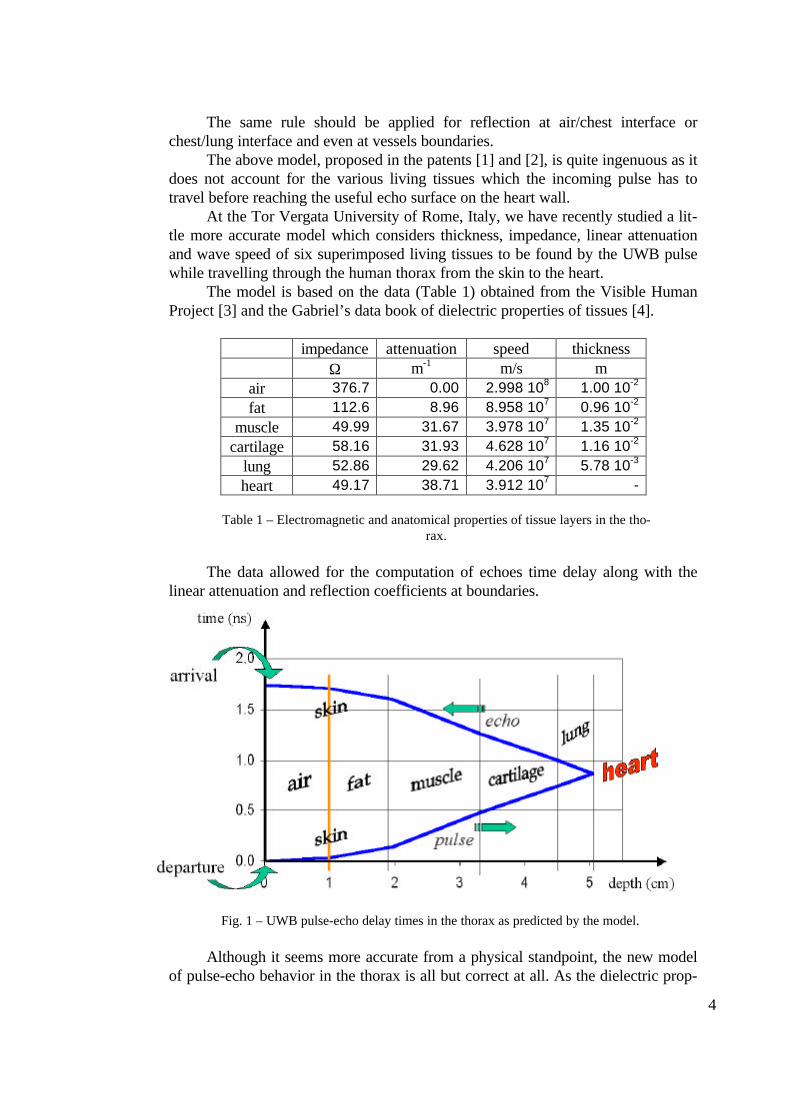
The same rule should be applied for reflection at air/chest interface orchest/lung interface and even at vessels boundaries.
The above model, proposed in the patents [1] and [2], is quite ingenuous as itdoes not account for the various living tissues which the incoming pulse has totravel before reaching the useful echo surface on the heart wall.
At the Tor Vergata University of Rome, Italy, we have recently studied a lit-tle more accurate model which considers thickness, impedance, linear attenuationand wave speed of six superimposed living tissues to be found by the UWB pulsewhile travelling through the human thorax from the skin to the heart.
The model is based on the data (Table 1) obtained from the Visible HumanProject [3] and the Gabriel’s data book of dielectric properties of tissues [4].
impedance attenuation speed thicknessΩ m-1 m/s m
air 376.7 0.00 2.998 108 1.00 10-2
fat 112.6 8.96 8.958 107 0.96 10-2
muscle 49.99 31.67 3.978 107 1.35 10-2
cartilage 58.16 31.93 4.628 107 1.16 10-2
lung 52.86 29.62 4.206 107 5.78 10-3
heart 49.17 38.71 3.912 107 -
Table 1 – Electromagnetic and anatomical properties of tissue layers in the tho-rax.
The data allowed for the computation of echoes time delay along with thelinear attenuation and reflection coefficients at boundaries.
Fig. 1 – UWB pulse-echo delay times in the thorax as predicted by the model.
Although it seems more accurate from a physical standpoint, the new modelof pulse-echo behavior in the thorax is all but correct at all. As the dielectric prop-

5
erties used were those measured on actual living tissues using a continuous wave at1500 MHz [4], the model remains intrinsically wrong.
Indeed for an effective model to be developed, we need ultra wide-band di-electric properties, not narrow band ones (although in the microwave region). Thismeans that a convolution method, or a Finite Differences Time Domain techniquelike that already employed in UWB antenna calculation [5], should be used.
Also, both the real part and the imaginary one of the reflection coefficients atthe boundaries should be considered, this is because the UWB receiving correlator,working in the time domain, is, by design, strongly sensitive to phase errors.
Another point to be addressed is that of the receiving correlator itself. Weneed to assess what amount is actually its phase sensitivity.
Fig. 2 – Model predicted attenuation of pulse-echo intensity travelling from the transmittingantenna to the receiving antenna. Each step accounts for echo at the boundary. Decreasing
of the curve accounts for linear attenuation in the tissue (imaginary part of reflection coeffi-cient and multiple reflections are ignored).
In a nutshell: we know that a heart motion-related signal is obtained out of aUWB radar device aimed at the thorax, but what are we actually measuring? Towhat extent correlator performances, pulse shape and tissues properties affect theintensity and morphology of the recovered signal?
These problems ask for some clear answers to reach an adequate physicalunderstanding of medical UWB radar and subsequent clinical viability and accept-ability of the technique. Accurate modeling of the phenomena with correct and ex-tended electromagnetic measures should help advancement of science in this field.
This evidence opens up a whole new area for the non-invasive measures andmonitoring of human body functions. In practice any object of adequate size can bemonitored. Vocal cords, vessels, bowels, heart, lung, chest, bladder and fetus aregood candidates for the UWB radar probing. As a matter of fact radar monitoring
6
of human physiologic functions was considered as early as the 70’s [6], [7], [8],but any further development was impeded by the cumbersome and expensive tech-nology of those times.
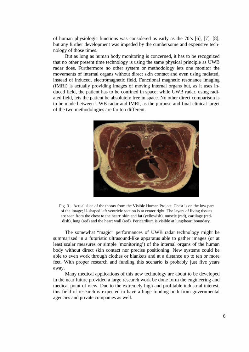
But as long as human body monitoring is concerned, it has to be recognizedthat no other present time technology is using the same physical principle as UWBradar does. Furthermore no other system or methodology lets one monitor themovements of internal organs without direct skin contact and even using radiated,instead of induced, electromagnetic field. Functional magnetic resonance imaging(fMRI) is actually providing images of moving internal organs but, as it uses in-duced field, the patient has to be confined in space; while UWB radar, using radi-ated field, lets the patient be absolutely free in space. No other direct comparison isto be made between UWB radar and fMRI, as the purpose and final clinical targetof the two methodologies are far too different.
Fig. 3 – Actual slice of the thorax from the Visible Human Project. Chest is on the low partof the image; U-shaped left ventricle section is at center right. The layers of living tissuesare seen from the chest to the heart: skin and fat (yellowish), muscle (red), cartilage (red-dish), lung (red) and the heart wall (red). Pericardium is visible at lung/heart boundary.
The somewhat “magic” performances of UWB radar technology might besummarized in a futuristic ultrasound-like apparatus able to gather images (or atleast scalar measures or simple ‘monitoring’) of the internal organs of the humanbody without direct skin contact nor precise positioning. New systems could beable to even work through clothes or blankets and at a distance up to ten or morefeet. With proper research and funding this scenario is probably just five yearsaway.
Many medical applications of this new technology are about to be developedin the near future provided a large research work be done form the engineering andmedical point of view. Due to the extremely high and profitable industrial interest,this field of research is expected to have a huge funding both from governmentalagencies and private companies as well.
7
3. THE BIOMEDICAL APPLICATIONS OF UWB RADAR TECHNIQUE
By using a particular non-resonating antennas (to avoid ringing), the MIRwas operated as a cardiovascular monitor to detect cardiac contractions, arterialwall motion and a breath monitor to detect respiratory movements.
Furthermore MIR system, used as electromagnetic sensors (EM-sensors) areexperimented for assessing vocal cord activity and speech parameters.
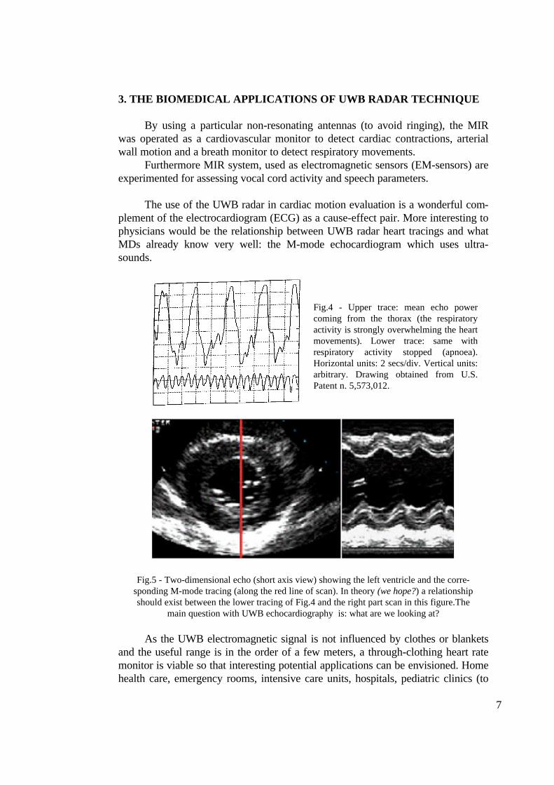
The use of the UWB radar in cardiac motion evaluation is a wonderful com-plement of the electrocardiogram (ECG) as a cause-effect pair. More interesting tophysicians would be the relationship between UWB radar heart tracings and whatMDs already know very well: the M-mode echocardiogram which uses ultra-sounds.
Fig.4 - Upper trace: mean echo powercoming from the thorax (the respiratoryactivity is strongly overwhelming the heartmovements). Lower trace: same withrespiratory activity stopped (apnoea).Horizontal units: 2 secs/div. Vertical units:arbitrary. Drawing obtained from U.S.Patent n. 5,573,012.
Fig.5 - Two-dimensional echo (short axis view) showing the left ventricle and the corre-sponding M-mode tracing (along the red line of scan). In theory (we hope?) a relationshipshould exist between the lower tracing of Fig.4 and the right part scan in this figure.The
main question with UWB echocardiography is: what are we looking at?
As the UWB electromagnetic signal is not influenced by clothes or blanketsand the useful range is in the order of a few meters, a through-clothing heart ratemonitor is viable so that interesting potential applications can be envisioned. Homehealth care, emergency rooms, intensive care units, hospitals, pediatric clinics (to
8
alert for the Sudden Infant Death Syndrome, SIDS), rescue operations (to look forsome heart beating under ruins, or soil, or snow) or law enforcement are just someof potential areas of application.
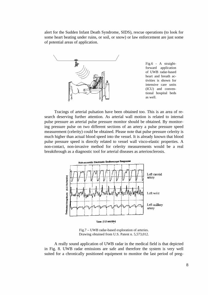
Fig.6 - A straight-forward applicationof UWB radar-basedheart and breath ac-tivities is shown forintensive care units(ICU) and conven-tional hospital bedsas well.
Tracings of arterial pulsation have been obtained too. This is an area of re-search deserving further attention. As arterial wall motion is related to internalpulse pressure an arterial pulse pressure monitor should be obtained. By monitor-ing pressure pulse on two different sections of an artery a pulse pressure speedmeasurement (celerity) could be obtained. Please note that pulse pressure celerity ismuch higher than actual blood speed into the vessel. It is already known that bloodpulse pressure speed is directly related to vessel wall visco-elastic properties. Anon-contact, non-invasive method for celerity measurements would be a realbreakthrough as a diagnostic tool for arterial diseases as arteriosclerosis.
Fig.7 – UWB radar-based exploration of arteries.Drawing obtained from U.S. Patent n. 5,573,012.
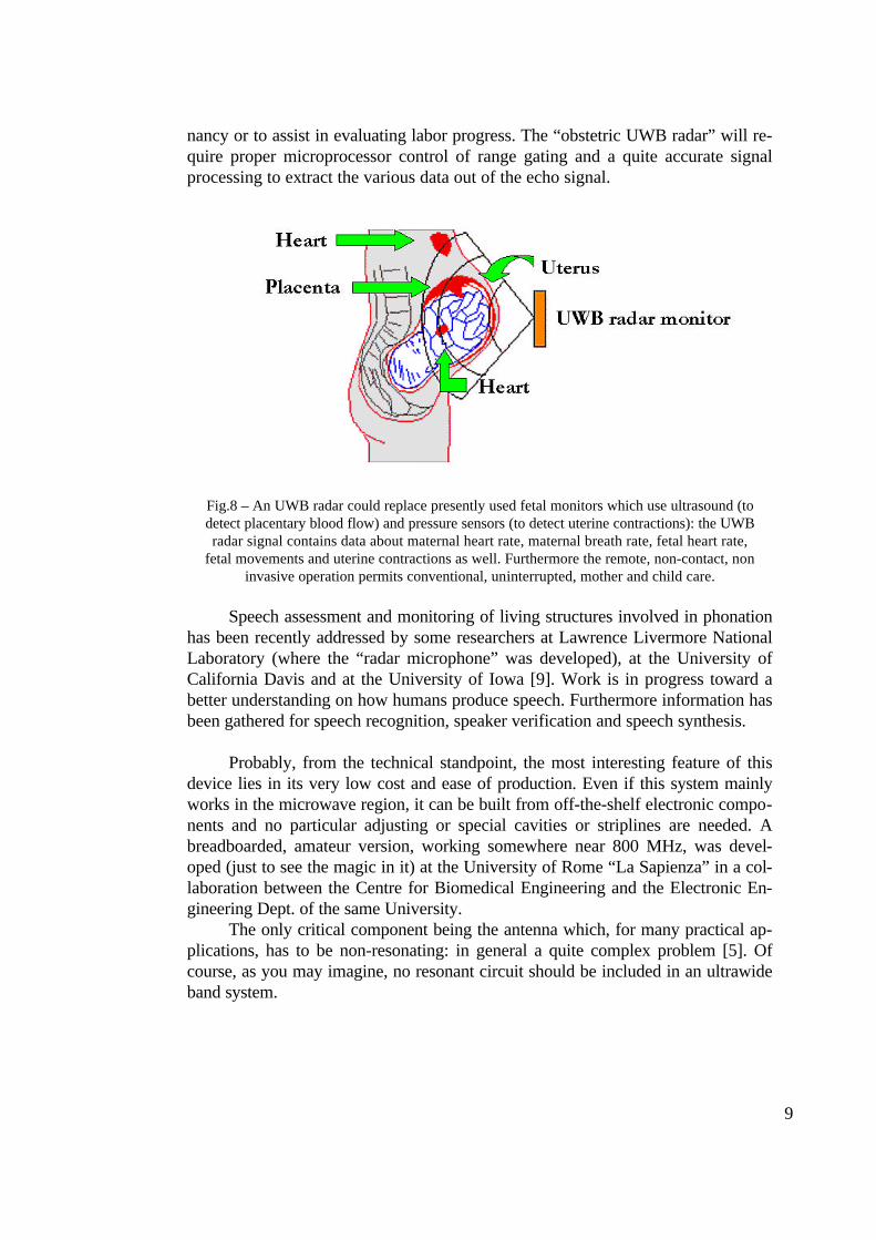
A really sound application of UWB radar in the medical field is that depictedin Fig. 8. UWB radar emissions are safe and therefore the system is very wellsuited for a chronically positioned equipment to monitor the last period of preg-
9
nancy or to assist in evaluating labor progress. The “obstetric UWB radar” will re-quire proper microprocessor control of range gating and a quite accurate signalprocessing to extract the various data out of the echo signal.
Fig.8 – An UWB radar could replace presently used fetal monitors which use ultrasound (todetect placentary blood flow) and pressure sensors (to detect uterine contractions): the UWBradar signal contains data about maternal heart rate, maternal breath rate, fetal heart rate,
fetal movements and uterine contractions as well. Furthermore the remote, non-contact, noninvasive operation permits conventional, uninterrupted, mother and child care.
Speech assessment and monitoring of living structures involved in phonationhas been recently addressed by some researchers at Lawrence Livermore NationalLaboratory (where the “radar microphone” was developed), at the University ofCalifornia Davis and at the University of Iowa [9]. Work is in progress toward abetter understanding on how humans produce speech. Furthermore information hasbeen gathered for speech recognition, speaker verification and speech synthesis.
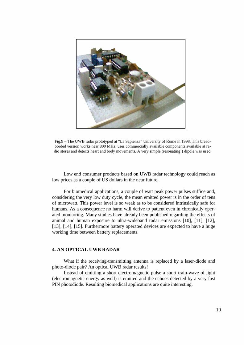
Probably, from the technical standpoint, the most interesting feature of thisdevice lies in its very low cost and ease of production. Even if this system mainlyworks in the microwave region, it can be built from off-the-shelf electronic compo-nents and no particular adjusting or special cavities or striplines are needed. Abreadboarded, amateur version, working somewhere near 800 MHz, was devel-oped (just to see the magic in it) at the University of Rome “La Sapienza” in a col-laboration between the Centre for Biomedical Engineering and the Electronic En-gineering Dept. of the same University.
The only critical component being the antenna which, for many practical ap-plications, has to be non-resonating: in general a quite complex problem [5]. Ofcourse, as you may imagine, no resonant circuit should be included in an ultrawideband system.
10
Fig.9 – The UWB radar prototyped at “La Sapienza” University of Rome in 1998. This bread-borded version works near 800 MHz, uses commercially available components available at ra-dio stores and detects heart and body movements. A very simple (resonating!) dipole was used.
Low end consumer products based on UWB radar technology could reach aslow prices as a couple of US dollars in the near future.
For biomedical applications, a couple of watt peak power pulses suffice and,considering the very low duty cycle, the mean emitted power is in the order of tensof microwatt. This power level is so weak as to be considered intrinsically safe forhumans. As a consequence no harm will derive to patient even in chronically oper-ated monitoring. Many studies have already been published regarding the effects ofanimal and human exposure to ultra-wideband radar emissions [10], [11], [12],[13], [14], [15]. Furthermore battery operated devices are expected to have a hugeworking time between battery replacements.
4. AN OPTICAL UWB RADAR
What if the receiving-transmitting antenna is replaced by a laser-diode andphoto-diode pair? An optical UWB radar results!
Instead of emitting a short electromagnetic pulse a short train-wave of light(electromagnetic energy as well) is emitted and the echoes detected by a very fastPIN photodiode. Resulting biomedical applications are quite interesting.
11
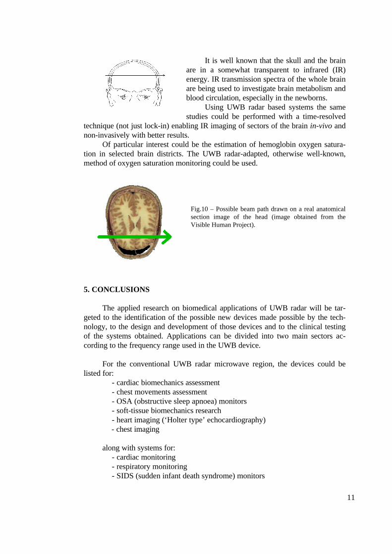
It is well known that the skull and the brainare in a somewhat transparent to infrared (IR)energy. IR transmission spectra of the whole brainare being used to investigate brain metabolism andblood circulation, especially in the newborns.
Using UWB radar based systems the samestudies could be performed with a time-resolved
technique (not just lock-in) enabling IR imaging of sectors of the brain in-vivo andnon-invasively with better results.
Of particular interest could be the estimation of hemoglobin oxygen satura-tion in selected brain districts. The UWB radar-adapted, otherwise well-known,method of oxygen saturation monitoring could be used.
Fig.10 – Possible beam path drawn on a real anatomicalsection image of the head (image obtained from theVisible Human Project).
5. CONCLUSIONS
The applied research on biomedical applications of UWB radar will be tar-geted to the identification of the possible new devices made possible by the tech-nology, to the design and development of those devices and to the clinical testingof the systems obtained. Applications can be divided into two main sectors ac-cording to the frequency range used in the UWB device.
For the conventional UWB radar microwave region, the devices could belisted for:
- cardiac biomechanics assessment- chest movements assessment- OSA (obstructive sleep apnoea) monitors- soft-tissue biomechanics research- heart imaging (‘Holter type’ echocardiography)- chest imaging
along with systems for:- cardiac monitoring- respiratory monitoring- SIDS (sudden infant death syndrome) monitors
12
- vocal tract studying
If an IR laser diode is used as the antenna, a more common radar is obtained(actually an hybrid between a narrow band and a wide band radar) which emits ashort packet of electromagnetic waves whose echoes are sampled using a ‘conven-tional’ UWB receiver equipped with a PIN photodiode. A series of possible de-vices can be listed for the IR region:
- non-invasive biochemical study of soft tissues (exp. brain)- non-invasive study of metabolic processes- IR spectral imaging.
Let’s conclude with a dream: that of non-invasive, continuous (Holter-type),monitoring of cardiac output. With acceptable limitations, it could be in fact possi-ble by measuring, with an UWB radar, the difference in distance between the ante-rior and posterior left ventricle walls. This could give an estimation of ventricle’svolume variations during the heart cycle and eventually give systolic ejection vol-umes and cardiac output. With the rapid technology advancements we are experi-encing these times, it only needs to have a dream for that becomes true.
6. REFERENCES
[1] McEwan: “Body monitoring and imaging apparatus and method”. United States Pat-ent 5,766,208 Jun. 16, 1998.
[2] McEwan: “Body monitoring and imaging apparatus and method”. United States Pat-ent 5,573,012 Nov. 12, 1996.
[3] Chang Y.J.: “The NPAC Visible Human Viewer”. Syracuse University, NY.www.npac.syr.edu/projects/vishuman/VisibleHuman.html
[4] Gabriel C.: “Compilation of the Dielectric Properties of Body Tissues at RF and Mi-crowave Frequencies”. Physics Department, King's College London, London WC2R2LS, UK. Armstrong Laboratory (AFMC), Occupational and Environmental HealthDirectorate, Radiofrequency Radiation Division, 2503 D Drive, Brooks Air ForceBase, TX, 78235-5102. Report: AL/OE-TR-1996-0037
[5] Bernardi P., Cavagnaro M., Ferigo D., Marzano F.S., Pettinelli E., Pierdicca N., PisaS., Piuzzi E., Staderini E.M. and Tombolelli F.: “Il metodo FDTD applicato allo studiodi radar impulsati”. Atti “FDday98” Giornata Naz. sul Metodo FDTD. Eds. Bardati F.and Marroceo G. “Tor Vergata” University of Rome. Dip. Inf. Sist. e Prod. April 24th
1998.[6] Caro C.G. and Bloice J.A.: “Contactless apnoea detector based on radar”. Lancet
1971 Oct 30; 2(7731):959-961.[7] Franks C.I., Brown B.H. and Johnston D.M.: “Contactless respiration monitoring of
infants”. Med. Biol. Eng. 1976 May; 14(3):306-318.[8] Franks C.I., Watson J.B., Brown B.H. and Foster E.F.: “Respiratory patterns and
risk of sudden unexpected death in infancy”. Arch. Dis. Child 1980 Aug; 55(8):595-604.

13
[9] Holzrichter J.F., Burnett G.C., Ng L.C. and Lea W.A.: “Speech articulator measure-ments using low power EM-wave sensors [letter]”. J. Acoust. Soc. Am., 1998 Jan,103:1, 622-5.
[10] Cleary S.F., Nickless F., Liu L.M. and Hoffman R.: “Studies of exposure of rab-bits to electromagnetic pulsed fields”. Bioelectromagnetics 1980; 1(3):345-352.
[11] D’Andrea J.A., Cobb B.L. and Lorge J.O.: “Lack of behavioral effects in therhesus monkey: high peak microwave pulses at 1.3 GHz”. Bioelectromagnetics 1989;10(1):65-76.
[12] Walters T.J., Mason P.A., Sherry C.J., Steffen C. and Merritt J.H.: “No detect-able bioeffects following acute exposure to high peak power ultra-wide band electro-magnetic radiation in rats”. Aviat. Space Environ. Med. 1995 Jun; 66(6):562-7.
[13] Sherry C.J., Blick D.W., Walters T.J., Brown G.C. and Murphy M.R.: “Lack ofbehavioral effects in non-human primates after exposure to ultrawideband electromag-netic radiation in the microwave frequency range”. Radiat. Res. 1995 Jul; 143(1):93-7.
[14] Merritt J.H., Kiel J.L. and Hurt W.D.: “Considerations for human exposure stan-dards for fast-rise-time high-peak-power electromagnetic pulses”. Aviat. Space Envi-ron. Med. 1995 Jun; 66(6):586-9.
[15] Jauchem J.R., Seaman R.L., Lehnert H.M., Mathur S.P., Ryan K.L:, Frei M.R.and Hurt W.D.: “Ultra-wideband electromagnetic pulses: lack of effects on heart rateand blood pressure during two-minute exposures of rats”. Bioelectromagnetics 1998;19(5):330-3
[16] Azevedo S.G., McEwan T.E., “Micropower Impulse Radar,” Energy and Tech-nology Review, UCRL-52000-96-1 1996; 1-2:16-13.
[17] “Algorithms for Synthetic Aperture Radar Imagery II,” SPIE Proceedings 1995;2487.
[18] “Algorithms for Synthetic Aperture Radar Imagery III,” SPIE Proceedings 1996;2757.
Related Documents