UvA-DARE is a service provided by the library of the University of Amsterdam (http://dare.uva.nl) UvA-DARE (Digital Academic Repository) Malaria during pregnancy in Rwanda Rulisa, S. Link to publication Citation for published version (APA): Rulisa, S. (2014). Malaria during pregnancy in Rwanda General rights It is not permitted to download or to forward/distribute the text or part of it without the consent of the author(s) and/or copyright holder(s), other than for strictly personal, individual use, unless the work is under an open content license (like Creative Commons). Disclaimer/Complaints regulations If you believe that digital publication of certain material infringes any of your rights or (privacy) interests, please let the Library know, stating your reasons. In case of a legitimate complaint, the Library will make the material inaccessible and/or remove it from the website. Please Ask the Library: http://uba.uva.nl/en/contact, or a letter to: Library of the University of Amsterdam, Secretariat, Singel 425, 1012 WP Amsterdam, The Netherlands. You will be contacted as soon as possible. Download date: 30 Jun 2018

Welcome message from author
This document is posted to help you gain knowledge. Please leave a comment to let me know what you think about it! Share it to your friends and learn new things together.
Transcript

UvA-DARE is a service provided by the library of the University of Amsterdam (http://dare.uva.nl)
UvA-DARE (Digital Academic Repository)
Malaria during pregnancy in Rwanda
Rulisa, S.
Link to publication
Citation for published version (APA):Rulisa, S. (2014). Malaria during pregnancy in Rwanda
General rightsIt is not permitted to download or to forward/distribute the text or part of it without the consent of the author(s) and/or copyright holder(s),other than for strictly personal, individual use, unless the work is under an open content license (like Creative Commons).
Disclaimer/Complaints regulationsIf you believe that digital publication of certain material infringes any of your rights or (privacy) interests, please let the Library know, statingyour reasons. In case of a legitimate complaint, the Library will make the material inaccessible and/or remove it from the website. Please Askthe Library: http://uba.uva.nl/en/contact, or a letter to: Library of the University of Amsterdam, Secretariat, Singel 425, 1012 WP Amsterdam,The Netherlands. You will be contacted as soon as possible.
Download date: 30 Jun 2018

Malaria during pregnancy in Rwanda
83
CHAPTER 5
Fetal and Maternal Hemodynamics in Acute Malariaduring Pregnancy
Stephen Rulisa1,2, Nadine Kaligirwa 3, Steven Agaba 3, PlacideKarangayire3, Petra F. Mens 2,4, Peter J. de Vries 2,5
1. National University of Rwanda, University Teaching Hospital of Kigali, Kigali, Rwanda
2. Center for Poverty Related Communicable Diseases (CPCD), Academic Medical Center,Amsterdam, The Netherlands
3. Center for Treatment and Research on AIDS, Kigali, Rwanda
4. Royal Tropical Institute/Koninklijk Instituut voor de Tropen (KIT), Amsterdam, TheNetherlands
5. Division of Infectious Diseases, Academic Medical Center, Amsterdam, The Netherlands
Published in: International Journal of Gynecology and Obstetrics 2012, 119 66–69
Malaria during pregnancy in Rwanda
84
Abstract
Objective: To measure maternal and fetal hemodynamics during acute malaria in pregnancy.
Methods: Time courses of maternal heart rate (MHR), maternal blood pressure (BP), and fetal
heart rate (FHR) were performed until 56 days after initiation of anti malarial treatment with
artemether–lumefantrine. Women with malaria were hospitalized for at least 3 days until
recovery.
Results: Mean baseline characteristics of pregnant women with malaria (n=38) versus pregnant
women without malaria (n=39) were as follows: gestational age (28.8 vs 24.6 weeks; P=0.006);
maximum FHR (165.3 vs 158.3 beats per minute [bpm]; P=0.054); minimum FHR (137.6 vs 128.7
bpm; P=0.016); mean BP (74.7 vs 80.9 mm Hg; P=0.001); pulse pressure (40.3 vs 42.1 mm Hg;
P=0.300); andMHR (107.4 vs 81.3 bpm; Pb0.001). The geometric mean parasite count was 13 795
per L. Complete time courses were collected from a subgroup of participants. For women with
malaria, maternal body temperature and BP normalized within 24 hours and after 72 hours,
respectively. The MHR among pregnant women without malaria showed a physiologic increase
during pregnancy of approximately 7 bpm between days 0 and 56. The mean FHR among women
with malaria normalized after 72 hours.
Conclusion: Acute malaria induces maternal and fetal hemodynamic changes.

Malaria during pregnancy in Rwanda
85
Introduction
Malaria in pregnancy (MiP) causes over 10 000 maternal deaths worldwide each year [1]. The
deleterious effects of MiP are different for chronic and acute malaria. In high transmission areas,
chronic infection is the leading cause of anemia during pregnancy, low birth weight, fetal anemia,
and fetal death [1,2]. In low transmission areas such as Rwanda, MiP presents as an acute febrile
disease [3]. Physiologic hemodynamic changes associated with pregnancy include increased
cardiac output, heart rate, and stroke volume [4,5]. These reversible adaptations are most
pronounced in the first and second trimesters, subsiding after delivery. The maternal heart rate
(MHR) increases after 2–5 weeks of pregnancy and stabilizes in the third trimester at
approximately 15–20 beats per minute (bpm) higher than the rate observed among non pregnant
women. These changes are compatible with decreased vascular resistance, probably triggered by
hormonal changes. Blood pressure (BP) drops as utero–placental circulation expands and
stabilizes by the third trimester. Increased blood volume is not matched by increased
erythropoiesis, which leads to anemia. The acute effects of MiP include hemodynamic changes
and spiking fever, which threaten fetal survival and induce premature labor. Acute malaria affects
the microcirculation of vital organs; this effect can extend to the placenta during pregnancy [5].
Placental malaria is associated with increased BP before delivery in primigravidae but not in
multigravidae [6]. Whether acute MiP also increases BP is not known. TheMHR typically increases
during acute malaria, parallel to increased body temperature and other signs of acute disease,
and infected red blood cells undergo hemolysis, which contributes to anemia in pregnancy [5,7,8].
Data on fetal wellbeing, fetal circulation, and recovery time after a period of acute stress such as
exposure to MiP are limited [9]. A need exists to further study maternal and fetal hemodynamics
during acute MiP in regions like Rwanda where changing endemicity alters the presentation of
MiP from chronic infection to acute disease. The aim of the present study was, therefore, to
evaluate maternal and fetal hemodynamic parameters among pregnant Rwandan women with
and without acute malaria.
Malaria during pregnancy in Rwanda
86
Materials and methods
A prospective cohort study was conducted to compare maternal and fetal hemodynamic
characteristics among pregnant women who (AL) and pregnant women without malaria. The
present study was conducted at Rwamagana District Hospital and Health Centre located in the
Eastern Province, Rwanda, between November 1, 2007, and February 30, 2010. The intensity of
malaria transmission in this province is variable, hypoendemic, and subject to seasonal
fluctuations. The study protocol was approved by the Rwandan National Ethics Committee; all
participants gave written informed consent. Inclusion criteria for pregnant women with malaria
were gestational age (GA) exceeding 12 weeks (late gestation was not an exclusion criterion); age
older than 18 years; symptomatic and microscopically confirmed malaria; and willingness to
provide written informed consent. The GA was estimated from the last menstrual period and/or
by ultrasoundmeasurement. Exclusion criteria were nonviable fetus; living outside the catchment
area; and probable non compliance with study protocols. Pregnant women without malaria and
without history of treatment with AL in the current pregnancy who attended the prenatal care
clinics of Rwamagana Health Center were invited to participate as a comparison group. These
womenwerematched by GA and gravidity to the pregnantwomenwithmalaria. After enrollment,
women with malaria were admitted to the hospital for 3 days and thereafter followed on an
outpatient basis. To confirm malaria and identify the species, thick blood smears were examined
by an experienced microscopy technician. For women in the comparison group, malaria was
excluded by microscopy after which the baseline measurements were conducted. Patients with
malaria were treated with 6 doses of AL. Each dose comprised 4 tablets (20 mg of artemether and
120mg of lumefantrine per tablet), administered under supervision by a study nurse or physician.
The time of the first dose was recorded as t=0 (day 0); consecutive doses were given at 8, 24, 36,
48, and 60 hours. Microscopic evaluation of thick blood smears was repeated every 4 hours within
the first 24 hours of treatment and every 8 hours thereafter, until at least 72 hours after initiation
of therapy or when parasite clearance was confirmed. Patients with malaria were clinically
examined every 8 hours after enrollment, for 3 consecutive days. Fetal viability, GA, biometry,
and fetal heart rate (FHR)were measured for all fetuses using portable ultrasonography
conducted by a trained medical doctor. The first ultrasound examination was performed at t=0,
prior to the first administration of AL. Cardiotocography (CTG) registrations were conducted every

Malaria during pregnancy in Rwanda
87
4 hours on the first day of AL treatment, and every 8 hours for another 2 days. Fever (>38 °C) was
treated with paracetamol. Maternal vital signs, including BP, pulse rate, respiratory rate, and
temperature were measured upon enrollment for both groups of women. Routine blood tests
(full blood count and HIV testing) and urine tests (urinalysis for cytology) were also performed at
enrollment. For women with malaria, the blood count was repeated on day 3. All women were
followed up on an outpatient basis on days 7, 14, 21, 28, 35, 42, 49, and 56. At each follow up
visit, BP, pulse rate, respiratory rate, and temperature were measured and CTG and/or
ultrasonography performed. Participants were reimbursed 2000 Rwandan francs (approximately
US $3.5) for each visit. Participants who missed a visit were called or visited by the study nurse to
reschedule their appointments. Emergency medical care during the study period was managed
according to local standards. Cardiotocography was recorded following standard procedures for
30 minutes using a portable BD4002 fetal monitor (Huntleigh, Cardiff, UK). Recordings were
stored electronically; the minimum, maximum, and mean FHR values were recorded separately
in the case report form. Printed hardcopies were visually inspected to determine the mean FHR
from CTG tracing segments that covered at least 10 consecutive minutes. A normal FHR was
considered to be 120–160 bpm. The mean FHR was considered pathologic if deviations less than
2 bpm or greater than 25 bpmwere found during a 2 minute recording. An FHR less than 120 bpm
or greater than 160 bpm for at least 2 consecutive minutes was considered an abnormal
bradycardia or tachycardia, respectively. Abnormalities were managed according to local
guidelines, including labor induction when deemed necessary. Statistical tests were performed
using SPSS version 16 (IBM, Armonk, NY, USA). Continuous and numeric variables, age, and FHR
were checked for normality. If not normally distributed, these values were either transformed or
non parametric statistics were used. Statistical evaluations for parasite count were based on log
transformations. At baseline, associations between numeric variables were tested for the entire
cohort using Pearson r correlation coefficients. The Spearman test was used for non parametric
data, maternal or fetal hemodynamics (heart rate, BP), temperature, and parasite count. To
determine differences between woman with and without malaria, FHR, MHR, BP, and
temperature were compared using the Student t test for parametric tests and Mann–Whitney
tests for non parametric tests. These data were presented graphically. Differences within and
between the 2 groups over time were analyzed first by visual inspection of mean data plots and
their 95% confidence intervals. Significant differences were confirmed using repeated
Malaria during pregnancy in Rwanda
88
measurements analysis (generalized linear model). A P value below 0.05 was considered
statistically significant.
Results
In total, 40 patients with acute malaria and 40 women without malaria were invited to participate
in the present study. Malaria could not be microscopically confirmed for 1 patient; 1 patient with
malaria was excluded owing to intrauterine fetal death; and 1 woman without malaria withdrew
on day 1 of the study. The initial analysis was, therefore, performed on 38 women with malaria
and 39 women without malaria (Table 5.1). Details of the follow up analysis are provided in Table
5.2.

Malaria during pregnancy in Rwanda
89
Table 5.1: Patient characteristics at enrollment. a
Variable Pregnant women Mean difference P value
With malaria(n=38) Withoutmalaria(n=39)
Median age, years (range) 24.7(18.0 39.0) 24.6(18.0 36.0) 2.1(1.5 3.0) 0.900b
Body weight 57.5(55.3 59.7) 58.6(55.9 64.4) 2.6 to 4.3 0.200b
US estimated gestionational age 28.8(26.6 30.9) 24.6(22.4 26.7) 7.2 to 1.3 0.006b
Trimester,2nd/3rd 12/26 26/13 0.004c
Parity,primi/secundi/multi/unknown 16/8/11/3 19/8/10/2 0.800c
Geometric mean parasite count,
per μl
13795(7910 24058) NA
Mean axillary temperature,oc (range) 36.8(34.9 38.8) 36.0(34.0 37.4) 1.2 to 0.32 0.001b
Fetal heart rate, bpm
Mean 151.4(145.7 157.2) 115.4(95.7 135.0) 56.4 to 15.6 0.001b
Maximum 165.3(158.8 171.7) 158.3(155.3 161.4) 14.0 to 0.1 0.054b
Minimum 137.6(131.0 144.1) 128.7(125.8 131.7) 16.0 to1.8 0.016b
Maternal blood pressure,
mmHg
Mean 74.7(72.5 77.0) 80.9(78.0 83.9) 3.1 11.7 0.001b
Systolic 101.6(98.8 104.4) 109.0(105.6 112.4) 3.5 11.7 0.001b
Diastolic 61.3(59.0 63.6) 66.9(63.9 69.9) 1.9 9.3 0.004b
Pulse pressure 40.3(37.9 42.6) 42.1(39.8 44.3) 1.42 to 5.0 0.300 b
Maternal heart rate, bpm 107.4(102.2 112.7) 81.3(77.6 84.9) 32.4 to 19.9 < 0.001b
Hemoglobin value,mmol/L 9.5(8.5 10.5;n=10)d 11.0(9.6 12.5;n=14)d 1.5( 0.1 to 3.1) 0.060b
Abbreviations: bpm, beats per minute; CI, confidence internal; NA, not applicable; US,ultrasound a Values are given as number or mean (95 % CI) unless otherwise indicated.b Studentt test.c X2 test.d hemoglobin was not always measured exactly at t<0, owing to logisticconstraints, unless anemia was clinically suspected .Only values obtained at t<0, just before drugadministration, are presented.

Malaria during pregnancy in Rwanda
90
Of the patients with malaria, 2 women delivered on study day 2 (GA 37 weeks and 39 weeks,
respectively); 1 delivered on study day 3 (GA 36 weeks); 1 delivered on study day 7 (GA 38 weeks);
and 1 delivered between study days 3 and 7 (GA 30 weeks). No premature deliveries occurred in
the comparison group during the entire follow up period. As a consequence, 28 women with
malaria and 29 women without malaria had a complete series of CTG recordings through to study
day 56. Although the initial number was 38 women with and 39 without, a complete series was
available only for 28 women with and 29 women without malaria. Patient characteristics at
enrollment are shown in Table 5.1. No significant differences were detected between the 2 groups
with respect to age, body weight, and parity. Despite matching to GA and gravidity, the mean
ultrasound estimated GA was significantly higher for the group with malaria than for the control
group (28.8 weeks versus 24.6 weeks; P=0.006). All women with malaria were infected with a
single species (Plasmodium falciparum), which cleared with a mean time of 25.6 hours (maximum
clearance time 56.0 hours) after initiation of AL therapy. Parasite recurrence was experienced by
4 patients experienced parasite recurrence (1 patient at day 42 and 3 patients at day 56). All 4
patients were re treated with AL and responded favorably. For patients with malaria, significant
positive correlations were found at baseline between maternal body temperature and MHR
(Pearson r=0.393; P=0.015); minimum FHR (Pearson r=0.522; P=0.001); maximum FHR (Pearson
r=0.545, P=0.001); and mean FHR (Pearson r=0.602, Pb0.001). No correlations were detected
between parasite count and any of the maternal or fetal hemodynamic parameters at baseline.
The MHR was significantly higher among women with malaria than among women without
malaria until study day 28 (Figure. 5.1; orange line). The MHR of patients with malaria stabilized
at day 7 at a rate approximately 15 bpm higher than that of the comparison group and stayed at
this level until day 56. The mean difference between days 7 and 56 was 2.2 bpm (95%CI, –3.1 to
7.6; P=0.400, paired Student t test). Although the MHR of the comparison group also increased
over time, it remained consistently lower than that of the patients with malaria through day 28.
Even after controlling for the 4 week difference in GA between the 2 groups, it still took
approximately 3 weeks for theMHR of patients withmalaria to return to the normal range (Figure.
5.2). The mean difference in MHR on day 21 for patients with malaria patients compared with day
49 of the control group was 5.9 bpm (95% CI, 0.3–11.6; P=0.041, Student t test). No significant
correlation was uncovered in the women with malaria between the hemoglobin values at
enrollment or on day 3, or their differences, and the MHR on day 56 (Table 5.1). Creatinine and
Malaria during pregnancy in Rwanda
91
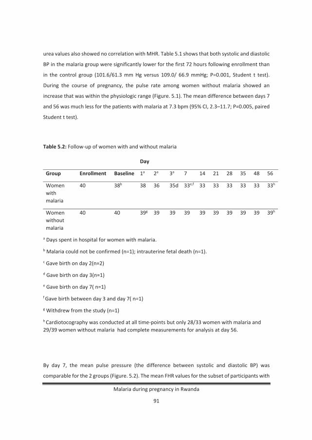
urea values also showed no correlation with MHR. Table 5.1 shows that both systolic and diastolic
BP in the malaria group were significantly lower for the first 72 hours following enrollment than
in the control group (101.6/61.3 mm Hg versus 109.0/ 66.9 mmHg; P=0.001, Student t test).
During the course of pregnancy, the pulse rate among women without malaria showed an
increase that was within the physiologic range (Figure. 5.1). The mean difference between days 7
and 56 was much less for the patients with malaria at 7.3 bpm (95% CI, 2.3–11.7; P=0.005, paired
Student t test).
Table 5.2: Follow up of women with and without malaria
Day
Group Enrollment Baseline 1a 2a 3a 7 14 21 28 35 48 56
Womenwithmalaria
40 38b 38 36 35d 33e,f 33 33 33 33 33 33h
Womenwithoutmalaria
40 40 39g 39 39 39 39 39 39 39 39 39h
a Days spent in hospital for women with malaria.
b Malaria could not be confirmed (n=1); intrauterine fetal death (n=1).
c Gave birth on day 2(n=2)
d Gave birth on day 3(n=1)
e Gave birth on day 7( n=1)
f Gave birth between day 3 and day 7( n=1)
g Withdrew from the study (n=1)
h Cardiotocography was conducted at all time points but only 28/33 women with malaria and29/39 women without malaria had complete measurements for analysis at day 56.
By day 7, the mean pulse pressure (the difference between systolic and diastolic BP) was
comparable for the 2 groups (Figure. 5.2). The mean FHR values for the subset of participants with

Malaria during pregnancy in Rwanda
92
a full series of repeated measurements until day 56 (28 patients with malaria and 29 women
without malaria) are shown in Figure. 5.1. The mean GA of the 28 patients included in Figure. 5.2
was slightly higher than that of the whole group of 38 patients (30.2 weeks versus 28.8 weeks).
The mean GA of the 28 control participants included in Figure. 5.1 was not different for the whole
group of 39 control participants (24.8 weeks versus 24.6 weeks). Fig. 1 illustrates that the FHR of
women with malaria returned to normal within 1 day, followed by a short period during which
the FHR was lower than that of control group. After 1 week, the FHR was within the normal range.
The range between highest and lowest observed FHR values during 30minutes of CTG registration
was comparable between the 2 groups.
Figure 5.1: Fetal and maternal heart rate over time
Legend: Maximum (red round markers), minimum (blue diamonds) and mean (green triangles)fetal heart, measured during 30 minutes CTG registrations, during and after treatment withartemether lumefantrine for P. falciparummalaria. The mean maternal heart rate is indicated by

Malaria during pregnancy in Rwanda
93
the orange line (no markers). The error bars indicate the 95% confidence intervals of the mean.The gray bands indicate the 95% confidence intervals of the mean of the measurements inhealthy pregnant women
Discussion
The principal finding of the present study is that acute malaria during pregnancy induces changes
in fetal and maternal circulatory indicators which normalize after the initiation of therapy with
AL, albeit at a different pace. No significant differences in outcome were detected between the 2
groups. One premature delivery occurred at 30 weeks after initiation of AL but was not clearly
caused by malaria or treatment. The prolonged tachycardia observed in women following an
episode of acute malaria (MHR b90 bpm until day 56) was a key finding of the present study. The
biologic explanation for this phenomenon is unclear. Anemia and hypovolemia [10] were not
apparent causes but neither could be excluded because blood tests were not performed after
study day 3. The combination of rapidly normalizing blood pressure
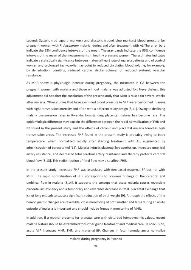
Figure 5.2: Maternal blood pressure over time
Malaria during pregnancy in Rwanda
94
Legend: Systolic (red square markers) and diastolic (round blue markers) blood pressure forpregnant women with P. falciparum malaria, during and after treatment with AL.The error barsindicate the 95% confidence intervals of the mean. The gray bands indicate the 95% confidenceintervals of the mean of the measurements in healthy pregnant women. The estimates indicatesindicate a statistically significance between maternal heart rate of malaria patients and of controlwomen and prolonged tachycardia may point to reduced circulating blood volume; for example,by dehydration, vomiting, reduced cardiac stroke volume, or reduced systemic vascularresistance.
As MHR shows a physiologic increase during pregnancy, the mismatch in GA between the
pregnant women with malaria and those without malaria was adjusted for. Nevertheless, this
adjustment did not alter the conclusion of the present study that MHR is raised for several weeks
after malaria. Other studies that have examined blood pressure in MiP were performed in areas
with high transmission intensity and often with a different study design [8,11]. Owing to declining
malaria transmission rates in Rwanda, longstanding placental malaria has become rare. The
epidemiologic difference may explain the difference between the rapid normalization of FHR and
BP found in the present study and the effects of chronic and placental malaria found in high
transmission areas. The increased FHR found in the present study is probably owing to body
temperature, which normalized rapidly after starting treatment with AL, augmented by
administration of paracetamol [12]. Malaria induces placental hypoperfusion, increased umbilical
artery resistance, and decreased fetal cerebral artery resistance and thereby protects cerebral
blood flow [8,13]. This redistribution of fetal flow may also affect FHR.
In the present study, increased FHR was associated with decreased maternal BP but not with
MHR. The rapid normalization of FHR corresponds to previous findings of the cerebral and
umbilical flow in malaria [8,14]. It supports the concept that acute malaria causes reversible
placental insufficiency and a temporary and reversible decrease in fetal–placental exchange that
is not long enough to cause a significant reduction of birth weight [9]. Although the effects of the
hemodynamic changes are reversible, close monitoring of both mother and fetus during an acute
episode of malaria is important and should include frequent monitoring of MHR.
In addition, if a mother presents for prenatal care with disturbed hemodynamic values, recent
malaria history should be established to further guide treatment and medical care. In conclusion,
acute MiP increases MHR, FHR, and maternal BP. Changes in fetal hemodynamics normalize

Malaria during pregnancy in Rwanda
95
within 1 week; however, MHR remains elevated for several weeks. The pathogenesis of the
observed fetal and maternal hemodynamic changes requires further study because such changes
might provide additional explanations of fetal distress during MiP.
Acknowledgments
The study was part of the Infectious Diseases Network for Treatment and Research in Africa(INTERACT) program, which received funding from The Netherlands Organization for ScientificResearch (Nederlandse Organisatie voor Wetenschappelijk) Global Health (NWO Wotro) in theNetherlands African partnership for capacity development and clinical interventions againstpoverty related diseases (NACCAP) program (W 070520100).

Malaria during pregnancy in Rwanda
96
References
1. Brabin B. Fetal anaemia in malarious areas: its causes and significance. Ann Trop Paediatr1992;12(3):303–10.
2. Matteelli A,Donato F, Shein A,Muchi JA, Abass AK, MarianiM, et al.Malarial infection andbirthweight in urban Zanzibar, Tanzania. Ann Trop Med Parasitol 1996;90(2): 125–34.
3. Rulisa S, Mens PF, Karema C, Schallig HD, Kaligirwa N, Vyankandondera J, et al. Malaria has noeffect on birth weight in Rwanda. Malar J 2009;8:194.
4. Hunter S, Robson SC. Adaptation of the maternal heart in pregnancy. Br Heart J1992;68(6):540–3.
5. Muthusamy A, Achur RN, Valiyaveettil M, Botti JJ, Taylor DW, Leke RF, et al. Chondroitinsulfate proteoglycan but not hyaluronic acid is the receptor for the adherence of Plasmodiumfalciparum infected erythrocytes in human placenta, and infected red blood cell adherence upregulates the receptor expression. Am J Pathol 2007;170(6):1989–2000.
6. Muehlenbachs A, Mutabingwa TK, Edmonds S, Fried M, Duffy PE. Hypertension and maternalfetal conflict during placental malaria. PLoS Med 2006;3(11):e446.
7. Tobal N, Chevillot M, Himily V, Perrotin F, Lansac J, Arbeille P. Doppler monitoring of fetalcirculation from multiple arteries over several days to improve evaluation of fetal prognosis. JRadiol 2002;83(12 Pt 2):1943–51.
8. Carles G, Tobal N, Marret H, Arbeille P. Acute reversible placental dysfunction and abnormalfetal heart rate at delivery. Eur J Obstet Gynecol Reprod Biol 2003;110(2):136–42.
9. Arbeille P, Caries G, Tobal N, Herault S, Georgescus M, Bousquet F, et al. Fetal flowredistribution to the brain in response to malaria infection: does protection of the fetus againstmalaria develop over time? J Ultrasound Med 2002;21(7):739–46.
10. Meuris S, Piko BB, Eerens P, Vanbellinghen AM, Dramaix M, Hennart P. Gestational malaria:assessment of its consequences on fetal growth. Am J Trop Med Hyg 1993;48(5):603–9.
11. Ndao CT, Dumont A, Fievet N, Doucoure S, Gaye A, Lehesran JY. Placental malarial infectionas a risk factor for hypertensive disorders during pregnancy in Africa: a case–control study in anurban area of Senegal, West Africa. Am J Epidemiol 2009;170(7):847–53.
12. Macaulay JH, Randall NR, Bond K, Steer PJ. Continuous monitoring of fetal temperature bynoninvasive probe and its relationship to maternal temperature, fetal heart rate, and cordarterial oxygen and pH. Obstet Gynecol 1992;79(3):469–74.

Malaria during pregnancy in Rwanda
97
13. Arbeille P, Carles G, Georgescu M, Tobal N, Herault S, Bousquet F, et al. Consequences ofreduced umbilical and increased foetal cerebral flow during malaria crisis on foetal behaviour.Parasitology 2003;126(Pt 6):513–9.
14. Dorman EK, Shulman CE, Kingdom J, Bulmer JN, Mwendwa J, Peshu N, et al. Impaireduteroplacental blood flow in pregnancies complicated by falciparum malaria. Ultrasound ObstetGynecol 2002;19(2):165–70.
Related Documents