ARTICLE Received 25 May 2016 | Accepted 30 Dec 2016 | Published 15 Feb 2017 Usp9x regulates Ets-1 ubiquitination and stability to control NRAS expression and tumorigenicity in melanoma Harish Potu 1 , Luke F. Peterson 1 , Malathi Kandarpa 1 , Anupama Pal 1 , Hanshi Sun 2 , Alison Durham 3 , Paul W. Harms 4 , Peter C. Hollenhorst 5 , Ugur Eskiocak 6,w , Moshe Talpaz 1 & Nicholas J. Donato 7 ETS transcription factors are commonly deregulated in cancer by chromosomal translocation, overexpression or post-translational modification to induce gene expression programs essential in tumorigenicity. Targeted destruction of these proteins may have therapeutic impact. Here we report that Ets-1 destruction is regulated by the deubiquitinating enzyme, Usp9x, and has major impact on the tumorigenic program of metastatic melanoma. Ets-1 deubiquitination blocks its proteasomal destruction and enhances tumorigenicity, which could be reversed by Usp9x knockdown or inhibition. Usp9x and Ets-1 levels are coincidently elevated in melanoma with highest levels detected in metastatic tumours versus normal skin or benign skin lesions. Notably, Ets-1 is induced by BRAF or MEK kinase inhibition, resulting in increased NRAS expression, which could be blocked by inactivation of Usp9x and therapeutic combination of Usp9x and MEK inhibitor fully suppressed melanoma growth. Thus, Usp9x modulates the Ets-1/NRAS regulatory network and may have biologic and therapeutic implications. DOI: 10.1038/ncomms14449 OPEN 1 Department of Internal Medicine/Division of Hematology/Oncology, University of Michigan School of Medicine and Comprehensive Cancer Center, Ann Arbor, Michigan 48109, USA. 2 Department of Periodontics and Oral Medicine, University of Michigan School of Dentistry, Ann Arbor, Michigan 48109, USA. 3 Department of Dermatology, University of Michigan School of Medicine, Ann Arbor, Michigan 48109, USA. 4 Departments of Pathology and Dermatology, University of Michigan School of Medicine, Ann Arbor, Michigan 48109, USA. 5 Department of Biochemistry and Molecular Biology, Medical Sciences Program, Indiana University Bloomington, 1001 Third St, Bloomington, Indiana 47405, USA. 6 Children’s Research Institute and Department of Pediatrics, Howard Hughes Medical Institute, University of Texas Southwestern Medical Center, Dallas, Texas 75390, USA. 7 Department of Pharmacology, University of Michigan School of Medicine, Ann Arbor, Michigan 48109, USA. w Present address: Compass Therapeutics, 450 Kendall Street, Cambridge, Massachusetts 02142, USA. Correspondence and requests for materials should be addressed to N.J.D. (email: [email protected]). NATURE COMMUNICATIONS | 8:14449 | DOI: 10.1038/ncomms14449 | www.nature.com/naturecommunications 1

Welcome message from author
This document is posted to help you gain knowledge. Please leave a comment to let me know what you think about it! Share it to your friends and learn new things together.
Transcript

ARTICLE
Received 25 May 2016 | Accepted 30 Dec 2016 | Published 15 Feb 2017
Usp9x regulates Ets-1 ubiquitination and stabilityto control NRAS expression and tumorigenicityin melanomaHarish Potu1, Luke F. Peterson1, Malathi Kandarpa1, Anupama Pal1, Hanshi Sun2, Alison Durham3,
Paul W. Harms4, Peter C. Hollenhorst5, Ugur Eskiocak6,w, Moshe Talpaz1 & Nicholas J. Donato7
ETS transcription factors are commonly deregulated in cancer by chromosomal translocation,
overexpression or post-translational modification to induce gene expression programs
essential in tumorigenicity. Targeted destruction of these proteins may have therapeutic
impact. Here we report that Ets-1 destruction is regulated by the deubiquitinating enzyme,
Usp9x, and has major impact on the tumorigenic program of metastatic melanoma. Ets-1
deubiquitination blocks its proteasomal destruction and enhances tumorigenicity, which could
be reversed by Usp9x knockdown or inhibition. Usp9x and Ets-1 levels are coincidently
elevated in melanoma with highest levels detected in metastatic tumours versus normal skin
or benign skin lesions. Notably, Ets-1 is induced by BRAF or MEK kinase inhibition, resulting in
increased NRAS expression, which could be blocked by inactivation of Usp9x and therapeutic
combination of Usp9x and MEK inhibitor fully suppressed melanoma growth. Thus, Usp9x
modulates the Ets-1/NRAS regulatory network and may have biologic and therapeutic
implications.
DOI: 10.1038/ncomms14449 OPEN
1 Department of Internal Medicine/Division of Hematology/Oncology, University of Michigan School of Medicine and Comprehensive Cancer Center,Ann Arbor, Michigan 48109, USA. 2 Department of Periodontics and Oral Medicine, University of Michigan School of Dentistry, Ann Arbor, Michigan 48109,USA. 3 Department of Dermatology, University of Michigan School of Medicine, Ann Arbor, Michigan 48109, USA. 4 Departments of Pathology andDermatology, University of Michigan School of Medicine, Ann Arbor, Michigan 48109, USA. 5 Department of Biochemistry and Molecular Biology, MedicalSciences Program, Indiana University Bloomington, 1001 Third St, Bloomington, Indiana 47405, USA. 6 Children’s Research Institute and Department ofPediatrics, Howard Hughes Medical Institute, University of Texas Southwestern Medical Center, Dallas, Texas 75390, USA. 7 Department of Pharmacology,University of Michigan School of Medicine, Ann Arbor, Michigan 48109, USA. w Present address: Compass Therapeutics, 450 Kendall Street, Cambridge,Massachusetts 02142, USA. Correspondence and requests for materials should be addressed to N.J.D. (email: [email protected]).
NATURE COMMUNICATIONS | 8:14449 | DOI: 10.1038/ncomms14449 | www.nature.com/naturecommunications 1

Recent progress has been made in targeting pathwaysactivated by mutations in metastatic melanoma, and theseadvances have led to major improvements in patient
treatment and survival1. However, many biological and clinicalcharacteristics of melanoma are still unknown and currenttargeted therapies (BRAF and/or MEK inhibitors) are onlyeffective in a subset of patients and typically for a limited duration(4–12 months)2. Combination kinase inhibitor therapy cancircumvent or delay resistance and reactivation of immuneresponsiveness has shown some promising results. However,these therapies are only effective in 30–40% of patients andserious side effects (that is, auto-immunity) limit sustainedclinical benefit, highlighting the need for novel strategies thatcould add to existing therapies3. Adjoined to that need, is the lackof understanding of some of the basic biology of melanoma,particularly what underlies the progression to metastatic diseaseafter driver mutations are in place. Some recent studies haveprovided insight and have suggested that age, environmentalfactors and diet may underlie the transition1,4,5.
The ubiquitin-proteasome system (UPS) has received con-siderable attention as a source of new drug targets because of theclinical success of 20S proteasome inhibitors in specific cancers.The UPS has multiple components that are consideredtargetable6,7. Among them are deubiquitinases (DUBs):enzymes that mediate removal of ubiquitin monomers orpolymers from target proteins, and are major regulators of theUPS. Many DUBs demonstrate specificity for proteins involved indisease-associated pathways and are deregulated in diseaseby mutations, altered expression or post-translationalmodification8–10. Ubiquitin specific peptidase 9, X-linked(Usp9x), also known as FAF; FAM; DFFRX and MRX99, is ahigh MW DUB that has been shown to be over-expressed inseveral cancers, but can have both positive and negative impacton tumorigenicity, depending on the cancer type and diseasemodel studied11–16. Usp9x deubiquitinates proteins essential intumour cell signalling and survival, protecting some of them fromproteasomal destruction14,15,17.
The ETS (E26 transformation-specific or E-twenty-six; basedon the gene transduced by the leukaemia virus, E26) transcriptionfactor family is composed of 28 members, which recognize aDNA binding sequence minimally consisting of GGA(A/T)18–20.Specific members of this highly conserved family are frequentlyactivated by chromosomal translocation, overexpression andstabilization (by altered ubiquitination) and are essential intumorigenesis21. For example, FLI1 and ERG are overexpressedin Ewing sarcoma and prostate cancer as a consequence ofchromosomal translocation and are key drivers of thesemalignancies22,23. Ets-1, and other family members, areoverexpressed and regulated (positively and negatively) byphosphorylation, sumoylation and ubiquitination associatedwith specific signalling events24–27. Phosphorylation of specificETS proteins mediated by an aberrant RAS/RAF/MEK/ERKsignalling pathway provides one mechanism for promoting geneexpression essential in driving the cancer phenotype anddominant negative versions of ETS genes can block oncogenicRAS/ERK tumorigenicity19,28. Ets-1 overexpression has beendocumented in many invasive and metastatic cancers, includingbreast, lung, colon, pancreatic and thyroid cancer25,29–34,where Ets-1 drives gene expression associated withcellular differentiation, migration, proliferation, survival andangiogenesis. Members of the ETS transcription factor familyare considered excellent therapeutic targets but most targetingapproaches have failed35.
This report provides evidence of an essential role for Usp9x inmelanoma because of its regulation of Ets-1 protein levels.Through Usp9x-mediated, site-specific deubiquitination, Ets-1
proteasomal destruction is inhibited, resulting in Ets-1 accumula-tion and increased melanoma tumorigenicity, which could beblocked by inhibition of Usp9x activity or knockdown of Ets-1.We also determined that Ets-1 expression was negativelyregulated by BRAF and/or MEK kinase activity and inhibitionof this pathway increased Ets-1 expression to increase NRASlevels by activating the NRAS promoter. Since NRAS mutationsare common (15–20%) in melanoma patients (and other cancersincluding multiple myeloma, lymphoma, lung, thyroid andcolorectal cancer36) and its continual expression is essential forNRAS mutant melanoma cell growth and survival37,38, NRASmutant tumours were highly dependent on Usp9x. Thus, weprovide evidence that Usp9x plays an important role in Ets-1regulation and melanoma tumorigenicity, in part through NRAStranscription which may be of particular importance in tumoursdriven by NRAS mutation.
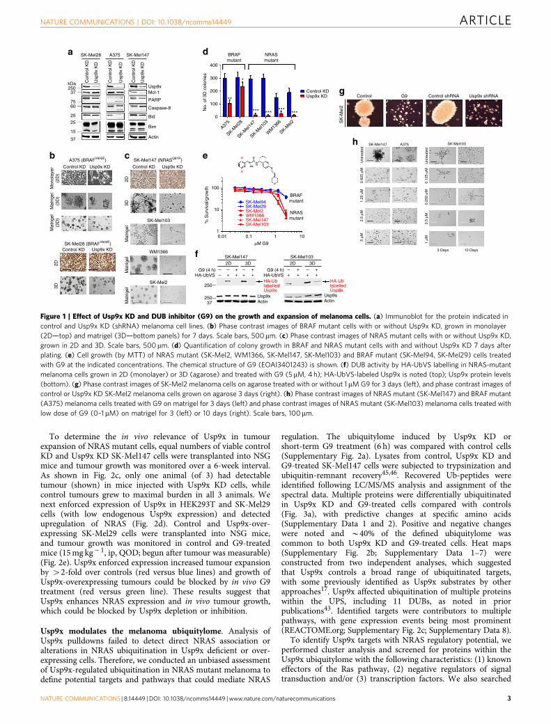
ResultsUsp9x is required for in vivo melanoma growth. We andothers previously described Usp9x activity and expression inmelanoma10,39 and sought to define its role in primary andmetastatic disease. Initially, we depleted Usp9x using a previouslycharacterized shRNA knockdown (KD) vector40 in threemelanoma cell lines with distinct driver mutations (BRAFmutant: SK-Mel28, A375; NRAS mutant: SK-Mel147) andmetastatic efficiencies (highly metastatic: A375, SK-Mel147) andcompared biological effects to control cells. Usp9x knockdown(KD) modestly reduced the steady-state level of the anti-apoptoticprotein Mcl-1 (a previously defined Usp9x substrate14), activatedcaspase cleavage (Fig. 1a) and reduced tumour growth understandard monolayer growth conditions (2D). However, Usp9xKD significantly impaired 3D melanoma growth, which is a betterdiscriminator of the malignant and benign phenotype41,42
(Fig. 1b,c). Usp9x depletion blocked expansive tumour growthin matrigel, particularly in tumours with NRAS mutations(Fig. 1c,d). To assess clinical relevance, we examined melanomachemosensitivity to our recently described small molecule Usp9xinhibitor (G9)39,43 and detected moderately greater sensitivity inNRAS versus BRAF mutant lines (Fig. 1e). Tumour cells grownin 3D had higher levels of Usp9x activity/expression thanthose measured in 2D cultures (confirmed in additional celllines—Supplementary Fig. 1a) and G9 inhibited Usp9x activity incells from either culture condition (Fig. 1f). Both Usp9x KD andG9 blocked anchorage-independent melanoma growth (Fig. 1g)and G9 dose-dependently inhibited melanoma growth inmatrigel (Fig. 1h), with nM sensitivity against NRAS mutantcells (SK-Mel103; IC50 B300 nM), suggesting that Usp9x plays arole in tumour expansion, particularly in tumours with an NRASmutation.
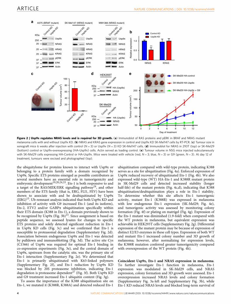
To further elucidate the role of Usp9x in melanoma andexamine the sensitivity of NRAS mutant tumours to Usp9x KDand inhibition, we first assessed the effects of Usp9x KD onspecific RAS proteins in highly metastatic NRAS and BRAFmutant melanomas. Usp9x KD reduced NRAS protein levels inboth NRAS and BRAF mutant cells with little to no effect onHRAS or KRAS expression (Fig. 2a). Previous studies demon-strated that continual expression of mutant NRAS was essentialfor NRAS mutant melanoma survival37,44, and we confirmed thatdependence in NRAS KD studies (Supplementary Fig. 1b). Usp9xKD suppressed NRAS, but not KRAS gene expression (Fig. 2b).Thus, Usp9x-mediated regulation of NRAS expression inmelanoma, particulalrly in NRAS mutant cells, may partlyunderly their dependence on Usp9x for continual expansionand survival. However, Usp9x may alter other components withinthe RAS signalling pathway as we detected a paradoxical increasein ERK activation in Usp9x KD cells.
ARTICLE NATURE COMMUNICATIONS | DOI: 10.1038/ncomms14449
2 NATURE COMMUNICATIONS | 8:14449 | DOI: 10.1038/ncomms14449 | www.nature.com/naturecommunications
To determine the in vivo relevance of Usp9x in tumourexpansion of NRAS mutant cells, equal numbers of viable controlKD and Usp9x KD SK-Mel147 cells were transplanted into NSGmice and tumour growth was monitored over a 6-week interval.As shown in Fig. 2c, only one animal (of 3) had detectabletumour (shown) in mice injected with Usp9x KD cells, whilecontrol tumours grew to maximal burden in all 3 animals. Wenext enforced expression of Usp9x in HEK293T and SK-Mel29cells (with low endogenous Usp9x expression) and detectedupregulation of NRAS (Fig. 2d). Control and Usp9x-over-expressing SK-Mel29 cells were transplanted into NSG mice,and tumour growth was monitored in control and G9-treatedmice (15 mg kg� 1, ip, QOD; begun after tumour was measurable)(Fig. 2e). Usp9x enforced expression increased tumour expansionby 42-fold over controls (red versus blue lines) and growth ofUsp9x-overexpressing tumours could be blocked by in vivo G9treatment (red versus green line). These results suggest thatUsp9x enhances NRAS expression and in vivo tumour growth,which could be blocked by Usp9x depletion or inhibition.
Usp9x modulates the melanoma ubiquitylome. Analysis ofUsp9x pulldowns failed to detect direct NRAS association oralterations in NRAS ubiquitination in Usp9x deficient or over-expressing cells. Therefore, we conducted an unbiased assessmentof Usp9x-regulated ubiquitination in NRAS mutant melanoma todefine potential targets and pathways that could mediate NRAS
regulation. The ubiquitylome induced by Usp9x KD orshort-term G9 treatment (6 h) was compared with control cells(Supplementary Fig. 2a). Lysates from control, Usp9x KD andG9-treated SK-Mel147 cells were subjected to trypsinization andubiquitin-remnant recovery45,46. Recovered Ub-peptides wereidentified following LC/MS/MS analysis and assignment of thespectral data. Multiple proteins were differentially ubiquitinatedin Usp9x KD and G9-treated cells compared with controls(Fig. 3a), with predictive changes at specific amino acids(Supplementary Data 1 and 2). Positive and negative changeswere noted and B40% of the defined ubiquitylome wascommon to both Usp9x KD and G9-treated cells. Heat maps(Supplementary Fig. 2b; Supplementary Data 1–7) wereconstructed from two independent analyses, which suggestedthat Usp9x controls a broad range of ubiquitinated targets,with some previously identified as Usp9x substrates by otherapproaches17. Usp9x affected ubiquitination of multiple proteinswithin the UPS, including 11 DUBs, as noted in priorpublications43. Identified targets were contributors to multiplepathways, with gene expression events being most prominent(REACTOME.org; Supplementary Fig. 2c; Supplementary Data 8).
To identify Usp9x targets with NRAS regulatory potential, weperformed cluster analysis and screened for proteins within theUsp9x ubiquitylome with the following characteristics: (1) knowneffectors of the Ras pathway, (2) negative regulators of signaltransduction and/or (3) transcription factors. We also searched
kDa
SK-Mel28a d
g
he
f
cb
Con
trol
KD
Usp
9x K
D
Con
trol
KD
Usp
9x K
D
Con
trol
KD
Usp
9x K
D
Usp9xMcl-1
PARP
Caspase-8
Bid
Bim
Actin
100
BRAFmutant
SK-Mel1472D 3D
G9 (4 h)
2D 3DSK-Mel103
SK-Mel147 SK-Mel103
3 Days 10 Days
Unt
reat
ed0.
625
μM1.
25 μ
M2.
5 μM
5 μM
Unt
reat
ed0.
125
μM0.
250
μM0.
5 μM
1 μM
A375
NRASmutant
CI O
O
O
SK-Mel94SK-Mel29SK-Mel2WM1366SK-Mel147SK-Mel103
N
N CN
NH
CI
% S
urvi
val/g
row
th10
10.01 0.1 1
μM G9
10
A375 SK-Mel147
25037
7560
25
25
15
37
400
BRAFmutant
NRASmutant
300
Control KDUsp9x KD
200
100
A375 (BRAFV600E) SK-Mel147 (NRASQ61R)
Control KD
Mon
olay
er(2
D)
2D
2D3D
3D
Mat
rigel
(3D
)M
atrig
el(3
D)
Usp9x KD
SK-Mel28 (BRAFV600E)Control KD Usp9x KD
Control KD
SK-Mel103
WM1366
SK-Mel2
Usp9x KD
**
*** *** *** ***
*
No.
of 3
D c
olon
ies
SK
-Mel
2
Control G9 Control shRNA Usp9x shRNA
0
A375
SK-Mel2
8
SK-Mel1
47
SK-Mel1
03
SK-Mel2
WM
1366
Mat
rigel
Mat
rigel
Mat
rigel
HA-UbVSG9 (4 h) – – –
– + ++
+ ++ ––
+ ++
+ ++
HA-UbVS
Usp9xActin
Usp9xActin
250
250
37
HA-UblabelledUsp9x
HA-UblabelledUsp9x
Figure 1 | Effect of Usp9x KD and DUB inhibitor (G9) on the growth and expansion of melanoma cells. (a) Immunoblot for the protein indicated in
control and Usp9x KD (shRNA) melanoma cell lines. (b) Phase contrast images of BRAF mutant cells with or without Usp9x KD, grown in monolayer
(2D—top) and matrigel (3D—bottom panels) for 7 days. Scale bars, 500mm. (c) Phase contrast images of NRAS mutant cells with or without Usp9x KD,
grown in 2D and 3D. Scale bars, 500 mm. (d) Quantification of colony growth in BRAF and NRAS mutant cells with and without Usp9x KD 7 days after
plating. (e) Cell growth (by MTT) of NRAS mutant (SK-Mel2, WM1366, SK-Mel147, SK-Mel103) and BRAF mutant (SK-Mel94, SK-Mel29) cells treated
with G9 at the indicated concentrations. The chemical structure of G9 (EOAI3401243) is shown. (f) DUB activity by HA-UbVS labelling in NRAS-mutant
melanoma cells grown in 2D (monolayer) or 3D (agarose) and treated with G9 (5 mM, 4 h); HA-UbVS-labeled Usp9x is noted (top); Usp9x protein levels
(bottom). (g) Phase contrast images of SK-Mel2 melanoma cells on agarose treated with or without 1 mM G9 for 3 days (left), and phase contrast images of
control or Usp9x KD SK-Mel2 melanoma cells grown on agarose 3 days (right). (h) Phase contrast images of NRAS mutant (SK-Mel147) and BRAF mutant
(A375) melanoma cells treated with G9 on matrigel for 3 days (left) and phase contrast images of NRAS mutant (SK-Mel103) melanoma cells treated with
low dose of G9 (0–1 mM) on matrigel for 3 (left) or 10 days (right). Scale bars, 100mm.
NATURE COMMUNICATIONS | DOI: 10.1038/ncomms14449 ARTICLE
NATURE COMMUNICATIONS | 8:14449 | DOI: 10.1038/ncomms14449 | www.nature.com/naturecommunications 3
the ubiquitylome for proteins known to interact with Usp9x orbelonging to a protein family with a domain recognized byUsp9x. Specific ETS proteins emerged as possible contributors asseveral members have an essential role in tumorigenicity andembryonic development19,20,24,33. Ets-1 is both responsive to anda target of the RAS/MEK/ERK signalling pathway24, and othermembers of the ETS family (that is, ERG, FLI1, FEV) have beenshown to associate with and be deubiquitinated by Usp9x(ERG)15. Ub-remnant analysis indicated that both Usp9x KD andinhibition of activity with G9 increased Ets-1 (and its isoform),Ets-2, ETV2 and/or GABPa ubiquitination specifically withintheir ETS domain (K388 in Ets-1), a domain previously shown tobe recognized by Usp9x (Fig. 3b)15. Since assignment is based onpeptide sequence, we assessed lysates for changes to specificETS proteins and solely detected significant reduction in Ets-1in Usp9x KD cells (Fig. 3c) and we confirmed that Ets-1 issusceptible to proteasomal degradation (Supplementary Fig. 2d).Association between endogenous Usp9x and Ets-1 was detectedby pulldown and immunoblotting (Fig. 3d). The active site Cys(C1566) of Usp9x was required for optimal Ets-1 binding inco-expression experiments (Fig. 3e), and the central domain ofUsp9x, upstream from the catalytic site, was the primary site ofEts-1 interaction (Supplementary Fig. 2e). We determined thatEts-1 is primarily ubiquitinated with K63-linked polymers(Supplementary Fig. 2f), and Ets-1 reduction by Usp9x KDwas blocked by 20S proteasome inhibition, indicating Ets-1degradation is proteasome dependent27 (Fig. 3f). Both Usp9x KDand G9 treatment increased Ets-1 ubiquitin content (Fig. 3g).
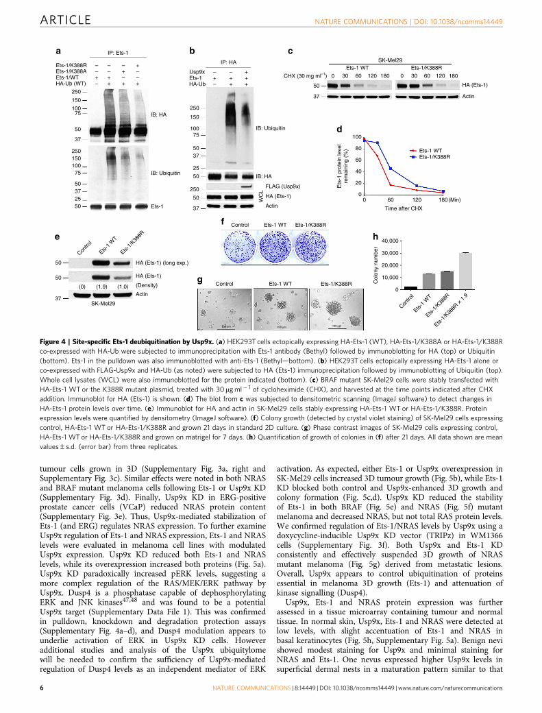
To assess the importance of the K388 ubiquitination site onEts-1, we mutated it (K388R, K388A) and detected reduced Ets-1
ubiquitination compared with wild-type protein, indicating K388serves as a site for ubiquitination (Fig. 4a). Enforced expression ofUsp9x reduced recovery of ubiquitinated Ets-1 (Fig. 4b). We alsoexpressed wild-type (WT) HA-Ets-1 and K388R mutant proteinin SK-Mel29 cells and detected increased stability (longerhalf-life) of the mutant protein (Fig. 4c,d), indicating that K388ubiquitination/deubiquitination plays a role in Ets-1 stability.To determine whether this site affects Ets-1 tumorigenicactivity, mutant Ets-1 (K388R) was expressed in melanomawith low endogenous Ets-1 expression (SK-Mel29; Fig. 4e),and tumorigenic activity was assessed by monitoring colonyformation (Fig. 4f) or plating on matrigel (Fig. 4g). Expression ofthe Ets-1 mutant was diminished (1.9-fold) when compared withthe WT protein in melanoma, but equivalent expression wasachievable in HEK293T cells (Supplementary Fig. 2g). Differentialexpression of the mutant protein may be because of expression ofdistinct E2/E3 enzymes in these cell types. Expression of both WTand mutant Ets-1 increased colony number and 3D growth ofmelanoma; however, after normalizing for expression levels,the K388R mutation conferred greater tumorigenicity comparedwith overexpression of the WT protein (Fig. 4h).
Coincident Usp9x, Ets-1 and NRAS expression in melanoma.To further investigate Ets-1 function in melanoma, Ets-1expression was modulated in SK-Mel29 cells, and NRASexpression, colony formation and 3D growth were assessed. Ets-1overexpression increased NRAS levels and colony formation(Supplementary Fig. 3a-left and Supplementary Fig. 3b), whileEts-1 KD reduced NRAS levels and blocked long-term survival of
A375 (BRAF mutant)
KDa
KDaHA (Usp9x)
NRAS25020
37
KDa25020
37
Contro
l KD
Contro
l KD Con
trol
Contro
l
Contro
l KD
Contro
l KD
Usp9x
KD
Usp9x
KD HA-U
sp9x
HA-Usp
9x
Usp9x
KD
Usp9x
KD
Contro
l KD
Control KDUsp
9x K
D
Usp9x KD
**
Usp9x
NRAS
HRAS
KRAS
pERK
ERK
Actin
100
200
SK-Mel29 HA-control
SK-Mel29 HA-Usp9x
SK-Mel29 HA-Usp9x + G9
150
100
50
50
0 2 4 6 8 10
Day of treatment
12Tu
mou
r vo
lum
e (m
m3 )
Tum
our
volu
me
(cm
3 ) 80
60
40
20
0
Usp9x
NRAS
HRAS
KRAS
pERK
ERK
Actin
Actin
HA (Usp9x)
NRAS
Actin
Usp9x1.0
0.5
0.0NRAS KRAS
Rel
ativ
e ex
pres
sion
NRAS
HRAS
KRAS
pERK
ERK
Actin
250
20
20
20
3737
37
SK-Mel147 (NRAS mutant) WM1366 (NRAS mutant)
HEK293T
SK-Mel29
a
c d e
b
Figure 2 | Usp9x regulates NRAS levels and is required for 3D growth. (a) Immunoblot of RAS proteins and pERK in BRAF and NRAS mutant
melanoma cells with and without Usp9x KD. (b) NRAS and KRAS gene expression in control and Usp9x KD SK-Mel147 cells by RT-PCR. (c) Tumour size in
xenograft mice 6 weeks after injection with control (N¼ 3) or Usp9x (N¼ 3) KD SK-Mel147 cells. (d) Immunoblot for NRAS in 293T (top) or SK-Mel29
(bottom) control or Usp9x-overexpressing (HA-Usp9x) cells. Actin served as loading control. (e) Tumour volume in NSG mice injected subcutaneously
with SK-Mel29 cells expressing HA-Control or HA-Usp9x. Mice were treated with vehicle (red, N¼ 3; blue, N¼ 3) or G9 (green, N¼ 3). At day 12 of
treatment, tumours were excised and photographed (top).
ARTICLE NATURE COMMUNICATIONS | DOI: 10.1038/ncomms14449
4 NATURE COMMUNICATIONS | 8:14449 | DOI: 10.1038/ncomms14449 | www.nature.com/naturecommunications

Relative
Row min
a b
c
e f
g
dC
ontr
ol-1
Con
trol
-2
Usp
9x K
D-1
Usp
9x K
D-2
G9-
1
G9-
2
Row max Ets-1 K*3881 331
MNYEK*LSR
Contro
l KD
Usp9x
KD
Usp9xInput Usp9x
IB: Ets-1
IB: Usp9x
IB: Usp9x
Ets-1
Ets-2
GABPα
NRAS
Actin
FLAG-Usp9x/CDM
FLAG-Usp9x/E5FLAG-Usp9x/E1 CDM
FLAG-Usp9x/E1HA-Ets-1
150 kDa
100 kDa
75 kDa
50 kDa
50 kDa
Usp9x W/T
Usp9x CDM
Usp9x E1
Usp9x E1 CDM
Usp9x E5
N C
CN
N
N
N
Input
Input
–
–––
+
––+
+
+––
+
–+–
+
–––
+
––+
+
+––
+
–+–
+
– +– + –+ + +
– – +– + –+ + +
IP: HA
IP: HA
IB: FLAG (Usp9x) MG132
250
50
37
– –+ +
Control KD Usp9x KD
Usp9x
Ets-1
Actin
IP: FLAG IP: FLAG
IB: FLAG IB: FLAG
IB: HA(Ub-Ets-1)
IB: HA(Ub-Ets-1)
IB: HA (Ets-1)
IB: FLAG(Usp9x)
kDa Contro
l KD
Contro
l
G9Usp9x
KD
Usp9xActin
Input
250150100
50
37
25
50
25037
75
Ets-1binding
++
++
++
–
+/–
Usp9x Cysteine
Cysteine
Alanine
Alanine
Histidine
UCH
U
U
UCH
Histidine
2,575
1,593
1,593
386
2,575
IB: HA (Ets-1)
FLAG-Usp9xHA-Ets-1
MNYEK*LSR HumanMouseRatChimpanzeeRhesus
MNYEK*LSR
MNYEK*LSRMNYEK*LSR
MNYEK*LSR
415 441
N CPNT
kDakDa
50
250
250
lgG
Input Ets-1lgG
lP
lP
250
50
50
50
20
37
250 kDa
50 kDa
TAD ETS
Figure 3 | Usp9x deubiquitinates Ets-1 and regulates its degradation. (a) Heat maps of differentially ubiquitinated proteins. NRAS mutant SK-Mel147
cells were exposed to control and Usp9x KD or G9 treatment as noted. The number of unique peptides and proteins reproducibly detected is shown.
(b) Schematic diagram of the human Ets-1 protein showing the PNT (pointed domain, aa 53–136), TAD (transactivation domain, aa 137–242) and ETS
domains. The putative site of ubiquitination (MNYEK*LSR) in human Ets-1 is shown and is conserved in mammalian species (right). (c) Immunoblot of
ETS family proteins and NRAS in NRAS mutant melanoma cells with and without Usp9x KD. Actin served as a loading control. (d) Reciprocal
immunoprecipitation of Usp9x and Ets-1 with endogenous Ets-1 and Usp9x in NRAS mutant SK-Mel2 cells. Immunoblotting was performed to detect Ets-1
or Usp9x in pulldowns and a portion of the input sample. (e) Top—Ectopically expressed FLAG-Usp9x (full-length) or FLAG-Usp9x-CDM (catalytic
domain mutant, C1566A) was co-expressed with HA-Ets-1 in HEK293T cells. HA (Ets-1) immunoprecipitation was followed by immunoblotting of FLAG
(Usp9x—top) or HA (Ets-1—bottom). Input lysate was also immunoblotted. Center—Ectopically expressed FLAG-Usp9x deletion constructs (FLAG-Usp9x
E1, FLAG-Usp9x E1/CDM (catalytic domain mutant—C1566A), FLAG-Usp9x E5 (C-terminal deletion)) (illustrated in the bottom panel) were co-expressed
with HA-Ets-1 in HEK293T cells. HA (Ets-1) immunoprecipitation was followed by FLAG (Usp9x) or HA (Ets-1) immunoblotting. Input lysate was also
immunoblotted. Bottom—Map and summary of the Usp9x deletion constructs and their Ets-1 binding activity. The position of the ubiquitin C-terminal
hydrolase (UCH) in the catalytic domain is shown by bold letters. Numbers and letters designate highlighted amino acids. (f) Immunoblot for Usp9x, Ets-1
and actin in control and Usp9x KD WM1366 NRAS mutant cells treated±MG132 for 8 h (10 mM). (g) HEK293T cells ectopically expressing FLAG-Ets-1 and
HA-Ubiquitin were subjected to control or Usp9x KD (left) or treated with vehicle or G9 (2.5mM, 6 h—right). FLAG immunoprecipitation was followed by
HA blotting to detect Ub-Ets-1 levels. Immunoblot for FLAG (Ets-1) in the pulldowns (top) and input lysate (Usp9x and actin—bottom) is shown.
NATURE COMMUNICATIONS | DOI: 10.1038/ncomms14449 ARTICLE
NATURE COMMUNICATIONS | 8:14449 | DOI: 10.1038/ncomms14449 | www.nature.com/naturecommunications 5
tumour cells grown in 3D (Supplementary Fig. 3a, right andSupplementary Fig. 3c). Similar effects were noted in both NRASand BRAF mutant melanoma cells following Ets-1 or Usp9x KD(Supplementary Fig. 3d). Finally, Usp9x KD in ERG-positiveprostate cancer cells (VCaP) reduced NRAS protein content(Supplementary Fig. 3e). Thus, Usp9x-mediated stabilization ofEts-1 (and ERG) regulates NRAS expression. To further examineUsp9x regulation of Ets-1 and NRAS expression, Ets-1 and NRASlevels were evaluated in melanoma cell lines with modulatedUsp9x expression. Usp9x KD reduced both Ets-1 and NRASlevels, while its overexpression increased both proteins (Fig. 5a).Usp9x KD paradoxically increased pERK levels, suggesting amore complex regulation of the RAS/MEK/ERK pathway byUsp9x. Dusp4 is a phosphatase capable of dephosphorylatingERK and JNK kinases47,48 and was found to be a potentialUsp9x target (Supplementary Data File 1). This was confirmedin pulldown, knockdown and degradation protection assays(Supplementary Fig. 4a–d), and Dusp4 modulation appears tounderlie activation of ERK in Usp9x KD cells. Howeveradditional studies and analysis of the Usp9x ubiquitylomewill be needed to confirm the sufficiency of Usp9x-mediatedregulation of Dusp4 levels as an independent mediator of ERK
activation. As expected, either Ets-1 or Usp9x overexpression inSK-Mel29 cells increased 3D tumour growth (Fig. 5b), while Ets-1KD blocked both control and Usp9x-enhanced 3D growth andcolony formation (Fig. 5c,d). Usp9x KD reduced the stabilityof Ets-1 in both BRAF (Fig. 5e) and NRAS (Fig. 5f) mutantmelanoma and decreased NRAS, but not total RAS protein levels.We confirmed regulation of Ets-1/NRAS levels by Usp9x using adoxycycline-inducible Usp9x KD vector (TRIPz) in WM1366cells (Supplementary Fig. 3f). Both Usp9x and Ets-1 KDconsistently and effectively suspended 3D growth of NRASmutant melanoma (Fig. 5g) derived from metastatic lesions.Overall, Usp9x appears to control ubiquitination of proteinsessential in melanoma 3D growth (Ets-1) and attenuation ofkinase signalling (Dusp4).
Usp9x, Ets-1 and NRAS protein expression was furtherassessed in a tissue microarray containing tumour and normaltissue. In normal skin, Usp9x, Ets-1 and NRAS were detected atlow levels, with slight accentuation of Ets-1 and NRAS inbasal keratinocytes (Fig. 5h, Supplementary Fig. 5a). Benign nevishowed modest staining for Usp9x and minimal staining forNRAS and Ets-1. One nevus expressed higher Usp9x levels insuperficial dermal nests in a maturation pattern similar to that
IP: Ets-1a b c
d
e
f
g
h
IP: HA
CHX (30 mg ml–1) 0
50
37
100
Ets-1 WTEts-1/K388R
Ets-1/
K388R
80
60
Ets
-1 p
rote
in le
vel
rem
aini
ng (
%)
40
20
00 60 120
Time after CHX
180(Min)
30 60
Ets-1 WT Ets-1/K388RSK-Mel29
120 180 0 30 60 120
HA (Ets-1)
Actin
180
– –– –
–
–
+ ++ + +
++
++ + +
+ +
–––
–––
Ets-1/K388REts-1/K388AEts-1/WT Ets-1
Usp9x
HA-UbHA-Ub (WT)
250
IB: HA
IB: HA
FLAG (Usp9x)
HA (Ets-1)
Actin
WC
L
IB: Ubiquitin
IB: Ubiquitin
Ets-1
Ets-1
WT
Contro
l
Ets-1/
K388R
Ets-1/
K388R
× 1
.9
Ets-1
WT
Contro
l
150
10075
250
150
10075
50
37
25015010075
50372550
50
50
37SK-Mel29
Actin(0) (1.9) (1.0)
HA (Ets-1) (long exp.)
Control
Control
100 μm 100 μm 100 μm
Ets-1 WT
Ets-1 WT
Ets-1/K388R
Ets-1/K388R
40,000
30,000
20,000
10,000
0
Col
ony
num
ber
HA (Ets-1)
(Density)
5037
250
50
37
25
50
Figure 4 | Site-specific Ets-1 deubiquitination by Usp9x. (a) HEK293T cells ectopically expressing HA-Ets-1 (WT), HA-Ets-1/K388A or HA-Ets-1/K388R
co-expressed with HA-Ub were subjected to immunoprecipitation with Ets-1 antibody (Bethyl) followed by immunoblotting for HA (top) or Ubiquitin
(bottom). Ets-1 in the pulldown was also immunoblotted with anti-Ets-1 (Bethyl—bottom). (b) HEK293T cells ectopically expressing HA-Ets-1 alone or
co-expressed with FLAG-Usp9x and HA-Ub (as noted) were subjected to HA (Ets-1) immunoprecipitation followed by immunoblotting of Ubiquitin (top).
Whole cell lysates (WCL) were also immunoblotted for the protein indicated (bottom). (c) BRAF mutant SK-Mel29 cells were stably transfected with
HA-Ets-1 WT or the K388R mutant plasmid, treated with 30 mg ml� 1 of cycloheximide (CHX), and harvested at the time points indicated after CHX
addition. Immunoblot for HA (Ets-1) is shown. (d) The blot from c was subjected to densitometric scanning (ImageJ software) to detect changes in
HA-Ets-1 protein levels over time. (e) Immunoblot for HA and actin in SK-Mel29 cells stably expressing HA-Ets-1 WT or HA-Ets-1/K388R. Protein
expression levels were quantified by densitometry (ImageJ software). (f) Colony growth (detected by crystal violet staining) of SK-Mel29 cells expressing
control, HA-Ets-1 WT or HA-Ets-1/K388R and grown 21 days in standard 2D culture. (g) Phase contrast images of SK-Mel29 cells expressing control,
HA-Ets-1 WT or HA-Ets-1/K388R and grown on matrigel for 7 days. (h) Quantification of growth of colonies in (f) after 21 days. All data shown are mean
values±s.d. (error bar) from three replicates.
ARTICLE NATURE COMMUNICATIONS | DOI: 10.1038/ncomms14449
6 NATURE COMMUNICATIONS | 8:14449 | DOI: 10.1038/ncomms14449 | www.nature.com/naturecommunications

kDaCon
trol K
D
Usp9x
KD
HA-Con
trol HA-Controla
e
h
i
kl
j
fg
b c d
HA-Control
Control KD
Control KD
Ets-1 KD
Ets-1 KD
P=0.0483 P=0.0195 P=0.0223
300
h-sc
ore 200
100
0
300
5
Rel
ativ
e pr
otei
n ex
pres
sion
4
3
2
1
0
n=4 n=4n=4
n=17n=17n=7 n=7n=11 n=11
n=17n=7 n=11
h-sc
ore 200
100
0
300
h-sc
ore 200
100
0
300h-
scor
e 200
100
0
300
h-sc
ore 200
100
0
300
h-sc
ore 200
100
0
Ets-1
KD
Ets-1
KD
HA-Usp
9x
Contro
l KD
Usp9x
KD
Contro
l KD
Usp9x
KD
Contro
l KD
Usp9x
KD
Contro
l KD
Usp9x
KD
HA-Usp9x
Usp9x KD
Usp9x
Ets-1
NRAS
Pan-RAS
Actin
Actin
Usp9x
Usp9x
Usp9x
Usp9x
Patient #
MetastaticPrimary
Usp9x0.0275
Primar
y
Met
asta
tic
Primar
y
Met
asta
tic
Primar
y
Met
asta
tic
Ets-1
Ets-1
Ets-10.0256
NRAS
NRAS
NRAS0.7642
Skin
Nevus
Nevus
Tissue typeTissue typeTissue type
Nevus
Primarymelanoma
Melanoma, cutaneousMelanoma, non-cutaneousMelanoma, metastatic
Mela
nom
a
Nevus
Mela
nom
a
Nevus
Mela
nom
a
Metastaticmelanoma
Ets-1
Ets-1
NRAS
NRAS
A E I
B F J
C G K
D H L
Actin
HA-Control
SK-Mel2 (NRAS Mutant)
NRAS MutantBRAF Mutant
SK-Mel147 SK-Mel2SK-Mel28A375
+Control KD +Ets-1 KDHA-Usp9x
HA-Usp9x
1,000 cells
2,000 cellsHA-Usp9x
FLAG-Control FLAG-Ets-1
Usp9x
Ets-1
NRAS
pERK
Actin
SK-Mel147 SK-Mel29
25050
20
37
37
kDa250
50
20
20
37
kDa250
50
20
37
kDa
250
1 2 3 4 5 6 7 8 9 2 3 4 5 6 7 8 91
50
20
37
Figure 5 | Usp9x overexpression in tumours correlates with increased Ets-1 and NRAS protein expression. (a) Immunoblot for Usp9x, Ets-1, NRAS,
pERK and actin in control and Usp9x KD SK-Mel147 cells and HA-Control and HA-Usp9x-overexpressing SK-Mel29 cells. (b) Phase contrast images of
SK-Mel29 cells expressing HA-Control or HA-Usp9x and grown on matrigel for 7 days (top) or SK-Mel29 cells expressing Flag-Control or Flag-Ets-1 and
grown on matrigel for 7 days (bottom). Scale bars, 100mm. (c) Phase contrast images of HA-Control and HA-Usp9x expressing SK-Mel29 cells alone or
with Ets-1 KD grown on matrigel for 7 days. Scale bars, 100 mm. (d) Colony growth (detected by crystal violet staining) of SK-Mel29 cells expressing HA-
Control or HA-Usp9x after 21 days in standard 2D culture (left) or after Ets-1 KD before plating (right). (e) Immunoblot for Usp9x, Ets-1, NRAS and actin in
BRAF mutant cell lines 5 days after Usp9x KD. (f) Immunoblot for Usp9x, Ets-1, NRAS, Pan-RAS and actin in NRAS mutant cell lines after 5 days of KD.
(g) Phase contrast images of NRAS-mutant SK-Mel2 cells with or without Usp9x KD and Ets-1 KD and grown in 3D (matrigel) for 7 days. Scale bars,
500mm (100mm inset). (h) Immunostaining for Usp9x, Ets-1 and NRAS in normal skin, benign nevi, primary melanoma and metastatic melanoma (insets
show whole tissue microarray). Scale bars, 20mm. (i,j) Quantitation of Usp9x, Ets-1 and NRAS immunohistochemical staining by multiplying staining
percentage (0–100%) by staining intensity on a numerical scale (none¼ 1, weak¼ 2, moderate¼ 3, strong¼4). (k) Immunoblot for Usp9x, Ets-1, NRAS
and actin in nine primary and nine metastatic melanoma tumours. (l) Quantification of Usp9x, Ets-1 and NRAS expression in immunoblots from nine
primary and nine metastatic melanoma patient tumours.
NATURE COMMUNICATIONS | DOI: 10.1038/ncomms14449 ARTICLE
NATURE COMMUNICATIONS | 8:14449 | DOI: 10.1038/ncomms14449 | www.nature.com/naturecommunications 7
described for HMB45 (refs 49,50), and Usp9x/Ets-1/NRASstaining co-localized in this sample (Supplementary Fig. 5b,yellow versus red arrows). There was co-incident and significantoverexpression of Usp9x, Ets-1 and NRAS in melanomaversus nevi (Fig. 5i), but Usp9x expression was not notablydifferent between primary and metastatic melanoma (Fig. 5j;Supplementary Fig. 5a). Analysis of fresh tumour tissue fromprimary or metastatic sites (Supplementary Table 1) byimmunoblotting suggested that Usp9x positivity was morecommon in metastatic (8/9) than primary tumour (3/9) andcorrelated with higher Ets-1 (or its isoform) levels in mostUsp9x-expressing tumours (Fig. 5k). NRAS levels trended towardhigher expression in Ets-1/Usp9x-positive samples, but didnot reach statistical significance (Fig. 5l). Melanoma tumourspre-characterized as efficient metastasizers51 showed higherexpression of Usp9x, Ets-1 and NRAS protein than thosewith inefficient metastatic activity (Supplementary Fig. 5c).Assessment of high-resolution images suggested that Ets-1 waslocalized in both the cytoplasm and nucleus, particularly intumour tissues (Supplementary Fig. 5d) as previously noted withother ETS proteins52,53. Altogether, these results suggest thatUsp9x overexpression is an early event in expansion of primaryand metastatic melanoma, involving stabilization of Ets-1 toamplify NRAS expression.
Usp9x stabilizes Ets-1 to induce NRAS expression. To define amechanism for regulation of NRAS expression by Usp9x inmelanoma, we examined the effect of Usp9x (or Ets-1) on NRASpromoter activity. Previous ChIP-SEQ studies in other cell lines(Supplementary Fig. 6) confirmed multiple ETS sites in theNRAS promoter region. We cloned the NRAS promoter fromSK-Mel147 cells and established a luciferase reporter construct.In two melanoma cell lines (SK-Mel29, WM1366; Fig. 6a,b),Usp9x activated NRAS promoter activity by B2-fold, while Ets-1expression increased promoter activity by 42.5-fold. ChIP-SEQdefined 5 ETS sites (designated E1M through E5M) on the NRASpromoter (Fig. 6c), which were individually mutated to definetheir involvement in ETS responsiveness. E1M, E2M, E3M andE4M point mutations suppressed ETS promoter activity (Fig. 6d),suggesting cooperation between sites. Mutation of E5M hadminimal effect. To assess the effect of Usp9x knockdown on Ets-1levels on chromatin, chromatin-immunoprecipitation of Ets-1and NRAS promoter PCR were performed (ChIP-PCR) onnuclear extracts from control and Usp9x KD WM1366 cells.Usp9x KD markedly reduced the recovery of Ets-1 bound to theNRAS promoter (Fig. 6e). Thus, Ets-1 appears to mediate NRASexpression by binding multiple sites in the NRAS promoter and issubject to regulation by Usp9x.
Usp9x is a valid tumour target in melanoma. In addition totheir role in tumorigenicity and NRAS regulation, Usp9x andEts-1 may control responsiveness to kinase inhibition. We notedconstitutive overexpression of nuclear Ets-1 in a melanomacell model of vemurafenib resistance54 and previously reportedthat G9 overcame this resistance via DUB inhibition39
(Supplementary Fig. 7a–c). Recent publications have describeddownregulation of several ETS family proteins following kinaseinhibition, but specific upregulation of Ets-1 has been noted incells treated with a BRAF inhibitor (Supplementary Fig. 7d–f),suggesting a distinct regulatory mechanism exists for Ets-1(refs 55–57). Short-term inhibition of MEK or BRAF kinaseactivity with small molecules (PD 0325901, vemurafenib) blockedERK activation but increased Ets-1 and NRAS expressionin BRAF-mutant SK-Mel29 cells (Supplementary Fig. 7g),suggesting that MEK inhibition reverses a negative feedback
loop suppressing Ets-1 expression55,56. We confirmed that bothMEK- (PD) and BRAF- (vemurafenib) inhibition increased Ets-1gene and protein expression in a time-dependent fashion(Fig. 7a–e) and also increased NRAS promoter activity (Fig. 7f).Usp9x KD blocked kinase inhibitor-induced Ets-1 and NRASexpression (Fig. 7g) and correlated with greater cell growthinhibition (Fig. 7h) and apoptosis (Fig. 7i) than that activated bykinase inhibition alone. Ets-1 KD caused similar changes in cellstreated with kinase inhibitor (Fig. 7j).
To determine whether Usp9x-targeting agents could haveclinical value in melanoma patients, we evaluated G9 activity inan in vivo model of NRAS mutant melanoma. G9 rapidly reducedEts-1 protein levels in NRAS mutant cells (Fig. 8a). Miceinoculated with NRAS mutant SK-Mel147 cells were treated withG9, PD or their combination, and tumour growth was assessedover a 3-week treatment interval. Both G9 and PD reducedtumour growth (Fig. 8b), but tumour cells refractory to eitheragent began to emerge by the end of the treatment interval(Fig. 8b, right). Combined G9 and PD treatment completelyblocked tumour growth measured in vivo, (Fig. 8b, right)which was confirmed by end of study assessment of tumourweight (Fig. 8c) and appearance (Fig. 8d). To further assess theclinical potential of DUB inhibition in melanoma therapy,tumour derived from a patient with NRAS mutant melanoma(M405—Supplementary Fig. 5c) was established in NSG mice andtreated with vehicle or G9. G9 treatment blocked tumour growth,assessed by tumour volume (Fig. 8e) and end of study tumoursize (Fig. 8f) and weight (Fig. 8g) measurements. In addition,Ets-1 protein levels were significantly reduced in tumours fromG9-treated mice (Fig. 8h,i). These results suggest that DUBinhibition can suppress tumour growth and enhance theantitumor activity of kinase inhibitors by reducing Ets-1 proteincontent and NRAS expression in melanoma.
DiscussionUsp9x has been shown to be overexpressed or mutated in severalcancers, but its effects on tumorigenesis have been difficult todefine, possibly because of the context-specific function of itsmany substrates17. We noted that melanoma was unexpectedlydependent on Usp9x for 3D growth and in vivo expansion, withpotential Usp9x addiction noted in NRAS mutant melanoma. Wefound that Usp9x KD or inhibition induced major changes in themelanoma ubiquitylome when assessed by ubiquitin-remnantenrichment, suggesting that modification of multiple proteinscould underlie the observed effects of Usp9x on melanoma.However, each potential modification needs to be validatedas Ub-peptide sequence information alone does not fullydiscriminate between ‘hits’ and true or effector substrates, asnoted with specific members of the ETS family (Fig. 3c) in thisstudy. Within this hit list, we identified Ets-1 as a Usp9x substrateand key mediator of Usp9x dependence in melanoma. We furtherdemonstrated that Ets-1 promotes NRAS gene expression, whichmay at least partly underlie the high sensitivity of melanoma toUsp9x inhibition and Ets-1 depletion. Since NRAS mutationsoccur in a broad range of tumour types38, those regulated byEts-1 (or other member of the ETS family) may be treatablethrough Usp9x inhibition. Indeed, previous reports haveshown Usp9x deubiquitinates and stabilizes ERG, and ourpreviously described DUB inhibitor (WP1130) demonstratedanti-tumour efficacy in ERG-driven prostate cancer15. TheUsp9x-deubiquitation site on Ets-1 (K388) shares sequenceidentity with previously defined sites of interaction betweenETS proteins and Usp9x, suggesting that Usp9x may stabilizeother ETS family members (ERG, FLI1, FEV) throughthis specific recognition motif (MNY(D/E)K*LSR)15. Additionalstudies are needed to confirm this. It is worth noting that
ARTICLE NATURE COMMUNICATIONS | DOI: 10.1038/ncomms14449
8 NATURE COMMUNICATIONS | 8:14449 | DOI: 10.1038/ncomms14449 | www.nature.com/naturecommunications

non-mutant NRAS is also transcriptionally activated by Ets-1 andcontrollable by Usp9x. Thus, tumours dependent on elevatedwild-type NRAS expression (for example, basal-like breastcancer)58 may also be highly responsive to Usp9x inhibition.Other RAS regulatory proteins were also detected in the Usp9xubiquitylome (that is, RIN, RSU1)59,60 and may contribute to theeffects of Usp9x inhibition on the NRAS pathway. However,regulation of specific ETS proteins by Usp9x may also haveimplications outside the NRAS regulatory network. For example,ETS proteins can bind to mutated upstream promoters of criticalgenes (that is, hTERT) and may also underlie the biologicalimportance of Usp9x in melanoma and other tumours30,31.
Analysis of the Usp9x ubiquitylome predicted a diverse groupof substrates, including a number of targets within the UPS, butwhether these are valid targets or are regulated directly orindirectly by Usp9x requires further investigation. As we recentlynoted, inactivation of Usp9x leads to expression of a closelyrelated enzyme (Usp24) as a compensatory mechanism43. Toaccount for dynamic changes caused by Usp9x KD, we comparedthe ubiquitylome generated after Usp9x KD to that induced by
our recently characterized DUB inhibitor with activityagainst Usp9x (ref. 43). About 40% of targets were common toboth conditions, including some previously defined by otherapproaches (Supplementary Data File 6). One common target,Ets-1, was pursued based on its biologic role in tumour expansionand involvement in the RAS/MEK/ERK pathway. Dusp4 wasselected based on similar criterion. The ubiquitylomes generatedwith G9 and Usp9x KD probably had incomplete overlap becauseG9 targets other DUBs, including Usp24 and Usp5 (refs 39,43).UbiScan analysis did not capture all previously defined Usp9xtargets, perhaps because of limitations of the technique ordifferences in gene expression in the cell type examined here. Inaddition, protein ubiquitination and turnover may have kineticsthat cannot be fully resolved by single time point studies andknockdowns performed in one cell line. Definitive identificationof substrates for Usp9x and other UPS proteins in specifictissues will require a combination of genetic and biochemicalapproaches.
Our studies indicate that Usp9x may be a good therapeutictarget in melanoma because of its effects on tumour expansion,
d
100,000
120,000
80,000
Rel
ativ
e lu
cife
rase
act
ivity
60,000
40,000
20,000
––––– –
––––
+ ++
+
+ +
++
+ + +
++
–––– –
––
– ––
–
–– –
–––– –
––––
–––FLAG-Ets-1NRAS promoter (WT)NRAS promoter E1M
NRAS promoter E3MNRAS promoter E4M
NRAS promoter E2M
NRAS promoter E5M
SK-MeI29
a bWM1366
kDa
250
50
37
kDa
25050
37
5,000
150,000
100,000
50,000
4,000
3,000
2,000
1,000
Rel
ativ
e lu
cife
rase
act
ivity
Rel
ativ
e lu
cife
rase
act
ivity
+ ++
+
+–
–––
NRAS promoterEts-1 (FLAG)Usp9x (FLAG)
+ ++
+
+–
–––
NRAS promoterEts-1 (FLAG)Usp9x (FLAG)
Usp9x (FLAG)
Ets-1 (FLAG)
Actin
Usp9x (FLAG)
Ets-1 (FLAG)
Actin
c
e WM1366 (NRAS mutant)
300 bp
200 bp
Control KD
IP: IgG
2.0
Inpu
tEm
pty
Empt
y
1.5
Rel
. pro
mot
eren
richm
ent
1.0
0.0
0.5
IP: Ets-1 IP: IgG IP: Ets-1
Usp9x KD
NRAS Promoter
E1M
E2M
E3M
E4M E5M
210
280
350
420
490
560
630
700
1 2 3 4 5 6 7 8
5′
5′
o
o
5′o
5′o
5′o
5′o
5′o
5′o
5′o
Figure 6 | Ets-1 activates the proximal NRAS promoter. (a) Immunoblot for FLAG in BRAF mutant SK-Mel29 cells (express low endogenous Usp9x
and Ets-1 levels) stably transfected with FLAG-Usp9x or FLAG-Ets-1 (top). Relative luciferase units (firefly/Renilla) in lysates from SK-Mel29 cells
expressing (48 h) the proximal NRAS promoter, FLAG-Ets-1 or FLAG-Usp9x (bottom). (b) Immunoblot for FLAG in NRAS mutant WM1366 cells expressing
FLAG-Ets-1 or FLAG-Usp9x (top). Relative luciferase units (firefly/Renilla) in lysates from WM1366 cells expressing the proximal NRAS promoter,
FLAG-Ets-1 or full-length FLAG-Usp9x (bottom). (c) Proximal NRAS promoter sequence cloned from NRAS mutant SK-Mel147 cells, highlighting 5 putative
ETS sites (designated E1M through E5M) derived from ChIP-SEQ analysis in other cell lines and visual inspection of the sequence. The consensus ETS
binding sequence is highlighted below (boxed). (d) Relative luciferase units (firefly/Renilla) in lysates from SK-Mel29 cells expressing FLAG-Ets-1
and the proximal NRAS promoter (WT) or point mutants of each ETS putative binding site in the promoter region (E1M, E2M, E3M, E4M and E5M).
(e) DNA-protein crosslinks from control and Usp9x KD cells were subjected to immunoprecipitation (as noted) before being used to prime a PCR reaction
to detect the NRAS promoter. PCR products are shown (top) and compared with the input fraction (unfractionated DNA–protein complexes). Relative
enrichment of the NRAS promoter for each condition is graphed below and represents the ave.±s.d. of three independent experiments.
NATURE COMMUNICATIONS | DOI: 10.1038/ncomms14449 ARTICLE
NATURE COMMUNICATIONS | 8:14449 | DOI: 10.1038/ncomms14449 | www.nature.com/naturecommunications 9
regulation of Ets-1 stability, NRAS expression and response tokinase inhibitors. However, other Usp9x substrates may also add(for example, Mcl-1) or diminish (for example, Dusp4) anti-tumour activity of Usp9x inhibition and will need to be furtherexamined in melanoma and other tumours. In melanoma, bothMEK and BRAF inhibition led to an induction of Ets-1 andNRAS expression that could be blocked by Usp9x inhibition.Combined kinase and DUB inhibition was effective in completely
suppressing NRAS-mutant melanoma in vivo, suggestingcombination therapy may prevent resistance mediated by Ets-1induction. Usp9x inhibition is expected to add to the treatmentoptions for patients with Ets-1-overexpressing tumours, particu-larly when used in rational, biologically based combinations.Equally attractive, Usp9x inhibition may be an effective means oftargeting NRAS-mutant and -dependent tumours, a goal that hasbeen particularly elusive with other approaches.
1.50
1.25
1.00
0.75
0.50
0.25
0.00
Rel
ativ
e fo
ld-e
xpre
ssio
n1.25
1.00
0.75
0.50
0.25
0.00
Rel
ativ
e fo
ld-e
xpre
ssio
n
1.50
1.75
1.25
1.00
0.75
0.50
0.25
0.00
Rel
ativ
e fo
ld-e
xpre
ssio
n
0.5 μM PD0325901 (WM1366) 0.5 μM PD0325901 (WM1366) 1 μM Vemurafenib (SK-Mel29)
Ets-1 Ets-2 GABPA Dusp4 Ets-1 Ets-2 GABPA Dusp4 Ets-1 Ets-2 GABPA Dusp4
0 1 2 3 h0 h PD (0.5 μM) 0 4 24 48 h PD (0.5 μM)Ets-1NRASpERK
Actin
PD (1 μM, 24 h)
Ets-1
NRAS
Usp9x
Pan-RAS
NRAS (long exp)
pERK
cPARP
Actin
(0.5 μM PD)
Ets-1
NRAS
Pan-RAS
cPARP
Actin
pERK
Ets-1
ActinDusp4
1 h2 h3 h
0 h1 h2 h3 h
0 h4 h24 h48 h
50kDa
20
37
37
50kDa
3737
– + – +kDa
25050
20
20
20
37
7537
SK-Mel147Control KD Usp9x KD
SK-Mel147
SK-Mel147 (NRAS mutant)
Control KD
ControlKD
DMSO
Annexin V
PD0325901
Usp9x KDWM1366 (NRAS mutant)
Control KD Ets-1 KD
Usp9xKD
WM1366
Rel
ativ
e lu
cife
rase
uni
ts 15,000
10,000
5,000
0+ +– +
NRAS promoterPD (24 h)
Vehicle
PD0325901(1 μM)
104
103
102
101
100
104103102101100
104
103
102
101
100
104103102101100
104
103
102
101
100
104103102101100
103
104
102
101
100
104103102101100
6.17 4.39
84.4 5.03
8.15 18.4
62.4 11.0 17.1 8.81
10.8 63.3
42.9 12.0
10.7 34.3
0 4 24 48 0 4 24 48 hkDa
7550
20
20
37
37
a b c d e
jihgf
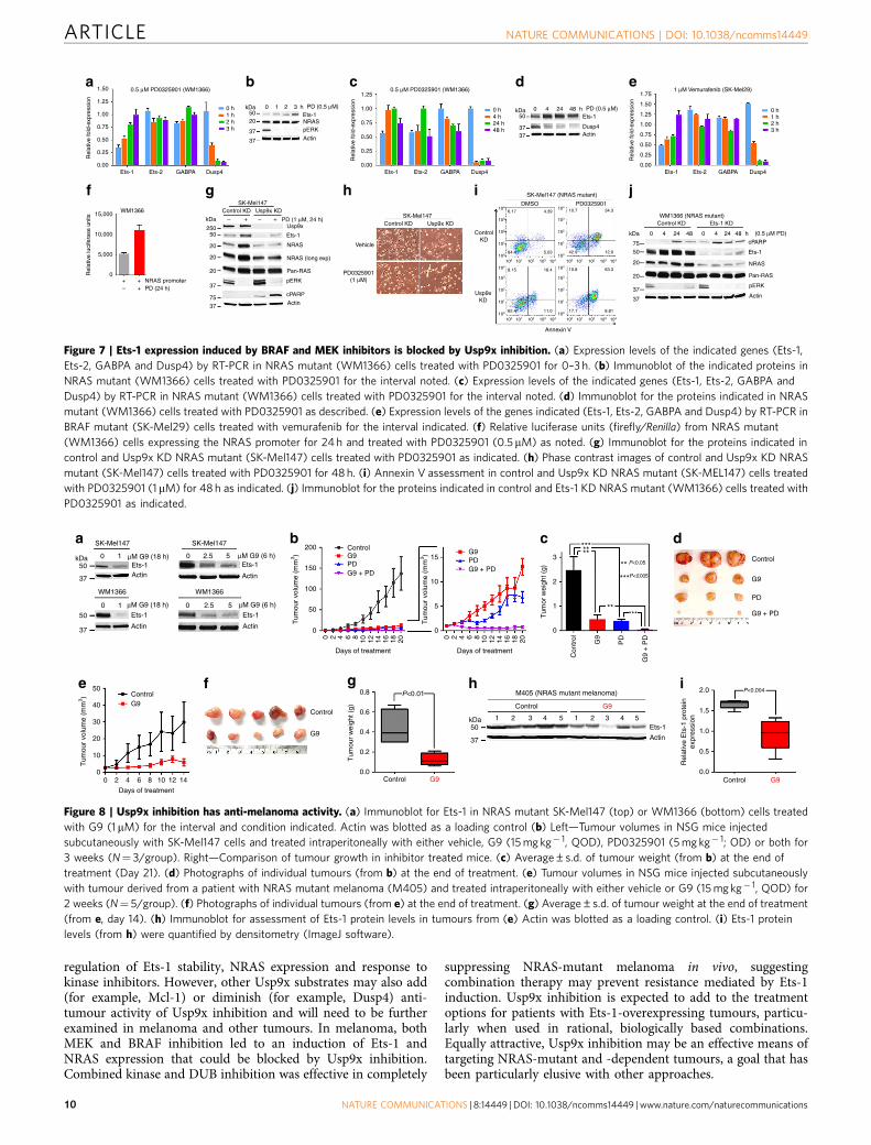
Figure 7 | Ets-1 expression induced by BRAF and MEK inhibitors is blocked by Usp9x inhibition. (a) Expression levels of the indicated genes (Ets-1,
Ets-2, GABPA and Dusp4) by RT-PCR in NRAS mutant (WM1366) cells treated with PD0325901 for 0–3 h. (b) Immunoblot of the indicated proteins in
NRAS mutant (WM1366) cells treated with PD0325901 for the interval noted. (c) Expression levels of the indicated genes (Ets-1, Ets-2, GABPA and
Dusp4) by RT-PCR in NRAS mutant (WM1366) cells treated with PD0325901 for the interval noted. (d) Immunoblot for the proteins indicated in NRAS
mutant (WM1366) cells treated with PD0325901 as described. (e) Expression levels of the genes indicated (Ets-1, Ets-2, GABPA and Dusp4) by RT-PCR in
BRAF mutant (SK-Mel29) cells treated with vemurafenib for the interval indicated. (f) Relative luciferase units (firefly/Renilla) from NRAS mutant
(WM1366) cells expressing the NRAS promoter for 24 h and treated with PD0325901 (0.5mM) as noted. (g) Immunoblot for the proteins indicated in
control and Usp9x KD NRAS mutant (SK-Mel147) cells treated with PD0325901 as indicated. (h) Phase contrast images of control and Usp9x KD NRAS
mutant (SK-Mel147) cells treated with PD0325901 for 48 h. (i) Annexin V assessment in control and Usp9x KD NRAS mutant (SK-MEL147) cells treated
with PD0325901 (1 mM) for 48 h as indicated. (j) Immunoblot for the proteins indicated in control and Ets-1 KD NRAS mutant (WM1366) cells treated with
PD0325901 as indicated.
SK-Mel147a b c d
e f g h i
SK-Mel147
WM1366 WM1366
kDa μM G9 (18 h)
μM G9 (18 h)
50 Ets-1Actin
μM G9 (6 h)Ets-1
Actin
Ets-1
Actin
μM G9 (6 h)Ets-1
Actin
0 1
0 0 2.5 51
37
kDa50
37
50
37
0 2.5 5200
50 0.8 2.0
1.5
1.0
0.5
0.0
1 2 3 4 5 1 2 3 4 5Ets-1
Actin
P<0.01 P<0.004
0.6
0.4
0.2
0.0
ControlG9
Control
Control
G9
G9 Control G9
Control
M405 (NRAS mutant melanoma)
G940
30
20
10
00 2 4 6 8 10 12 14
ControlG9 G9
PD PD
G9 + PD G9 + PD150
15 3P<0.05
***
P<0.005
***
******
**
2
1
0
10
Control
G9
PD
G9 + PD5
0
100
Tum
our
volu
me
(mm
3 )
Tum
our
volu
me
(mm
3 )
Tum
our
volu
me
(mm
3 )
Tum
or w
eigh
t (g)
Tum
our
wei
ght (
g)
Rel
ativ
e E
ts-1
pro
tein
expr
essi
on
50
0
0 2 4 6 8 10 12 14 16 18 20
Days of treatment
Days of treatment
0 2 4 6 8 10 12 14 16 18 20
Con
trol G9
PD
G9
+ P
DDays of treatment
***
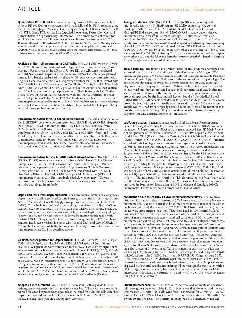
Figure 8 | Usp9x inhibition has anti-melanoma activity. (a) Immunoblot for Ets-1 in NRAS mutant SK-Mel147 (top) or WM1366 (bottom) cells treated
with G9 (1mM) for the interval and condition indicated. Actin was blotted as a loading control (b) Left—Tumour volumes in NSG mice injected
subcutaneously with SK-Mel147 cells and treated intraperitoneally with either vehicle, G9 (15 mg kg� 1, QOD), PD0325901 (5 mg kg� 1; OD) or both for
3 weeks (N¼ 3/group). Right—Comparison of tumour growth in inhibitor treated mice. (c) Average±s.d. of tumour weight (from b) at the end of
treatment (Day 21). (d) Photographs of individual tumours (from b) at the end of treatment. (e) Tumour volumes in NSG mice injected subcutaneously
with tumour derived from a patient with NRAS mutant melanoma (M405) and treated intraperitoneally with either vehicle or G9 (15 mg kg� 1, QOD) for
2 weeks (N¼ 5/group). (f) Photographs of individual tumours (from e) at the end of treatment. (g) Average±s.d. of tumour weight at the end of treatment
(from e, day 14). (h) Immunoblot for assessment of Ets-1 protein levels in tumours from (e) Actin was blotted as a loading control. (i) Ets-1 protein
levels (from h) were quantified by densitometry (ImageJ software).
ARTICLE NATURE COMMUNICATIONS | DOI: 10.1038/ncomms14449
10 NATURE COMMUNICATIONS | 8:14449 | DOI: 10.1038/ncomms14449 | www.nature.com/naturecommunications

MethodsCell culture. A375, SK-Mel2, WM1366 (ATCC), SK-Mel28, SK-Mel29, SK-Mel147and SK-Mel103 cell lines were provided by Dr Monique Verhaegen (University ofMichigan, Ann Arbor, MI, USA). The A375R (vemurafenib-resistant) cell line wasa kind gift from Dr Juxiang Cao (Boston University School of Medicine, Boston,Massachusetts, USA). HEK293T cells were primarily maintained in Dulbecco’sModified Eagle’s Medium (DMEM). The VCaP2 (prostate) cell line was providedby Dr Arul Chinnaiyan (University of Michigan, Ann Arbor, MI, USA) andcells were cultured in DMEM Glutamax. All media was supplemented with10% heat-inactivated FBS (Atlanta Biological), 2 mM L-glutamine and1% penicillin/streptomycin (GIBCO). Tet Free FBS was from Omega Scientific, Inc.
Antibodies. Primary antibodies used in this study include: NRAS, Pan-RAS,HRAS, Dusp4, Ubiquitin (total), Ets-2, GABPAa, Mcl-1, HA, b-Actin, ERK2(total) (Santa Cruz); KRAS (Calbiochem, OP24); AF-6, Usp9x, Ets-1 (BethylLaboratories); pERK, Caspase8, PARP, BID, BIM (Cell Signaling); ERG (Abcam);HA (Roche); FLAG (Sigma). Blots were developed with ECL substrate (Pierce)and imaged on X-ray film (BioExpress). Antibody catalogue numbers and theirdilutions are included in Supplementary Table 2.
Three-dimensional cultures (3D). Equal numbers of viable control, KD andoverexpressing cells from each cell type (1,000 cells per well or as indicated) weregrown on growth factor-reduced Matrigel (Catalogue # 354230; BD transduction)for 7 days61. Phase contrast images were acquired at � 5 or � 10 resolution on aLeica inverted microscope. For cells treated with small molecule inhibitors, mediawas exchanged every 3 days. To quantify the number of colonies, total numbers ofcolonies from 2 to 3 wells of an 8-well chamber slide were counted using phasecontrast images acquired at � 5 resolution. For spheroid culture, 106 cells wereplated in complete media on 100 mm dishes coated with 1% agarose. The cells wereallowed to grow for 2–3 days. The spheroids were collected, pelleted, lysed in lysisbuffer and subjected to immunoblot analysis.
Assessment of the Usp9x ubiquitylome (UbiScan). Sample preparation andmass spectrometry. Cells were collected in Urea Lysis Buffer (20 mM HEPES(pH 8.0), 9.0 M urea, 1 mM sodium orthovanadate (activated), 2.5 mM sodiumpyrophosphate, 1 mM �-glycerol-phosphate) and processed by Cell SignalingTechnology using the Ubiquitin Branch Motif Antibody (CST cat. #3925)45,46
for PTMScan analysis. Lysates were sonicated, centrifuged at 20,000 g for 15 minand ‘cleared’ protein extracts were reduced (with DTT), carboxamidomethylated(with iodoacetamide) and normalized for total protein before tryptic digestion(Worthington, cat. #LS003740). Peptides were enriched by solid-phase extractionwith Sep-Pak C18 classic cartridges (Waters cat. #WAT051910), lyophilized andre-dissolved. Slurries of the Ubiquitin Branch Motif Antibody were used to recoverubiquitin-remnant peptides, which were eluted from antibody-resin with 0.15%trifluoroacetic acid (100 ml total volume). Peptides were desalted on Empore C18
(Sigma) packed tips and eluted with 40% acetonitrile in 0.1% TFA, then loadeddirectly onto a 10 cm� 75 mm PicoFrit capillary column packed with Magic C18AQ reverse-phase resin. The column was developed with a linear gradient ofacetonitrile in 0.125% formic acid, delivered at 280 nl min� 1 over a 90-mininterval. Analytical replicates were generated by running duplicate samples toincrease the number of MS/MS identifications from each sample. A LTQ-OrbitrapVelos mass spectrometer running Xcalibur 2.0.7 SP1 was used to collect tandemmass spectra by the top 20 method, a dynamic exclusion repeat count of 1, andrepeat duration of 30 s. A singly charged polysiloxane ion m/z¼ 371.101237 wasused for real time recalibration of mass error. SEQUEST and the Core platformfrom Harvard University were used to evaluate MS/MS spectra and files weresearched against the NCBI Homo sapiens FASTA Database updated on 27 June2011 containing 34,899 forward and 34,899 reverse sequences. Precursor ion massaccuracy of ±5 p.p.m., and 1 Da for product ions was allowed. Protease specificitywas limited to trypsin, with at least one tryptic (K- or R-containing) terminusrequired per peptide and a maximum of four mis-cleavages. Methionine residueoxidation and the di-glycine (K-GG) remnant was allowed on lysine residues andcysteine carboxamidomethylation was specified as a static modification. Falsediscovery rates were estimated using reverse decoy databases and filtered using a5% FDR in the Linear Discriminant module of Core. We also filtered for thepresence of the K-GG motif in peptides.
Label-free quantitation. All quantitative results were generated using ProgenesisV4.1 (Waters Corporation) or XCalibur 2.0.7 SP1 to extract the integrated peakarea of the corresponding peptide assignments according to previously publishedprotocols45,46. The Progenesis software incorporates a chromatographic alignment(or time warping) algorithm that performs multiple binary comparisons togenerate an overall clustering strategy for the complete data set of all identifiedpeptides on the basis of mass precision. Extracted ion chromatograms for peptideions that changed in abundance between samples were manually reviewed toensure accurate quantitation either in Progenesis or using XCalibur software(version 2.0.7 SP1, Thermo Scientific). This eliminated the possibility that theautomated process selected the wrong chromatographic peak from which to derivethe corresponding intensity measurement. Peak areas were normalized using a log2median normalization strategy in Progenesis45,46.
shRNA-mediated gene knockdown. Melanoma cells were infected with thelentiviral expression system for short hairpin RNA (shRNA) against humanpLVX-Usp9x, kindly provided by Dr Dzwokai Ma (University of California,Santa Barbara)40. For NRAS and control KD: pGIPZ Control, pGIPZ-NRAS-1, andpGIPZ-NRAS-2 were obtained from Open Biosystems. Open Biosystems TRIPZcontrol (clone ID: RHS4743) and TRPIZ human Usp9x (clone ID: V3THS320834)doxycycline-inducible shRNA vectors were also used in melanoma cells.Doxycycline at 1 mg ml� 1 was used to induce shRNA expression.
Ets-1 shRNA was kindly provided by coauthor, Dr Peter C. Hollenhorst(Indiana University, Bloomington, Indiana). HEK293T cells were transfected withthe lentiviral packaging vectors pMD2.G and psPax2 (Addgene) together with theshRNA vectors to produce virus using PolyFect as described by the manufacturer(QIAGEN). The medium was changed to DMEM with 10% fetal bovine serum, andafter 48 h, viral supernatant was collected. Viral supernatant containing 4 mg ml� 1
of Polybrene (Sigma-Aldrich) was added to each melanoma cell line. Cells withstable KD were selected with puromycin.
Chemical reagents. EOAI3402143 (referred to as G9) was synthesized andprovided by Cheminpharma (Branford, CT). Other reagents used in this studywere obtained from the following sources: hemagglutinin-tagged ubiquitin vinylmethyl sulfone (HA-UbVS; Boston Biochem); vemurafenib (PLX4032; ChemieTek); PD 0325901 (Cayman Chemical). All reagents were made up and storedfrozen as 10 mM stock solutions.
Crystal violet colony staining. Equal numbers of viable SK-Mel29 (or A375)cells with modified gene expression were grown in 6-well plates for 3 weeks andsubjected to crystal violet staining (3.7% paraformaldehyde (PFA), 0.05% CrystalViolet in distilled water (filter at 0.45 um)) for 20 min at room temperature. Theplate was photographed by scanning.
DUB-labelling assays. To assay DUB activity, melanoma cells were lysed in DUBbuffer (50 mM Tris pH 7.2, 5 mM MgCl2, 250 mM sucrose, protease inhibitorcocktail (Roche), 1 mM NaF and fresh 1 mM PMSF) for 10 min at 4 �C, followed bybrief sonication. The lysates were centrifuged at 20,000 g for 10 min, and thesupernatants (20 mg) were incubated with 2 mM of HA-UbVS for 75 min at37 �C, followed by boiling in reducing sample buffer and resolving bySDS–polyacrylamide gel electrophoresis (SDS–PAGE). DUBs were detectedby HA immunoblotting62.
Lysate preparation and western blotting. Total cell lysates were prepared bysonicating and boiling cell pellets in � 1 Laemmli-reducing sample buffer.Detergent-soluble cell lysates were prepared by lysing cells in cold isotonic lysisbuffer (10 mM Tris–HCl, pH 7.5, 0.1% Triton X-100, 150 mM NaCl, proteaseinhibitor cocktail and 1 mM PMSF) for 15 min on ice and centrifuging for 10 minat 20,000 g. The clarified supernatant was used as the detergent-soluble cell fraction.Primary and metastatic melanoma tumours were isolated from patients, and asmall portion was sliced, minced and snap frozen with liquid nitrogen followed byhomogenization in lysis buffer. Lysates were electrophoresed (SDS–PAGE gels) andtransferred to nitrocellulose membranes (Whatmann). Proteins were detected byimmunoblotting. Uncropped western blots of key figures are presented inSupplementary Fig. 8.
Plasmids. For overexpressing Usp9x, p3xFlag-Usp9x was created by 3-waycloning using PCR to amplify a 320 bp N-terminal fragment of Usp9x with theStuI site in Usp9x. Forward: 50- tgtacgaagcttacagccacgactcgtggctc-30 ; Reverse:50ggaaccacccatcgaggcc-30 . The PCR product was cut with HindIII and StuI.pCDNA5-TAP-Usp9x was cut with StuI and NotI. These fragments were ligatedinto p3XFlag-CMV10 (Sigma) linearized with HindIII/NotI. A PCR was performedwith forward primer 50-gctctagatctatggactacaaagacc-30 and the reverse primerdescribed above. This product was cut with BglII and StuI, and ligated togetherwith the StuI/BamHI fragment from p3XFlag-Usp9x together with MIGR1linearized with BglII. pcDNA3-Usp9x-HA was kindly provided by Dr Dzwokai Ma(University of California, Santa Barbara)40. 3xFlag-Ets-1 and pGL4.25 were kindlyprovided by Dr Peter C. Hollenhorst (Indiana University, Bloomington, Indiana)24.HA-Ets-1 (WT) was kindly provided by William G. Kaelin, Jr. (Dana-FarberCancer Institute, Boston). Approximately, 5 mg of each pCDNA3 andpCDNA3-Usp9x-HA plasmid, and 2 mg of p3xFlag-Ets-1 WT, HA-Ets-1WT andHA-Ets-1/K388R were used for overexpression in SK-Mel29 and A375 cells.
MTT assay. Cells were seeded in a 96-well plate at 5,000 per well in the presence ofthe indicated concentration of compound for 3 days in a CO2 incubator at 37 �C.Twenty microliters of 5 g l� 1 MTT solution was added to each well for 2 h at 37 �C.The cells were then lysed in 10% SDS buffer, and absorbance at 570 nm relative to areference wavelength of 630 nm was determined with a microplate reader. Toexamine proliferation using the MTT assay, cells were plated in triplicate andprocessed for MTT assay as described above.
NATURE COMMUNICATIONS | DOI: 10.1038/ncomms14449 ARTICLE
NATURE COMMUNICATIONS | 8:14449 | DOI: 10.1038/ncomms14449 | www.nature.com/naturecommunications 11
Quantitative RT-PCR. Melanoma cells were grown on 100 mm dishes with orwithout PD 0325901 or vemurafenib for 0–48 h followed by RNA isolation usingthe RNeasy kit (Qiagen, Valencia, CA). Samples for qRT-PCR were prepared with� 1 SYBR Green PCR Master Mix (Applied Biosystems, Foster City, CA) andprimers listed in Supplementary Information. The primers were optimized foramplification under the following reaction conditions: denaturing at 95 �C for10 min, followed by 40 cycles of 95 �C for 15 s and 60 �C for 1 min. Melting curveswere analysed for all samples after completion of the amplification protocol.GAPDH was used as the housekeeping gene for control expression. All RT-PCRprimers were purchased from RealTimePrimers.com.
Analysis of Ets-1 ubiquitination in 293T cells. HEK293T cells grown in DMEMwith 10% FBS were co-transfected with Flag-Ets-1 and HA-ubiquitin expressionplasmids. For analysis of the effects of Usp9x KD, the cells were transfectedwith shRNAs against Usp9x or a non-targeting shRNA for 72 h before plasmidtransfection. For the analysis of the effects of G9, cells were co-transfected withFlag-Ets-1 and HA-ubiquitin (WT) expression vectors for 40 h, then treated withG9 (2.5 mM) for 5 h. Cells were lysed in 1% NP-40, 1% SDS, 2 mM EDTA, 1 mMNEM (fresh) and 25 mM Tris–HCl, pH 7.5, boiled for 20 min, and then dilutedwith 10 volumes of immunoprecipitation buffer (lysis buffer with 1% NP-40).Lysate of 500 mg was immunoprecipitated with anti-FLAG overnight and thenwith 30 ml protein A/G for 2 h at 4 �C. The beads were washed five times withimmunoprecipitation buffer and 0.1 M NaCl. Western blot analysis was performedwith anti-HA or ubiquitin antibody to detect ubiquitinated Ets-1. Usp9x, FLAGand actin were probed by immunoblotting.
Immunoprecipitation for K63-linked ubiquitination. To assess ubiquitination ofEts-1, HEK293T cells were co-transfected with FLAG-Ets-1, pRK5-HA-ubiquitin(WT), pRK5-HA-Ub/K48 only or pRK5-HA-Ub/K63 only (obtained fromDr Vaibhav Kapuria (University of Lausanne, Switzerland)), and after 48 h, cellswere lysed in 1% NP-40, 1% SDS, 2 mM EDTA, 1 mM NEM (fresh) and 10 mMTris–HCl, pH 7.5. Lyses were boiled for 20 min and then diluted with 10 volumesof immunoprecipitation buffer (lysis buffer with 1% NP-40). FLAG wasimmunoprecipitated as described above. Western blot analysis was performedwith anti-HA or ubiquitin antibody to detect ubiquitinated Ets-1.
Immunoprecipitation for Ets-1/K388 mutant ubiquitination. The Ets-1/K388(K388A, K388R) mutant was generated using a Quickchange II Site-Directedmutagenesis Kit on the HA-Ets-1 construct (Agilent Technologies). Primersets used in mutagenesis are provided in Supplementary Table 3. To assessubiquitination of Ets-1, HEK293T cells were co-transfected with HA-Ets-1,HA-Ets-1/K388A or HA-Ets-1/K388R with pRK5-HA-ubiquitin (WT), andimmunoprecipitation with Ets-1 antibody (Bethyl, Montgomery, TX) wasperformed as described above. Western blot analysis was performed with theanti-HA and ubiquitin antibody.
Usp9x and Ets-1 immunoprecipitation. For immunoprecipitation of endogenousUsp9x, SK-Mel2 cells were lysed in lysis buffer (25 mM HEPES (pH 7.5), 400 mMNaCl, 0.5% IGEPAL CA-630, 5% glycerol, protease inhibitors and 1 mM freshPMSF). The soluble fraction of the lysate (1 mg) was diluted to adjust NaCl andIGEPAL CA-630 concentrations to 100 mM and 0.125%, respectively. Preclearedlysates were incubated with a rabbit control IgG or anti-Usp9x antibody (5 mg)(Bethyl) at 4 �C for 3 h with rotation, followed by immunoprecipitation withProtein A/G PLUS Agarose (Santa Cruz Biotechnolgy) beads at 4 �C for 1 h withrotation. Beads were washed five times with 100 mM NaCl and 0.1% IGEPAL CA-630 and boiled in Laemmli buffer for Western blot analysis. Anti-Ets-1 was used toimmunoprecipitate Ets-1 as described above.
Co-immunoprecipitation for Usp9x and Ets-1. FLAG-Usp9x WT, FLAG-Usp9x-CDM, FLAG-Usp9x E1, FLAG-Usp9x E1M, FLAG-Usp9x E5 (ref. 43) andHA-Ets-1 WT plasmids were transfected into HEK293T cells. Forty-eight hoursafter transfection, cells were lysed in lysis buffer (25 mM HEPES (pH 7.5), 400 mMNaCl, 0.5% IGEPAL CA-630, 1 mM NEM (fresh) 1 mM DTT, 5% glycerol andprotease inhibitors) and the soluble fraction of the lysate was diluted to adjust NaCland IGEPAL CA-630 concentrations to 100 mM and 0.125%, respectively. Lysate of0.5 mg was immunoprecipitated with anti-HA (Ets-1) overnight and then with40ml protein A/G for 2 h at 4 �C. Beads were washed five times with 100 mM NaCland 0.1% IGEPAL CA-630, and boiled in Laemmli buffer for Western blot analysis.Western blot analysis was performed with anti-FLAG antibody (Usp9x).
Apoptosis measurement. An Annexin V-fluorescein isothiocyanate (FITC)staining assay was performed as previously described43. The cells were seeded insix-well plates and exposed to compounds as indicated for 48 h. The cells were thentrypsinized, washed with cold PBS, and stained with Annexin V-FITC for 10 minon ice. Positive cells were detected by flow cytometry.
Xenograft studies. NSG (NOD/SCID/IL2r-g (null)) mice were injectedmid-dorsally with 3� 105 BRAF mutant SK-Mel29 expressing HA-control,HA-Usp9x cells, or 5� 105 NRAS mutant SK-Mel 147 cells in 0.1 ml ofMatrigel/DMEM suspension. 5� 105 M405 (NRAS mutant) patient-derivedmelanoma tumour cells51 in 0.1 ml of Matrigel/L15 suspension were alsoinoculated in NSG mice. Tumours were allowed to reach about 10 mm3, afterwhich mice were tumour-size matched and assigned to treatment groups consistingof vehicle, PD 0325901 or G9 as indicated. G9 and PD 0325901 were administeredin DMSO: PEG300 (1:1) by i.p. injection every other day at 15 mg kg� 1 for G9 andevery day for PD 0325901 at 5 mg kg� 1. Tumour size was monitored by calipersevery other day using the following formula: volume¼ (width)2� length� height/2.Animal weight was also recorded every other day.
Tissue banking. The tissue bank protocol used for this study was developed andapproved jointly by the clinical director of the University of Michigan (UM)melanoma program, UM Cancer Center director of tissue procurement, UM chiefof anatomic pathology, and UM director of the section of dermatopathology. Theprotocol was developed to avoid any compromise in patient care, pathologicdiagnosis, tumour staging, or treatment. Patient confidentiality was maintainedby password and firewall-protected access to all pertinent databases. Melanomaspecimens were obtained with informed consent from all patients according toprotocols approved by the Institutional Review Board of UM Medical School(HUM00102527). All patients included in this study had stage II or III melanomaproven by biopsy (most often needle core). A small (typically 2–6 mm) tissuesample was obtained from surgically resected tumours. Most of the melanomas inthis study were regional stage III lymph node or skin/soft tissue disease withpalpable, clinically enlarged node(s) or soft tissues.
Luciferase assays. Luciferase assays used a Dual Luciferase Reporter AssaySystem (Promega) according to the manufacturer instructions. NRAS promotersequences (733 bp) from the NRAS mutant melanoma cell line SK-Mel147 werecloned upstream of the firefly luciferase-pGL3-Basic (Promega) plasmid cut withHind III and XhoI (Forward Primer- 50-AGACTCGAGGAGGAGTGCC-30 -XhoI,Reverse Primer- 50-GATCAAGCTTAAATGTTGGAGACCCCGGAA-30—HindIII)and site-directed mutagenesis of promoter and expression constructs wereperformed using the Quickchange Lightning Multi Site-Directed mutagenesis Kit(Agilent Technologies). Primer sets used in mutagenesis are provided inSupplementary Table 4. Positive clones were confirmed by the UM sequence core.Melanoma SK-Mel29 and WM1366 cells were plated at B50% confluence in a6-well plate (3� 105 cells per well) 24 h before transfection. Cells were transfectedwith 1 mg of each p3xFlag, p3xFlag-Usp9x, p3xFlag-Ets-1, HA-Ets-1, wild-type(WT) or point mutant NRAS promoter constructs (WT, E1M, E2M, E3M, E4Mand E5M), 2 mg of firefly and 200 ng of Renilla plasmid using PolyFect TransfectionReagent (Qiagen). After 48 h, media was removed, and cells were washed two timeswith � 1 PBS, resuspended in 500 ml � 1 PLB, disrupted by one freeze/thaw cycle(� 80 �C) and dissociated with a BD 1 ml 26G syringe. Luciferase activity wasmeasured in 20ml of cell lysate using a BD PharMingen (Monolight 3010C)luminometer. Firefly values were normalized to Renilla values.
Melanoma tissue microarray (TMA) immunohistochemistry. For immuno-histochemical analysis, tissue microarrays (TMA) were used, containing 36 cases ofmelanoma and 12 cases of normal and non-melanoma tumour tissues of the skin induplicates (96 cores) (Catalogue No.: Z7020108, BioChain Institute, Inc.). All thetissues were from surgical resection. They were fixed in 10% neutral-bufferedformalin for 24 h. Ninety-four cores consisted of 8 normal skin, 8 benign nevi, 4cases of non-melanoma skin cancer basal cell carcinoma (BCC), 4 cases non-melanoma skin cancer squamous cell carcinoma (SCC), 48 malignant melanomasand 24 metastatic melanomas. Immunohistochemistry was also performed onindividual slides for Usp9x, Ets-1 and NRAS. Formalin-fixed, paraffin sections werecut at 5 microns and rehydrated to water. Heat-induced epitope retrieval wasperformed with FLEX TRS high pH retrieval buffer (9.01) for 20 min. After per-oxidase blocking, the antibody was applied at room temperature for 60 min. TheFLEX HRP EnVision System was used for detection. DAB chromagen was thenapplied for 10 min. Slides were counterstained with Harris Hematoxylin for 5 s andthen dehydrated and coverslipped. Tumour content of each core or slide wasverified by H&E staining. Immunohistochemistry was performed using anti-Usp9x(1:1,000, Abcam), Ets-1 (1:500, Bethyl) and NRAS (1:150, Origene, clone 5G7).Slides were scored by a UM dermatologist and pathologist (Dr Paul WilliamHarms) for percentage of positive cells and intensity of staining. All positive casesdisplayed nuclear and cytoplasmic staining. Photomicrographs were taken with aSPOT Insight Colour camera (Diagnostic Instruments) on an Olympus BX41microscope with Olympus UPlanFL � 10 and � 40, � 200 and � 400 objectivesusing SPOT Basic software.
Immunofluorescence. BRAF mutant A375 parental and vemurafenib-resistantcells were grown on 6-well slides for 24 h. Media was then decanted and the wellswere washed 3� with PBS. Cells were fixed in methanol for 20 min at � 20 �C,washed 3� with PBS and blocked for 1 h at room temperature in PBS with 0.3%Tween-20 and 5% BSA. The primary antibody was Ets-1 (Bethyl), which was
ARTICLE NATURE COMMUNICATIONS | DOI: 10.1038/ncomms14449
12 NATURE COMMUNICATIONS | 8:14449 | DOI: 10.1038/ncomms14449 | www.nature.com/naturecommunications
diluted 1:100 in PBS with 0.3% Tween-20 and 1% BSA (antibody dilution buffer)and incubated overnight at 4 �C. After 3� washes in PBS, Alexa Fluor 488anti-Rabbit secondary antibody (Life Technologies) was added at 1:500 in antibodydilution buffer and with DAPI incubated for 1 h at room temperature. After5� washes in PBS, slides were coverslipped with ProLong Gold anti-fadereagent (Invitrogen). Images were acquired using an Olympus Fluo View 500.A representative image from each sample is shown.
Chromatin immunoprecipitation assay (ChIP). WM1366 cells were seeded at adensity of 5� 107 in 150 mm dishes and protein/DNA cross-linking was inducedwith formaldehyde at 1% final concentration at room temperature for 15 min.Crosslinking was terminated by the addition of 1/10 volume 1.25 M glycine for5 min at room temperature followed by cell lysis (1% SDS, 10 mM EDTA, 50 mMTris, pH 8) for 10 min and sonication (Misonix, Microson Ultrasonic CellDisruptor (20 s on, 40 off, 10 amplitude for 30 min) resulting in an averagechromatin fragment size of 300 bp. DNA–protein complexes were immunopreci-pitated with 5 mg of rabbit Ets-1 (Bethyl) or 5 mg rabbit IgG antibody (Santa Cruz)overnight at 4 �C (1:10 ml volume) in dilution buffer; (20 mM Tris at pH 8, 2 mMEDTA, 150 mM NaCl, 0.01% Triton X-100, 0.01% SDS, protease inhibitors) androtated overnight and then with 50 ml Dynabeads for 2 h at 4 �C. Beads werewashed with low salt (0.1% SDS, 1% Triton X-100, 2 mM EDTA, 20 mM Tris atpH 7.5, 150 mM NaCl), high salt (0.1% SDS, 1% Triton X-100, 2 mM EDTA,20 mM Tris at pH 7.5, 500 mM NaCl), LiCl (250 mM LiCl, 1% NP-40, 1%deoxycholic acid, 1 mM EDTA, 10 mM Tris at pH 7.5) and TE wash buffer (10 mMTris, pH 7.5, 1 mM EDTA) twice. Beads were resuspended with 250ml of freshelution buffer (1% SDS, 50 mM NaHCO3). Elutes were resuspended with 5 M NaCl(10 ml in 250 ml of elute) and incubated at 65 �C overnight, followed by 10mg ml� 1
RNase A addition and incubated for 30 min at 37 �C. ChIP DNA was purified usinga Quick PCR Purification Kit (Qiagen). Primers used for NRAS promoter detection(244 bp): forward primer (5-GTAGCCGCCTGGTTACTG-3), reverse primer(5-CCCAGAGATCAAAACCTC-3). RT-PCR was performed as described above.
Statistical analysis. All statistical analysis was carried out using GraphPad Prismsoftware (GraphPad Prism 6 and GraphPad InStat3). For quantitative data,treatment groups were reported as mean±s.d. and compared using the unpairedStudent’s t-test. Usp9x, Ets-1 and NRAS expression values were categorized intolow/moderate (o300 product score) and high (4300 product score). Statisticalsignificance was established at Pr0.05 unless otherwise noted. Data points areshown as the mean±s.d.
Institutional approval. Protocols utilizing animals were reviewed and approved bythe University Animal Care and Use Committee (University of Michigan). Allpatient samples were obtained through signed informed consent using a protocolreviewed and approved by the Institutional Review Board (University of Michigan).
Data availability. Mass spectrometry proteomics data have been deposited withthe ProteomeXchange Consortium via the PRIDE partner repository with thedata set identifier PXD005417 (ref. 63). Access details include: Website:http://www.ebi.ac.uk/pride, Project name: Ubiquitin remnant analysis inmelanoma post inhibition of Usp9x. Project accession: PXD005417. Project DOI:Not applicable. G9 may be made available through a materials transfer agreement(MTA). All other remaining data are available within the Article and itsSupplementary Files, or available from the authors on request.
References1. Merlino, G. et al. The state of melanoma: challenges and opportunities. Pigment
Cell Melanoma Res. 29, 404–416 (2016).2. Flaherty, K. T. et al. Combined BRAF and MEK inhibition in melanoma with
BRAF V600 mutations. New Engl. J. Med. 367, 1694–1703 (2012).3. Orloff, M., Weight, R., Valsecchi, M. E. & Sato, T. Immune check point
inhibitors combination in melanoma: worth the toxicity? Rev. Recent Clin.Trials 11, 81–86 (2016).
4. Piskounova, E. et al. Oxidative stress inhibits distant metastasis by humanmelanoma cells. Nature 527, 186–191 (2015).
5. Kaur, A. et al. sFRP2 in the aged microenvironment drives melanomametastasis and therapy resistance. Nature 532, 250–254 (2016).
6. Crawford, L. J. & Irvine, A. E. Targeting the ubiquitin proteasome system inhaematological malignancies. Blood Rev. 27, 297–304 (2013).
7. Shen, M., Schmitt, S., Buac, D. & Dou, Q. P. Targeting the ubiquitin-proteasome system for cancer therapy. Exp. Opin. Ther. Targets 17, 1091–1108(2013).
8. Clague, M. J. et al. Deubiquitylases from genes to organism. Physiol. Rev. 93,1289–1315 (2013).
9. Clague, M. J., Coulson, J. M. & Urbe, S. Cellular functions of the DUBs. J. CellSci. 125, 277–286 (2012).
10. Luise, C. et al. An Atlas of altered expression of deubiquitinating enzymes inhuman cancer. PLoS ONE 6, e15891 (2011).
11. Cox, J. L. et al. Context-dependent function of the deubiquitinating enzymeUSP9X in pancreatic ductal adenocarcinoma. Cancer Biol. Therapy 15,1042–1052 (2014).
12. Peng, J. et al. USP9X expression correlates with tumor progression and poorprognosis in esophageal squamous cell carcinoma. Diagnos. Pathol. 8, 177(2013).
13. Perez-Mancera, P. A. et al. The deubiquitinase USP9X suppresses pancreaticductal adenocarcinoma. Nature 486, 266–270 (2012).
14. Schwickart, M. et al. Deubiquitinase USP9X stabilizes MCL1 and promotestumour cell survival. Nature 463, 103–107 (2010).
15. Wang, S. et al. Ablation of the oncogenic transcription factor ERG bydeubiquitinase inhibition in prostate cancer. Proc. Natl Acad. Sci. 111,4251–4256 (2014).
16. Wang, Y. et al. Elevated expression of USP9X correlates with poor prognosis inhuman non-small cell lung cancer. J. Thor. Dis. 7, 672–679 (2015).
17. Murtaza, M., Jolly, L. A., Gecz, J. & Wood, S. A. La FAM fatale: USP9X indevelopment and disease. Cell Mol. Life Sci.: CMLS 72, 2075–2089 (2015).
18. Hollenhorst, P. C., McIntosh, L. P. & Graves, B. J. Genomic and biochemicalinsights into the specificity of ETS transcription factors. Ann. Rev. Biochem. 80,437–471 (2011).
19. Kar, A. & Gutierrez-Hartmann, A. Molecular mechanisms of ETS transcriptionfactor-mediated tumorigenesis. Critic. Rev. Biochem. Mol. Biol. 48, 522–543(2013).
20. Findlay, V. J., LaRue, A. C., Turner, D. P., Watson, P. M. & Watson, D. K. inAdvances in Cancer Research Vol. 119 (eds Kenneth, D. T. & Paul, B. F.), 1–61(Academic Press, 2013).
21. Oikawa, T. & Yamada, T. Molecular biology of the Ets family of transcriptionfactors. Gene 303, 11–34 (2003).
22. Tomlins, S. A. et al. Recurrent fusion of TMPRSS2 and ETS transcription factorgenes in prostate cancer. Science 310, 644–648 (2005).
23. Erkizan, H. V., Uversky, V. N. & Toretsky, J. A. Oncogenic partnerships:EWS-FLI1 protein interactions initiate key pathways of Ewing’s sarcoma. Clin.Cancer Res.: Off. J. Am. Assoc. Cancer Res. 16, 4077–4083 (2010).
24. Plotnik, J. P., Budka, J. A., Ferris, M. W. & Hollenhorst, P. C. ETS1 is agenome-wide effector of RAS/ERK signaling in epithelial cells. Nucleic AcidsRes. 42, 11928–11940 (2014).
25. Lu, G. et al. Phosphorylation of ETS1 by Src family kinases prevents itsrecognition by the COP1 tumor suppressor. Cancer Cell 26, 222–234 (2014).
26. Vitari, A. C. et al. COP1 is a tumour suppressor that causes degradation of ETStranscription factors. Nature 474, 403–406 (2011).
27. Ji, Z. et al. Regulation of the Ets-1 transcription factor by sumoylation andubiquitinylation. Oncogene 26, 395–406 (2006).
28. Hollenhorst, P. C. RAS/ERK pathway transcriptional regulation throughETS/AP-1 binding sites. Small GTPases 3, 154–158 (2012).
29. Dittmer, J. The role of the transcription factor Ets1 in carcinoma. Semin.Cancer Biol. 35, 20–38 (2015).
30. Horn, S. et al. TERT promoter mutations in familial and sporadic melanoma.Science 339, 959–961 (2013).
31. Huang, F. W. et al. Highly recurrent TERT promoter mutations in humanmelanoma. Science 339, 957–959 (2013).
32. Li, Y., Luo, H., Liu, T., Zacksenhaus, E. & Ben-David, Y. The ets transcriptionfactor Fli-1 in development, cancer and disease. Oncogene 34, 2022–2031(2015).
33. Rothhammer, T. et al. The Ets-1 transcription factor is involved in thedevelopment and invasion of malignant melanoma. Cell Mol. Life Sci. 61,118–128 (2004).
34. Bell, R. J. et al. Cancer. The transcription factor GABP selectively binds andactivates the mutant TERT promoter in cancer. Science 348, 1036–1039 (2015).
35. Cooper, C. D., Newman, J. A. & Gileadi, O. Recent advances in the structuralmolecular biology of Ets transcription factors: interactions, interfaces andinhibition. Biochem. Soc. Trans. 42, 130–138 (2014).
36. Baines, A. T., Xu, D. & Der, C. J. Inhibition of Ras for cancer treatment: thesearch continues. Fut. Med. Chem. 3, 1787–1808 (2011).
37. Eskandarpour, M. et al. Suppression of oncogenic NRAS by RNA interferenceinduces apoptosis of human melanoma cells. Int. J. Cancer 115, 65–73(2005).
38. Mandala, M., Merelli, B. & Massi, D. Nras in melanoma: targeting theundruggable target. Crit. Rev. Oncol./Hematol. 92, 107–122 (2014).
39. Potu, H. et al. Usp5 links suppression of p53 and FAS levels in melanoma to theBRAF pathway. Oncotarget 5, 5559–5569 (2014).
40. Xu, Z. et al. Identification of a deubiquitinating enzyme as a novelAGS3-interacting protein. PLoS ONE 5, e9725 (2010).
41. Lee, G. Y., Kenny, P. A., Lee, E. H. & Bissell, M. J. Three-dimensional culturemodels of normal and malignant breast epithelial cells. Nat. Methods 4,359–365 (2007).
42. Pal, A. & Kleer, C. G. Three dimensional cultures: a tool to study normal acinararchitecture vs malignant transformation of breast cells. J. Vis. Exp. 86, e51311(2014).
NATURE COMMUNICATIONS | DOI: 10.1038/ncomms14449 ARTICLE
NATURE COMMUNICATIONS | 8:14449 | DOI: 10.1038/ncomms14449 | www.nature.com/naturecommunications 13
43. Peterson, L. F. et al. Targeting deubiquitinase activity with a novelsmall-molecule inhibitor as therapy for B-cell malignancies. Blood 125,3588–3597 (2015).
44. Eskandarpour, M., Huang, F., Reeves, K. A., Clark, E. & Hansson, J. OncogenicNRAS has multiple effects on the malignant phenotype of human melanomacells cultured in vitro. Int. J. Cancer 124, 16–26 (2009).
45. Guo, A. et al. Immunoaffinity enrichment and mass spectrometry analysis ofprotein methylation. Mol. Cell. Proteom. 13, 372–387 (2014).
46. Ryder, D. J. et al. Identification of the acetylation and ubiquitin-modifiedproteome during the progression of skeletal muscle atrophy. PLoS ONE 10,e0136247 (2015).
47. Cagnol, S. & Rivard, N. Oncogenic KRAS and BRAF activation of theMEK/ERK signaling pathway promotes expression of dual-specificityphosphatase 4 (DUSP4/MKP2) resulting in nuclear ERK1/2 inhibition.Oncogene 32, 564–576 (2013).
48. Keyse, S. M. Dual-specificity MAP kinase phosphatases (MKPs) and cancer.Cancer Metastasis Rev. 27, 253–261 (2008).
49. Baehner, F. L. et al. The impact of primary melanoma thickness andmicroscopic tumor burden in sentinel lymph nodes on melanoma patientsurvival. Ann. Surg. Oncol. 19, 1034–1042 (2012).
50. Yaziji, H. & Gown, A. M. Immunohistochemical markers of melanocytictumors. Int. J. Surg. Pathol. 11, 11–15 (2003).
51. Quintana, E. et al. Human melanoma metastasis in NSG mice correlates withclinical outcome in patients. Sci. Transl. Med. 4, 159ra149 (2012).
52. Prescott, J. D., Koto, K. S., Singh, M. & Gutierrez-Hartmann, A. The ETStranscription factor ESE-1 transforms MCF-12A human mammaryepithelial cells via a novel cytoplasmic mechanism. Mol. Cell Biol. 24,5548–5564 (2004).
53. Schedin, P. J. et al. ESX induces transformation and functional epithelial tomesenchymal transition in MCF-12A mammary epithelial cells. Oncogene 23,1766–1779 (2004).
54. Liu, F. et al. Stat3-targeted therapies overcome the acquired resistanceto vemurafenib in melanomas. J. Investig. Dermatol. 133, 2041–2049 (2013).
55. Joseph, E. W. et al. The RAF inhibitor PLX4032 inhibits ERK signaling andtumor cell proliferation in a V600E BRAF-selective manner. Proc. Natl Acad.Sci. 107, 14903–14908 (2010).
56. Lito, P. et al. Relief of profound feedback inhibition of mitogenic signaling byRAF inhibitors attenuates their activity in BRAFV600E melanomas. Cancer Cell22, 668–682 (2012).
57. Pratilas, C. A. et al. V600EBRAF is associated with disabled feedback inhibitionof RAF-MEK signaling and elevated transcriptional output of the pathway.Proc. Natl Acad. Sci. USA 106, 4519–4524 (2009).
58. Zheng, Z. Y. et al. Wild-Type N-Ras, overexpressed in basal-like breast cancer,promotes tumor formation by inducing IL-8 secretion via JAK2 activation. CellRep. 12, 511–524 (2015).
59. Bliss, J. M., Venkatesh, B. & Colicelli, J. The RIN family of Ras effectors.Methods Enzymol. 407, 335–344 (2006).
60. Dougherty, G. W., Jose, C., Gimona, M. & Cutler, M. L. The Rsu-1-PINCH1-ILK complex is regulated by Ras activation in tumor cells. Eur. J. Cell Biol. 87,721–734 (2008).
61. Pal, A., Huang, W., Toy, K. A. & Kleer, C. G. CCN6 knockdowndisrupts acinar organization of breast cells in three-dimensional culturesthrough up-regulation of type III TGF-beta receptor. Neoplasia 14, 1067–1074(2012).
62. Kapuria, V. et al. Deubiquitinase inhibition by small-molecule WP1130 triggersaggresome formation and tumor cell apoptosis. Cancer Res. 70, 9265–9276(2010).
63. Vizcaino, J. A. et al. ProteomeXchange provides globally coordinatedproteomics data submission and dissemination. Nat. Biotechnol. 32, 223–226(2014).
AcknowledgementsWe thank Jessica Mercer for editing the manuscript and Jeffrey Silva and Matthew Stokes(Cell Signaling Technology Inc.) for analysis and discussion of the Usp9x ubiquitylome.We also thank Arul Chinnaiyan, Monique Verhaegen (University of Michigan, AnnArbor, MI, USA) for kindly providing cell lines and William G. Kaelin, Jr (Dana-FarberCancer Institute, Boston) for kindly providing Ets-1 HA plasmid. Vaibhav Kapuria(University of Lausanne, Switzerland) kindly provided pRK5-HA-Ub WT, K48 and K68plasmids and Yihong Liu provided technical assistance with this study. We also thankPinki Chowdhury (University of Michigan, Ann Arbor, MI, USA) for kindly providingtechnical assistance for ChIP analysis. We thank Nisha Meireles, Clinical ResearchSpecialist, Multidisciplinary Cutaneous Oncology Program, for database and datamanagement. We thank the ENCODE project consortium and Richard Myers for the useof data sets. We also would like to thank the patients who agreed to be part of anIRB-approved translational study. We acknowledge support from the Allen H. BlondyResearch Fund for Melanoma (to M.T., H.P.), The Harry J. Lloyd Charitable Trustand the Michigan Translational Research and Commercialization (MTRAC) program(to N.J.D.).
Author contributionsH.P., L.F.P., M.K., A.P. and H.S. performed the research and analysed the data. P.W.H.analysed TMA and protein expression in clinical samples. P.C.H., U.E., A.D. and M.T.contributed materials. H.P. and N.J.D. designed the study, analysed the data and wrotethe manuscript. All authors contributed to data review and provided comments on themanuscript.
Additional informationSupplementary Information accompanies this paper at http://www.nature.com/naturecommunications
Competing financial interests: A patent (Patent No: US 8,809,377 B2, Date of Patent:Aug. 19, 2014) covering the synthesis and use of G9 has been filed with L.F.P., M.T. andN.J.D. as authors and constitutes a competing financial interest. The remaining authorsdeclare no competing financial interests.
Reprints and permission information is available online at http://npg.nature.com/reprintsandpermissions/
How to cite this article: Potu, H. et al. Usp9x regulates Ets-1 ubiquitination and stabilityto control NRAS expression and tumorigenicity in melanoma. Nat. Commun. 8, 14449doi: 10.1038/ncomms14449 (2017).
Publisher’s note: Springer Nature remains neutral with regard to jurisdictional claims inpublished maps and institutional affiliations.
This work is licensed under a Creative Commons Attribution 4.0International License. The images or other third party material in this
article are included in the article’s Creative Commons license, unless indicated otherwisein the credit line; if the material is not included under the Creative Commons license,users will need to obtain permission from the license holder to reproduce the material.To view a copy of this license, visit http://creativecommons.org/licenses/by/4.0/
r The Author(s) 2017
ARTICLE NATURE COMMUNICATIONS | DOI: 10.1038/ncomms14449
14 NATURE COMMUNICATIONS | 8:14449 | DOI: 10.1038/ncomms14449 | www.nature.com/naturecommunications
Related Documents