Add the following: ▲ á 127 ñ FLOW CYTOMETRIC ENUMERATION OF CD34+ CELLS INTRODUCTION The CD34 antigen is expressed on the surface of almost all human hematopoietic stem and progenitor cells. The absolute number of CD34+ hematopoietic stem cells (HSCs) has been shown to correlate with in vitro colony-forming unit (CFU) assay activity and with clinical engraftment in hematopoietic grafts prepared from peripheral blood, bone marrow, and cord blood sources. The single-platform, flow cytometric CD34+ cell enumeration method described here is based on a clinical laboratory protocol, 1 established by the International Society of Hematotherapy and Graft Engineering (ISHAGE), now known as the Inter- national Society for Cellular Therapy (ISCT). This protocol can be used to enumerate CD34+ cells in samples of peripheral blood, leukapheresis products, bone marrow, and cord blood. CD34+ cell enumeration by flow cytometry is a rare event anal- ysis, which requires specific gating instructions that are provided in this chapter. Furthermore, the USP CD34+ Cell Enumera- tion System Suitability RS has been developed to assess the reagents and ensure the correct gating during data acquisition and analysis. IDENTIFICATION OF CD34+ HEMATOPOIETIC STEM CELLS CD34+ cell enumeration by flow cytometry is a rare event analysis. For the analysis of CD34+ HSCs, cell samples are stained with fluorescently labeled antibodies against both the HSC antigen CD34 and the pan-leukocyte antigen CD45. Five parame- ters —forward light scatter (FSC), side light scatter (SSC), CD34 staining, CD45 staining, and viability dye staining are com- bined in a sequential, or Boolean, gating strategy to identify viable CD34+ cells. CD34+ HSCs have FSC and SSC characteristics similar to lymphocytes; expressing both CD45 and CD34, and exhibiting dim CD45 expression and low SSC characteristics. Viability dye does not stain live cells, allowing the exclusion of dead cells from the analysis of viable cell preparations. For analysis of nonviable, fixed (preserved) cell preparations such as the USP CD34+ Cell Enumeration System Suitability Reference Standard (RS), either the flow cytometer viable cell analysis gate is fully opened to include all cells, or the Viability dye is omitted from the analysis. ENUMERATION CONSIDERATIONS The CD34+ cell enumeration method described here relies on the use of synthetic fluorescent microspheres (counting beads) as internal enumeration controls. Homogeneous counting beads are added to the cell sample at a known concentration and volume, or the counting beads may be procured, pre-aliquoted, and lyophilized in special sample tubes, in which all sub- sequent cell staining and processing steps are conducted. To avoid the loss of counting beads, the wash steps are omitted, and protein-containing sample buffers are used. An ammonium chloride-based, red blood cell (RBC)-lysing protocol is used for fresh cell preparations; no lysing is necessary for frozen, thawed, or fixed cell preparations. After the cells are stained and processed, the counting beads and cells are simultaneously analyzed on a flow cytometer. The number of CD34+ cells/ mL in the cell sample can be directly calculated by comparing the absolute number of target CD34+ cells and the number of counting beads detected in the same data file. The USP CD34+ Cell Enumeration System Suitability RS is used to verify that the correct reagents and flow cytometer gating parameters were used. EQUIPMENT SPECIFICATIONS The following equipment is needed: • A pipettor capable of accurately reverse-pipetting microliter volumes • A flow cytometer with the following minimum specifications (see Flow Cytometry á1027 ñ): • Detection capabilities for FSC; SSC; “green” fluorescence emission (range, 510–550 nm); “yellow” fluorescence emis- sion (range, 560–590 nm); and “red” fluorescence emission (>600 nm) • Light-scatter and fluorescence measurements that are correlated to time • Light-scatter resolution allowing for the identification of lymphocytes, monocytes, granulocytes, and fluorescent counting beads • Detection and data acquisition rates of at least 5000 cells/s • A 488-nm laser excitation source • Fluorescence intensity sensitivity allowing for the detection of cellular autofluorescence • Analysis software, including a data file structure in the Flow Cytometry Standard (FCS) or equivalent, with spectral overlap correction capability 1 Sutherland DR, Keeney M, Gratama JW. Enumeration of CD34+ hematopoietic stem and progenitor cells. Curr Protoc Cytom. 2003;6:6.4.1–6.4.24. USP 40 Biological Tests / á127ñ Flow Cytometric Enumeration of CD34+ Cells 1

Welcome message from author
This document is posted to help you gain knowledge. Please leave a comment to let me know what you think about it! Share it to your friends and learn new things together.
Transcript

Add the following:
▲á127ñ FLOW CYTOMETRIC ENUMERATION OF CD34+ CELLS
INTRODUCTION
The CD34 antigen is expressed on the surface of almost all human hematopoietic stem and progenitor cells. The absolutenumber of CD34+ hematopoietic stem cells (HSCs) has been shown to correlate with in vitro colony-forming unit (CFU) assayactivity and with clinical engraftment in hematopoietic grafts prepared from peripheral blood, bone marrow, and cord bloodsources. The single-platform, flow cytometric CD34+ cell enumeration method described here is based on a clinical laboratoryprotocol,1 established by the International Society of Hematotherapy and Graft Engineering (ISHAGE), now known as the Inter-national Society for Cellular Therapy (ISCT). This protocol can be used to enumerate CD34+ cells in samples of peripheralblood, leukapheresis products, bone marrow, and cord blood. CD34+ cell enumeration by flow cytometry is a rare event anal-ysis, which requires specific gating instructions that are provided in this chapter. Furthermore, the USP CD34+ Cell Enumera-tion System Suitability RS has been developed to assess the reagents and ensure the correct gating during data acquisition andanalysis.
IDENTIFICATION OF CD34+ HEMATOPOIETIC STEM CELLS
CD34+ cell enumeration by flow cytometry is a rare event analysis. For the analysis of CD34+ HSCs, cell samples are stainedwith fluorescently labeled antibodies against both the HSC antigen CD34 and the pan-leukocyte antigen CD45. Five parame-ters—forward light scatter (FSC), side light scatter (SSC), CD34 staining, CD45 staining, and viability dye staining are com-bined in a sequential, or Boolean, gating strategy to identify viable CD34+ cells.
CD34+ HSCs have FSC and SSC characteristics similar to lymphocytes; expressing both CD45 and CD34, and exhibiting dimCD45 expression and low SSC characteristics. Viability dye does not stain live cells, allowing the exclusion of dead cells fromthe analysis of viable cell preparations. For analysis of nonviable, fixed (preserved) cell preparations such as the USP CD34+Cell Enumeration System Suitability Reference Standard (RS), either the flow cytometer viable cell analysis gate is fully openedto include all cells, or the Viability dye is omitted from the analysis.
ENUMERATION CONSIDERATIONS
The CD34+ cell enumeration method described here relies on the use of synthetic fluorescent microspheres (countingbeads) as internal enumeration controls. Homogeneous counting beads are added to the cell sample at a known concentrationand volume, or the counting beads may be procured, pre-aliquoted, and lyophilized in special sample tubes, in which all sub-sequent cell staining and processing steps are conducted. To avoid the loss of counting beads, the wash steps are omitted, andprotein-containing sample buffers are used. An ammonium chloride-based, red blood cell (RBC)-lysing protocol is used forfresh cell preparations; no lysing is necessary for frozen, thawed, or fixed cell preparations.
After the cells are stained and processed, the counting beads and cells are simultaneously analyzed on a flow cytometer. Thenumber of CD34+ cells/mL in the cell sample can be directly calculated by comparing the absolute number of target CD34+cells and the number of counting beads detected in the same data file. The USP CD34+ Cell Enumeration System Suitability RSis used to verify that the correct reagents and flow cytometer gating parameters were used.
EQUIPMENT SPECIFICATIONS
The following equipment is needed:• A pipettor capable of accurately reverse-pipetting microliter volumes• A flow cytometer with the following minimum specifications (see Flow Cytometry á1027ñ):
• Detection capabilities for FSC; SSC; “green” fluorescence emission (range, 510–550 nm); “yellow” fluorescence emis-sion (range, 560–590 nm); and “red” fluorescence emission (>600 nm)
• Light-scatter and fluorescence measurements that are correlated to time• Light-scatter resolution allowing for the identification of lymphocytes, monocytes, granulocytes, and fluorescent
counting beads• Detection and data acquisition rates of at least 5000 cells/s• A 488-nm laser excitation source• Fluorescence intensity sensitivity allowing for the detection of cellular autofluorescence• Analysis software, including a data file structure in the Flow Cytometry Standard (FCS) or equivalent, with spectral
overlap correction capability
1 Sutherland DR, Keeney M, Gratama JW. Enumeration of CD34+ hematopoietic stem and progenitor cells. Curr Protoc Cytom. 2003;6:6.4.1–6.4.24.
USP 40 Biological Tests / á127ñ Flow Cytometric Enumeration of CD34+ Cells 1

FLOW CYTOMETER INSTRUMENT SETUP AND CONSIDERATIONS
NOTE—For general information, see á1027ñ and the flow cytometer manufacture's recommendations.Ensure that the sheath fluid receptacle is filled, the waste fluid receptacle is empty, and all caps are tightly closed. Verify that
the laser power, system vacuum, and pressure are properly set.Using fluorochrome-conjugated calibration microspheres, verify proper system alignment by measuring the average detec-
tion rate, mean fluorescence, mean FSC, mean SSC, and the respective calculated coefficient of variance (CV) values as meas-ured by each detector. All values should fall within the ranges recommended by the manufacturer of the flow cytometer in-strument. Adjust the detector threshold to ensure that all counting bead events are included.
Use either antibody-binding microspheres or preserved cells, such as the USP CD34+ Cell Enumeration System Suitability RS,to establish a matrix of detector values while adjusting for spectral overlap (compensation). Note that although either micro-spheres or cells can be stained with anti-CD34 phycoerythrin (PE) and anti-CD45 fluorescein isothiocyanate (FITC) antibodies,only fresh cells can be stained with Viability dye.
REAGENTS
Phosphate-buffered saline (PBS): 138 mM sodium chloride, 2.7 mM potassium chloride, 8 mM dibasic sodium phos-phate, and 1.47 mM monobasic potassium phosphate. [NOTE—pH 7.0–7.4. If needed, adjust pH with hydrochloric acid.]Dilution buffer: 1% (w/v) bovine serum albumin or human serum albumin in PBS10X Stock ammonium chloride RBC lysis buffer: 1.5 M ammonium chloride, 0.01 M sodium bicarbonate, and 0.01 Methylenediaminetetraacetic acid (EDTA)2
Diluted (1X) RBC lysis buffer: 10X Stock ammonium chloride RBC lysis buffer diluted 1:10 with waterAnti-CD34 PE: PE-conjugated mouse IgG1 anti-human CD34 antibody (clones QBEnd10, 8G12, 581, Birma K3, or equiv-alent class II or class III PE-conjugated anti-CD34 antibodies), appropriately titratedAnti-CD45 FITC: FITC-conjugated mouse IgG1 anti-human CD45 antibody (clones J33, T29/33, or HLE-1 or equivalentFITC-conjugated anti-CD45 antibodies), appropriately titratedViability dye: 1 mg/mL of 7-aminoactinomycin D (7-AAD) or equivalent, freshly diluted from a stock solution of 100mg/mL3
Instrument calibration beads: Fluorochrome-conjugated instrument calibration microspheres4
Suspended counting beads: Fluorochrome-conjugated counting beads, formulated in liquid suspension5
Lyophilized counting bead tube: Fluorochrome-conjugated counting beads, formulated and lyophilized in a countingtube6
System suitability standard USP CD34+ Cell Enumeration System Suitability RS: Reconstitute the entire vial of USPCD34+ Cell Enumeration System Suitability RS with 0.5 mL of distilled water.Cell samples: Fresh, fixed, or cryopreserved and thawed cell suspensions containing CD34+ cells, with a minimum of100,000 nucleated cells/sample. Cell samples may include peripheral blood, leukapheresis product, cord blood, or bonemarrow.
SAMPLE PREPARATION
Use reverse-pipetting techniques for all sample dilutions and transfers. Dilute Cell samples with Dilution buffer to obtain a to-tal nucleated cell concentration of 10–30 × 106 cells/mL. Add Anti-CD45 FITC and Anti-CD34 PE to a 12-mm × 75-mm polystyr-ene tube (or a Lyophilized counting bead tube), followed by Viability dye to a final concentration of 1 mg/mL. Add 100 mL ofwell-mixed Cell samples to the bottom of the tube, and mix. Incubate for 20 min protected from light. Add 2 mL of Diluted(1X) RBC lysis buffer to fresh samples or add 2 mL of Dilution buffer to a fixed or frozen and thawed sample; vortex to mix. Forfresh Cell samples, incubate for 10 min protected from light; for fixed or frozen then thawed Cell samples, no incubation time isneeded. Place samples on ice. Samples should be analyzed within 1 h of lysis.
Immediately before acquiring data on the flow cytometer, pipet 100 mL of well-mixed Suspended counting beads (if not usingLyophilized counting bead tubes) to the prepared Cell samples described above. Cap the tube, and gently mix by inversion. Donot add counting beads to Cell samples intended to be used for adjustment of the instrumentation compensation matrix.
Immediately proceed to flow acquisition to collect a minimum of 75,000 CD45+ events and a minimum of 100 CD34+ cells.Acquire and analyze data by creating regions and logical gates (manually, or by using software appropriate for the flow cytom-eter), as described in Table 1.
2 A suitable fixative-free RBC lysis reagent can be obtained from BD Biosciences: BD Pharm LyseTM Lysing Buffer Catalog No. 555899, BD Ammonium ChlorideLysing Solution (10X) Catalog No 344563, or equivalent.3 Suitable reagents can be obtained from BD Biosciences as part of a kit, Catalog No. 344563, or BD Biosciences Catalog No. 555899, or equivalent.4 A suitable reagent can be obtained from BD Biosciences: Catalog No. 641319, or equivalent.5 Suitable reagents can be obtained from Dako: Catalog No. K2370 (in a kit) or Dako Catalog No. S2366, or equivalent.6 A suitable reagent can be obtained from BD Biosciences: Catalog No. 340334.
2 á127ñ Flow Cytometric Enumeration of CD34+ Cells / Biological Tests USP 40

System Suitability Requirements
Use the USP CD34+ Cell Enumeration System Suitability RS to verify that the gating strategy described below allows thedetection and quantitation of CD34+ HSCs. Note that for fixed Cell samples, the viability gate (R1 in Table 1) must be openedto include all events; alternatively, the Viability dye can be omitted. Calculate the number of CD34+ HSCs/mL in USP CD34+Cell Enumeration System Suitability RS.
Acceptance Criteria
The results should fall within the range provided in the USP CD34+ Cell Enumeration System Suitability RS certificate.
DATA ACQUISITION AND ANALYSIS
For flow acquisition, collect a minimum of 75,000 CD45+ events and a minimum of 100 CD34+ cells. Acquire and analyzedata by creating regions and logical gates (manually, or using software appropriate for the flow cytometer) as described inTable 1. The best results are obtained when cell and bead data (events) are displayed as bivariate dot plots. Table 1 containsrepresentative dot plots and gating strategies for fresh cell samples. Dot plot displays may vary depending on the cell sample,flow cytometer, and software used. Troubleshooting guidelines can be found in Table 2.
Table 1. Dot Plot Descriptions and Gating Instructions
Step Format Purpose Gating InstructionsRepresentative Dot Plots and Gating
Strategies
1
SSC (linear)vs. Viabilitydye fluores-cence (log)
Defineviable cells
• Display all unselected events. Createregion R1 around events with little-to-nofluorescence.
• Included in the region R1: viable cells,debris.
• Excluded from the region R1: dead anddying cells, counting beads.
2
SSC (linear)vs. CD45fluores-cence (log)
Defineviable CD45+leukocytesandlymphocytes
• Display R1-selected events. Create regionR2 around events with CD45+fluorescence, excluding debris (i.e., eventswith very low SSC). Within R2, createregion R3 around events with high CD45+fluorescence and low SSC.
• Included in the region R2: viable CD45+leukocytes.
• Included in the region R3: viablelymphocytes.
• Excluded from the region R2: cell debris,platelets, and unlysed RBCs.
• Excluded from the region R3:granulocytes, monocytes, and othernon-lymphocytes.
3
SSC (linear)vs. CD34fluores-cence (log)
Define viableCD34+ cells
• Display R2-selected events. Create regionR4 around events with CD34+fluorescence and low SSC.
• Included in the region R4: viable CD34+cells.
• Excluded from the region R4: CD34- cells.
USP 40 Biological Tests / á127ñ Flow Cytometric Enumeration of CD34+ Cells 3
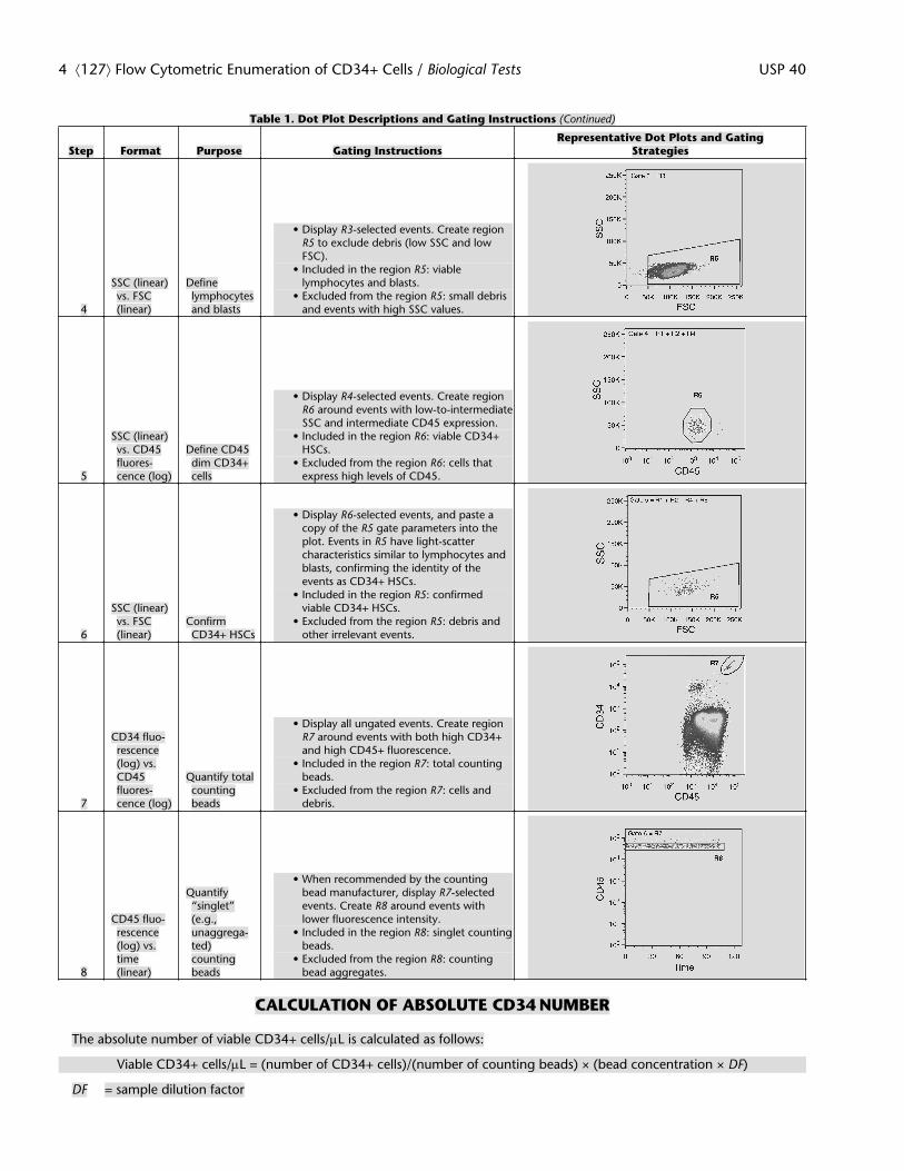
Table 1. Dot Plot Descriptions and Gating Instructions (Continued)
Step Format Purpose Gating InstructionsRepresentative Dot Plots and Gating
Strategies
4
SSC (linear)vs. FSC(linear)
Definelymphocytesand blasts
• Display R3-selected events. Create regionR5 to exclude debris (low SSC and lowFSC).
• Included in the region R5: viablelymphocytes and blasts.
• Excluded from the region R5: small debrisand events with high SSC values.
5
SSC (linear)vs. CD45fluores-cence (log)
Define CD45dim CD34+cells
• Display R4-selected events. Create regionR6 around events with low-to-intermediateSSC and intermediate CD45 expression.
• Included in the region R6: viable CD34+HSCs.
• Excluded from the region R6: cells thatexpress high levels of CD45.
6
SSC (linear)vs. FSC(linear)
ConfirmCD34+ HSCs
• Display R6-selected events, and paste acopy of the R5 gate parameters into theplot. Events in R5 have light-scattercharacteristics similar to lymphocytes andblasts, confirming the identity of theevents as CD34+ HSCs.
• Included in the region R5: confirmedviable CD34+ HSCs.
• Excluded from the region R5: debris andother irrelevant events.
7
CD34 fluo-rescence(log) vs.CD45fluores-cence (log)
Quantify totalcountingbeads
• Display all ungated events. Create regionR7 around events with both high CD34+and high CD45+ fluorescence.
• Included in the region R7: total countingbeads.
• Excluded from the region R7: cells anddebris.
8
CD45 fluo-rescence(log) vs.time(linear)
Quantify“singlet”(e.g.,unaggrega-ted)countingbeads
• When recommended by the countingbead manufacturer, display R7-selectedevents. Create R8 around events withlower fluorescence intensity.
• Included in the region R8: singlet countingbeads.
• Excluded from the region R8: countingbead aggregates.
CALCULATION OF ABSOLUTE CD34 NUMBER
The absolute number of viable CD34+ cells/mL is calculated as follows:
Viable CD34+ cells/mL = (number of CD34+ cells)/(number of counting beads) × (bead concentration × DF)
DF = sample dilution factor
4 á127ñ Flow Cytometric Enumeration of CD34+ Cells / Biological Tests USP 40

The number of CD34+ cells is determined from the viable CD34+ cells (region R5 from Table 1, Step 6). Depending on therecommendations of the counting bead manufacturer, the number of counting beads is determined from either the totalcounting beads (region R7 from Table 1, graph 7) or the singlet counting beads (region R8 from Table 1, graph 8). The beadconcentration is specified by the counting bead manufacturer.
Table 2. Troubleshooting Guidelines (see also á1027ñ)
Problems Possible Reasons Solutions Comments
Unable to clearly define the beadgate (R7) in Table 1, graph 7 dueto fluorescent debris
Degraded fresh cell sample; fixedsample; too many platelets; in-complete RBC lysis; nucleatedRBCs (e.g., in cord blood)
Use a far-red fluorescence channel(>600 nm), instead of FITC, togate counting beads.
Counting beads are highly fluores-cent in all channels. Autofluores-cence interference decreases dra-matically at longer wavelengths(>600 nm).
Unable to find CD34+ population inTable 1, graph 3
Incorrect compensation settings forPE and Viability dye
Repeat the spectral compensationsetup procedure, and adjust set-tings accordingly.
Make sure that counting beads arenot in the compensation matrixsample, because they can interferewith compensation settings.
Unsure of gating for singlet or totalbeads
High levels of counting bead aggre-gates
Follow the counting bead manufac-turer's recommendation for includ-ing or excluding aggregates.
Counting bead concentration cal-culations will vary by manufactur-er.
Want to use a different CD34 anti-body clone
Multi-parametric flow cytometryconsiderations
Alternative antibody clones must becarefully validated against thespecified CD34 antibodies. Choosea class II or class III antibody thatdetects all CD34 isoforms and gly-coforms.
Class I antibodies do not detect allCD34 glycoforms.
Want to use a different CD45 anti-body clone
Multi-parametric flow cytometryconsiderations
Alternative antibody clones must becarefully validated against thespecified CD45 antibodies. Choosean antibody that detects all iso-forms and all glycoforms of CD45.
—
ADDITIONAL REQUIREMENTS
USP Reference Standards á11ñ
USP CD34+ Cell Enumeration System Suitability RS▲USP40
USP 40 Biological Tests / á127ñ Flow Cytometric Enumeration of CD34+ Cells 5
Related Documents