“Using Advanced Technology to Better Diagnose Somatic Dysfunction” Joint Presentation The American Academy of Osteopathy 2017 Convocation The Balance Point: Bringing the Science and Art of Osteopathic Medicine Together Colorado Springs, CO March 24, 2017 Larry Smarr, PhD. Director, California Institute for Telecommunications and Information Technology Harry E. Gruber Professor, Dept. of Computer Science and Engineering, UCSD And Michael Kurisu, D.O. Clinical Director – UCSD Center for Integrative Medicine Director of Clinical Training – Osteopathic Center San Diego 1

Welcome message from author
This document is posted to help you gain knowledge. Please leave a comment to let me know what you think about it! Share it to your friends and learn new things together.
Transcript

“Using Advanced Technology
to Better Diagnose Somatic Dysfunction”
Joint Presentation
The American Academy of Osteopathy 2017 Convocation
The Balance Point: Bringing the Science and Art of Osteopathic Medicine Together
Colorado Springs, CO
March 24, 2017
Larry Smarr, PhD.
Director, California Institute for Telecommunications and Information Technology
Harry E. Gruber Professor, Dept. of Computer Science and Engineering, UCSD
And
Michael Kurisu, D.O.
Clinical Director – UCSD Center for Integrative Medicine
Director of Clinical Training – Osteopathic Center San Diego 1
Osteopathic Medicine
is Both an Art and a Science
Science Art

A Common Philosophy
Between the Science of System Dynamics and Osteopathy
“Multi-Component, Nonlinear,
Dynamic, Adaptive Systems”
Body is Unit
Body is Self Healing
Structure and Function
are Interrelated
Tenets of Osteopathy
Hawley and Smarr 1985

Larry is Not Your “Typical” Patient:
From Supercomputing to Quantified Self
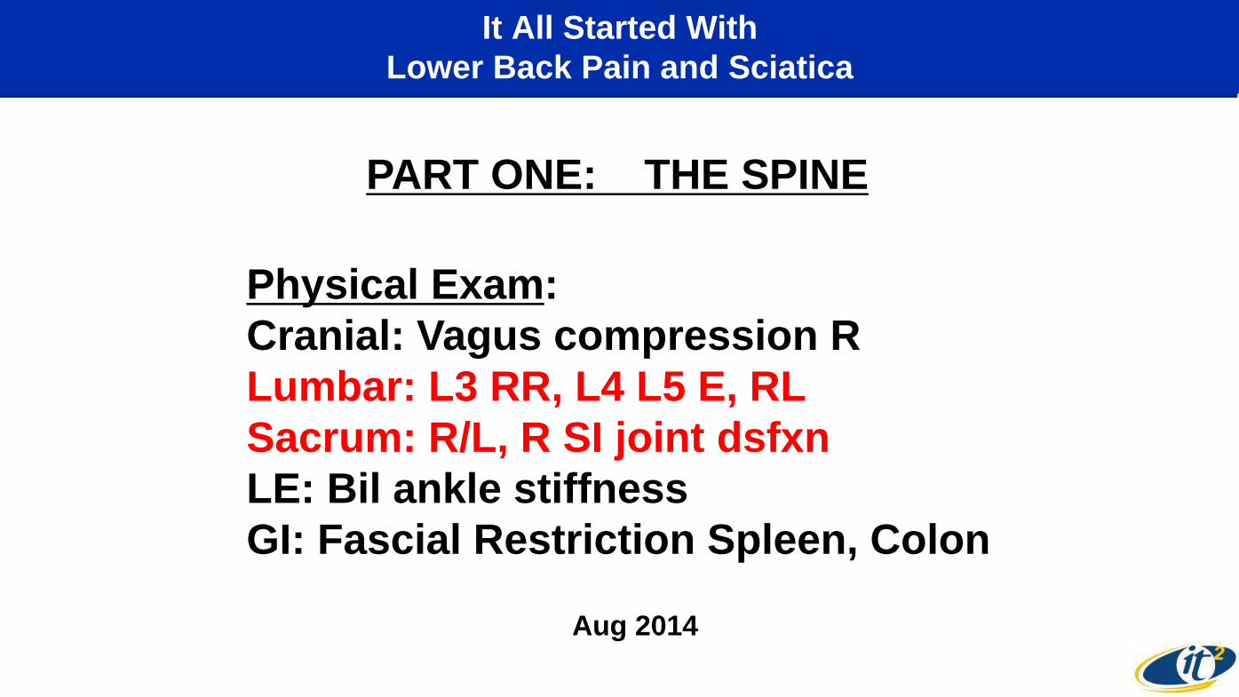
It All Started With
Lower Back Pain and Sciatica
Physical Exam:
Cranial: Vagus compression R
Lumbar: L3 RR, L4 L5 E, RL
Sacrum: R/L, R SI joint dsfxn
LE: Bil ankle stiffness
GI: Fascial Restriction Spleen, Colon
PART ONE: THE SPINE
Aug 2014

But There Was a Deeper Problem:
Thoracic/Cervical Scoliosis of LS Spine
4/15/2011 2/28/2012 5/2/2014
Note
Curvature
“You will notice
that there is a substantial
scoliotic curvature of your
upper mid-thoracic
and lower cervical spine.”
-David Wing,
Calit2 EPARC
8/18/2014
10/23/2014
2D CT Scans
At Calit2
Using GE Lunar
Densitometer

MRI Shows Central Canal Stenosis
L4-L5: Circumferential disc bulge extends slightly inferiorly in an area of deficiency of the
posterior aspect of the L5 superior endplate. Combined with moderate facet arthropathy and
ligamentum flavum redundancy, there is severe central canal stenosis with near
complete effacement of Cerebrospinal Fluid (CSF) and mass effect on the cauda
equina. Severe bilateral foraminal stenosis, with compression of the exiting bilateral
L4 nerve roots.
MRI Images from Cynthia Santillan, MD,
UCSD Radiology October 30, 2015

Images courtesy of Christine Chung MD, UCSD MSK Imaging Research Lab (www.MSKMRI.com)
T2 with fat suppression: makes fat dark and fluid bright (emphasizes disc and fluid around spinal cord/roots)
T1 has contrast with fat bright(shows bone and alignment well)
Advanced MRI:
Adjusting T1/T2 to Bring Out Regions of Interest

Validating My Osteopathic Findings
with 3D MRI Virtual Reality
Visualizations from MRI by Jurgen Schulze, Calit2, UCSD

Mike Kurisu Examining Larry Smarr’s Spine
in the Calit2 Virtual Reality CAVE
Visualizations from MRI by Jurgen Schulze, Calit2, UCSD

Combining an Osteopathic Examination
With 3D Visualization of the Patient
Recreating the Anatomy Lesson
Using 3D “Visible Larry” to Guide DO Manipulation
March 29, 2016
Calit2
This is what we will be doing in our breakout session

Internal Spinal Abnormalities Detected
Using Spiral Technology Adapted from Mass Market Gaming Technologies
Slides from: Spencer Stein, President, Spiral Therapy, Inc &
Stephen Moxey MPT, OCS, FAAOMPT 2017
The Inside/Out Comparison
Discussion at breakout session

March 2016 February 2017
Tracking Somatic Dysfunction
with External Spiral Technology
Slides from: Spencer Stein, President, Spiral Therapy, Inc &
Stephen Moxey MPT, OCS, FAAOMPT 2017

Tracking Body Shape Data February 2017
Slide from: Spencer Stein, President, Spiral Therapy, Inc &
Stephen Moxey MPT, OCS, FAAOMPT 2017
Metabolic
Syndrome
Tracking
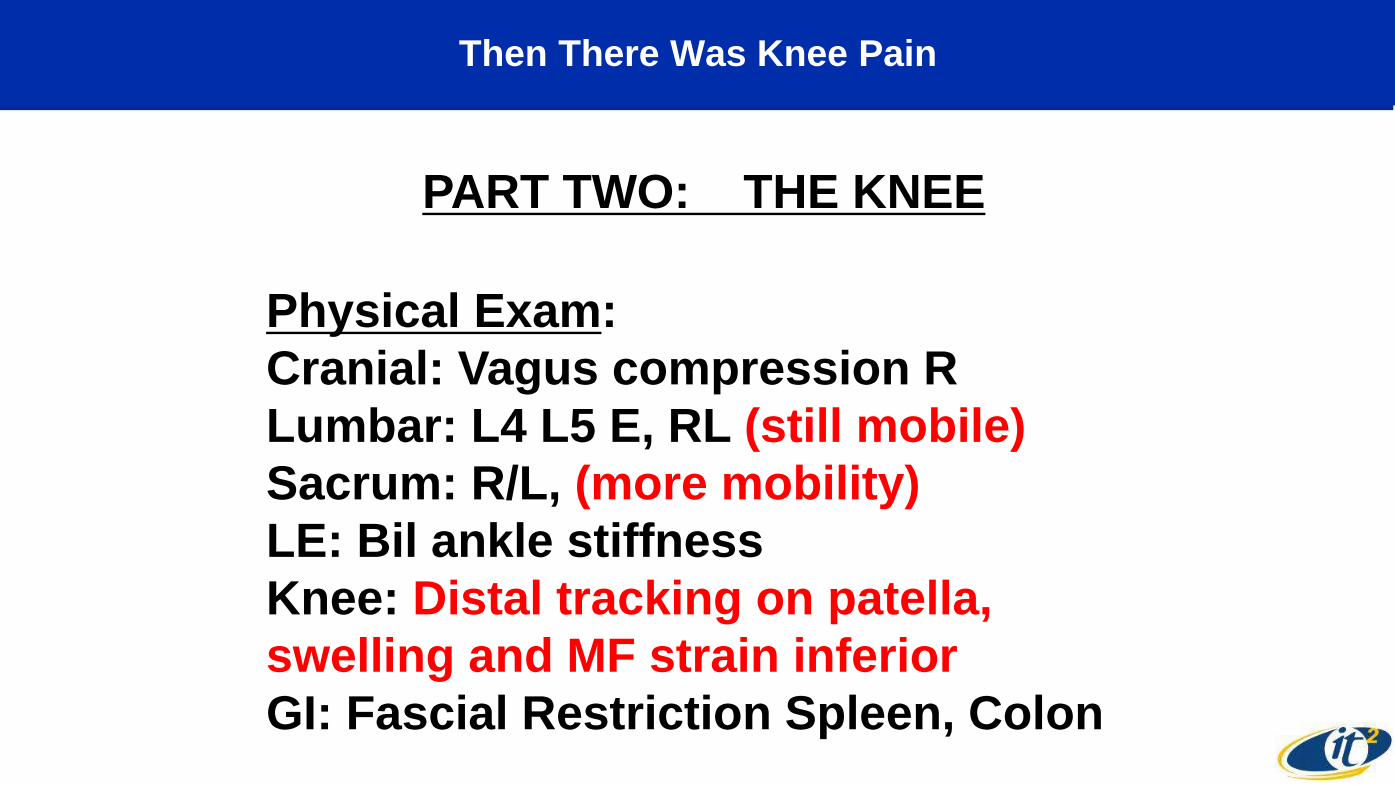
Then There Was Knee Pain
PART TWO: THE KNEE
Physical Exam:
Cranial: Vagus compression R
Lumbar: L4 L5 E, RL (still mobile)
Sacrum: R/L, (more mobility)
LE: Bil ankle stiffness
Knee: Distal tracking on patella,
swelling and MF strain inferior
GI: Fascial Restriction Spleen, Colon
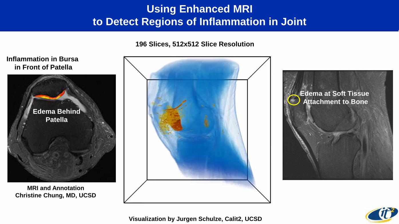
Using Enhanced MRI
to Detect Regions of Inflammation in Joint
MRI and Annotation
Christine Chung, MD, UCSD
Visualization by Jurgen Schulze, Calit2, UCSD
Inflammation in Bursa
in Front of Patella
Edema at Soft Tissue
Attachment to Bone
196 Slices, 512x512 Slice Resolution
Edema Behind
Patella

Finally, I Could Detect Abdominal Dysfunction
Physical Exam:
Cranial: Vagus compression bilaterally
Lumbar: L5 compressed on R
Sacrum: stabalized
LE: Bil ankle stiffness R>L
GI: Fascial Restriction Spleen, moderately
distended descending colon, warmth, mass
effect felt sigmoid junction with increasing
density
PART THREE: THE ABDOMEN

A Decade Ago I Started Tracking My Internal Biomarkers
To Understand My Body’s Dynamics
Calit2 64 Megapixel Display Wall
My Quarterly
Data Collection

Only One of My Blood Measurements
Was Far Out of Range--Indicating Chronic Inflammation
Normal Range <1 mg/L
27x Upper Limit
Complex Reactive Protein (CRP) is a Blood Biomarker
for Detecting Presence of Inflammation
Episodic Peaks in Inflammation
Followed by Spontaneous Drops
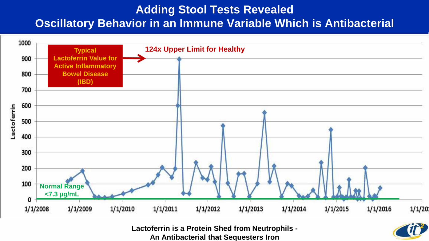
Adding Stool Tests Revealed
Oscillatory Behavior in an Immune Variable Which is Antibacterial
Normal Range
<7.3 µg/mL
124x Upper Limit for Healthy
Lactoferrin is a Protein Shed from Neutrophils -
An Antibacterial that Sequesters Iron
Typical
Lactoferrin Value for
Active Inflammatory
Bowel Disease
(IBD)

Descending Colon
Sigmoid Colon
Threading Iliac Arteries
Major Kink
Confirming the IBD (Colonic Crohn’s) Hypothesis:
Finding the “Smoking Gun” with MRI Imaging
I Obtained the MRI Slices
From UCSD Medical Services
and Converted to Interactive 3D
Working With Calit2 Staff
Transverse ColonLiver
Small Intestine
Diseased Sigmoid ColonCross Section
MRI Jan 2012
Severe Colon
Wall Swelling
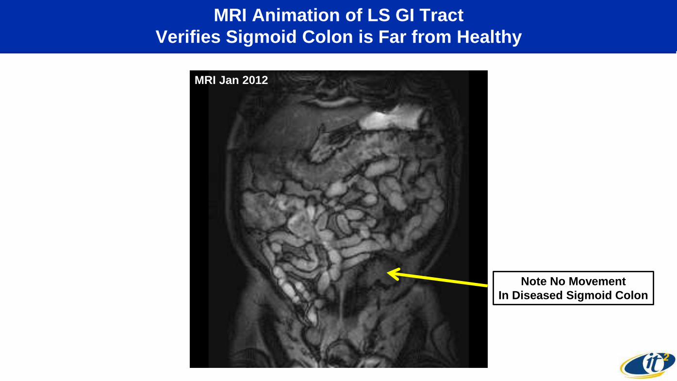
MRI Animation of LS GI Tract
Verifies Sigmoid Colon is Far from Healthy
Note No Movement
In Diseased Sigmoid Colon
MRI Jan 2012

Why Did I Have IBD?
I Found I Had a SNP Associated with Crohn’s Disease
From www.23andme.com
SNPs Associated with CD
Polymorphism in
Interleukin-23 Receptor Gene
— 80% Higher Risk
of Pro-inflammatory
Immune Response
NOD2
IRGM
ATG16L1
~200 Human DNA SNPs
Associated with IBD.
Analysis Indicated
I Had Colonic Crohn’s,
Not Ileal Crohn’s

To Understand the Interaction of Genetics and the Immune System
We Must Consider the Human Microbiome
Your Microbiome is
Your “Near-Body” Environment
and its Cells
Contain 100x as Many DNA Genes
As Your Human DNA-Bearing Cells
Your Body Has 10 Times
As Many Microbe Cells As
DNA-Bearing Human Cells
Inclusion of the Microbiome Genomics
Will Radically Alter Medicine

We Found Major State Shifts in Microbial Ecology Phyla
Between Healthy and Three Forms of IBD
Most
Common
Microbial
Phyla
Average HE
Average
Ulcerative Colitis
Average LS
Colonic Crohn’s Disease
Average
Ileal Crohn’s Disease
Our Team Used 25 CPU-Years
to Compute
Comparative Gut Microbiomes
Starting From
2.7 Trillion DNA Bases
of My Samples
and Healthy and IBD Controls
Discussion at breakout session

The Microbiome–Gut–Brain Axis
Provides New Systemic Insights into Shifts in Behavior and Disease
Source: Montiel-Castro, et al.
Frontiers in Integrative Neuroscience 2013

Using 3D “Visible Larry”
to Guide Abdominal DO Manipulation
March 29, 2016
Calit2

Full Body CAT Scan at mm Resolution, Including Virtual Colonoscopy
June 2016 Convinced Me Time Had Come for Surgery
No Air
Source: June 2016
Dr. Harvey Eisenberg,
Body Scan Intl., Irvine, CA
Classic
Colonic Crohn’s
Stricture

Pre-Surgery Mental/Emotional Preparation
Over Thanksgiving 2016

I Had Been Giving Tours of “Visible Larry” for Years:
In Calit2’s Virtual Reality StarCAVE
3D Volumetric
Visualization
Created by
Calit2’s Jurgen
Schulze
from January
2012 MRI

QI’s Jurgen Schulze Converted Abdominal MRI Slices
to 3D Organ Segmentation for Surgical Pre-Planning
MRI Slice from Dr. Cynthia Santillan 3D Organ Segmentation Made by Dr. Jurgen Schulze
from Dr. Santillan’s 150-Slice MRI
Images of Dr. Smarr’s Abdomen
To Support Sigmoid Colon Resection Surgery

Pre-Surgical Planning in QI Virtual Reality
on Friday November 25, 2016
Dr. Ramamoorthy in
QI Virtual Reality CAVE
Exploring Dr. Smarr’s Colon
With Dr. Schulze’s Software

Using QI Organ Segmentation in Jacobs OR
on Tuesday November 29, 2016
Dr. Smarr
With da Vinci Robot
Arms Inside Him
OR Team Using Large Screens
To Watch Dr. Schulze’s da Vinci Images
Dr. Ramamoorthy Operating
Da Vinci Xi Robot During Surgery
Dr. Schulze Rotating 3D Organs To Match Up
With da Vinci Arms and Internal Camera
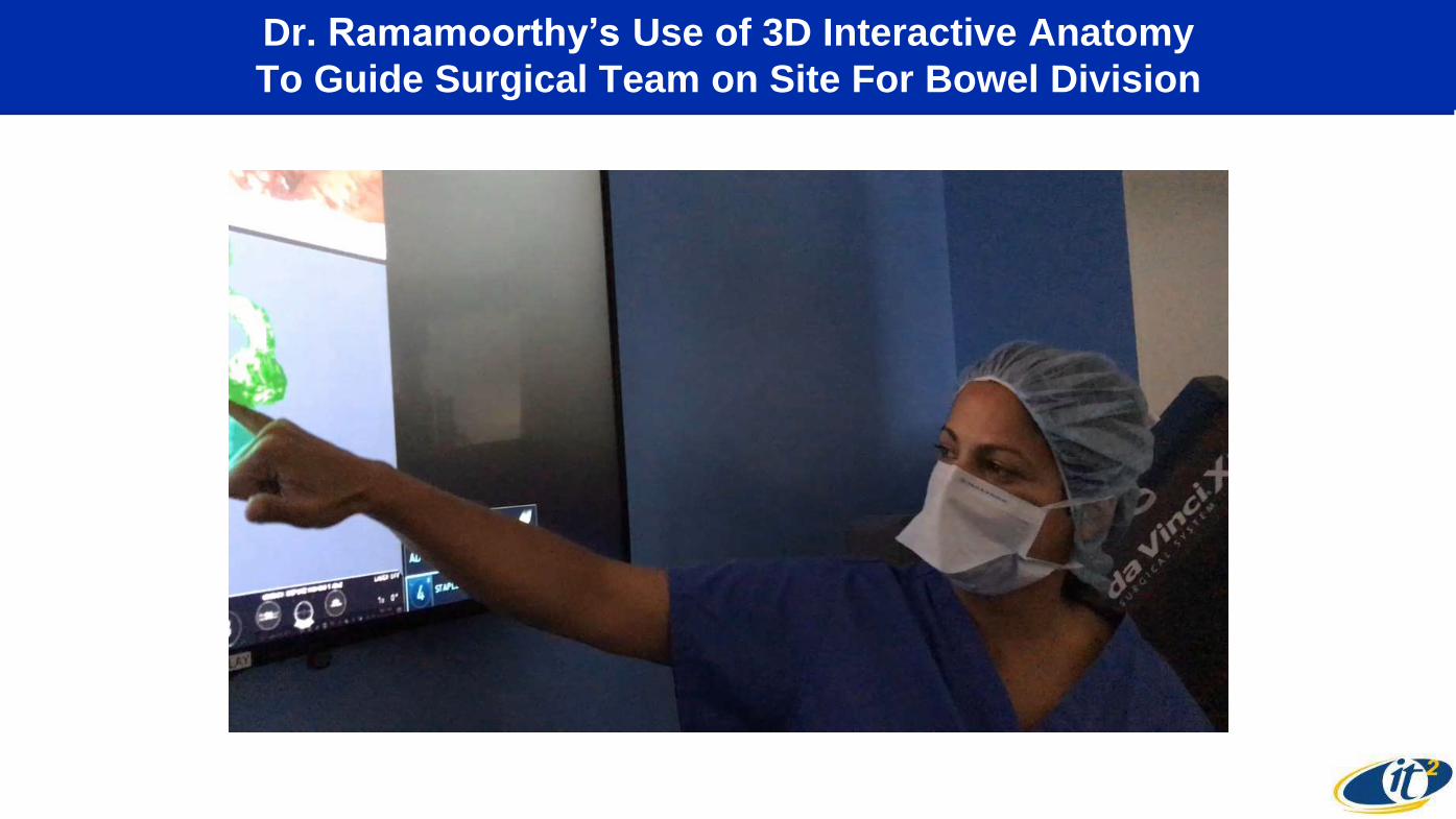
Dr. Ramamoorthy’s Use of 3D Interactive Anatomy
To Guide Surgical Team on Site For Bowel Division

Surgery Day:
Worlds Apart Yet Connected
Dr. Smarr Dr. Kurisu
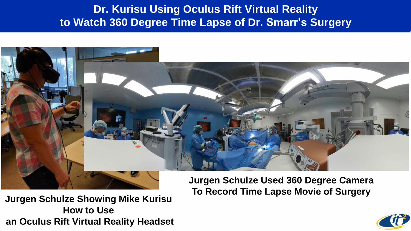
Dr. Kurisu Using Oculus Rift Virtual Reality
to Watch 360 Degree Time Lapse of Dr. Smarr’s Surgery
Jurgen Schulze Showing Mike Kurisu
How to Use
an Oculus Rift Virtual Reality Headset
Jurgen Schulze Used 360 Degree Camera
To Record Time Lapse Movie of Surgery
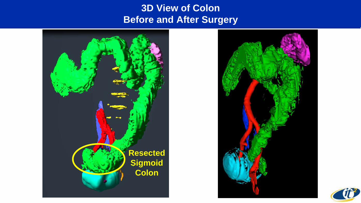
3D View of Colon
Before and After Surgery
Resected
Sigmoid
Colon

Blood hs-CRP on Log Scale:
Use as Post-Surgery Biomarker of Success?
Surgery
Healthy Range

Stool Lactoferrin on Log Scale:
Dramatic Drop After Surgery
Surgery
Healthy Range
1800x Lower
Than Highest Value

Quantified Recovery (Steps Walked Per Day) -
Recovered to Pre-Surgery Level in Two Weeks
10,000
Steps
Su
rgery
Left
JM
C
5 Miles
Per Day
Dec 14Nov 29
Mike Tx Larry at home
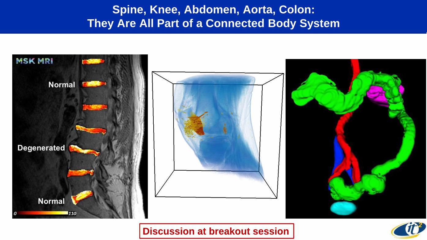
Spine, Knee, Abdomen, Aorta, Colon:
They Are All Part of a Connected Body System
Discussion at breakout session

From Digitally-Enabled Future Patient
to Digitally-Enabled Future Doctor
Related Documents


