23/12/2015 1 Useful clues in assessing blistering disorders Dr Saleem Taibjee [email protected] Consultant Dermatologist & Dermatopathologist Dorset County Hospital Inflammatory dermatoses!! Temptation = Look at clinical request form and agree / try to fit with clinical suggestion ‘Blind’ reporting • Look at slide ‘blind’ first without looking at request form Inflammatory algorithms in textbooks Intra-epidermal blistering 3 major mechanisms (Weedon) • 1. Intercellular oedema (spongiosis) - eczema • 2. Intracellular oedema (ballooning) - viral • 3. Acantholysis - pemphigus • (4. Individual cell necrosis – EM/SJS/TEN) In some diseases more than one mechanism

Welcome message from author
This document is posted to help you gain knowledge. Please leave a comment to let me know what you think about it! Share it to your friends and learn new things together.
Transcript

23/12/2015
1
Useful clues in assessing
blistering disorders
Dr Saleem Taibjee [email protected]
Consultant Dermatologist & Dermatopathologist
Dorset County Hospital
Inflammatory dermatoses!!
Temptation =
Look at clinical request form
and agree / try to fit
with clinical suggestion
‘Blind’ reporting • Look at slide ‘blind’ first without looking at request form
Inflammatory algorithms in textbooks
Intra-epidermal blistering
3 major mechanisms (Weedon)
• 1. Intercellular oedema (spongiosis) - eczema
• 2. Intracellular oedema (ballooning) - viral
• 3. Acantholysis - pemphigus
• (4. Individual cell necrosis – EM/SJS/TEN)
In some diseases more than one mechanism

23/12/2015
2
Blistering – clinical pitfalls
• Blistering is often a secondary event
– e.g. cellulitis, vasculitis
• It can be difficult to determine the clinical
level of the split
• Blistering maybe absent or subtle in true
blistering disorders
Frozen Section

23/12/2015
3
This talk:
Blistering – histological pitfalls
• 1. You can’t see a blister! – Additional levels
– 6 histological clues
• 2. You can see a blister, but doesn’t seem to fit! – Diverse histological patterns in blistering disorders
– Drug reaction
– Dermatitis artefacta
– Rarer condition
You can’t see a blister:
‘invisible dermatosis’
Consider:
• Dermatitis herpetiformis
• Pemphigus foliaceous
• Grover’s disease
• Additional levels often necessary
– Serial sections
Case 1 • 5 month history of erythematosquamous eruption
with erosions on face, scalp, forehead and neck
Clue 1
“Floating or absent stratum corneum”
Floating or absent stratum corneum sign
• Clue to superficial (subcorneal) acantholytic blistering
– Pemphigus foliaceous
– Bullous impetigo
– Staphylococcal scalded skin syndrome
– Peeling skin syndrome
– Psoriatic erythroderma
– Artefact

23/12/2015
4
Clue 2 Acro-eccrocentric acantholytic granular cells
- Geometric hyperchromatic cells
- Clue to pemphigus foliaceous
Helmut Kerl, BSD meeting 2012
Immunofluorescence
• Intraepidermal intercellular pattern
Floating or absent stratum corneum sign

23/12/2015
5
Folliculocentric acantholytic granular cells
Erosions and blistering subtle!

23/12/2015
6
Pemphigus foliaceous vs vulgaris
Pemphigus
foliaceous
Pemphigus
vulgaris
Clinical morphology
Blisters and erosions subtle
Painful flaccid blistering
Mucosal involvement
Uncommon
Usual
Histology
Subcorneal acantholysis,
may be subtle
Suprabasal acantholysis
with adnexal involvement
Indirect
immunofluorescence
Stains human skin strongly
Stains monkey
oesophagus strongly
ELISA
Dsg-1 ++, Dsg-3 -
Dsg-1 +/-, Dsg-3 ++
Case 2
• 6-year-old child
– 7 days history of bullous
lesions right leg
?Exaggerated response to
insect bites
?Bullous disease of childhood
Floating or absent stratum corneum sign

23/12/2015
7
Bullous impetigo
Staphylococcal Scalded Skin Syndrome
• Widespread blisters
• Exfoliative toxin produced by Staphylococcus aureus
• Organism not identifiable/cultured from affected skin
Case 3
• Itchy rash
?scabies ?other
23/12/2015
8
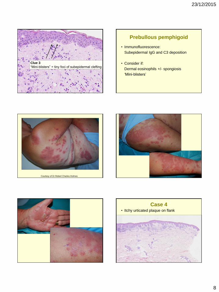
Clue 3
“Mini-blisters” = tiny foci of subepidermal clefting
Prebullous pemphigoid
• Immunofluorescence:
Subepidermal IgG and C3 deposition
• Consider if:
Dermal eosinophils +/- spongiosis
‘Mini-blisters’
Courtesy of Dr Robert Charles-Holmes
Case 4 • Itchy urticated plaque on flank

23/12/2015
9
Clue 4
Micro-Nikolsky sign
= subepidermal cleft at edge of biopsy
Micro-Nikolsky sign: D Metze, ISDP Barcelona 2011
Shearing force of rotation of punch produces blister at edge
Case 5 • F78 - Itchy red papules
Solar lentigo-like elongation of rete ridges
23/12/2015
10
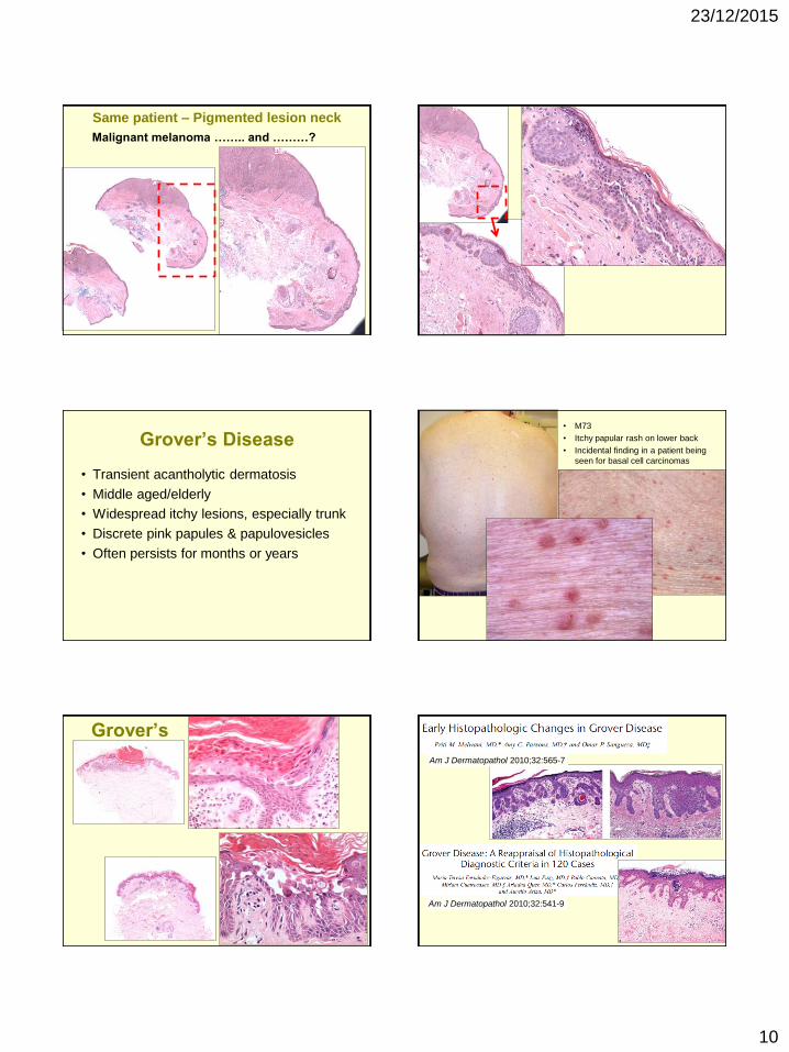
Same patient – Pigmented lesion neck
Malignant melanoma …….. and ………?
Grover’s Disease
• Transient acantholytic dermatosis
• Middle aged/elderly
• Widespread itchy lesions, especially trunk
• Discrete pink papules & papulovesicles
• Often persists for months or years
• M73
• Itchy papular rash on lower back
• Incidental finding in a patient being
seen for basal cell carcinomas
Grover’s
Am J Dermatopathol 2010;32:565-7
Am J Dermatopathol 2010;32:541-9

23/12/2015
11
Clue to Grover’s disease
• Late lesions have
elongated rete ridges and
may resemble solar lentigo
Case 6 • F60. 5/12 erythematous scaly patch with
scarring, alopecia and milia. ? BCC
Clue 6 • Milia with scarring =
Clue to deeper sub-epidermal blistering disorder
Micro-Nikolsky sign

23/12/2015
12
Be brave!
Richard Carr’s report (summary):
Dense lymphoplasmacytic infiltrate with vascular proliferation and papillary dermal fibrosis. Occasional eosinophils. Subepidermal clefting over a wide area, especially in specimen B.
Although regression of tumour is a possibility we have also considered cicatricial pemphigoid.
Brunstig-Perry pemphigoid
• Immunofluorescence:
Linear IgG deposition along
basement membrane zone
• Variant of cicatricial pemphigoid
– Lesions limited to forehead & scalp
Milia with scarring =
Clue to sub-epidermal blistering
Pseudoporphyria caused by naproxen
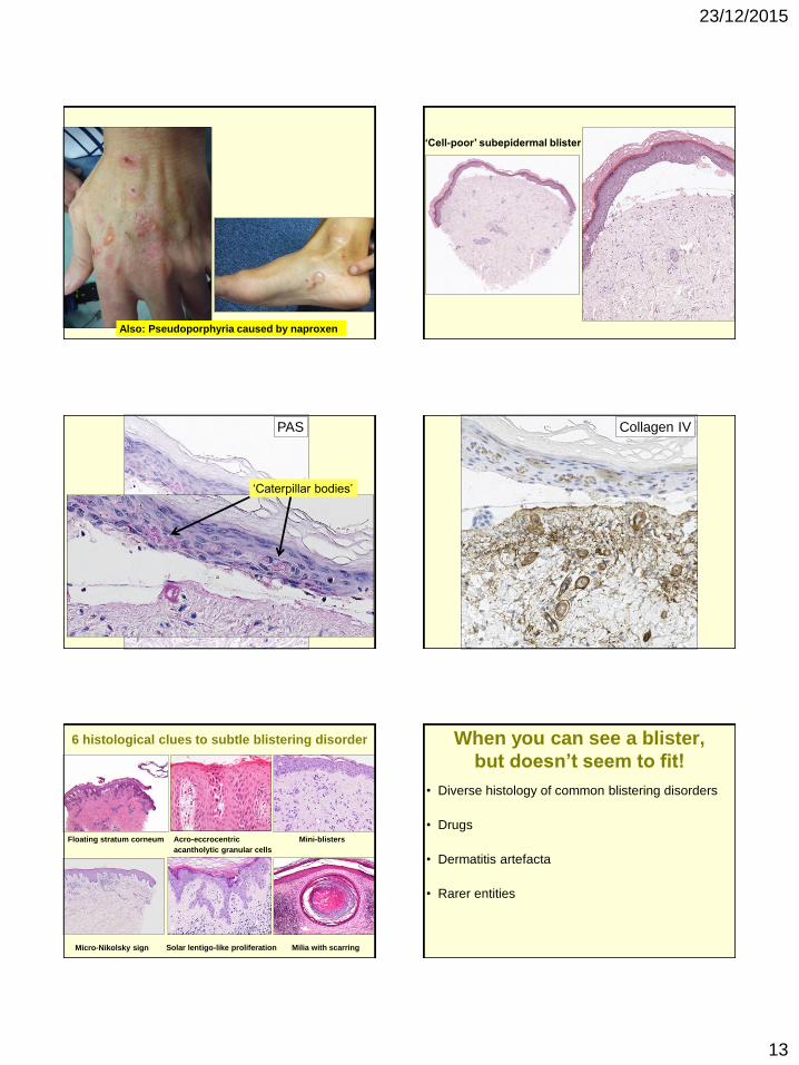
23/12/2015
13
Also: Pseudoporphyria caused by naproxen
‘Cell-poor’ subepidermal blister
PAS
‘Caterpillar bodies’
Collagen IV
6 histological clues to subtle blistering disorder
Micro-Nikolsky sign
Floating stratum corneum Acro-eccrocentric
acantholytic granular cells
Mini-blisters
Solar lentigo-like proliferation Milia with scarring
When you can see a blister,
but doesn’t seem to fit!
• Diverse histology of common blistering disorders
• Drugs
• Dermatitis artefacta
• Rarer entities

23/12/2015
14
Diverse histology of common blistering disorders
Case 7
• Itchy rash trunk & legs
ST8 (12-12138A)
Grover’s histology Patterns:
• DARIER-like – Acantholytic dyskeratosis
• HAILEY-HAILEY-like – Prominent acantholysis throughout epidermis
• PEMPHIGUS VULGARIS-like – Suprabasal clefting with sparse acantholytic
dyskeratosis
• SPONGIOTIC – Acantholytic cells within spongiotic foci
ST9 (12-12138B)

23/12/2015
15
Diverse histology of common blistering disorders
Case 8
• Itchy vesicles knees & elbows
Case 8
Prominent acantholysis is rarely a sign of DH

23/12/2015
16
Papillary microabscess, but also acantholysis
Dermatitis herpetiformis – the usual
Papillary dermal microabscess Subepidermal blister with neutrophils
Case 9
Flask sign = spongiotic blistering Mixed histological pattern = clue to drug reaction
Flask sign = spongiotic blistering
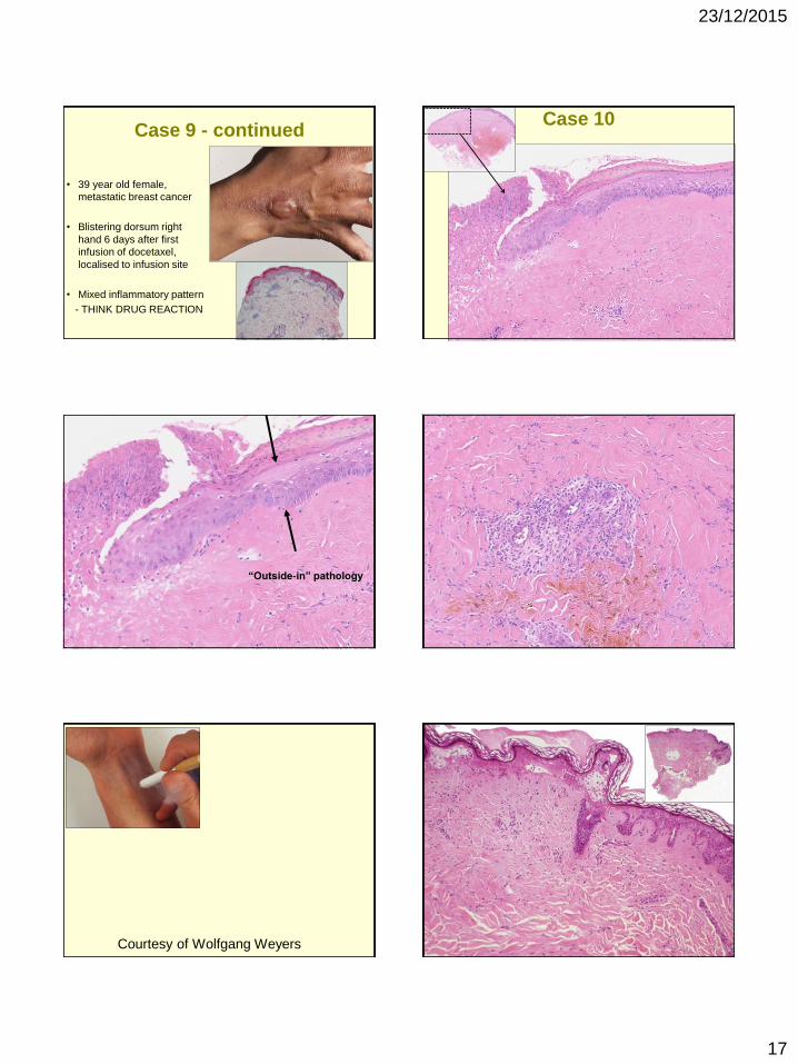
23/12/2015
17
Case 9 - continued
• 39 year old female,
metastatic breast cancer
• Blistering dorsum right
hand 6 days after first
infusion of docetaxel,
localised to infusion site
• Mixed inflammatory pattern
- THINK DRUG REACTION
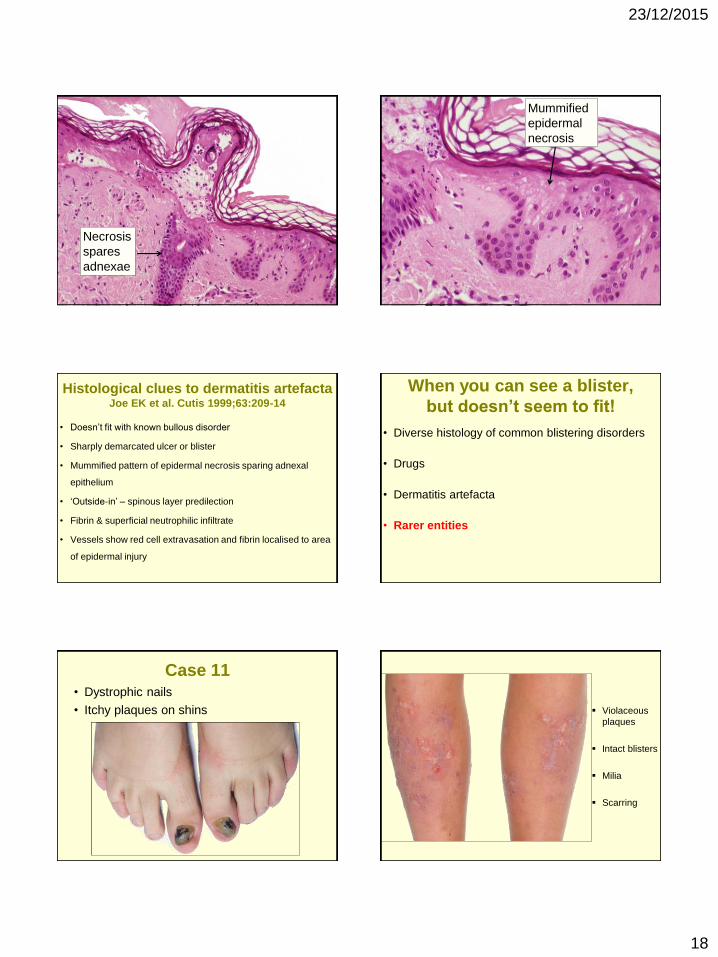
Case 10
“Outside-in” pathology
Courtesy of Wolfgang Weyers
23/12/2015
18
Necrosis
spares
adnexae
Mummified
epidermal
necrosis
Histological clues to dermatitis artefacta Joe EK et al. Cutis 1999;63:209-14
• Doesn’t fit with known bullous disorder
• Sharply demarcated ulcer or blister
• Mummified pattern of epidermal necrosis sparing adnexal
epithelium
• ‘Outside-in’ – spinous layer predilection
• Fibrin & superficial neutrophilic infiltrate
• Vessels show red cell extravasation and fibrin localised to area
of epidermal injury
When you can see a blister,
but doesn’t seem to fit!
• Diverse histology of common blistering disorders
• Drugs
• Dermatitis artefacta
• Rarer entities
Case 11 • Dystrophic nails
• Itchy plaques on shins Violaceous
plaques
Intact blisters
Milia
Scarring
23/12/2015
19
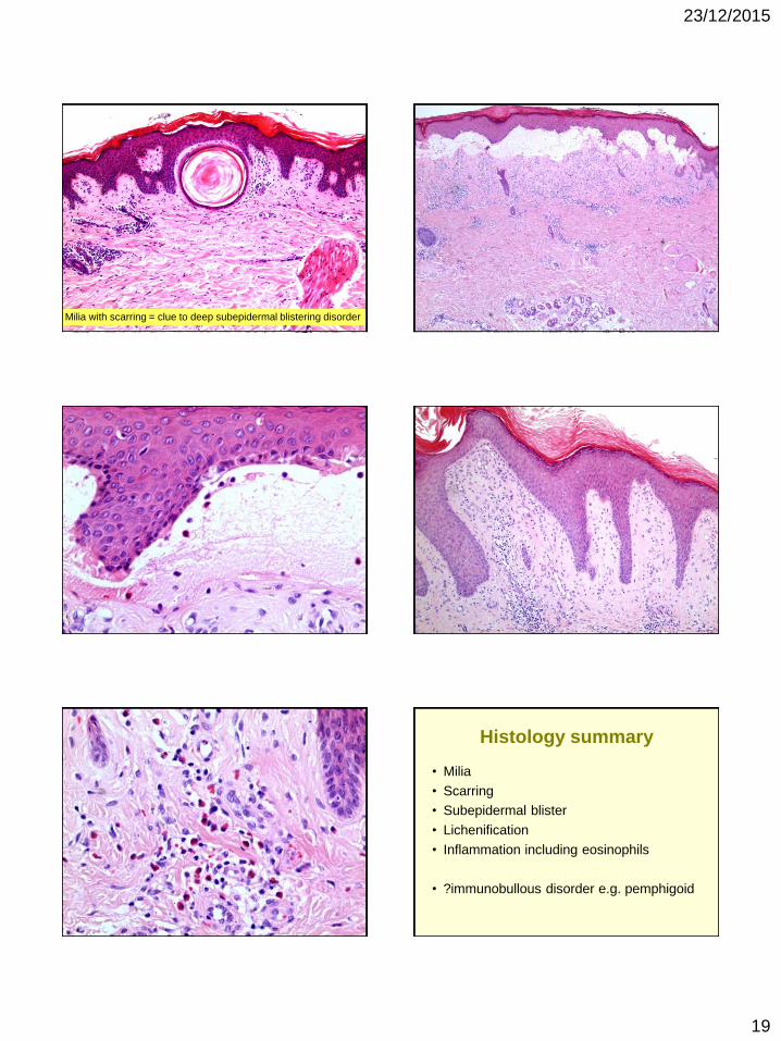
Milia with scarring = clue to deep subepidermal blistering disorder
Histology summary
• Milia
• Scarring
• Subepidermal blister
• Lichenification
• Inflammation including eosinophils
• ?immunobullous disorder e.g. pemphigoid

23/12/2015
20
Fibrinogen IgG
Courtesy of Balbir Bhogal, St John’s Institute of Dermatology
Family history
? ? ?
? ?
Mutation analysis of COL7A1
(+/-) c.6215A>G,p.Gln2072Arg, exon 74
→ Dominant dystrophic epidermolysis bullosa pruriginosa
1st described by Kuske in 1946¹
Term EB ‘pruriginosa’ coined by McGrath et al in 1994²
Rare subtype of DEB³
Autosomal dominant / autosomal recessive / sporadic
Late onset disease not uncommon
1. Kuske H. Dermatologica 1946; 91: 304–5
2. McGrath et al. Br J Dermatol 1994; 130: 617–25
3. Schumann H et al. Br J Dermatol 2008;159:464-469
Normal skin DDEB
Blister
EB pruriginosa – learning points
Hereditary condition BUT………..
- Late onset
- Significant intra-familial variability
Unusual clinical features
- Pruritic
- May lack blisters
Histological features
- Milia with scarring +/- blister
- Inflammation including eosinophils
23/12/2015
21
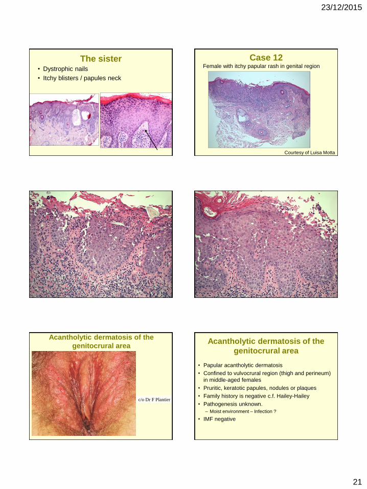
The sister • Dystrophic nails
• Itchy blisters / papules neck
Case 12 Female with itchy papular rash in genital region
Courtesy of Luisa Motta
Acantholytic dermatosis of the
genitocrural area
c/o Dr F Plantier
Acantholytic dermatosis of the
genitocrural area
• Papular acantholytic dermatosis
• Confined to vulvocrural region (thigh and perineum)
in middle-aged females
• Pruritic, keratotic papules, nodules or plaques
• Family history is negative c.f. Hailey-Hailey
• Pathogenesis unknown.
– Moist environment – Infection ?
• IMF negative

23/12/2015
22
Hailey-Hailey
Hailey-Hailey: ‘dilapidated brick wall’
Summary
Blind reporting
When you can’t see a blister!
– Serial sections / levels
– 6 histological clues
When you can see a blister, but doesn’t seem to fit!
– Diverse histology of common blistering disorders
– Drug reactions
– Dermatitis artefacta
– Rarer entities
British Society for Dermatopathology
http://www.britsocdermpath.co.uk
British Society for Dermatopathology Annual Meeting on Tues 5 July 2016
at the ICC Birmingham
Closing date for abstracts is 8 Feb 2016
Abstract submission via BAD website at www.bad.org.uk/events/annualmeeting
The BSD Self-assessment meeting is Mon 4 July at same venue
6th British Society for Dermatopathology Annual Trainees’ Workshop
3 September 2015 in Bristol
President: Dr Paul Craig
Acknowledgements • Richard Carr
• Marie-Anne Brundler
• Jerry Marsden
• Luisa Motta
• Robert Charles-Holmes
• Sivanie Vivehanantha
• Andrew Ilchyshyn
• Wolfgang Weyers
• Claudia Roberts

Related Documents