Use of response surface methodology to examine chitinase regulation in the basidiomycete Moniliophthora perniciosa Maı´za Alves LOPES a,b , Dayane Santos GOMES a , Maria Gabriela BELLO KOBLITZ b , Carlos Priminho PIROVANI a , Ju ´lio CE ´ ZAR DE MATTOS CASCARDO a , Aristo ´teles GO ´ ES-NETO b , Fabienne MICHELI a,c, * a Laborato ´rio de Geno ˆmica e Expressa ˜o Ge ˆnica, DCB, Universidade Estadual de Santa Cruz, Rodovia Ilhe ´us-Itabuna, Km 16, 45650-000 Ilhe ´us-BA, Brazil b Laborato ´rio de Pesquisa em Microbiologia, DCBio, Universidade Estadual de Feira de Santana, Km 3, BR 116 (norte), 44031-460 Feira de Santana-BA, Brazil c UMR DAP, Cirad, Avenue Agropolis TA80/02, 34398 Montpellier cedex 5, France article info Article history: Received 26 January 2007 Received in revised form 11 September 2007 Accepted 31 October 2007 Corresponding Editor: Gareth W. Griffith Keywords: Basidiomycota Central composite design Chitinase Mycelial growth Secretion abstract We report here the first analysis of chitinase regulation in Moniliophthora perniciosa, the causal agent of the witches’ broom disease of cacao. A multivariate statistical approach was employed to evaluate the effect of several variables, including carbon and nitrogen sources and cultivation time, on M. perniciosa non-secreted (detected in mycelium, i.e. in symplasm and cell wall) and secreted (detected in the culture medium) chitinase activities. Non-secreted chitinase activity was enhanced by peptone and chitin and repressed by glucose. Chitinase secretion was increased by yeast extract alone or in combination with other nitrogen sources, and by N-acetylglucosamine, and repressed in presence of chitin. The best cultivation times for non-secreted and secreted chitinase activities were 30 and 20 d, respectively. However, chitinase activity was always higher in the mycelium than in the culture medium, suggesting a relatively poor chitinase secretion activity. Conversely, higher mycelial growth was observed when the activity of the non-secreted chitinase was at its lowest, i.e. when the fungus was grown on glucose and yeast extract as sources of carbon and nitrogen, respectively. Conversely, the induction of non-secreted chitinase activity by chitin decreased the mycelium growth. These results suggest that the culture medium, by the induction or repression of chitinases, affected the hyphal growth. Thus, as an essential component of M. perniciosa growth, chitinases may be a potential target for strategies to control disease. ª 2007 The British Mycological Society. Published by Elsevier Ltd. All rights reserved. Introduction Chitinases (EC 3.2.1.14), belonging to the family of glycosyl hydrolases, catalyse the hydrolysis of chitin, a linear homo- polymer of b-1,4-linked N-acetyl-D-glucosamine (NAG) residues. Chitinases play an important physiological and ecological role in ecosystems as recyclers of chitin by generat- ing carbon and nitrogen sources (Cohen-Kupiec & Chet 1998). Chitinase production occurs naturally in a wide variety of organisms, such as microorganisms containing chitin, * Corresponding author. Laborato ´ rio de Geno ˆ mica e Expressa ˜o Ge ˆ nica, DCB, Universidade Estadual de Santa Cruz, Rodovia Ilhe ´ us- Itabuna, Km 16, 45650-000 Ilhe ´ us-BA, Brasil. E-mail address: [email protected] journal homepage: www.elsevier.com/locate/mycres mycological research 112 (2008) 399–406 0953-7562/$ – see front matter ª 2007 The British Mycological Society. Published by Elsevier Ltd. All rights reserved. doi:10.1016/j.mycres.2007.10.017

Welcome message from author
This document is posted to help you gain knowledge. Please leave a comment to let me know what you think about it! Share it to your friends and learn new things together.
Transcript
m y c o l o g i c a l r e s e a r c h 1 1 2 ( 2 0 0 8 ) 3 9 9 – 4 0 6
journa l homepage : www.e l sev i er . com/ loca te /mycres
Use of response surface methodology to examine chitinaseregulation in the basidiomycete Moniliophthora perniciosa
Maıza Alves LOPESa,b, Dayane Santos GOMESa, Maria Gabriela BELLO KOBLITZb,Carlos Priminho PIROVANIa, Julio CEZAR DE MATTOS CASCARDOa, AristotelesGOES-NETOb, Fabienne MICHELIa,c,*aLaboratorio de Genomica e Expressao Genica, DCB, Universidade Estadual de Santa Cruz, Rodovia Ilheus-Itabuna,
Km 16, 45650-000 Ilheus-BA, BrazilbLaboratorio de Pesquisa em Microbiologia, DCBio, Universidade Estadual de Feira de Santana, Km 3, BR 116 (norte),
44031-460 Feira de Santana-BA, BrazilcUMR DAP, Cirad, Avenue Agropolis TA80/02, 34398 Montpellier cedex 5, France
a r t i c l e i n f o
Article history:
Received 26 January 2007
Received in revised form
11 September 2007
Accepted 31 October 2007
Corresponding Editor:
Gareth W. Griffith
Keywords:
Basidiomycota
Central composite design
Chitinase
Mycelial growth
Secretion
* Corresponding author. Laboratorio de GeItabuna, Km 16, 45650-000 Ilheus-BA, Brasil.
E-mail address: [email protected]/$ – see front matter ª 2007 The Bdoi:10.1016/j.mycres.2007.10.017
a b s t r a c t
We report here the first analysis of chitinase regulation in Moniliophthora perniciosa, the
causal agent of the witches’ broom disease of cacao. A multivariate statistical approach
was employed to evaluate the effect of several variables, including carbon and nitrogen
sources and cultivation time, on M. perniciosa non-secreted (detected in mycelium, i.e. in
symplasm and cell wall) and secreted (detected in the culture medium) chitinase activities.
Non-secreted chitinase activity was enhanced by peptone and chitin and repressed by
glucose. Chitinase secretion was increased by yeast extract alone or in combination with
other nitrogen sources, and by N-acetylglucosamine, and repressed in presence of chitin.
The best cultivation times for non-secreted and secreted chitinase activities were 30 and
20 d, respectively. However, chitinase activity was always higher in the mycelium than
in the culture medium, suggesting a relatively poor chitinase secretion activity. Conversely,
higher mycelial growth was observed when the activity of the non-secreted chitinase was
at its lowest, i.e. when the fungus was grown on glucose and yeast extract as sources of
carbon and nitrogen, respectively. Conversely, the induction of non-secreted chitinase
activity by chitin decreased the mycelium growth. These results suggest that the culture
medium, by the induction or repression of chitinases, affected the hyphal growth. Thus,
as an essential component of M. perniciosa growth, chitinases may be a potential target
for strategies to control disease.
ª 2007 The British Mycological Society. Published by Elsevier Ltd. All rights reserved.
Introduction
Chitinases (EC 3.2.1.14), belonging to the family of glycosyl
hydrolases, catalyse the hydrolysis of chitin, a linear homo-
polymer of b-1,4-linked N-acetyl-D-glucosamine (NAG)
nomica e Expressao Gen
rritish Mycological Society
residues. Chitinases play an important physiological and
ecological role in ecosystems as recyclers of chitin by generat-
ing carbon and nitrogen sources (Cohen-Kupiec & Chet 1998).
Chitinase production occurs naturally in a wide variety of
organisms, such as microorganisms containing chitin,
ica, DCB, Universidade Estadual de Santa Cruz, Rodovia Ilheus-
. Published by Elsevier Ltd. All rights reserved.
400 M. A. Lopes et al.
bacteria, arthropods, vertebrates, and plants. The physiologi-
cal function of chitinases depends on their source. In bacteria,
chitinases play a role in nutrition by degrading chitin, which is
used as a source of both carbon and nitrogen by the cell
(Wiwat et al. 2002). In plants, most chitinases are induced by
stress factors, such as infection by pathogens containing
chitin, and are considered pathogenesis-related proteins
(Hamel & Bellemare 1995; Krishnaveni et al. 1999; Regalado
et al. 2000). In organisms containing chitin in their cell wall
or in other defined structures, such as in fungi, chitinases
are known to be involved in spore germination, hyphal growth
and branching, development of mycelium and reproductive
structures, cell separation, autolysis, and parasitism (Chet
et al. 1997; De Marco et al. 2000; Flach et al. 1992; Gooday
1990, 1997; Kuranda & Robbins 1991; Zellinger et al. 1999).
Production of microbial chitinases has received worldwide
attention in both industrial and scientific communities,
mainly because of its wide spectrum of applications. Chitin
has a broad range of applications in biochemical, food, and
various chemical industries: it has antimicrobial, anticholes-
terol, and antitumour activities. It is also used in wastewater
treatment, drug delivery, wound healing, and dietary fibre
(Dahiya et al. 2006 for review). Previous studies have investi-
gated substrate type in order to optimize chitinase production;
these have involved several chitinases-producing microor-
ganisms, such as Verticillium lecanii (Liu et al. 2003), Trichoderma
harzianum (Nampoothiri et al. 2004), Streptomyces sp. (Nawani &
Kapadnis 2005), and Penicillium chrysogenum (Patidar et al.
2005). The concept of response surface methodology (RSM)
has eased development processes and has been of significant
use in industry. Recent studies have indicated the use of RSM
for analysing the effects of different factors on proteolytic ac-
tivity (Gobbetti et al. 1999) and optimizing enzyme production
(Nawani & Kapadnis 2005; Souza et al. 1999). However, the reg-
ulation and capacity for chitinase secretion by basidiomycetes
under different culture conditions is, to our knowledge, still
unknown. For this reason we investigated chitinase regulation
in Moniliophthora perniciosa syn. Crinipellis perniciosa; (Aime &
Phillips-Mora 2005), the causal agent of the witches’ broom
disease, one of the most important diseases of cacao in the
western hemisphere (Purdy & Schmidt 1996; Rocha et al.
1993). The disease shows two distinct stages: a biotrophic
and a necrotrophic/saprotrophic phase. In the biotrophic
phase, the fungus causes hypertrophy and hyperplasia of
the tissues, loss of apical dominance, and proliferation of ax-
illary shoots, which results in the formation of abnormal
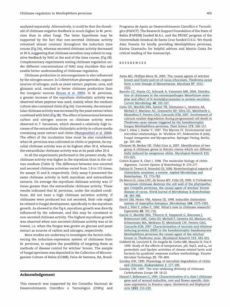
Table 1 – Variables for screening the five-level central compos
Variable �1.68 �1
Carbon source Chitin Chitin (0.5 %)/
glucose (0.5 %)
Nitrogen
source
NH4H2PO4 Yeast extract
(0.5 %)/NH4H2PO4
(0.1 %)
Incubation
time (days)
10 14
stems (green broom). In the second stage, the fungus changes
to the saprotrophic phase and causes necrosis and death of
infected tissues distal from the original infection site, produc-
ing a dry broom (Ceita et al. 2007; Garcia et al. 2007). Basidio-
mata production and spore formation occur on infected
necrotic tissue (Silva et al. 2002; Scarpari et al. 2005).
In order to evaluate M. perniciosa as an industrial source of
chitinases, as well as to explore the possibility of targeting chi-
tinases for disease control, the present study evaluated, using
the concept of RSM, the influence of different carbon and
nitrogen sources and the cultivation time on the activity of
non-secreted or secreted chitinases from the fungus.
Materials and methods
Culture conditions
Moniliophthora perniciosa strain ALF553 (CCBM000257, UEFS,
Feira de Santana, Brazil) was grown on agar containing
10 g l�1 glucose, 1 g l�1 NH4H2PO4, 0.2 g l�1 KCl, 0.2 g l�1
MgSO4.7H2O, 5 g l�1 yeast extract, 1 g l�1 CuSO4.5H2O and 1 g l�1
ZnSO4.7 H2O at 25 �C for 15 d prior to its use for inoculum
development for chitinase production. Liquid culture medium
containing 10 g l�1 carbon source, 6 g l�1 nitrogen source, 0.2 g
l�1 KCl, 0.2 g l�1 MgSO4.7H2O, 1 g l�1 CuSO4.5H2O, and 1 g l�1
ZnSO4.7H2O was used for inoculum development and for
chitinase production. A 1 cm diam disc of fungal mycelium
was taken from the solid culture, transferred to liquid
medium, and incubated at 25 �C without agitation, for
10–30 d, as described in the experimental design.
Experimental design
A three-variable, five-level central composite design (CCD)
leading to 17 sets of experiments (assays), performed in dupli-
cate, was used to verify the best culture medium conditions to
induce the production of chitinases, as well as to analyse the
mycelium growth. The independent variables consisted of
different incubation times and sources of carbon and nitrogen
(Table 1). The results (dependent variables) were the mea-
sured chitinase activity in the mycelium (non-secreted pro-
teins, i.e. symplastic and cell wall proteins) and in the
culture medium (secreted proteins) and the measured myce-
lium growth. The liquid culture medium used was the same
as described above. The incubation was carried out at 25 �C
ite design
0 þ1 þ1.68
Glucose N-acetyl
glucosamine (0.5 %)/
glucose (0.5 %)
N-acetyl
glucosamine
Yeast extract Peptone (0.5 %)/
yeast extract
(0.1 %)
Peptone
20 26 30

Chitinase regulation in Moniliophthora perniciosa 401
without agitation. The software STATISTICA 6.0 (StatSoft,
Tulsa, OK) was used to generate the design matrix and to
analyse the results. Table 2 presents the design matrix and re-
sponses (results) obtained for chitinase activity in the myce-
lium and in the culture medium, and for the mycelium
growth.
Protein extraction and quantification
Mycelium grown on liquid medium was collected, washed
three times with 35 ml cold Milli-Q water (Millipore, Billerica,
MA), dried on filter paper, and ground in liquid nitrogen. The
powder was transferred to a 2 ml tube, and to each 100 mg
of powder, 1.25 ml of 10 mM phosphate buffer (pH 6) contain-
ing 1 % ascorbic acid was added. The tubes were vortexed
and centrifuged at 20,000 g for 10 min at 4 �C. The superna-
tant, containing the mycelium proteins, was collected and
stored at 4 �C. The culture medium, on which Moniliophthora
perniciosa was grown, was filtered on Miracloth (Calbiochem,
San Diego, CA). The filtrate, containing the secreted proteins
from M. perniciosa, was collected and stored at 4 �C. The con-
centration of the proteins from both the mycelium extract
and culture medium was estimated by a based Bradford assay
(Biorad, Hercules, CA) using bovine serum albumin (BSA) as
the standard protein.
Determination of the chitinase activity
Colorimetric assays of chitinase activity were performed in
triplicate using the purple dye-labelled biopolymeric sub-
strate, CM-chitin-RBV (Loewe Biochemical, Saulerlach,
Germany). Two hundred microlitres of CM-chitin-RBV
(2 mg ml�1) were mixed with 300 ml mycelium extract or cul-
ture medium and 300 ml of 50 mM acetate buffer (pH 5). The
Table 2 – Experimental design and results of the central comp
Assay Variable
Carbonsource
Nitrogensource
Incubationtime
1 þ1 þ1 þ1
2 þ1 þ1 �1
3 þ1 �1 þ1
4 þ1 �1 �1
5 �1 þ1 þ1
6 �1 þ1 �1
7 �1 �1 þ1
8 �1 �1 �1
9 þ1.68 0 0
10 �1.68 0 0
11 0 þ1.68 0
12 0 �1.68 0
13 0 0 þ1.68
14 0 0 �1.68
15 0 0 0
16 0 0 0
17 0 0 0
Experiments were performed in triplicate.
mixture was incubated at 37 �C for 3 h. The reaction was
stopped by the addition of 200 ml of 2 M HCl. Samples were
cooled on ice for 15 min, then centrifuged at 20,000 g for
10 min to remove the non-degraded substrate. The superna-
tant was collected and the assay was performed spectrophoto-
metrically at 550 nm (Cary� 100 UV–visible spectrophotometer;
Varian, Palo Alto, CA). Chitinase activity was measured in
units per nanogram of protein per hour. One unit of chitinase
activity corresponded to an increase in absorbance of 0.1
(Ramırez et al. 2004).
Determination of the mycelium growth
The mycelium growth was obtained by quantifying the myce-
lium mass. Three replicates of the Moniliophthora perniciosa my-
celium disc for each assay were collected, quickly dried on
filter paper, and separated from the medium layer using a razor
blade. The mycelium was weighed after 4 h drying at 60 �C.
Results
Central composite design
Based on the identification of variables by the three-level frac-
tional factorial, a CCD was developed for variables that signif-
icantly affected chitinase activity in the mycelium and in the
culture medium. Table 2 lists the coded values of the levels
of the variables selected in the CCD, as well as the responses
generated by the CCD, such as chitinase activity in the myce-
lium and in the culture medium, and mycelium growth.
A quadratic type model was generated, and all the effects
were considered significant (P< 0.05). The model has a relatively
osite design
Chitinase activity(UA ng�1 protein h�2)
Mycelium dryweight (mg)
Mycelium Culture medium
100.22 11.09 99
93.99 26.22 109
74.13 11.61 116
78.1 16.04 60
161.21 3.03 90
76.77 4.3 115
84.48 12.84 92
51.86 0.451 116
52.53 52.14 29
189.61 4.06 29
47.5 8.37 116
0 0 0
60.6 24.29 159
39.29 2.85 57
61.81 34.28 208
59.87 13.15 206
63.5 32.5 201

402 M. A. Lopes et al.
high R2 value (0.771 for the mycelium, 0.766 for the culture me-
dium and 0.789 for the mycelium growth), indicating that 77,
76, and 79 %, respectively, of the variations between the
assays were due to the differences between the tested culture
conditions. Although the F-value was not predictive, it could
be considered significant at a confidence level of 80 %, mean-
ing that the model is descriptive of the data obtained. The pure
error and the lack-of-fit values were not significant (Table 3).
Effect of nitrogen source on non-secretedand secreted chitinase activities
Non-secreted and secreted chitinase activities from Monilioph-
thora perniciosa were monitored using inorganic (NH4H2PO4)
and organic (yeast extract and peptone) nitrogen sources.
The inorganic source NH4H2PO4 used alone was insufficient
to allow the growth and survival of M. perniciosa, and there-
fore, no chitinase activity was observed in the corresponding
mycelium and culture medium (Table 2, assay 12). Among
the organic nitrogen sources and compared with the pep-
tone–yeast extract combination or yeast extract alone,
peptone enhanced non-secreted chitinase activity when com-
bined with chitin as carbon source (Fig 1A). However, chiti-
nase secretion in the culture medium greatly increased in
the presence of yeast extract alone, and significant responses
were also obtained when yeast extract was applied in combi-
nation with another nitrogen source (peptone or NH4H2PO4;
Fig 1B). This last result was obtained in culture medium for
cultures containing NAG (Fig 1B) as the carbon source.
Effect of carbon source on non-secretedand secreted chitinase activities
Non-secreted and secreted chitinase activities from Monilioph-
thora perniciosa were monitored using different carbon sour-
ces: chitin, glucose, and NAG. Non-secreted chitinase
activity was enhanced by chitin (peak production at 30 d cul-
tivation) and repressed by glucose regardless of the duration
of incubation. Non-secreted chitinase activity at 10 d occurred
in the presence of NAG with a higher yield (180 UA ng�1 pro-
tein h�2) than that obtained in the presence chitin (150 UA
ng�1 protein h�2; Fig 2A). Secretion of chitinase in the culture
medium was higher in the presence of NAG and lower in the
presence of chitin regardless of the duration of incubation,
even at the height of secretion, which occurred at 20 d. The
presence of glucose did not affect the recovery of chitinase
activity from the culture medium as strongly as it did in the
mycelium (Fig 2B).
Table 3 – Characteristics of the models used in the centralcomposite design
ModelF-value
R2 Lack-of-fitvalue
Chitinase activity Mycelium 3.41 0.771 71� 10�6
Culture medium 2.45 0.766 5� 10�6
Dry weight Mycelium 2.91 0.789 13� 10�3
Effect of incubation time on chitinase production
Non-secreted and secreted chitinase activities were analysed
after different incubation times, from 10 to 30 d. In the
mycelium, chitinase activity tended to increase with longer
incubation periods. Regarding the culture medium containing
peptone and chitin, peak activity was observed at 30 d (Fig 3A).
The highest chitinase secretion in the medium culture
occurred after 20 d incubation; longer or shorter incubation
periods were shown to be less effective (Fig 3B).
Effect of nitrogen source, carbon source,and incubation time on mycelium growth
The mycelium growth was quantified by weighing the dry my-
celium. The mycelium growth increased when yeast extract
and glucose were used as nitrogen and carbon sources,
respectively (Fig 4A); in these conditions, the highest growth
was obtained after 20 d incubation (Fig 4B,C). The lowest
growth was observed when the fungus was incubated with
chitin and NAG as carbon sources (Fig 4A,B), with NH4H2PO4
and peptone as nitrogen sources (Fig 4A and C), and at short
(10 d) and long (30 d) incubation times (Fig 4B,C). Moreover,
the mycelium cultivated in medium containing chitin (assay
10) was thinner and more dispersed than the mycelium culti-
vated on medium containing glucose, which was thicker and
more compacted (data not shown).
Discussion
Several prokaryotic and eukaryotic microorganisms are
potential producers of chitinolytic enzymes, and medium
composition can significantly affect their production (Nawani &
Kapadnis 2005; Souza et al. 2005). The use of statistical
methods based on experimental design, such as the response
surface methodology, enabled the evaluation of the simulta-
neous effects of culture medium components and of variables
(e.g. incubation time) on the enzymatic production. The use of
these methods enables a better understanding of the possible
interactions between evaluated variables (Nawani & Kapadnis
2005). The model developed here enabled the analysis of the
influence of both carbon and nitrogen sources and incubation
time on the chitinase activity of Moniliophthora perniciosa. The
regulation of the synthesis of chitinases is still little
understood (Souza et al. 2005), even for organisms considered
high chitinase producers, such as Trichoderma harzianum
(Nampoothiri et al. 2004). To our knowledge, this is the first
analysis of chitinase regulation in M. perniciosa and, more gen-
erally, in basidiomycetes.
This study focused on the activity of two chitinases: the
non-secreted and secreted. The intracellular localization of
chitinases has been previously demonstrated by Takaya
et al. (1998), and it seems that the greater portion of chitinases
remains in the cell wall where they could act on chitin (located
only in cell wall) and participate in cell wall modification dur-
ing cell growth and morphogenesis (Choquer et al. 2007). Some
of these chitinases may be covalently linked to the wall struc-
ture (Iranzo et al. 2002). Here, we assumed that non-secreted

Fig 1 – Surface plot of chitinase activity of Moniliophthora perniciosa as a function of nitrogen and carbon sources (in coded
values). (A) Mycelium. (B) Culture medium.
Chitinase regulation in Moniliophthora perniciosa 403
chitinases included intracellular chitinases (whose role and
localization remains to be resolved) and those tightly linked
to the cell wall. As suggested in other works, chitinases,
even from the same group, may be differentially regulated
(Choquer et al. 2007).
Chitin, glucose, and N-acetylglucosamine were used to
evaluate the effects of the carbon sources on non-secreted
and secreted chitinase activities. In cultures supplemented
by chitin, an increase in non-secreted chitinase activity was
observed at 30 d. However, at 10 d, non-secreted chitinase ac-
tivity was slightly higher with NAG (180 UA ng�1 protein h�2)
than with chitin (150 UA ng�1 protein h�2; Fig 2A). As chitin
is insoluble, the fungus is unable to utilize it without previous
hydrolysis into NAG or soluble oligomers. Although non-
secreted chitinase activity remained almost constant in the
presence of NAG throughout the induction time course, it sig-
nificantly increased in presence of chitin, suggesting that non-
secreted activity at 30 d may be induced by chitooligomers
and not by NAG. In support of this hypothesis, a plasma
membrane receptor has recently been discovered on plant
Fig 2 – Surface plot of chitinase activity of Moniliophthora pernicio
(A) Mycelium. (B) Culture medium.
cells, which recognizes chitin fragments for defence signalling
(Kaku et al. 2006; Choquer et al. 2007). Conversely, when the
medium was supplemented with glucose, non-secreted chiti-
nase activity was repressed whatever the duration of incuba-
tion (Fig 2A). However, glucose did not have a stronger
repressive action on the secreted chitinase activity (Fig 2B),
suggesting that these two categories of chitinases may be
differently regulated in the presence of glucose as the carbon
source. Similar results were obtained for other fungi, such as
Trichoderma harzianum (Nawani & Kapadnis 2005; Ulhoa &
Peberdy 1991) and Colletotrichum gloesporioides (Souza et al.
2005). However, the increase of the chitinase activity may
also be influenced by other carbon sources, such as NAG.
Barreto et al. (2004) demonstrated that NAG both up- and
down-regulated chitinase production in Metarhizium aniso-
pliae; when present in low concentrations (�0.05 %), NAG
induced the production and secretion of chitinase, but when
present in higher concentrations, it repressed chitinolytic
activity. In Aspergillus fumigatus (Escott et al. 1998) and Tricho-
derma harzianum (Nawani & Kapadnis 2005; Ulhoa & Peberdy
sa as a function of time and carbon source (in coded values).

Fig 3 – Surface plot of chitinase activity of Moniliophthora perniciosa as a function of nitrogen source and time (in coded
values). (A) Mycelium. (B) Culture medium.
404 M. A. Lopes et al.
1991), NAG repressed chitinase activity by negative feedback.
In M. perniciosa, NAG did not appear to exert an effect on
non-secreted chitinase activity (see discussion above); how-
ever, it did induce extracellular chitinolytic activity (Fig 2B).
Considering the relatively high concentration of NAG (0.5 %)
used in our experiments, the increase in activity of the
Fig 4 – Surface plot of Moniliophthora perniciosa mycelium dry w
a function of carbon source and time; (C) as a function of nitrog
secreted chitinase in M. perniciosa at 20 d was quite unex-
pected. However, because of the lack of information regarding
the regulation of chitinolytic activities in basidiomycetes, it
could be speculated that the regulation of the chitinase path-
way is very different from those fungi already described, espe-
cially when secreted and non-secreted chitinase activities are
eight: (A) as a function of carbon and nitrogen sources; (B) as
en source and time.
Chitinase regulation in Moniliophthora perniciosa 405
analysed separately. Alternatively, it could be that the thresh-
old of chitinase negative feedback is much higher in M. perni-
ciosa than in other fungi. The latter hypothesis may be
supported by the fact that non-secreted chitinase activity
remained almost constant throughout the induction time
course (Fig 2A), whereas secreted chitinase activity decreased
at 30 d, suggesting that chitinase secretion may submit to neg-
ative feedback by NAG at the end of the time course, (Fig 2B).
Complementary experiments testing chitinase regulation un-
der different concentrations of NAG may elucidate this and
enable better understanding of chitinase regulation.
Chitinase production in microorganisms is also influenced
by the nitrogen source. In Colletotrichum gloeosporioides, organic
sources of nitrogen, such as yeast extract, peptone, urea, and
glutamic acid, resulted in better chitinase production than
the inorganic sources (Souza et al. 2005). In M. perniciosa,
a greater increase of the mycelium chitinolytic activity was
observed when peptone was used, mainly when the medium
culture also contained chitin (Fig 3A). Conversely, the extracel-
lular chitinase activity was increased by the yeast extract when
combined with NAG (Fig 3B). The effect of interactions between
carbon and nitrogen sources on chitinase activity were
observed in T. harzanium, which showed a relatively high in-
crease of the extracellular chitinolytic activity in culture media
containing yeast extract and chitin (Nampoothiri et al. 2004).
The effect of the incubation time must be also considered;
when M. perniciosa was cultivated on chitin or peptone, its my-
celial chitinase activity was at its highest after 30 d, whereas
the extracellular chitinase activity was at its peak after 20 d.
In all the experiments performed in the present study, the
chitinase activity was higher in the mycelium than in the cul-
ture medium (Table 2). The difference between non-secreted
and secreted chitinase activities varied from 1.8 to 115 times,
for assays 15 and 8, respectively. Only assay 9 presented the
same chitinase activity in both mycelium and extracellular
extracts. On average the mycelium chitinase activity was 17
times greater than the extracellular chitinase activity. These
results indicated that M. perniciosa, under the studied condi-
tions, did not have a high chitinase secretion activity. If
chitinases were produced but not secreted, their role might
be related to fungal development, specifically to the mycelium
growth. As showed in the Fig 4, mycelium growth was directly
influenced by the substrate, and this may be correlated to
non-secreted chitinase activity. The highest mycelium growth
was observed when non-secreted chitinase activity was at its
lowest, i.e. when the fungus was grown on glucose and yeast
extract as sources of carbon and nitrogen, respectively.
More studies are underway to investigate the factors influ-
encing the induction–repression system of chitinases from
M. perniciosa, to explore the possibility of targeting them as
methods of disease control for witches’ broom. The sample
of fungal specimen was deposited in the Collection of Microor-
ganism Culture of Bahia (CCMB), Feira de Santana, BA, Brazil.
Acknowledgement
This research was supported by the Conselho Nacional de
Desenvolvimento Cientıfico e Tecnologico (CNPq) and
Programa de Apoio ao Desenvolvimento Cientıfico e Tecnolo-
gico (PADCT). The Research Support Foundation of the State of
Bahia (FAPESB) funded M.A.L. and the PROIIC program of the
Universidade Estadual de Santa Cruz funded D.S.G. We thank
Alan Pomela for kindly providing Moniliophthora perniciosa,
Karina Gramacho for helpful advices and Marcio Costa for
critical reading of the manuscript.
r e f e r e n c e s
Aime MC, Phillips-Mora W, 2005. The causal agents of witches’broom and frosty pod rot of cacao (chocolate, Theobroma cacao)form a new lineage of Marasmiaceae. Mycologia 97: 1012–1022.
Barreto CC, Staats CC, Schrank A, Vainstein MH, 2004. Distribu-tion of chitinases in the entomopathogen Metarhizium aniso-pliae and effect of N-Acetylglucosamine in protein secretion.Current Microbiology 48: 102–107.
Ceita GO, Macedo JNA, Santos TB, Alemanno L, Gesteira AS,Micheli F, Mariano AC, Gramacho KP, Silva DC, Meinhardt L,Mazzafera P, Pereira GAG, Cascardo JCM, 2007. Involvement ofcalcium oxalate degradation during programmed cell death inTheobroma cacao tissues triggered by the hemibiotrophicfungus Moniliophthora perniciosa. Plant Science 173: 106–117.
Chet I, Inbar J, Hadar Y, 1997. The Mycota IV, Enviromental andmicrobial relationships. In: Wicklow DT, Soderstrom B (eds),Fungal Antagonists and Mycoparasites. Springer-Verlag, Berlin,pp. 165–184.
Choquer M, Becker HF, Vidal-Cros A, 2007. Identification of twogroup A chitinase genes in Botrytis cinerea which are differen-tially induced by exogenous chitin. Mycological Research 111:615–625.
Cohen-Kupiec R, Chet I, 1998. The molecular biology of chitindigestion. Current Opinion of Biotechnology 9: 270–277.
Dahiya N, Tewari R, Hoondal GS, 2006. Biotechnological aspects ofchitinolytic enzymes: a review. Applied Microbiology andBiotechnology 71: 773–782.
De Marco JL, Lima LHC, de Sousa MV, Felix CR, 2000. A Trichodermaharzianum chitinase destroys the cell wall of the phytopatho-gen Crinipellis perniciosa, the causal agent of witches’ broomdisease of cocoa. World Journal of Microbiology & Biotechnology16: 383–386.
Escott GM, Hearn VM, Adams DJ, 1998. Inducible chitinolyticsystem of Aspergillus fumigatus. Microbiology 144: 1575–1581.
Flach J, Pilet F, Jolles P, 1992. What’s new in chitinase research?Experientia 48: 701–716.
Garcia O, Macedo JNA, Tiburcio R, Zapparoli G, Rincones J,Bittencourt LMC, Ceita GO, Micheli F, Gesteira AS, Mariano AC,Schiavinato MA, Medrano FJ, Meinhardt LW, Pereira GAG,Cascardo JCM, 2007. Characterization of necrosis and ethyleneinducing proteins (NEP) in the hemibiotrophic basidiomyceteMoniliophthora perniciosa the causal agent of the witches’broom in Theobroma cacao. Mycological Research 111: 443–455.
Gobbetti M, Lanciotti R, De Angelis M, Corbo MR, Massini R, Fox P,1999. Study of the effects of temperature, pH, NaCl, and aw onproteolytic and lipolytc activities of cheese-related lactic acidbacteria by quadratic response surface methodology. EnzymeMicrobial Technology 25: 795–809.
Gooday GW, 1990. Physiology of microbial degradation of chitinand chitosan. Biodegradation 1: 177–190.
Gooday GW, 1997. The ever-widening diversity of chitinase.Carbohydrates Europe 19: 18–22.
Hamel F, Bellemare G, 1995. Characterisation of a class I chitinasegene and of wound-inducible, root and flower-specific chiti-nase expression in Brassica napus. Biochemical and BiophysicalActa 1263: 212–220.

406 M. A. Lopes et al.
Iranzo M, Aguado C, Pallotti C, Canizares JV, Mormeneo S, 2002.The use of trypsin to solubilize wall proteins from Candidaalbicans led to the identification of the chitinase 2 as an en-zyme covalently linked to yeast wall structure. Research inMicrobiology 153: 227–232.
Kaku H, Nishizawa Y, Ishii-Minami N, Akimoto-Tomiyama C,Dohmae N, Takio K, Minami E, Shibuya N, 2006. Plant cellsrecognize fragments for defense signalling through a plasmamembrane receptor. Proceedings of the National Academy ofSciences, USA 103: 11086–11091.
Krishnaveni S, Lisng GH, Muthukrishnan S, Manickam A, 1999.Purification and partial characterization of chitinases fromsorghum seeds. Plant Science 144: 1–7.
Kuranda MJ, Robbins PW, 1991. Chitinase is required for cellseparation during growth of Saccharomyces cerevisae. Journal ofBiological Chemistry 266: 19758–19767.
Liu BL, Kao PM, Tzeng YM, Feng KC, 2003. Production of chitinasefrom Verticillium lecanii F091 using submerged fermentation.Enzyme and Microbial Technology 33: 410–415.
Nampoothiri KM, Baiju TV, Sandhya C, Sabu A, Szakacs G,Pandey A, 2004. Process optimization for antifungal chitinaseproduction by Trichoderma harzanium. Process Biochemistry 39:1583–1590.
Nawani NN, Kapadnis BP, 2005. Optimization of chitinase pro-duction using statistics based experimental designs. ProcessBiochemistry 40: 651–660.
Patidar P, Agrawal D, Banerjee T, Patil S, 2005. Optimization ofprocess parameters for chitinase production by soil isolates ofPenicillium chrysogenum under solid substrate fermentation.Process Biochemistry 40: 2962–2967.
Purdy LH, Schmidt RA, 1996. Status of cocoa witches broom:biology, epidemiology, and management. Annual Review ofPhytopathology 34: 573–594.
Ramırez MG, Avelizapa LIR, Avelizapa NGR, Camarillo RC, 2004.Colloidal chitin stained with Remazol Brilliant Blue RR, a usefulsubstrate to select chitinolytic microorganisms and to evaluatechitinases. Journal of Microbiological Methods 56: 213–219.
Regalado AP, Pinheiro C, Vidal S, Chaves I, Ricardo CP, Rodrigues-Pousada C, 2000. The Lupinus albus class-III chitinase gene, IF3,is constitutively expressed in vegetative organs and develop-ing seeds. Planta 210: 543–550.
Rocha HM, Miranda RAC, Sgrillo RB, Setubal RA, 1993. Witches’broom in Bahia, Brazil. In: Rudgard SA, Madison AC,Andebrhan T (eds), Disease and Management in Cocoa: compara-tive epidemiology of Witches’ Broom. Chapman & Hall, London,pp. 189–200.
Scarpari LM, Meinhardt LW, Mazzafera P, Pomella AWV,Schiavinato MA, Cascardo JMC, Pereira GAG, 2005. Biochemi-cal changes during the development of witches’ broom: themost important disease of cocoa in Brazil caused by Crinipellisperniciosa. Journal of Experimental Botany 56: 865–877.
Silva SD, Luz EDMN, Almeida OC, Gramacho K, Bezerra JL, 2002.Redescricao da sintomatologia causada por Crinipellis perni-ciosa em cacaueiro. Agrotropica 1: 1–23.
Souza MCO, Roberto IC, Milagres AMF, 1999. Solid-state fermen-tation for xylanase production by Thermoascus aurantiacususing response surface methodology. Applied Microbiology andBiotechnology 52: 768–772.
Souza RF, Soares RMA, Nascimento RP, Coelho RRR, Gomes RC,2005. Effect of different carbon sources on endochitinaseproduction by Colletotrichum gloeosporioides. Current Microbiol-ogy 51: 16–21.
Takaya N, Yamazaki D, Horiuchi H, Ohta A, Takagi M, 1998. In-tracellular chitinase gene from Rhizopus oligosporus: molecularcloning and characterization. Microbiology 144: 2647–2654.
Ulhoa CJ, Peberdy JF, 1991. Regulation of chitinase synthesis in Tri-choderma harzianum. Journal of General Microbiology 137: 2163–2169.
Wiwat C, Siwayaprahm P, Bhumiratana A, 2002. Cloning,sequencing, and expression of a chitinase from Bacilus circu-lans No. 4.1. Current Microbiology 44: 167–172.
Zellinger S, Galhaup C, Payer K, Woo SL, Mach RL, Fekete C,Lorito M, Kubicek CP, 1999. Chitinase gene expression duringinteraction of Trichoderma harzianum with its host. FungalGenetics and Biology 26: 131–140.
Related Documents


