analytica chimica acta 609 ( 2 0 0 8 ) 120–130 available at www.sciencedirect.com journal homepage: www.elsevier.com/locate/aca Use of polystyrene spin-coated compact discs for microimmunoassaying Jes ´ us Tamarit-L´ opez, Sergi Morais, Rosa Puchades ∗ , ´ Angel Maquieira ∗∗ Departamento de Qu´ ımica, Instituto de Qu´ ımica Molecular Aplicada, Universidad Polit´ ecnica de Valencia, Camino de Vera s/n, 46071 Valencia, Spain article info Article history: Received 11 October 2007 Received in revised form 17 December 2007 Accepted 19 December 2007 Published on line 3 January 2008 Keywords: Compact disc Polystyrene Microimmunoassay Chlorpyriphos abstract The analytical potential of polystyrene (PS) spin-coated modified compact discs (CDs) sur- face as platforms for the development of microarray immunoassays is presented. The surface maintained the optical characteristics of compact discs, obtaining a transparent and smooth film polymer of 70 nm thickness, the track being read ( 780 nm) without errors in a commercial CD reader/writer. The analytical capability of the methodology was demonstrated through an analysis of a neurotoxic compound (2560 spots per disc), reaching 0.08 gL −1 as limit of detection. These figures demonstrate the enormous potential of using PS spin-coated compact discs in combination with CD players as an easy-to-operate and portable device to develop lab-on-a-disc analytical applications. © 2007 Elsevier B.V. All rights reserved. 1. Introduction Compact discs (CDs) are made of a 1.2 mm thick polycarbon- ate (PC) substrate, covered with a reflective metallised film (aluminium, silver or gold) that is protected from oxidation by a lacquer resin [1]. The high optical quality of the poly- meric materials used to manufacturate CDs and digital video discs (DVDs) make these devices promising analytical plat- forms for microarraying purposes and molecular screening; they are a competitive alternative to standard microarray sup- ports made of glass [2,3]. Moreover, disc manufacturing is able to create disc-based microfluidic structures that use centrifu- gal force for fluid management in a lab-on-a-chip analytical system [4,5]. In a earlier publication, working with standard compact discs, Kido et al. explored the principle of CD-based microim- munoassay for the quantitative analysis of pesticide residues ∗ Corresponding author. ∗∗ Corresponding author. Tel.: +34 963877342; fax: +34 963879349. E-mail address: [email protected] ( ´ A. Maquieira). in water [6]. Recently, the use of disc surfaces (PC, poly(methyl methacrylate)) as a high-throughput screening platform has been reported, discriminating different types of Plum pox virus by single-nucleotide polymorphism analysis [7]. Other applications using compact disc technology for ana- lytical purposes have been developed in several patents and papers [8–15]. However, the development of analytical com- pact disc-based methods requires not only the use of standard CDs as analytical platforms, but also the compact disc-reader to fully exploit the potential of CD technology. In the above mentioned applications, the analytical platform, the detec- tor or both were not standard audio–video elements which complicates the use and applications of the technique. Recently, Potyrailo et al. [16] utilized the pickup head unit inside the optical disc drive as a laser scanner detector. Nev- ertheless, this study did not achieve a high-density analysis and the concentrations detected for calcium and other ionic 0003-2670/$ – see front matter © 2007 Elsevier B.V. All rights reserved. doi:10.1016/j.aca.2007.12.028

Welcome message from author
This document is posted to help you gain knowledge. Please leave a comment to let me know what you think about it! Share it to your friends and learn new things together.
Transcript
a n a l y t i c a c h i m i c a a c t a 6 0 9 ( 2 0 0 8 ) 120–130
avai lab le at www.sc iencedi rec t .com
journa l homepage: www.e lsev ier .com/ locate /aca
Use of polystyrene spin-coated compact discs formicroimmunoassaying
Jesus Tamarit-Lopez, Sergi Morais, Rosa Puchades ∗, Angel Maquieira ∗∗
Departamento de Quımica, Instituto de Quımica Molecular Aplicada, Universidad Politecnica de Valencia,Camino de Vera s/n, 46071 Valencia, Spain
a r t i c l e i n f o
Article history:
Received 11 October 2007
Received in revised form
17 December 2007
Accepted 19 December 2007
Published on line 3 January 2008
a b s t r a c t
The analytical potential of polystyrene (PS) spin-coated modified compact discs (CDs) sur-
face as platforms for the development of microarray immunoassays is presented. The
surface maintained the optical characteristics of compact discs, obtaining a transparent
and smooth film polymer of 70 nm thickness, the track being read (� 780 nm) without
errors in a commercial CD reader/writer. The analytical capability of the methodology was
demonstrated through an analysis of a neurotoxic compound (2560 spots per disc), reaching
0.08 �g L−1 as limit of detection. These figures demonstrate the enormous potential of using
Keywords:
Compact disc
Polystyrene
Microimmunoassay
PS spin-coated compact discs in combination with CD players as an easy-to-operate and
portable device to develop lab-on-a-disc analytical applications.
© 2007 Elsevier B.V. All rights reserved.
Recently, Potyrailo et al. [16] utilized the pickup head unit
Chlorpyriphos
1. Introduction
Compact discs (CDs) are made of a 1.2 mm thick polycarbon-ate (PC) substrate, covered with a reflective metallised film(aluminium, silver or gold) that is protected from oxidationby a lacquer resin [1]. The high optical quality of the poly-meric materials used to manufacturate CDs and digital videodiscs (DVDs) make these devices promising analytical plat-forms for microarraying purposes and molecular screening;they are a competitive alternative to standard microarray sup-ports made of glass [2,3]. Moreover, disc manufacturing is ableto create disc-based microfluidic structures that use centrifu-gal force for fluid management in a lab-on-a-chip analyticalsystem [4,5].
In a earlier publication, working with standard compactdiscs, Kido et al. explored the principle of CD-based microim-munoassay for the quantitative analysis of pesticide residues
∗ Corresponding author.∗∗ Corresponding author. Tel.: +34 963877342; fax: +34 963879349.
E-mail address: [email protected] (A. Maquieira).0003-2670/$ – see front matter © 2007 Elsevier B.V. All rights reserved.doi:10.1016/j.aca.2007.12.028
in water [6]. Recently, the use of disc surfaces (PC, poly(methylmethacrylate)) as a high-throughput screening platform hasbeen reported, discriminating different types of Plum poxvirus by single-nucleotide polymorphism analysis [7].
Other applications using compact disc technology for ana-lytical purposes have been developed in several patents andpapers [8–15]. However, the development of analytical com-pact disc-based methods requires not only the use of standardCDs as analytical platforms, but also the compact disc-readerto fully exploit the potential of CD technology. In the abovementioned applications, the analytical platform, the detec-tor or both were not standard audio–video elements whichcomplicates the use and applications of the technique.
inside the optical disc drive as a laser scanner detector. Nev-ertheless, this study did not achieve a high-density analysisand the concentrations detected for calcium and other ionic
t a 6
sc3i
ra[3bfhrpvrtntdafoL
asTteedtm
strn1
Lsm
c[ef
dacasbwaso
a n a l y t i c a c h i m i c a a c
pecies in water were in the mg L−1 range using specificolorimetric reagents. In this noteworthy approach, films ofmm × 4 mm are attached at the polycarbonate surface, mak-
ng water analysis easy and direct.Our research team has recently addressed the use of low
eflectivity compact discs (L-CDs) as analytical platforms withcommercial CD player as the detector to develop microassays
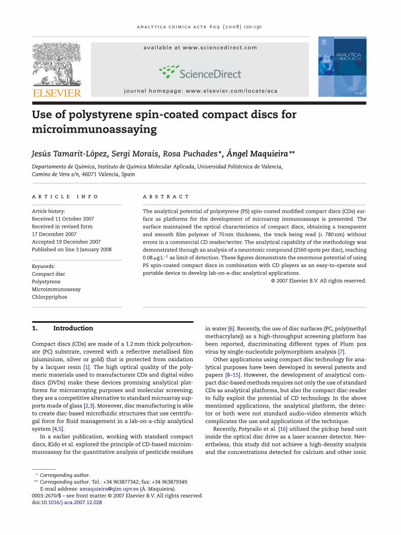
17]. With this approach, L-CDs have a gold layer that reflects0% of the light of the laser beam CD drive (� 780 nm), the resteing transmitted through the disc. The reflected light allowsor the following of the data track of the disc by the pickupead of the CD drive and, therefore, the entire disc can beead. On the other hand, the transmitted light is detected by alanar photodiode incorporated onto the CD drive which con-erts it into an electrical signal. In the absence of an analyticalesponse, about 70% of the laser light is transmitted throughhe surface of the L-CDs and is detected as a background sig-al. In contrast, for a positive signal, the optical properties ofhe disc are modified causing a variation in the light intensityetected by the photodiode, which is further related to thenalyte concentration. In this case, the microassays were per-ormed on the PC side of the L-CDs (down side of disc) andver a 0.6 mm thick PC film assembled onto the top of the-CDs.
One interesting approach that may open up the analyticalpplications of the CD is the modification of the original discurfaces by lacquing the gold layer of the disc with a polymer.his procedure should be compatible with the standard pro-
ocols employed to deposit probes of protein, nucleic acids,tc., by passive adsorption, covalently linkage or throughlectrostatic attraction. This procedure also resolves severalrawbacks, especially those related to the loss of the func-ional capabilities of the biomolecules passively adsorbed on
etallised surfaces [18].On the other hand, on the top side of the discs (metallised
urface) there is a major benefit in terms of spatial resolu-ion, because the laser light incident on the polycarbonate isefracted at a higher angle into the surface; thus, the origi-al incident spot of around 800 �m will be focused down on.05 �m at the metallised surface [19].
In this sense, we postulate the use of polymer spin-coated-CDs as analytical platform, in conjunction with a modifiedtandard CD drive detector, to develop high-density microim-unoassays.Spin-coating of polymer solutions onto flat substrates is a
ommon method to produce thin and uniform polymer films20]. This process is simple and inexpensive, and it has beenmployed to obtain suitable immobilization of biomoleculesor analytical purposes [21,22].
Polystyrene (PS) is also a common polymer used for theirect adsorption of protein in enzyme-linked immunosorbentssay (ELISA) plate methods, being compatible with most bio-hemical reagents. Indeed, PS films are frequently employeds a model system in protein adsorption studies since theirurface chemistry is well-known [23]. In spite of the loss ofiological activity of the proteins that are passively adsorbed,
hich may exceed 90%, the remaining activity is enough tollow the assay to work [24]. This procedure is simple andhould be considered as an alternative strategy before devel-ping other nonadsorptive immobilization procedures such
0 9 ( 2 0 0 8 ) 120–130 121
as covalent attachment or avidin–biotin interaction [25]. Thechoice of PS in this study is also supported by its chemical andoptical properties, such as transparency over a broad spectralrange (� > 290 nm) in addition to its good thermal and mechan-ical characteristics [26].
In this paper, the feasibility of developing microim-munoassays onto polystyrene spin-coated low reflectivitycompact discs is demonstrated. The optimal conditions andthe best chemical and optical characteristics relative topolystyrene deposition onto the surface are also evaluated.As a proof of concept, the determination of a neurotoxicorganophosphorous compound by an indirect competitivemicroimmunoassay is addressed. The detection is performedusing a modified CD reader/writer developing a sensitive andsimple analytical methodology.
2. Experimental
2.1. Chemicals
Buffers (coating buffer: 50 mM sodium carbonate buffer, pH9.6; printing buffer, PBS-T: 10 mM sodium phosphate buffer,150 mM NaCl, 0.01% (v/v) Tween 20, 5% (v/v) glycerol, pH7.2; MES: 50 mM 2-(N-morpholino)ethanesulfonate buffer pH6.0; 50 mM sodium acetate buffer pH 4.6) and washingsolutions were filtered through a 0.22 �m pore size nitrocel-lulose membrane from Whatman GmbH (Dassel, Germany)before use. Polystyrene pellets (MW 50, 250 and 575 kDa)and bovine serum albumin (BSA) were provided by AcrosOrganics (Geel, Belgium). Stock polystyrene solutions wereprepared in Dowanol PMA (1-methoxy-2-propanol acetate)obtained from Sigma–Aldrich (Madrid, Spain). Polyclonalantibody (C2-II) against chlorpyriphos, coating conjugate(OVA-triclopyr) and anti-BSA serum were previously obtainedand characterized by ELISA [27]. The chlorpyriphos standardwas purchased from Dr. Ehrenstorfer (Augsburg, Germany).Nanogold-labelled goat anti-rabbit immunoglobulin (GAR-Au) was supplied by Nanoprobes, Inc. (Yaphank, NY, USA).Glycerol, Tween 20, silver enhancer solution, peroxidase-labelled goat anti-rabbit immunoglobulin (GAR-HRP) ando-phenylenediamine (OPD) were provided by Sigma–Aldrich(Madrid, Spain). Cy5 labelled goat anti-rabbit immunoglobu-lin (GAR-Cy5) was purchased from Jackson ImmunoResearchLaboratories, Inc. (West Grove, PA, USA). All other chemicalsused were analytical grade.
2.2. Instrumentation
Low reflectivity standard compact discs were obtained fromMedia Corp. (Tau-Yuan Shien, China). Their reflectivity aver-age was 30.6% at 780 nm from the measurement of the opticalcharacteristics using a Dr. Schenk 146 scanner (Dr. SchenkGmbH, Planegg, Germany).
The disc microarrayer (model VP478ACD) with 457 �mdiameter pins was from V&P Scientific, Inc. (San Diego, CA,
USA). The CD arrayer is matched with four rows of fivepins with a spot pitch of 1125 �m, each pin carrying 6 nL ofreagent solution leaving a 3 nL drop on the CD that produces500 �m diameter spots. Array features allow for the printing
122 a n a l y t i c a c h i m i c a a c t a 6 0 9 ( 2 0 0 8 ) 120–130
Fig. 1 – (A) Scheme of the competitive immunoassay, showing the intensity of the spot formed over the spin-coated L-CDsurface related to the analyte concentration. (B) Scheme of the detection principle: (i) When the laser beam hits a spot withhigh immunoreaction response, light is not transmitted through the disc. (ii) Light is partially transmitted through the disc.The amount of light detected by the photodiode depends on the developing reaction extension. (iii) In absence of analytical
er be
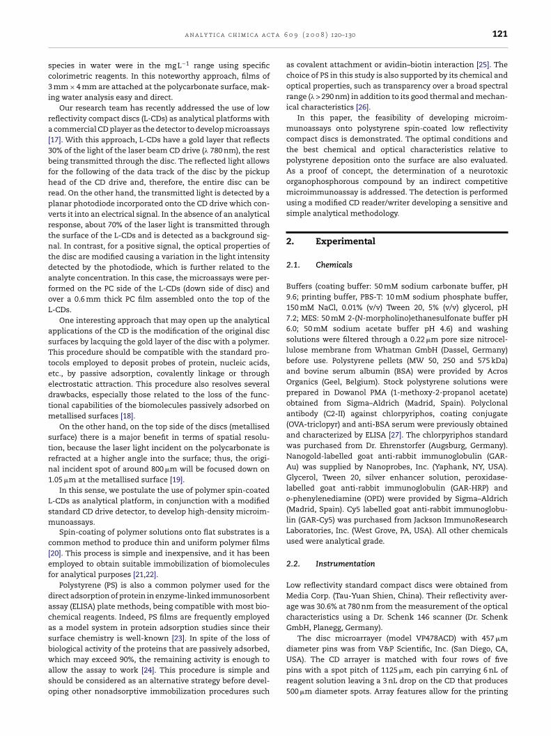
response (no immunoreaction product), about 70% of the lasit is detected as a background signal by the photodiode.of 320 spots per array and 8 arrays per disc (2560 spots perdisc).
Contact angle system OCA20 equipped with SCA20 soft-ware was from Dataphysics Instruments GmbH (Filderstadt,Germany). An 8453 diode array spectrophotometer from Agi-lent Technologies (Palo Alto, CA, USA) was employed tomeasure absorbance. Scanning electron microscope (SEM)images were obtained by an S-4500 SEM from Hitachi High-Technologies (Krefeld, Germany). A Nicolet Nexus Fouriertransform infra-red (FTIR) device, ZnSe window, from ThermoElectro Corporation (Waltham, MA, USA) with OMNIC 6.2software was used for attenuated total reflectance (ATR) mea-surements.
Atomic force microscope (AFM) images were obtainedin the air at room temperature using a Nanoscope IIIamicroscope (Digital Instruments, Santa Barbara, CA, USA).The contact mode was used with Si3N4 triangular levers(spring constant of 0.57 N m−1) for thickness measurements,
whereas, the tapping mode was used for cross-sectional pro-files and roughness measurements. Unless otherwise stated,the applied force was maintained at low values, and the scanrate was 2 Hz for contact mode and 1 Hz for tapping mode.am light is transmitted through the surface of the disc and
2.3. CD spin-coating
The gold surface of the L-CD was first conditioned by gentlywashing with 96◦ ethanol followed by rinsing with deionisedwater and dried with slight centrifugation. Afterwards, discswere spun at 840 rpm, using a laboratory centrifuge fromSelecta (Barcelona, Spain). Next, 1 mL of 3% (w/v) PS inDowanol was dispensed with a pipette near the internal radiusof the gold surface, so the polymer solution was evenly dis-tributed over the entire surface while rotating for 1 min at aconstant speed. The coated CD was then heated in an ovenat 60 ◦C for 30 min in order to remove any remaining sol-vent.
2.4. Protocol of microimmunoassaying
The immunoassays were based on an indirect competitive for-mat as shown in Fig. 1A. The PS lacqued CD was coated with
35 �L of conjugate solution (OVA-triclopyr at 4 mg L−1 in coat-ing buffer) and evenly distributed using 22 mm × 22 mm glasscover slips (eight sample areas per disc). The discs were thenset onto a CD box with a water-saturated filter paper for 16 h
t a 6
afc−
itdw(cti51w
2
ACdstd(sspwcrtta
F(m
a n a l y t i c a c h i m i c a a c
t 4 ◦C. Afterwards the CD was washed with deionised wateror 1 min and dried by slight centrifugation. The conjugateoated discs are stable for at least 3 months when stored at20 ◦C.
Next, the antibody solution (C2-II diluted 1:1600 in print-ng buffer) with or without chlorpyriphos was arrayed ontohe CD by stamping from a 384-well plate. After 5 min, theisc was washed with PBS-T for 1 min, rinsed with deionisedater, and dried out as before. Later, 35 �L of GAR-Au solution
1:100 in PBS-T) was dispensed onto each working area andovered with a glass cover slip. After 1 h at room temperature,he disc was washed, rinsed and dried as before. To detect themmunocomplex formation, the arrays were incubated with0 �L of a 1:1 (v/v) mixture of silver enhancer solution for2 min and covered as before. After washing with deionisedater and drying, the disc was read.
.5. CD detection system
scheme of the detection procedure is shown in Fig. 1B. TheD drive was from Plextor America (Fremont, CA, USA). Therive has an optical system with a laser (� 780 nm) to readtandard CDs and uses the servo focus/tracking system to cen-re and focus the beam on the spiral data track of the entireisc surface. A planar photodiode SLSD-71N6 from Silonex
Montreal, Canada), 25.4 mm long and 5.04 mm width, with apectral range between 400 and 1100 nm, maximum spectralensitivity of 0.55 A/W at 940 nm, was attached to the upperart of the CD reader, 2 mm above the L-CD, along the linehich is scanned by the laser beam during the reading pro-
ess. The position of the photodiode in combination with the
adial and rotational movement of the laser and disc, respec-ively, allows for the coverage of almost the entire disc area,he surface being large enough to simultaneously detect 8 sep-rate assays per disc, 2560 spots per disc (Fig. 2). The electricalig. 2 – (A) Example of spin-coated L-CD with 8 assay areas, each1 cm × 1 cm) showing different array densities: (B1) 4 matrices of
aximum density achieved by the current printing mode (2560 s
0 9 ( 2 0 0 8 ) 120–130 123
signal generated by the photodiode is digitalized by a 16-bitdata acquisition board DT9832A-02-OEM from Data Transla-tion Inc. (Menlo Park, CA, USA), stored in memory and thendeconvoluted into an image. The captured data are transferredto the computer through a universal serial bus 2.0 interface forits quantification.
The reading of assay results was performed using customsoftware written in Visual C+ (BioDisk) which simulates thewriting process of a 700 MB size file. This software also controlsthe data acquisition board, selecting the sampling frequency,size file, and writing the resulting data to the computer harddisc in an uncompressed binary format.
Reading areas of the discs were selected by trigger foot-prints of 3.5 cm on the outer rim of the CD, detected by areflective photosensor EE-SY125 from Omron (Schaumburg,IL, USA) which is integrated onto the custom-built electronicboard. The data captured from each detection area were rep-resented within a sector that is formed by a set of arcs centredover a radial direction, starting from the most inner and mov-ing toward the outer radius, so an image in a 16-bit grey-scalecode was obtained for every sample area.
A lineal velocity of 13 m s−1 (10×) was the highest read-ing speed, taking the CD player 6 min to scan the full disc.A sampling rate of 1 mega sample s−1 was selected for thedata acquisition board which provides two 16-bit analogicalto digital converters and an input voltage range of ±10 V.The maximum spatial resolution was, therefore, 13 �m; so,given a 450 �m diameter spot, the laser needs 35 �s to readit, reading 35 samples (pixels) per spot in the angular direc-tion. In the radial direction, according to the laser trackingsystem of a standard CD reader, a sample is taken each 1.6 �m
(track pitch for CDs), so 280 samples per spot are achieved.Considering the circular shape of the spots, more than 7500samples are taken for each one, ensuring an accurate identi-fication.with 16 matrices of 3 × 3 spots. (B) Disc sections2 × 2 spots, (B2) 4 matrices of 3 × 3 spots and (B3) thepots/disc).
a c t a
124 a n a l y t i c a c h i m i c a2.6. Imaging and data analysis
The Biodisk software allows for the image to be transferredonto a 16-bit tif format or bitmap. Due to the spatial differ-ence between samples taken horizontally (each 13 �m) andvertically (each 1.6 �m), a graphical adjustment was made todisplay a proportional X–Y image with Photoshop 7.0 fromAdobe Systems Inc. (San Jose, CA, USA). The quantificationwas made by GenePix software from Axon Inst. (Union City,CA, USA). Signal intensities (absolute signal) of each spotwere calculated by subtracting local background. Inhibitioncurves were mathematically analysed by fitting experimen-tal results to a sigmoidal four-parameter logistic equation. Allthe software implemented runs on a Windows-based personalcomputer.
2.7. Protein adsorption capacity
To test the protein adsorption capacity, 50 �L of BSA solution(4 mg L−1 in coating buffer) was dispensed, being evenly dis-tributed with glass cover slips onto the PS-coated disc andleft for 16 h at 4 ◦C. Afterwards, the CD was washed withdeionised water for 1 min and dried out by slight centrifuga-tion. Then, 50 �L of anti-BSA serum (1:100 dilution in printingbuffer) was dispensed and distributed with a glass cover slip.After 1 h, the CD was washed with PBS-T for 1 min, rinsedwith deionised water, and dried out. Next, 50 �L of GAR-HRPsolution (1:100 in PBS-T) were deposited onto each workingarea and distributed as before. After 1 h at room tempera-ture, the disc was washed, rinsed and dried as describedabove. Each working area was cut in a 14 mm × 12 mm pieceand introduced into a glass vial. Next, 2 mL of OPD solu-tion (2 g L−1 in citrate buffer pH 5.5) containing 0.8 �L of 33%H2O2 were added to the contents of each vial. After 4 min,1 mL of 2.5 M H2SO4 was added to stop the enzymatic reac-tion. Finally, the absorbance at 490 nm of 200 �L aliquots wasmeasured. The same procedure was carried out as controlon surfaces without BSA coating to provide the backgroundabsorbance.
3. Results and discussion
3.1. Spin-coating process
The spinning speed, solvent nature, molecular weight, as wellas the volume and concentration of polymer solution affectthe final thickness, smoothness, transparency and uniformityof the spin-coated film [28,29]. These factors were studied andoptimised using the absolute signal (AS) and signal to noiseratio (SNR) as selection criteria.
Dowanol was used as the solvent because it dissolvesPS properly [30] and the solutions can be applied withoutaffecting the physical and chemical properties of the disc com-ponents, PC and gold.
Regarding the spin speed, a range between 720 and 960 rpm
was studied, the optimal speed being 840 rpm. It was observedthat higher (960 rpm) and lower (720 rpm) spin speeds resultedin irregular covering and, therefore, unusable surfaces. Sub-sequently, using Dowanol as solvent and spinning the disc6 0 9 ( 2 0 0 8 ) 120–130
at 840 rpm, different volumes (1, 3 and 5 mL) of coating poly-mer were tested by applying 1% (w/v) PS solution (molecularweight of 250 kDa). As the spinning volume solution increased,a decrease in the AS and SNR was observed. The obtainedAS/SNR values were 15,600/5.7, 8500/3.2 and 5600/1.7 for 1,3 and 5 mL, respectively. Volumes below 1 mL (0.1, 0.25 and0.5 mL) were not enough to coat the whole CD surface so, con-sidering the highest signal and SNR values, 1 mL was selectedas the optimum volume.
Regarding the polymer concentration, 1, 3 and 5% (w/v)of 250 kDa PS coating solutions were tested; concentrationsabove 5% (w/v) were not completely dissolved and, conse-quently, were not tested. The AS and SNR values obtained(15,600/5.7, 20,000/6.2 and 2400/1.3) revealed that the bestcoating was achieved using 3% (w/v) PS concentration.
Further, three PS with different molecular weights (50, 250and 575 kDa) were tested under selected conditions (1 mL of3% (w/v) PS in Dowanol, spinning at 840 rpm) giving AS/SNRvalues of 14,300/3.7, 20,000/6.2 and 18,700/4.9, respectively.Thus, in order to obtain the best AS and SNR values in a non-competitive assay, 1 mL of 3% (w/v) PS 250 kDa in Dowanol wasselected for coating purposes.
3.2. Characterization of the polystyrene films
Polystyrene spin-coated surfaces were physico-chemicallycharacterized before immunoreagent adsorption using AFMimaging, contact angle and absorbance (780 nm) measure-ments.
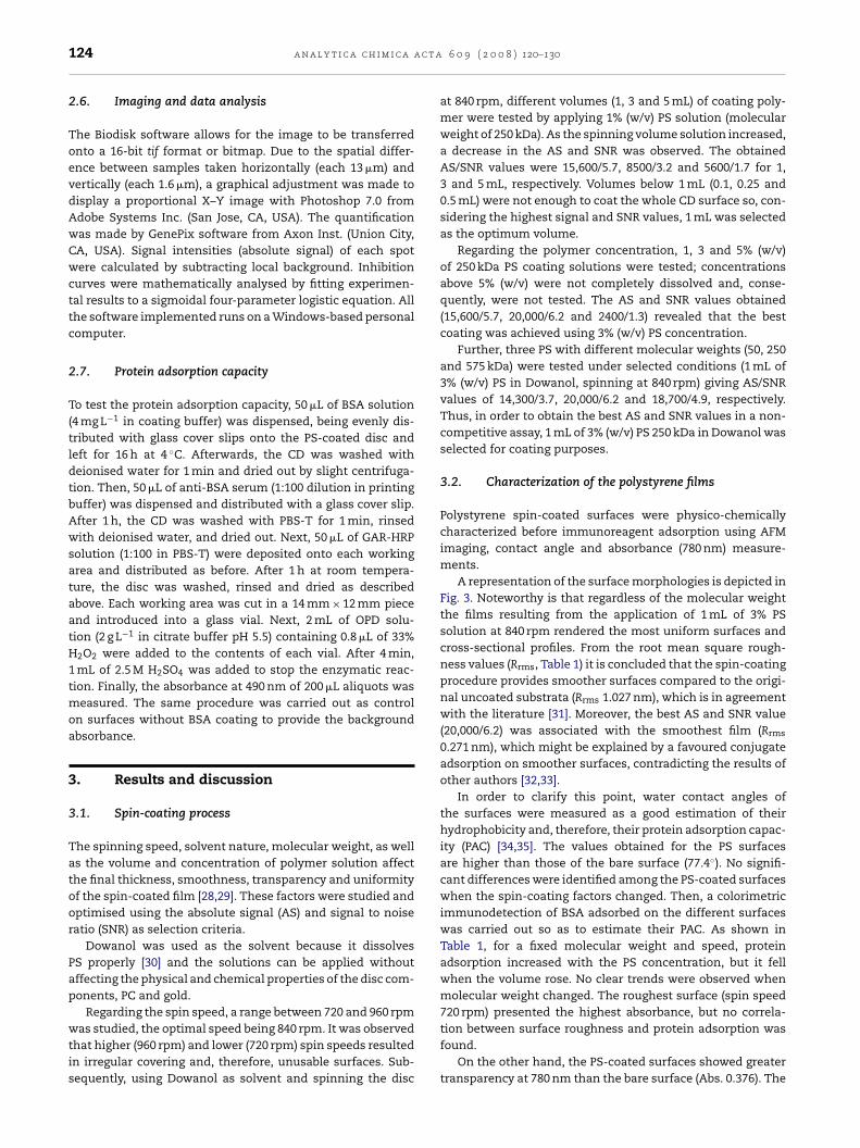
A representation of the surface morphologies is depicted inFig. 3. Noteworthy is that regardless of the molecular weightthe films resulting from the application of 1 mL of 3% PSsolution at 840 rpm rendered the most uniform surfaces andcross-sectional profiles. From the root mean square rough-ness values (Rrms, Table 1) it is concluded that the spin-coatingprocedure provides smoother surfaces compared to the origi-nal uncoated substrata (Rrms 1.027 nm), which is in agreementwith the literature [31]. Moreover, the best AS and SNR value(20,000/6.2) was associated with the smoothest film (Rrms
0.271 nm), which might be explained by a favoured conjugateadsorption on smoother surfaces, contradicting the results ofother authors [32,33].
In order to clarify this point, water contact angles ofthe surfaces were measured as a good estimation of theirhydrophobicity and, therefore, their protein adsorption capac-ity (PAC) [34,35]. The values obtained for the PS surfacesare higher than those of the bare surface (77.4◦). No signifi-cant differences were identified among the PS-coated surfaceswhen the spin-coating factors changed. Then, a colorimetricimmunodetection of BSA adsorbed on the different surfaceswas carried out so as to estimate their PAC. As shown inTable 1, for a fixed molecular weight and speed, proteinadsorption increased with the PS concentration, but it fellwhen the volume rose. No clear trends were observed whenmolecular weight changed. The roughest surface (spin speed720 rpm) presented the highest absorbance, but no correla-
tion between surface roughness and protein adsorption wasfound.On the other hand, the PS-coated surfaces showed greatertransparency at 780 nm than the bare surface (Abs. 0.376). The
a n a l y t i c a c h i m i c a a c t a 6 0 9 ( 2 0 0 8 ) 120–130 125
Fig. 3 – Atomic force microscope images and random cross-sectional profiles for different PS spin-coated metallised CDsurface. AFM images (500 nm × 500 nm; z-range 100 nm) are recorded in air. Cross-sections are taken along the horizontalwhite lines. (a) 1% (w/v), 1 mL, 840 rpm, PS 250 kDa; (b) 1% (w/v), 3 mL, 840 rpm, PS 250 kDa; (c) 1% (w/v), 5 mL, 840 rpm, PS250 kDa; (d) 3% (w/v), 1 mL, 840 rpm, PS 250 kDa; (e) 5% (w/v), 1 mL, 840 rpm, PS 250 kDa; (f) 3% (w/v), 1 mL, 720 rpm, PS250 kDa; (g) 3% (w/v), 1 mL, 960 rpm, PS 250 kDa; (h) 3% (w/v), 1 mL, 840 rpm, PS 50 kDa; (i) 3% (w/v), 1 mL, 840 rpm, PS 575 kDa.

126 a n a l y t i c a c h i m i c a a c t a 6 0 9 ( 2 0 0 8 ) 120–130
Table 1 – Physico-chemical performances of polystyrene spin-coated low reflectivity discs (L-CD)
Polystyrenecoatings
RoughnessRrms (nm)
Water contactangle (◦)
PACa (Absat 490 nm)
Transparency(Abs at 780 nm)
A B C D Mean S.D.
50 1 3 840 0.287 82.6 ± 2.9 0.215 0.218250 1 1 840 0.797 84.7 ± 2.2 0.215 0.324250 3 1 840 0.817 84.3 ± 1.9 0.168 0.329250 5 1 840 0.813 84.3 ± 1.3 0.148 0.325250 1 3 720 1.116 87.4 ± 2.4 0.407 0.248250 1 3 840 0.271 85.3 ± 1.0 0.238 0.197250 1 3 960 0.497 83.3 ± 1.5 0.267 0.233250 1 5 840 0.510 88.0 ± 2.0 0.335 0.352575 1 3 840 0.342 85.8 ± 1.5 0.181 0.219
A, molecular weight (kDa); B, volume (mL); C, concentration (%, w/v); D, spinning speed (rpm); Rrms, root mean square roughness of500 nm × 500 nm sections; Raw original low reflectivity disc (L-CD) characteristics: Rrms, 1.027 nm; PAC, no detected; transparency, 0.376; water
contact angle, 77.4 ± 2.0.a Protein adsorption capacity.most transparent surface was obtained by spinning the discwith 1 mL of PS 250 kDa, 3% (w/v) at 840 rpm (Table 1); this filmalso presented the best AS and SNR values as described above.This might indicate that the transparency of the film is a key
parameter in order to obtain high AS and SNR values using theCD drive as a detector.Since PS presents several bands in the infra-red spectralzone, such as the C–H stretching vibrations in the region
Fig. 4 – Polystyrene film thickness determination by (A) atomic foAFM: A 500 nm × 500 nm image was first recorded under high loaunder normal load. The cross-sections are taken along the black20 kV; zoom, 150,000×.
between 2700 and 3200 cm−1, FTIR-ATR spectroscopy wasused to determine certain modifications in the covering poly-mer. Results (data not shown) confirmed the presence ofpolystyrene on the surface. A match of 75.2% in terms of cor-
relation between the standard polystyrene and the substratespectra was obtained.To assess the thickness of polymer film, AFM imaging intwo operation modes was used. First, the polymer covering
rce microscopy, and (B) scanning electron microscopy.d, followed by recording a square of 3 �m × 3 �m imageand grey lines (z-range 100 nm). SEM: electron voltage,
t a 6
waLlttt6wA1pdloi
3c
Psctspcc
Flsb
Assay sensitivity, in terms of IC50 and SNR, was improved asTween concentration decreased (Table 2), but no competitionwas observed below 0.01%, this value being considered as the
a n a l y t i c a c h i m i c a a c
as scraped off of the substrate by scanning in contact modet a high force for short periods of time, creating a small hole.arger images of this area were then recorded under normaload. The height difference between the bottom of the hole andhe undamaged PS film was used as an estimation of the filmhickness. A mean value of 70 nm was obtained (Fig. 4A) underhe optimal conditions of spin-coating. On the other hand, a0 nm thick PS layer was estimated by SEM imaging (Fig. 4B),hich is in good agreement with the data obtained by AFM.lthough most manufacturers protect the metal layer with a0 �m thick film [36], the layer resulting from our spin-coatingrocedure is thick enough to protect the digital content of theisc. Indeed, and more importantly, the disc is read using the
aser beam (� 780 nm) by the CD player without errors. This is autstanding result since any disc modification must maintain
ts read-out capacity using the CD player as detector.
.3. Immunoassays on polystyrene spin-coatedompact discs
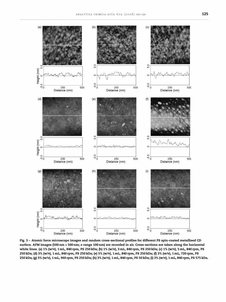
rotein adsorption onto polystyrene depends on reagentolution properties. Among these, the pH and the coatingonjugate concentration have a major effect [25,37]. First,he influence of the coating buffer pH on AS and SNR was
tudied. Sodium carbonate pH 9.6, phosphate pH 7.4, MESH 6.0 and acetate pH 4.6 buffers, were tested using aonstant coating conjugate concentration (4 mg L−1) in a non-ompetitive immunoassay. As shown in Fig. 5A, the highestig. 5 – Absolute signal (solid lines) and SNR values (dashines) obtained for (A) different pHs of the coating conjugateolution and for (B) different glycerol contents in printinguffer.
0 9 ( 2 0 0 8 ) 120–130 127
AS and SNR values were observed when the surface wascoated with sodium carbonate pH 9.6. At pH 4.6 no signal wasdetected.
To study the influence of coating conjugate concentrationon assay sensitivity, 8, 4 and 2 mg L−1 were tested, resultingin IC50 values of 13.6, 8.6 and 1.7 �g L−1, with SNR values of13.5, 6.5 and 3.0, respectively. Coating concentrations below2 mg L−1 gave inconsistent results and, therefore, coatingconjugate at 4 mg L−1 was used for further assay optimisa-tion.
To prevent protein denaturation in the coating process,glycerol at 1%, 2.5%, 5% and 10% (v/v) was added to the print-ing buffer as protective substance [38], which also preventsirregular-shaped dots. In general, the glycerol content in theprinting buffer (PBS-T 10 mM, 0.025% (v/v) Tween 20, pH 7.2),had a considerable effect on the AS, being higher for lowerconcentrations of glycerol (Fig. 5B). However, the best resultsin terms of SNR were obtained with 5% glycerol.
The presence of Tween 20 in the printing buffer helps toreduce non-specific signal and improves the wettability of thesample solution, resulting in a better spot homogeneity [39].
optimum.
Table 2 – Effect of printing buffer composition onsensitivity and signal to noise ratio (SNR) in achlorpyriphos competitive assay
Variable IC50 (�g L−1) SNR
Tween 20 (%)a
0.010 0.96 12.410.025 4.87 8.510.050 4.00 5.490.100 2.49 2.42
pHb
4.6 3.17 10.576.0 1.57 24.017.2 0.96 12.419.6 1.33 4.20
PBS (mM)c
0 13.77 6.231 2.54 8.965 3.19 6.32
10 0.96 12.4120 2.57 10.7845 2.81 10.30
Competition time (min)d
5 0.96 12.4115 0.99 10.6330 1.21 14.7060 1.36 24.3490 3.35 25.92
a PBS-T 10 mM, pH 7.2, competition time 5 min.b Sodium acetate pH 4.6, MES pH 6.0, phosphate pH 7.2 and sodium
carbonate pH 9.6 buffers at 10 mM with NaCl at 150 mM, 0.01%(v/v) Tween 20, competition time 5 min.
c PBS-T pH 7.2, 0.01% (v/v) Tween 20, competition time 5 min.d PBS-T 10 mM, pH 7.2, 0.01% (v/v) Tween 20.
128 a n a l y t i c a c h i m i c a a c t a
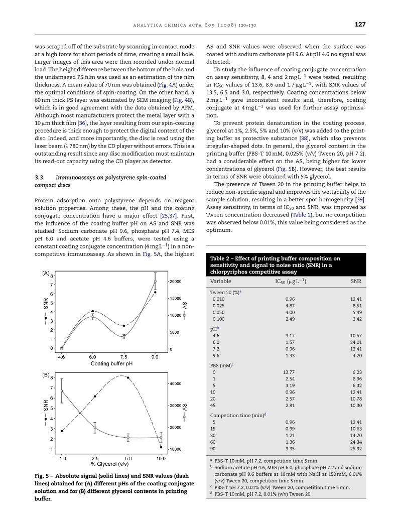
Fig. 6 – Dose–response curve (mean value ± S.D., n = 9)obtained for chlorpyriphos with the CD drive. Each panelrepresents a grey-scale image obtained for 0, 1 and256 �g L−1 of chlorpyriphos from a competitive
CD printers, different indexing shapes, etc.) will enhance, evenmore, the analytical potential of standard compact discs. Con-sidering the active surface, a circular spiral of 26,700 mm inlength, and a track pitch of 300 �m (centre to centre distance
Fig. 7 – Digital images of a parallel microimmunoassay forchlorpyriphos developed over PS-coated L-CDs obtainedfrom fluorescence scanner using GAR-Cy5 (A) and from CDreader detection using GAR-Au and silver enhancersolution (B). With PS-coated standard CDs (high reflectivity)spots were not detected with the CD reader (C). The assay isperformed printing eight arrays (concentrations) of 2 × 2spots (replicates). The numbers inside the panelscorrespond to chlorpyriphos concentration in �g L−1 of eacharray. Red colour in (A) is a false colour representing the
microimmunoassay on PS-coated L-CD.
The influence of the pH and ionic strength of the printingbuffer on IC50 values are shown in Table 2. The best sensitivitywas found using 10 mM phosphate buffer at pH 7.2 with Tween20 at 0.01% as printing buffer. Finally, different competitiontimes (5, 15, 30, 60 and 90 min) were tested to determine theinfluence on sensitivity and SNR. It was clear that longer incu-bation times increased the SNR of the assay, but a decreasein sensitivity (higher IC50) was observed (Table 2), so 5 minwas selected as the optimum competition time for laterassays.
3.4. Performance of the disc-reader
The chlorpyriphos dose–response curve for the optimal con-ditions, reading with a CD drive, is shown in Fig. 6 as are therepresentative grey-scale images of a competitive microim-munoassay arrays. The limit of detection (IC10) was 0.08 �g L−1
with a dynamic range between 0.20 and 7.0 �g L−1, correspond-ing to 80% and 20% inhibitory concentration, respectively.
On the other hand, the mean CV intradisc (calculated from16 spots randomly located over the disc) was 7.6%, and the CVinterdisc (calculated from three discs) was 12.7% in terms ofAS.
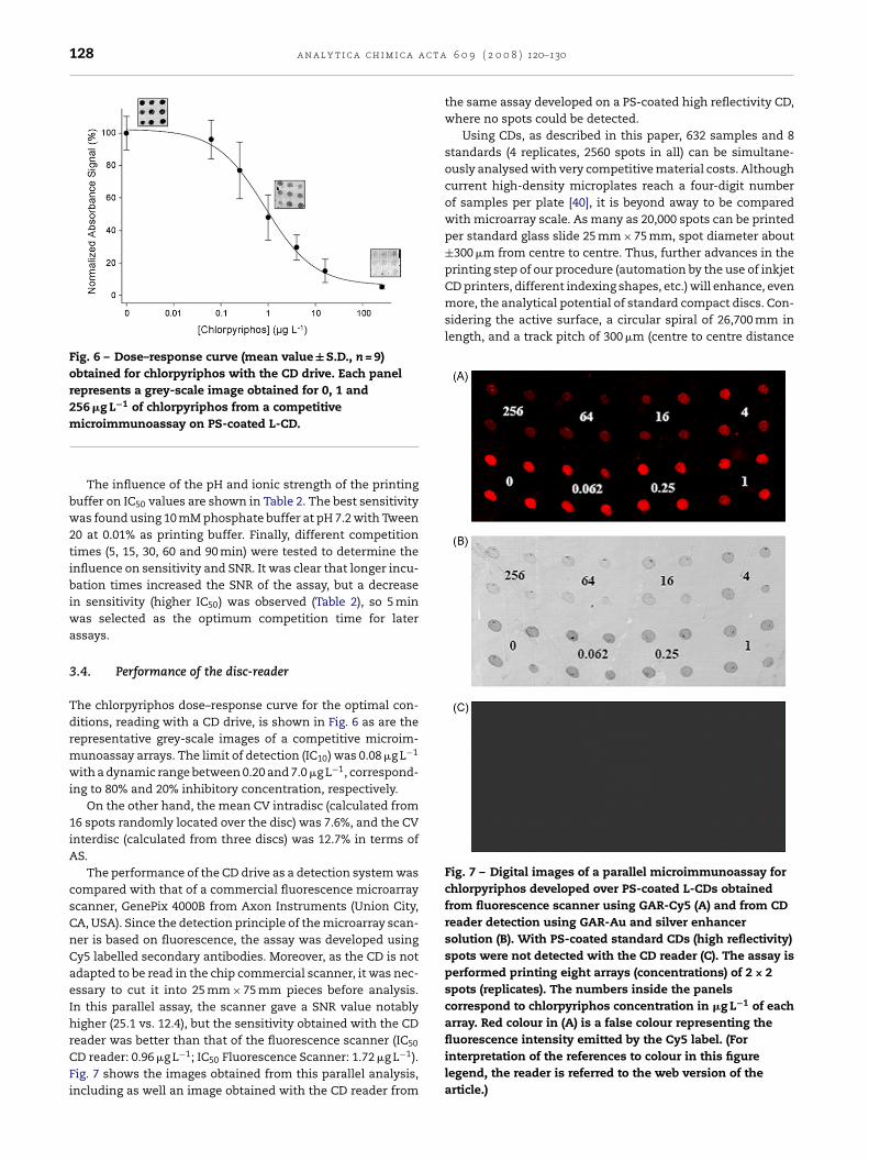
The performance of the CD drive as a detection system wascompared with that of a commercial fluorescence microarrayscanner, GenePix 4000B from Axon Instruments (Union City,CA, USA). Since the detection principle of the microarray scan-ner is based on fluorescence, the assay was developed usingCy5 labelled secondary antibodies. Moreover, as the CD is notadapted to be read in the chip commercial scanner, it was nec-essary to cut it into 25 mm × 75 mm pieces before analysis.In this parallel assay, the scanner gave a SNR value notablyhigher (25.1 vs. 12.4), but the sensitivity obtained with the CD
reader was better than that of the fluorescence scanner (IC50CD reader: 0.96 �g L−1; IC50 Fluorescence Scanner: 1.72 �g L−1).Fig. 7 shows the images obtained from this parallel analysis,including as well an image obtained with the CD reader from
6 0 9 ( 2 0 0 8 ) 120–130
the same assay developed on a PS-coated high reflectivity CD,where no spots could be detected.
Using CDs, as described in this paper, 632 samples and 8standards (4 replicates, 2560 spots in all) can be simultane-ously analysed with very competitive material costs. Althoughcurrent high-density microplates reach a four-digit numberof samples per plate [40], it is beyond away to be comparedwith microarray scale. As many as 20,000 spots can be printedper standard glass slide 25 mm × 75 mm, spot diameter about±300 �m from centre to centre. Thus, further advances in theprinting step of our procedure (automation by the use of inkjet
fluorescence intensity emitted by the Cy5 label. (Forinterpretation of the references to colour in this figurelegend, the reader is referred to the web version of thearticle.)
t a 6
bbu
4
IspiCMr
tcplwiatoe
sapidgb
wtcif
sliTa
bFps
A
TCgoc
r
a n a l y t i c a c h i m i c a a c
etween spots), nearly 100,000 spots of 150 �m diameter coulde applied and detected by the CD reader in only a few min-tes.
. Conclusions
n this study, the capability of modified standard compact discurfaces by spin-coated polystyrene as analytical platforms toerform microimmunoassays has been demonstrated. Work-
ng with polymer films onto the metallised layer of theD, the efficient immobilization of immunoprobes is proven.oreover, this procedure takes advantage of the best spatial
esolution in terms of laser focusing.The spin-coating approach described herein opens the door
o the use of other polymers that could improve the physi-al adsorption of the biomolecules and consequently assayerformances. On the other hand, the development of a cova-
ent immobilization of the probes is a promising alternativehen direct adsorption is not possible, or when an oriented
mmobilization of biomolecules or the reuse of the supportre desired. Thus, the polymers can be functionalised prioro the spin-coating process in order to develop covalent andriented immobilization of bioreceptors such as protein, DNA,tc.
Besides the simplicity of operation and management, ver-atility, low cost and portability, the CD player proved to bevery competitive detector system in comparison, for exam-le, to commercial microarray scanners. This is more relevant
f we consider that the compared transducers are based onifferent optical phenomena, the absorbance detectors beingenerally more limited in sensitivity than are luminescence-ased devices.
Moreover, the CD array methodology offers advantagesith respect to the conventional plate immunoassay. Indeed,
he CDs show high-throughput analytical capacity (in theurrent format 2560 spots per disc) as well as considerablemmunoreagent savings (between 2-fold and up to 10,000-foldor coating conjugate and primary antibody, respectively).
On the other hand, working with the laser diode as the onlyensing system, the laser light is focused onto the reflectiveayer inside the disc to a spot of approximately 2 �m, provid-ng phase control of the light that reaches the photo-detector.hese features are essential for the rejection of ambient lightnd light produced by irregularities on the disc surface.
To conclude, with this CD-based methodology, it is possi-le to perform highly sensitive multianalyte determinations.urther improvements, such as the automation of differentrocess steps, will result in a more integrated and competitiveystem.
cknowledgments
his work was supported by the projects BQU2003-02677 and
TQ2007-64735/BQU (CICYT, Spain). J.T.-L. acknowledges arant from the Spanish Ministerio de Educacion y Ciencia to carryut his Ph.D. research. The authors thank Debra Westall forarefully revising the manuscript.0 9 ( 2 0 0 8 ) 120–130 129
e f e r e n c e s
[1] IEC, International Standard IEC60908: AudioRecording—Compact Disc Digital Audio System,International Electrotechnical Commission, Geneva,Switzerland, 1999.
[2] T.D. Boone, A.J. Ricco, Z.H. Fan, H. Tan, H.H. Hooper, S.J.Williams, Anal. Chem. 74 (2002) 78A.
[3] C. Situma, Y. Wang, M. Hupert, F. Barany, R.L. McCarley, S.A.Soper, Anal. Biochem. 340 (2005) 123.
[4] M.J. Felton, Anal. Chem. 75 (2003) 302A.[5] Y.-K. Cho, J.-G. Lee, J.-M. Park, B.-S. Lee, Y. Lee, C. Ko, Lab
Chip 7 (2007) 565.[6] H. Kido, A. Maquieira, B.D. Hammock, Anal. Chim. Acta 411
(2000) 1.[7] S. Morais, R. Marco-Moles, R. Puchades, A. Maquieira, Chem.
Commun. 22 (2006) 2368.[8] J. Virtanen, US Patent 6 030 581 (2000).[9] B.D. Hammock, H. Kido, A. Maquieira, US Patent 6 342 395
(2002).[10] S. Iimura, H. Ogawa, US Patent 0 169 677 (2003).[11] J.J. La Clair, M.D. Burkart, Org. Biomol. Chem. 1 (2003) 3244.[12] D.D. Nolte, F.E. Regnier, Opt. Photon. News 15 (2004) 48.[13] I. Alexandre, Y. Houbion, J. Collet, J. Demarteau, J.L. Gala, J.
Remacle, Biotechniques 33 (2002) 435.[14] S.A. Lange, G. Roth, S. Wittermann, T. Lacoste, A. Vetter, J.
Grassle, S. Kopta, M. Kolleck, B. Breitinger, M. Wick, J.K.H.Horber, S. Dubel, A. Bernard, Angew. Chem., Int. Ed. 45 (2006)270.
[15] Y. Li, Z. Wang, L. Ou, H.Z. Yu, Anal. Chem. 79 (2007) 426.[16] R.A. Potyrailo, W.G. Morris, A.M. Leach, T.M. Sivavec, M.B.
Wisnudel, S. Boyette, Anal. Chem. 78 (2006) 5893.[17] S. Morais, J. Carrascosa, D. Mira, R. Puchades, A. Maquieira,
Anal. Chem. 79 (2007) 7628.[18] B.A. Snopok, K.V. Kostyukevych, G.V. Beketov, S.A. Zinio, Y.M.
Shirshov, E.F. Venger, S.V. Verevka, Semicond. Phys.,Quantum Electron. Optoelectron. 3 (2000) 59.
[19] J.A. Cope, Phys. Educ. 28 (1993) 15.[20] K. Norrman, A. Ghanbari-Siahkali, N.B. Larsen, Annu. Rep.
Prog. Chem. C 101 (2005) 174.[21] R. Jenison, H. La, A. Haeberli, R. Ostroff, B. Polisky, Clin.
Chem. 47 (2001) 1894.[22] S.L. Peterson, A. McDonald, P.L. Gourley, D.Y. Sasaki, J.
Biomed. Mater. Res. A 72 (2005) 10.[23] R.J. Green, J. Davies, M.C. Davies, C.J. Robert, S.J.B. Tendler,
Biomaterials 18 (1997) 405.[24] J.E. Butler, L. Ni, W.R. Brown, K.S. Joshi, J. Chang, B.
Rosenberg, E.W. Voss, Mol. Immunol. 30 (1993) 1165.[25] J.E. Butler, Methods 22 (2000) 4.[26] R.N. Nurmukhametov, L.V. Volkova, S.P. Kabanov, J. Appl.
Spectrosc. 73 (2006) 55.[27] E.M. Brun, M. Garces-Garcıa, R. Puchades, A. Maquieira, J.
Agric. Food Chem. 53 (2005) 9352.[28] S.A. Gupta, R.K. Gupta, Ind. Eng. Chem. Res. 37 (1998) 2223.[29] D.B. Hall, P. Underhill, J.M. Torkelson, Polym. Eng. Sci. 38
(1998) 2039.[30] A. Imre, W.A. van Hook, J. Phys. Chem. Ref. Data 25 (1996)
637.[31] F.A. Denis, P. Hanarp, D.S. Sutherland, Y.F. Dufrene,
Nanoletters 2 (2002) 1419.[32] D.S. Sutherland, M. Broberg, H. Nygren, B. Kasemo,
Macromol. Biosci. 1 (2001) 270.
[33] X. Jia, T.J. McCarthy, Polym. Prepr. (Am. Chem. Soc., Div.Polym. Chem.) 42 (2001) 153.[34] A. Martın-Rodrıguez, M.A. Cabrerizo-Vilchez, R.
Hidalgo-Alvarez, Colloids Surf. A 92 (1994) 113.[35] R.D. Tilton, C.R. Robertson, A.P. Gast, Langmuir 7 (1991) 2710.

a c t a
130 a n a l y t i c a c h i m i c a[36] T. Fuchs, Film Thickness Measurement: Application Note.Retrieved September 2007 fromhttp://www.applied-spectrocopy.com/ftm.htm.
[37] D. Song, D. Forciniti, J. Colloid Interf. Sci. 221 (2000) 25.
6 0 9 ( 2 0 0 8 ) 120–130
[38] G. MacBeath, S.L. Schreiber, Science 289 (2000) 1760.[39] W. Kusnezow, J.D. Hoheisel, J. Mol. Recognit. 16 (2003)
165.[40] B.J. Battersby, M. Trau, Trends Biotechnol. 20 (2002) 167.
Related Documents