Oliveira, E. de A. M Silva and S. O. De Paula R. S. Dias, M. R. Eller, V. S. Duarte, Â. L. Pereira, C. C. Silva, H. C. Mantovani, L. L. mastitis isolated from bovine Staphylococcus aureus Use of phages against antibiotic-resistant doi: 10.2527/jas.2012-5884 originally published online July 26, 2013 2013, 91:3930-3939. J ANIM SCI http://www.journalofanimalscience.org/content/91/8/3930 the World Wide Web at: The online version of this article, along with updated information and services, is located on www.asas.org by guest on September 15, 2014 www.journalofanimalscience.org Downloaded from by guest on September 15, 2014 www.journalofanimalscience.org Downloaded from

Welcome message from author
This document is posted to help you gain knowledge. Please leave a comment to let me know what you think about it! Share it to your friends and learn new things together.
Transcript

Oliveira, E. de A. M Silva and S. O. De PaulaR. S. Dias, M. R. Eller, V. S. Duarte, Â. L. Pereira, C. C. Silva, H. C. Mantovani, L. L.
mastitis isolated from bovineStaphylococcus aureusUse of phages against antibiotic-resistant
doi: 10.2527/jas.2012-5884 originally published online July 26, 20132013, 91:3930-3939.J ANIM SCI
http://www.journalofanimalscience.org/content/91/8/3930the World Wide Web at:
The online version of this article, along with updated information and services, is located on
www.asas.org
by guest on September 15, 2014www.journalofanimalscience.orgDownloaded from by guest on September 15, 2014www.journalofanimalscience.orgDownloaded from

3930
Use of phages against antibiotic-resistant Staphylococcus aureus isolated from bovine mastitis1
R. S. Dias,* M. R. Eller,* V. S. Duarte,* Â. L. Pereira,* C. C. Silva,‡ H. C. Mantovani,† L. L. Oliveira,* E. de A. M Silva,* and S. O. De Paula,*2
*Laboratory of Molecular Immunovirology, Department of General Biology, Federal University of Viçosa, Viçosa, Brazil; †Laboratory of Anaerobic Microorganisms, Department of Microbiology, Federal
University of Viçosa, Viçosa, Brazil; and ‡ Department of Microbiology, Federal University of Viçosa, Viçosa, Brazil
ABSTRACT: Bovine mastitis is the primary disease of dairy cattle worldwide and it causes large economic losses. Among several microorganisms that are the causative agents of this disease, Staphylococcus aureus is the most prevalent. Although antibiotic therapy is still the most widely used procedure for the treatment of bovine mastitis, alternative means of treatment are necessary due to the presence of antibiotic residues in milk, which is a growing concern because of its interference with the production of milk derivatives and the selection of resistant bacterial strains. The use of bacteriophages as a tool for the control of pathogens is an alternative treatment to antibiotic therapy. In this work, to obtain phages with the potential for use in phage therapy as a treatment for mastitis, we isolated and identified the bacteria from the milk of mastitis-
positive cows. A total of 19% of the animals from small and medium farms of the Zona da Mata Mineira, Brazil, was positive for bovine mastitis, and bacteria of the genus Staphylococcus were the most prevalent pathogens. The majority of the S. aureus isolates tested was resistant to penicillin and ampicillin. In parallel, we isolated 10 bacteriophages able to infect some of these S. aureus isolates. We determined that these phages contained DNA genomes of approximately 175 kb in length, and the protein profiles indicated the presence of 4 major proteins. Electron microscopy revealed that the phages are caudate and belong to the Myoviridae family. The isolates exhibited interesting features for their use in phage therapy such as a high lytic potential, a wide range of hosts, and thermostability, all of which favor their use in the field.
Key words: antibiotic-resistant Staphylococcus aureus, bovine mastitis, phages
© 2013 American Society of Animal Science. All rights reserved. J. Anim. Sci. 2013.91:3930–3939 doi:10.2527/jas2012-5884
INTRODUCTION
Mastitis, the most common disease of dairy herds worldwide (Fessler et al., 2010), can be caused by over 150 different microorganisms, such as Staphylococcus aureus, Streptococcus agalactiae,
Streptococcus dysgalactiae, Streptococcus uberis, Staphylococcus epidermidis, Corynebacterium bovis, Corynebacterium pyogenes, and Candida albicans. Among all of these microorganisms, the most important is S. aureus, which is a common cause of mastitis on dairy farms (Olde Riekerink et al., 2008) and the most frequently isolated pathogen in heifer mastitis (Delgado et al., 2011; Dufour et al., 2012). In Brazil, studies report the predominance of S. aureus over other disease-causing agents in all regions of the country (Brito, 1996). In recent years, the incidence of antibiotic-resistant bacteria has increased. In addition, the antibiotic residues in milk also interfere with and often prevent the production of milk derivatives (Pinto, 2001). Bacteriophages (here after referred to as “phages”) are natural bacterial viruses abundant in all environments, including the water, soil, and
1We thank to the Núcleo de Microscopia e Microanálise (NMM) of the Federal University of Viçosa for providing materials and technical support. This study was supported by grants from the Fundação de Amparo à Pesquisa do Estado de Minas Gerais (FAPEMIG) and PhD fellowships from the Coordenação de Aperfeiçoamento de Pessoal de Nível Superior (CAPES) and the ConselhoNacional de Desenvolvimento Científico e Tecnológico (CNPq) to ALP. The funders had no role in the study design, data collection, data analysis, decision to publish, or preparation of this manuscript.
2Corresponding author: [email protected] September 21, 2012.Accepted May 21, 2013.
by guest on September 15, 2014www.journalofanimalscience.orgDownloaded from

Phages against mastitis Staphylococcus aureus 3931
air (d’Herelle, 1917). Their ability to not only target and destroy a specific bacterium but also to replicate exponentially and specifically underscores their potential role in treating infectious diseases (d’Herelle, 1917; Carson et al., 2010). Therefore, phage therapy is an alternative to antibiotics when bacteria develop antibiotic resistance, and the use of these particles has been studied by a number of research groups to control a variety of mastitis-causing pathogens, mainly S. aureus (Gill et al., 2006; Garcia et al., 2009; Son et al., 2010; Kwiatek et al., 2012). In the present work, we isolated 10 lytic bacteriophages that belonged to the Myoviridae family. The isolates exhibit interesting features for their use in phage therapy such as thermostability, a high lytic potential, and a wide range of hosts, which renders them useful against the S. aureus antibiotic-resistant strains that cause bovine mastitis.
MATERIALS AND METHODS
All animal experiments were performed in accordance with protocols approved by the Federal University of Viçosa Institutional Animal Care and Use Committee.
Collection, Isolation, and Identification of the Bacterial Strains
A total of 607 animals (2,408 teats) from 24 farms were studied for mastitis occurrence using as criteria the occurrence of clinical symptoms (for clinical mastitis) and the method described by Barnum and Newbould (1961). The antibiotic therapy in these animals was excluded. From 250 teats that tested positive for mastitis, 10 mL of milk were collected and tested for the presence of S. aureus. Each milk sample was streaked onto sheep blood agar and McConkey agar and incubated at 37°C for 24 h. The colonies that grew on the plates were gram stained, and a catalase test was performed over any gram-positive cocci. The catalase-positive bacteria were subjected to the coagulase and acetoin tests, and the catalase-negative bacteria were tested with CAMP test. In the case of the gram-negative bacteria, the test was performed using the Bactray I kit (Laborclin, Pinhais, Paraná, Brazil). The characterization procedure of bacterial colony was performed as previously described by Brito (1999).
Antibiogram
The antimicrobial susceptibility test of the 20 S. aureus isolates was performed using the plate diffusion technique (Watts, 2008). The antibiotics used for antibiogram were ampicillin 10 μg/mL, gentamicin 10 μg/mL, cephalothin 30 μg/mL, ciprofloxacin 5
μg/mL, sulfazotrim 25 μg/mL (Sulakvelidze et al., 2001), amoxicillin/clavulanate 30 μg/mL, ceftriaxone 30 μg/mL, tetracycline 30 μg/mL, penicillin G 10 μg/mL, oxacillin 1 μg/mL, erythromycin 15 μg/mL(Olde Riekerink et al., 2008), vancomycin 30 μg/mL, clindamycin 2 μg/mL, rifampicin 5 μg/mL, and chloramphenicol 30 μg/mL. The results were interpreted according to the standards of the Clinical and Laboratory Standards Institute (Watts, 2008).
Phage Collection and Isolation
The samples of sewage water used for the phage isolation were obtained from the Sewage Treatment Station (SAAE) of Viçosa, Minas Gerais, Brazil. Phages were isolated and propagated from 9 L of water in broth-heart infusion (BHI) medium without enrichment, as described by (Tanji et al., 2008). The lysis plaques isolated from initial plating were collected and transferred to an Erlenmeyer flask containing the culture medium and the bacteria in the exponential growth phase. These mixtures were then incubated at 37°C and plated again. This procedure was repeated 3 times to ensure the isolation of phages. The S. aureus strain 3930, courtesy of Brazilian Agricultural Research Corporation (EMBRAPA) Gado de Leite, Minas Gerais, Brazil, was used such as positive control in these experiments.
Phage Genome Extraction
The phage genomes were extracted as described previously in Sambrook and Russell (2006). Briefly, 50 μL of each isolated phage was added to an equal volume of lysis buffer (10 mM Tris–Cl, pH 8.0, 1 mM EDTA, and 0.5% Tween 20) and 2 μL of proteinase K (50 μg/mL). The mixture was vortexed thoroughly and incubated at 55°C for 2 h. The samples were subsequently inactivated at 94°C for 10 min and purified by standard phenol-chloroform extraction and ethanol precipitation procedures. The pellet was dissolved in 30 μL of distilled water, and the isolated nucleic acids were separated using 0.8% agarose gel electrophoresis, stained with ethidium bromide, and analyzed under a UV light. To analyze the phage genome composition, 10 μL of each genomic extract was digested with deoxyribonuclease (DNase) I (1 µg/mL) or Ribonuclease (RNase) A (1 µg/mL) for 60 min at 37°C. The digested fragments were analyzed using 1.0% agarose gel electrophoresis as described above.
Evaluation of the Viral Genome Size
The length of the viral genome was estimated using pulsed-field gel electrophoresis using a Bio-Rad CHEF-DR III Pulsed Field Electrophoresis System (Bio-Rad, USA). Twenty microliters of the extracted genomic DNA
by guest on September 15, 2014www.journalofanimalscience.orgDownloaded from

Dias et al.3932
was added to 20 μL of 2.0% low melt agarose according to the manufacture’s recommendations, and lambda DNA concatemers were used as the molecular marker. The samples were subjected to pulsed-field gel electrophoresis on a 1.0% agarose gel at an angle of 120° with a switching interval of 5 to 200 sec at 14°C. The gel was stained with ethidium bromide and analyzed under a UV light.
Protein Profile Analysis
An aliquot of the viral suspensions (50 mL) was concentrated with NaCl 1.0 M and 10% polyethylene glycol 8000, centrifuged at 11,000 xg for 20 minutes, at 4°C, and resuspended in 1 mL of water. These aliquot were used in the protein analysis or electron microscopy studies. The proteins were precipitated with 10% trichloroacetic acid and washed 3 times in cold acetone, and the total phage protein content was analyzed using electrophoresis on 15% polyacrylamide gel under denaturing conditions (SDS-PAGE) at 190 V for 1 h. The broad range SDS-PAGE standard was used as molecular size marker, and the gel was stained with 3% ammoniacal silver.
Electron Microscopy
A 10 μL aliquot of the viral suspension prepared as described above was added to a 200 mesh grid that was previously covered with FormVar (Sigma-Aldrich, Inc. St. Louis, MO, USA) for 5 min. The excess liquid was removed with filter paper, and the grids were covered with 10 μL of 2% uranyl acetate for 20 s. The samples were analyzed in the Núcleo de Microscopia e Microanálise, Federal University of Viçosa/Brazil, using a Zeiss EM 109 (Carl-Zeiss, Oberkochen, Germany) transmission electron microscope operating at 80 kV. The data were analyzed to measure the overall viral length, the tail length, and the capsid diameter. When necessary, additional dilutions were performed to obtain greater quality images.
Test of the Phage Lytic Potential
In 4 flasks containing 50 mL of BHI medium each, 1 mL of a suspension containing the bacterium S. aureus 3930 in the exponential growth phase [optical density (OD)600 = 0.7] was added to each phage solution at a multiplicity of infection of 0.1. The mixtures were incubated at 37°C and 200 rpm, and aliquots were analyzed in a spectrophotometer (Thermo Fisher Scientific Inc., Waltham, MA, USA; OD600) at 1, 2, 3, 4, 5, 6, 7, 8, 9, 10, 11, 12, 16, 20, and 24 h postinfection. After, mitomycin C was added in 2 flasks at final concentration of 0.5 µg/mL to evaluate its effect on the viral lysis potential. A preparation without phage was used as negative controls.
Test of Phage Specificity
Each phage was added at high titer [1010 plaque forming units (PFU)/mL] to 20 different S. aureus strains in exponential phase growth and plated on BHI medium using the pour plate method (Sambrook and Russell, 2006). As a control, we plated the uninfected bacterial strains. After incubation at 37°C for 18 h, the number of plates exhibiting lysis was counted. The plates that did not exhibit any changes were scored with a minus sign (–). The plates containing lysis plaques were scored with positive signs, ranging from (+) to (++++), the number of which increased with the number of lysis plaques observed.
Test of Phage Thermostability
A 150 μL aliquot of the phage suspension was incubated at 70, 80, 90 or 100°C for 5 min. After, the phage titer was calculated as PFU per mL (PFU/mL). The titer of a phage suspension with no temperature treatment was calculated in parallel.
RESULTS
Identification and Characterization of the Bacterial Strains
A total of 250 milk samples were obtained from 65 different farms with isolated bacteria in 58.18% of samples. One hundred one bacteria were isolated and identified as S. aureus (28.52%), Staphylococcus sp. (coagulase negative; 14.83%), Streptococcus sp. (CAMP negative; 6.84%), a gram-positive Bacillus (3.42%), a yeast strain (2.28%), Escherichia coli (1.15%), and Streptococcus agalactiae (1.14%).
Twenty isolates of S. aureus were randomly selected and tested for antibiotic susceptibilities. Ampicillin resistance was exhibited by 60% of the isolates and 65% were resistant to penicillin. The majority of the isolates (95%) was susceptible to gentamicin (Table 1).
Phages Characterization
Ten phages were isolated from the sewage water samples and were named Ufv-aur2 to Ufv-aur11.
The viral genomes were extracted, and pulsed-field gel electrophoresis studies indicated that the phage genomes are approximately 175 kb in length (Fig. 1A). Aliquots were digested with DNase I or RNase A. Although DNase I digestion degraded all of the samples, RNase A digestion had no effect. These results indicate that the genomes were composed of DNA (Fig. 1B).
Gel electrophoresis (SDS-PAGE) indicated that all phages showed identical protein profiles, as represented by Ufv-aur2, Ufv-aur3, Ufv-aur4, Ufv-aur5, Ufv-aur6,
by guest on September 15, 2014www.journalofanimalscience.orgDownloaded from

Phages against mastitis Staphylococcus aureus 3933
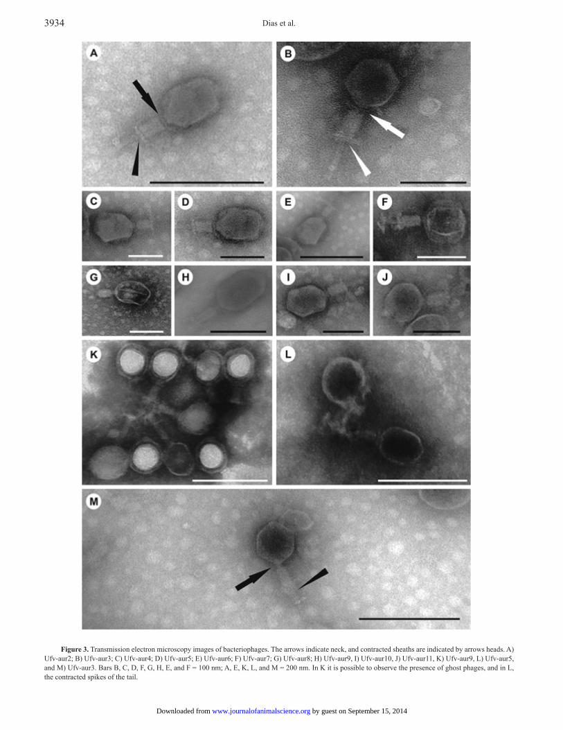
Ufv-aur7, Ufv-aur8, Ufv-aur9, Ufv-aur10, and Ufv-aur11 in Fig. 2. Transmission electron microscopy showed that the phages have an average size of approximately 200 nm, a 50-nm long contractile tail, and an icosahedral head that is about 90 nm in diameter. Interestingly, a basal plate was observed (Fig. 3).
Test of the Phage Lytic Potential
The S. aureus strain 3930 was subjected to infection by each phage, and the OD (OD600) of the culture medium was measured at different times of infection to
determine the bacterial growth. After 8 h of culture, all phages reduced S. aureus isolates growth (Fig. 4). In some cases, the addition of mitomycin C to the culture medium further decreased the detected OD values and delayed the subsequent growth compared with the samples containing bacteria plus phage (asterisks in Fig. 4). The OD values of the samples containing only the bacterial culture and mitomycin C were equivalent to the samples that contained only the bacterial culture (Fig. 4).
Table 1. Susceptibility of Staphylococcus aureus strains isolated of bovine mastitis infections to 15 different antimicrobials1
Isolate
AntibioticRIF AMC CRO CFL CIP SUT AMP TET ERI GEN PEN CLO OXA CLI VAN
S. aureus 2878 S S S S S S S S S S S S S S SS. aureus 3017 S S S S S S S S S S R S S S SS. aureus 3019 S S S S S S R R R S R S S I SS. aureus 3166 S S S S R S R S S S I S S S SS. aureus 3216 S S S S S S S S S S S S S I SS. aureus 3627 S S S S S S S S S S S S S I SS. aureus 3654 S S S S I S R S S S R S S I SS. aureus 3810 S S I S R S S S I S S S S S SS. aureus 3820 S S I S S S S S I S S S S S SS. aureus 3930 S S I S R S I S R S R S S I SS. aureus 1 S S S S S S S S S S R S S S SS. aureus 3 S S S S S S R S I S R S S R SS. aureus 9 R R S R S S R I I S R R S R SS. aureus 17 S S R S I S R R S R R S S S SS. aureus 49 R R I R S R R S R S R I R R RS. aureus 51 R R S R R R R S R S R I R R RS. aureus 53 I S S S S S R S S S R S R R RS. aureus 54 S S S S S S R S S S R I S S SS. aureus 56 S S S S S S R S I S S S S S SS. aureus 59 S S S S I S R R I S R S S I S
1Strains were spread on Müller Hinton agar plates and disks containing antibiotics were placed on culture. After incubation the halos of inhibition were measured and the results interpreted according to Clinical and Laboratory Standards Institute standards. AMP = ampicillin; GEN = gentamicin; CFL = cephalothin; CIP = ciprofloxacin; SUT = sulfazotrim; AMC = amoxicillin/clavulanic acid; CRO = ceftriaxone; TET = tetracycline; PEN = penicillin; OXA = oxacillin; ERI = erythromycin; VAN = vancomycin; CLI = clindamycin; RIF = rifampicin; CLO = chloramphenicol. S = represent sensitive strains, I = showed intermediate resistance, and R = resistant strains to antibiotics tested.
Figure 1. Pulsed-field gel electrophoresis of the viral genomes. Genomes were electrophoresed on 1% agarose gel in 0.5x Tris/Borate/EDTA (TBE) buffer. A) 1 = marker; 2 = genome Ufv-aur2; 3 = genome Ufv-aur3; 4 = genome Ufv-aur4; 5 = genome Ufv-aur5; 6 = genome Ufv-aur6; 7 = genome Ufv-aur7; 8 = genome Ufv-aur8; 9 = genome Ufv-aur9; 10 = genome Ufv-aur10; 11 = genome Ufv-aur11. B) Genomes treated with Ribonuclease (RNase) (R) and deoxyribonuclease (DNase) (D).
Figure 2. Analysis of the viruses total protein content via SDS-PAGE lines: Phages were concentrated with polietilenoglicol (10%). Phage proteins were precipitated with 10% trichloroacetic acid, washed 3 times in cold acetone, and electrophoresed on 13% polyacrylamide gel. Lines: 1 = molecular marker; 2 = Ufv-aur2; 3 = Ufv-aur3; 4 = Ufv-aur4; 5 = Ufv-aur5; 6 = Ufv-aur6; 7 = Ufv-aur7; 8 = Ufv-aur8; 9 = Ufv-aur9; 10 = Ufv-aur10; 11 = Ufv-aur11. The main proteins have a 50, 70, 100, and 140 kDa (arrows).
by guest on September 15, 2014www.journalofanimalscience.orgDownloaded from
Dias et al.3934
Figure 3. Transmission electron microscopy images of bacteriophages. The arrows indicate neck, and contracted sheaths are indicated by arrows heads. A) Ufv-aur2; B) Ufv-aur3; C) Ufv-aur4; D) Ufv-aur5; E) Ufv-aur6; F) Ufv-aur7; G) Ufv-aur8; H) Ufv-aur9, I) Ufv-aur10, J) Ufv-aur11, K) Ufv-aur9, L) Ufv-aur5, and M) Ufv-aur3. Bars B, C, D, F, G, H, E, and F = 100 nm; A, E, K, L, and M = 200 nm. In K it is possible to observe the presence of ghost phages, and in L, the contracted spikes of the tail.
by guest on September 15, 2014www.journalofanimalscience.orgDownloaded from

Phages against mastitis Staphylococcus aureus 3935
Test of Host Specificity
The phage isolates Ufv-aur2, Ufv-aur5, Ufv-aur6, and Ufv-aur11 were able to infect and lyse all mastitis-causing S. aureus isolates (Table 2). The phages Ufv-aur3 and Ufv-aur7 exhibited a smaller range of host specificity and were unable to infect at least 6 of the isolated strains.
Test of Phage Thermostability
The viral titer of each phage was measured after a heat treatment ranging from 70 to 100°C for 5 min (Fig. 5). The Ufv-aur2, Ufv-aur3, Ufv-aur8, and Ufv-aur11 phage isolates exhibited greater sensitivity to the thermal treatment relative to the others, as illustrated by a greater reduction compared with the initial titer. The 100°C treatment was able to reduce the viral titer of each phage to approximately 104 PFU/mL with the exception of the Ufv-aur9 phage, which displayed greater resistance to high temperatures.
DISCUSSION
In this work, we presented 10 new lytic bacteriophages isolated from sewage water sample that are able to infect and lyse mastitis-causing S. aureus isolates that were resistant to ampicillin and penicillin.
The antibiogram test showed that the S. aureus bacteria isolated from mastitis cases exhibited resistance to the antibiotics penicillin and ampicillin. The antibiotics that retained substantial inhibition were gentamicin (95%), sulfazotrim (90%), amoxicillin/clavulanate (85%), vancomycin (85%), cephalothin (85%), oxacillin (85%), chloramphenicol (80%), and tetracycline (80%). Although these antibiotics had an inhibitory effect on a majority of the tested isolates, none were able to kill all the tested bacteria. For all of the antibiotics tested, there were bacterial isolates that were resistant. The indiscriminate use of antibiotics has promoted an artificial selection of bacteria. For example, some isolates in this work are resistant to a number of the antibiotics tested. The isolate S. aureus 49 was resistant to 10 of 15 antibiotics tested, and S. aureus 51 was resistant to 11 antibiotics. Both isolates, S. aureus 49 and 51, and S. aureus 53 showed resistance to methicillin [methicillin-resistant Staphylococcus aureus (MRSA)]. Methicillin-resistant Staphylococcus aureus is a concern in hospitals and for nursing home professionals due to the low susceptibility of these organisms to most common antibiotics (Kurlenda and Grinholc, 2012). The traditional drug used for treating MRSA is vancomycin, but there is an increase in the percentage of infections caused by the MRSA displaying intermediate sensitivity or resistance to vancomycin (Hiramatsu et al., 1997).
These data further reinforce the search for alternative therapies, such as phage therapy, because most antibiotics are not effective in the treatment of human and diseases caused by these resistant strains.
The isolation of staphylococcal phages from domestic sewage is discredited by several authors because of the need for a potential host for viral persistence and the prevalence of Enterobacteriaceae in the sewage environment (Withey et al., 2005). It is believed that these phages may be more easily found in alternative environments, such as bodily fluids or hospital sewage. Our study contradicts these propositions because anti-S. aureus phages were easily isolated from domestic sewage, as also found by Synnottet al. (2009). In this study, the researches isolated a total of 52 phages from a domestic sewage of Tokyo, Japan. These experiments extended our knowledge of viral diversity and highlight the need to further explore the ecology of bacteriophages in diverse environments. The maintenance of viability during long-term storage is an important feature for phages with potential therapeutic use.
All of the phages isolated in this study possessed an approximately 175-kb sized DNA genome. Electron microscopy analysis illustrated that the phages were caudate and have an icosahedral head, a thin neck with a contractile tail, and an approximate size of 200 nm. These features were key for assignment the phages of this study to the Myoviridae family, order Caudovirales, whose T4 phage possesses double-stranded DNA of approximately 130 to 175 kb (O’Flaherty et al., 2004; Kwan et al., 2005). Therefore, based on these characteristics, the phages isolated in this work are more related to the genus T4-like viruses (Maniloff and Ackermann, 1998), but further genomic analysis will be performed to confirm this classification. Using SDS-PAGE, we verified that all phages have similar protein contents, including a dominant protein with a molecular weight of 50 kDa and secondary proteins with molecular weights of 70, 100, and 140 kDa. This was a preliminary test to screen if the phages were structurally different. Although all phages showed identical protein patterns and structures, a Random Amplification of Polymorphic DNA (RAPD-PCR) analysis indicated that they were not the same phage (data not shown). The therapeutic phages characterization is important because one of the major reasons for the failure of this type of therapy in the pre-antibiotic era is the application of noncharacterized bacteriophages, which led to inconsistent results (Verma et al., 2009).
When we tested the lytic potential of the phages isolated in this study, we verified that all of them were able to cause a decrease in the growth rate of S. aureus for up to 12 h of growth. However, in all tests the bacterial population infected by a phage became resistant after a number of infection cycles. This is quite expected and
by guest on September 15, 2014www.journalofanimalscience.orgDownloaded from

Dias et al.3936
Figure 4. Test of the phage lytic potential. Suspensions containing the bacterium Staphylococcus aureus 3930 in exponential growth phase were co-cultured with the different phages, with or without mitomycin. Aliquots were withdrawn at different times and analyzed in a spectrophotometer at a 600 nm. MitC is MitomycinC.
by guest on September 15, 2014www.journalofanimalscience.orgDownloaded from

Phages against mastitis Staphylococcus aureus 3937
is known as “co-evolution cycles.” In a few generations, the phage will be able to overlay the mechanism of bacterial resistance, returning to cause decreases in the bacterial population. Moreover, we verified that mitomycin C was a potent inducer of the lytic state for all of the isolates and that mitomycin C alone, in the absence of the isolated bacteriophages, was not able to cause reduction in DO similar to that observed when
the phage is added. Also discarding the possibility of a prophage being responsible for the suppression of bacteria growth (Little, 2005).
The phages Ufv-aur2, Ufv-aur5, Ufv-aur6, and Ufv-aur11 were capable of causing lysis in all of the tested isolates, and the phages Ufv-aur4, Ufv-aur8, Ufv-aur9, and Ufv-aur10 were 95% effective. However, the phages Ufv-aur3 and Ufv-aur7 caused lysis in only 70% of all the tested isolates. The phages Ufv-aur6 and Ufv-aur9 were able to cause a lysis in S. aureus 49 and S. aureus 51, respectively, both of which exhibited resistance to most of the antibiotics tested. These data confirm the feasibility of using phages as therapeutic agents because they are highly capable of killing antibiotic-resistant bacteria.
Bacteriophages have several features that make them potential therapeutic agents against infectious bacteria. One of these features is the highly specific and effective lysis of targeted pathogenic bacteria. However, this specificity results in a relatively narrow host range. For therapeutic use, some researchers believe that a specific pathogenic agent must be isolated and then tested for its phage susceptibility (Huff et al., 2004). However, this problem could be solved with the use of a phage cocktail effective against the most closely related pathogens.
The development of a phage therapeutic product with a wide antimicrobial range based on the most common associations of phages could be useful (Slopek et al., 1987; Smith et al., 1997; Carlton, 1999; Sulakvelidze et al., 2001; Huff et al., 2004; Oliveira et al., 2009). In addition, many studies have emphasized the lack of genetic diversity among multiple-drug-resistant bacteria. This fact may be due to clonal selection (Knight, 2002). This lack of variability means that the antibiotic-resistant bacteria are ideal targets for phage therapy ( Merril et al., 2006; Atterbury, 2009). Some results found in this work could suggest that the phages are distinct, once they present differences in their thermal resistances, host patterns, and rates of bacterial growth inhibitions.
The storage and handling of phages may represent a problem, depending on the viral specie used. Although some phages are stable and may be stored in a variety of conditions, others are fragile and need special attention when handled (Skurnik et al., 2007). Our experiments demonstrated that all of the studied species are thermostable due to the fact of all phages maintaining high titer after incubation at 100°C for 5 min. The Ufv-aur9 phage exhibited the most stability at high temperatures. This result showed that this phage was the best candidate for therapeutic use, given that processing conditions may require that phages are exposed to high temperatures. However, further studies evaluating the phage stability into storage conditions will be performed to strengthen these data.
Figure 5. Phage thermostability. The phages were subjected to 4 different heat treatments (70, 80, 90, and 100°C) for 5 min and then titrated to evaluate their resistance to high temperatures. An aliquot was kept unheated (un) as control. PFU is Plaque Forming Units.
by guest on September 15, 2014www.journalofanimalscience.orgDownloaded from

Dias et al.3938
LITERATURE CITEDAtterbury, R. J. 2009. Bacteriophage biocontrol in animals and meat
products. Microb. Biotechnol. 2:601–612. Barnum, D. A., and F. H. Newbould. 1961. The use of the California
mastitis test for the detection of bovine mastitis. Can. Vet. J. 2:83–90.Brito, M. A. V. P., and J. R. F. Brito. 1996. Produção científica
brasileira sobre mastite bovina. In: J. R. F. Brito and M. Bressan, editors, Controle integrado da mastite bovina. (In Portuguese.) Embrapa Dairy Cattle, Juiz de Fora, Brazil. p. 68–96.
Brito, M. A. V. P., and J. R. F. Brito. 1999. Diagnóstico microbiológico da mastite. No. 55. (In Portuguese.) Embrapa Dairy Cattle, Juiz de Fora, Brazil. p. 26.
Carlton, R. M. 1999. Phage therapy: Past history and future prospects. Arch. Immunol. Ther. Exp. (Warsz.) 47:267–274.
Carson, L., S. P. Gorman, and B. F. Gilmore. 2010. The use of lytic bacteriophages in the prevention and eradication of biofilms of Proteus mirabilis and Escherichia coli. FEMS Immunol. Med. Microbiol. 59:447–455.
Delgado, S., P. García, L. Fernández, E. Jiménez, M. Rodríguez-Baños, R. del Campo, and J. M. Rodríguez. 2011. Characterization of Staphylococcus aureus strains involved in human and bovine mastitis. FEMS Immunol. Med. Microbiol. 62:225–235.
d’Herelle, F. 1917. Sur un microbe invisible antagoniste des bacilles dysentkriques. Comptes Rendus de l’Académie des sciences 165:373–375.
Dufour, S., I. R. Dohoo, H. W. Barkema, L. Descôteaux, T. J. Devries, K. K. Reyher, J. P. Roy, and D. T. Scholl. 2012. Manageable risk factors associated with the lactational incidence, elimination, and prevalence of Staphylococcus aureus intramammary infections in dairy cows. J. Dairy Sci. 95:1283–1300.
Fessler, A., C. Scott, K. Kadlec, R. Ehricht, S. Monecke, and S. Schwarz. 2010. Characterization of methicillin-resistant Staphylococcus aureus st398 from cases of bovine mastitis. J. Antimicrob. Chemother. 65:619–625.
Garcia, P., C. Madera, B. Martinez, A. Rodriguez, and J. Evaristo Suarez. 2009. Prevalence of bacteriophages infecting Staphylococcus aureus in dairy samples and their potential as biocontrol agents. J. Dairy Sci. 92:3019–3026.
Gill, L. L., J. C. Pacan, M. E. Carson, K. E. Leslie, M. W. Griffiths, and P. M. Sabour. 2006. Efficacy and pharmacokinetics of bacteriophage therapy in treatment of subclinical Staphylococcus aureus mastitis in lactating dairy cattle. Antimicrob. Agents Chemother. 50:2912–2918.
Hiramatsu, K., H. Hanaki, T. Ino, K. Yabuta, T. Oguri, and F. C. Tenover. 1997. Methicillin-resistant Staphylococcus aureus clinical strain with reduced vancomycin susceptibility. J. Antimicrob. Chemother. 40:135–136.
Huff, W. E., G. R. Huff, N. C. Rath, J. M. Balog, and A. M. Donoghue. 2004. Therapeutic efficacy of bacteriophage and baytril (enrofloxacin) individually and in combination to treat colibacillosis in broilers. Poult. Sci. 83:1944–1947.
Knight, J. 2002. Superbugs reveal chink in armour. Nature 417:477.Kurlenda, J., and M. Grinholc. 2012. Alternative therapies in
Staphylococcus aureus diseases. Acta Biochim. Pol. 59:171–184.Kwan, T., J. Liu, M. DuBow, P. Gros, and J. Pelletier. 2005. The
complete genomes and proteomes of 27 Staphylococcus aureus bacteriophages. Proc. Natl. Acad. Sci. USA 102:5174–5179.
Kwiatek, M., S. Parasion, L. Mizak, R. Gryko, M. Bartoszcze, and J. Kocik. 2012. Characterization of a bacteriophage, isolated from a cow with mastitis, that is lytic against Staphylococcus aureus strains. Arch. Virol. 157:225–234.
Little, J. W. 2005. Lysogeny, prophage induction, and lysogenic conversion. In: M. K. Waldor, D. I. Friedman, and S. L. Adhya, editors, Phages; Their role in bacterial pathogenesis and biotechnology. ASM Press, Washington, DC. p. 37–54.
Table 2. Host specificity1
Isolate
PhageUfv-aur2 Ufv-aur3 Ufv-aur4 Ufv-aur5 Ufv-aur6 Ufv-aur7 Ufv-aur8 Ufv-aur9 Ufv-aur10 Ufv-aur11
Staphylococcus aureus 2878 ++++ ++++ ++++ ++++ ++ + ++++ +++ +++ ++S. aureus 3017 ++ +++ + + + ++ ++ ++ +++ ++S. aureus 3019 +++ +++ ++ +++ ++ + ++++ +++ +++ ++S. aureus 3166 ++++ +++ ++ ++++ +++ + +++ +++ +++ ++++S. aureus 3216 +++ +++ +++ +++ ++ + ++ +++ ++++ ++++S. aureus 3627 ++ +++ ++ ++ +++ + ++ +++ +++ +++S. aureus 3654 ++++ +++ ++++ ++++ ++++ + ++++ +++ ++++ ++++S. aureus 3810 ++ +++ + ++ + + +++ + ++ +S. aureus 3820 ++++ ++++ +++ +++ +++ ++ +++ +++ ++++ ++++S. aureus 3930 ++++ ++++ +++ ++++ ++++ +++ ++++ ++++ ++++ ++++S. aureus 1 +++ – +++ +++ +++ – +++ + ++++ ++++S. aureus 3 +++ – ++ + ++++ – – – + +S. aureus 9 ++ ++ + + +++ + + ++ – +S. aureus 17 + – ++ ++ ++++ – ++ + + +S. aureus 49 ++ + + + +++ + ++ ++ + ++S. aureus 51 ++ + – + ++ ++ + +++ + +S. aureus 53 + – + + ++ – + + + +S. aureus 54 + + ++ + + + + + + ++S. aureus 56 + – + + + – + ++ + ++S. aureus 59 ++ – + + + – +++ + + +++
1The infectivity of the phages was tested in different bacterial strains. The results are shown as scores (-) = no lysis; (+) = few lysis; (++) = moderate lysis; (+++) = marked lysis; (++++) = very marked lysis, related to the number of lysis plates, as shown in top of table.
by guest on September 15, 2014www.journalofanimalscience.orgDownloaded from

Phages against mastitis Staphylococcus aureus 3939
Maniloff, J., and H. W. Ackermann. 1998. Taxonomy of bacterial viruses: Establishment of tailed virus genera and the order caudovirales. Arch. Virol. 143:2051–2063.
Merril, C. R., D. Scholl, and S. Adhya. 2006. Phage therapy. In: R. Calendar, editor, Bacteriophages. Oxford Univ. Press, New York, NY. p. 725–741.
O’Flaherty, S., A. Coffey, R. Edwards, W. Meaney, G. F. Fitzgerald, and R. P. Ross. 2004. Genome of staphylococcal phage k: A new lineage of myoviridae infecting gram-positive bacteria with a low G+C content. J. Bacteriol. 186:2862–2871.
Olde Riekerink, R. G. M., H. W. Barkema, D. F. Kelton, and D. T. Scholl. 2008. Incidence rate of clinical mastitis on Canadian dairy farms. J. Dairy Sci. 91:1366–1377.
Oliveira, A., S. Sillankorva, R. Quinta, A. Henriques, R. Sereno, and J. Azeredo. 2009. Isolation and characterization of bacteriophages for avian pathogenic E. coli strains. J. Appl. Microbiol. 106:1919–1927.
Pinto, M. S., J. E. Faria, D. Message, S. T. A. Cassini, C. S. Pereira, and M. M. Gioso. 2001. Efeito de extratos de própolis verde sobre bactérias patogênicas isoladas do leite de vacas com mastite. (Effect of green propolis extracts on patogenic bacteria isolated from milk of cows with mastitis.) (In Portuguese.) Braz. J. Vet. Res. Anim. Sci. 38:278–283.
Sambrook, J., and D. W. Russell. 2006. The condensed protocols from molecular cloning: A laboratory manual. Cold Spring Harbor Laboratory Press, Cold Spring Harbor, NY.
Skurnik, M., M. Pajunen, and S. Kiljunen. 2007. Biotechnological challenges of phage therapy. Biotechnol. Lett. 29:995–1003.
Slopek, S., B. Weberdabrowska, M. Dabrowski, and A. Kucharewiczkrukowska. 1987. Results of bacteriophage treatment of suppurative bacterial infections in the years 1981–1986. Arch. Immunol. Ther. Ex. 35:569–583.
Smith, K. L., J. S. Hogan, and W. P. Weiss. 1997. Dietary vitamin E and selenium affect mastitis and milk quality. J. Anim. Sci. 75:1659–1665.
Son, J. S., E. B. Kim, S. J. Lee, S. Y. Jun, S. J. Yoon, S. H. Kang, and Y. J. Choi. 2010. Characterization of Staphylococcus aureus derived from bovine mastitis and isolation of two lytic bacteriophages. J. Gen. Appl. Microbiol. 56:347–353.
Sulakvelidze, A., Z. Alavidze, and J. G. Morris Jr. 2001. Bacteriophage therapy. Antimicrob. Agents Chemother. 45:649–659.
Synnott, A. J., Y. Kuang, M. Kurimoto, K. Yamamichi, H. Iwano, and Y. Tanji. 2009. Isolation from sewage influent and characterization of novel Staphylococcus aureus bacteriophages with wide host ranges and potent lytic capabilities. Appl. Environ. Microbiol. 75:4483–4490.
Tanji, Y., K. Hattori, K. Suzuki, and K. Miyanaga. 2008. Spontaneous deletion of a 209-kilobase-pair fragment from the Escherichia coli genome occurs with acquisition of resistance to an assortment of infectious phages. Appl. Environ. Microbiol. 74:4256–4263.
Verma, V., K. Harjai, and S. Chhibber. 2009. Characterization of a t7-like lytic bacteriophage of klebsiella pneumoniae b5055: A potential therapeutic agent. Curr. Microbiol. 59:274–281.
Watts, J. L. 2008. Performance Standards for Antimicrobial Disk and Dilution Susceptibility Tests for Bacteria Isolated from Animals; Approved Standard—Second Edition. NCCLS document M31-A2 [ISBN 1-56238-461-9]. NCCLS, 940 West Valley Road, Suite 1400, Wayne, Pennsylvania 19087-1898 USA, 2002.) volume 22, number 6. pages 1-12.
Withey, S., E. Cartmell, L. M. Avery, and T. Stephenson. 2005. Bacteriophages – Potential for application in wastewater treatment processes. Sci. Total Environ. 339:1–18.
by guest on September 15, 2014www.journalofanimalscience.orgDownloaded from

Referenceshttp://www.journalofanimalscience.org/content/91/8/3930#BIBLThis article cites 30 articles, 10 of which you can access for free at:
by guest on September 15, 2014www.journalofanimalscience.orgDownloaded from
Related Documents