+ Models FSI-5002; No of Pages 11 Use of multislice computed tomography in disaster victim identification—Advantages and limitations Martin Sidler a , Christian Jackowski a, * , Richard Dirnhofer a , Peter Vock b , Michael Thali a,b a Institute of Forensic Medicine, University of Bern, Buehlstrasse 20, CH-3012 Bern, Switzerland b Institute of Diagnostic Radiology, Inselspital, CH-3010 Bern, Switzerland Received 18 January 2006; received in revised form 4 June 2006; accepted 7 August 2006 Abstract After a mass fatality incident (MFI), all victims have to be rapidly and accurately identified for juridical reasons as well as for the relatives’ sake. Since MFIs are often international in scope, Interpol has proposed standard disaster victim identification (DVI) procedures, which have been widely adopted by authorities and forensic experts. This study investigates how postmortem multislice computed tomography (MSCT) can contribute to the DVI process as proposed by Interpol. The Interpol postmortem (PM) form has been analyzed, and a number of items in sections D and E thereof have been postulated to be suitable for documentation by CT data. CT scans have then been performed on forensic cases. Interpretation of the reconstructed images showed that indeed much of the postmortem information required for identification can be gathered from CT data. Further advantages of the proposed approach concern the observer independent documentation, the possibility to reconstruct a variety of images a long time after the event, the possibility to distribute the work by transmitting CT data digitally, and the reduction of time and specialists needed at the disaster site. We conclude that MSCT may be used as a valuable screening tool in DVI in the future. # 2006 Elsevier Ireland Ltd. All rights reserved. Keywords: Forensic science; Forensic radiology; Multislice computed tomography; Mass fatality; Disaster; Identification; Interpol; Virtual autopsy 1. Introduction In case of mass fatality incidents (MFIs), it is very important to identify the victims rapidly and accurately, both for juridical reasons and for the relatives to be able to mourn. The International Committee of the Red Cross’s contribution to the 2004 16th meeting of Interpol’s Standing Committee on Disaster Victim Identification states that ‘‘identification repre- sents the fulfilment of the right of human beings not to lose their identities after death and, overall, the right of families to know what has happened to their relatives in all circumstances’’ [1]. MFIs, whether natural disasters (such as floods), accidents (such as aircraft crashes) or outbreaks of violence (such as armed conflicts or acts of terrorism), are often international in scope, so that authorities and experts from several countries are involved in the actions taken in the aftermath. Fortunately, an internationally agreed upon standard exists about how to proceed to identify victims of MFIs: Interpol’s Disaster Victim Identification Guide [2], which is useful in any type of disaster, regardless of its cause and the dimension of the death toll. It ‘‘describes the three major stages in victim identification, namely: search for antemortem information for possible victims; recovery and examination of bodies to establish postmortem evidence from the deceased; comparison of antemortem and postmortem data’’ [3] and it ‘‘is the only international instrument found that specifically addresses concrete disaster victim identification techniques in disaster conditions’’ [3]. To facilitate the aforementioned third stage in DVI – comparison of antemortem (AM) and postmortem (PM) data – Interpol has devised a DVI form set, consisting of a yellow AM form, a pink PM form, and a Comparison Report (to be filled in when an identification has been established based on the match between one AM and one PM form). All three forms are available for download from Interpol’s website [4] in several languages as both a plain paper-and-pencil version (to be first printed, then filled in) and an electronic version (to be www.elsevier.com/locate/forsciint Forensic Science International xxx (2006) xxx–xxx * Corresponding author. Tel.: +41 31 631 84 12; fax: +41 31 631 38 33. E-mail address: [email protected] (C. Jackowski). 0379-0738/$ – see front matter # 2006 Elsevier Ireland Ltd. All rights reserved. doi:10.1016/j.forsciint.2006.08.004 Please cite this article as: Martin Sidler et al., Use of multislice computed tomography in disaster victim identification—Advantages and limitations, Forensic Science International (2006), doi:10.1016/j.forsciint.2006.08.004

Welcome message from author
This document is posted to help you gain knowledge. Please leave a comment to let me know what you think about it! Share it to your friends and learn new things together.
Transcript

+ Models
FSI-5002; No of Pages 11
www.elsevier.com/locate/forsciint
al xxx (2006) xxx–xxx
Forensic Science InternationUse of multislice computed tomography in disaster victim
identification—Advantages and limitations
Martin Sidler a, Christian Jackowski a,*, Richard Dirnhofer a,Peter Vock b, Michael Thali a,b
a Institute of Forensic Medicine, University of Bern, Buehlstrasse 20, CH-3012 Bern, Switzerlandb Institute of Diagnostic Radiology, Inselspital, CH-3010 Bern, Switzerland
Received 18 January 2006; received in revised form 4 June 2006; accepted 7 August 2006
Abstract
After a mass fatality incident (MFI), all victims have to be rapidly and accurately identified for juridical reasons as well as for the relatives’ sake.
Since MFIs are often international in scope, Interpol has proposed standard disaster victim identification (DVI) procedures, which have been
widely adopted by authorities and forensic experts. This study investigates how postmortem multislice computed tomography (MSCT) can
contribute to the DVI process as proposed by Interpol. The Interpol postmortem (PM) form has been analyzed, and a number of items in sections D
and E thereof have been postulated to be suitable for documentation by CT data. CT scans have then been performed on forensic cases.
Interpretation of the reconstructed images showed that indeed much of the postmortem information required for identification can be gathered from
CT data. Further advantages of the proposed approach concern the observer independent documentation, the possibility to reconstruct a variety of
images a long time after the event, the possibility to distribute the work by transmitting CT data digitally, and the reduction of time and specialists
needed at the disaster site. We conclude that MSCT may be used as a valuable screening tool in DVI in the future.
# 2006 Elsevier Ireland Ltd. All rights reserved.
Keywords: Forensic science; Forensic radiology; Multislice computed tomography; Mass fatality; Disaster; Identification; Interpol; Virtual autopsy
1. Introduction
In case of mass fatality incidents (MFIs), it is very important
to identify the victims rapidly and accurately, both for juridical
reasons and for the relatives to be able to mourn. The
International Committee of the Red Cross’s contribution to
the 2004 16th meeting of Interpol’s Standing Committee on
Disaster Victim Identification states that ‘‘identification repre-
sents the fulfilment of the right of human beings not to lose their
identities after death and, overall, the right of families to know
what has happened to their relatives in all circumstances’’ [1].
MFIs, whether natural disasters (such as floods), accidents
(such as aircraft crashes) or outbreaks of violence (such as
armed conflicts or acts of terrorism), are often international in
scope, so that authorities and experts from several countries are
involved in the actions taken in the aftermath. Fortunately, an
* Corresponding author. Tel.: +41 31 631 84 12; fax: +41 31 631 38 33.
E-mail address: [email protected] (C. Jackowski).
0379-0738/$ – see front matter # 2006 Elsevier Ireland Ltd. All rights reserved.
doi:10.1016/j.forsciint.2006.08.004
Please cite this article as: Martin Sidler et al., Use of multislice compu
limitations, Forensic Science International (2006), doi:10.1016/j.forscii
internationally agreed upon standard exists about how to
proceed to identify victims of MFIs: Interpol’s Disaster Victim
Identification Guide [2], which is useful in any type of disaster,
regardless of its cause and the dimension of the death toll. It
‘‘describes the three major stages in victim identification,
namely: search for antemortem information for possible
victims; recovery and examination of bodies to establish
postmortem evidence from the deceased; comparison of
antemortem and postmortem data’’ [3] and it ‘‘is the only
international instrument found that specifically addresses
concrete disaster victim identification techniques in disaster
conditions’’ [3]. To facilitate the aforementioned third stage in
DVI – comparison of antemortem (AM) and postmortem (PM)
data – Interpol has devised a DVI form set, consisting of a
yellow AM form, a pink PM form, and a Comparison Report (to
be filled in when an identification has been established based on
the match between one AM and one PM form). All three forms
are available for download from Interpol’s website [4] in
several languages as both a plain paper-and-pencil version (to
be first printed, then filled in) and an electronic version (to be
ted tomography in disaster victim identification—Advantages and
nt.2006.08.004
M. Sidler et al. / Forensic Science International xxx (2006) xxx–xxx2
+ Models
FSI-5002; No of Pages 11
filled in electronically and printed later). What makes the use of
these forms so recommendable is the fact that all versions share
an identical layout and numbering of items, many of which are
simply boxes that can be marked with a cross, allowing the
handling of reports compiled in foreign languages. Further-
more, database software has been developed specifically for
searching and matching AM and PM cases compiled with the
electronic versions of the forms.
After every MFI, the decision has to be made whether
autopsies should be performed on all victims. While this is
undoubtedly advisable at least in all cases in which a criminal
or terrorist background to the incident is suspected, it may not
always be necessary for the sake of establishing a victim’s
identity and cause of death. Nevertheless, Interpol, considering
DVI ‘‘an integral and essential part of the overall investigation
of the disaster’’ [2; p. 20], recommends performing autopsies
on all victims ‘‘not only for the identification and cause of death
aspects, but also to assist in preventing or minimizing the
effects of similar incidents in the future’’ [2; p. 20]. Indeed this
appears to be considered standard practice by DVI experts in
many countries where MFIs have occurred since the mid-1980s
[5–9]. However, national law may not deem autopsy of all
disaster victims mandatory, and thus even Interpol member
states do not always adhere to that recommendation, as
illustrated by the SAS SK 686 aircraft accident at the Milan
Linate airport on 8 October 2001, where the prosecutor in
charge ordered judicial autopsies of only 10 of the 118 victims
[10]. This example shows that the decision made on this
question after a given MFI is not simply a matter of the number
of victims. It must though be noted that all of [5–9] relate to
MFIs in which no more than 155 dead bodies were found. So
far, to the authors’ knowledge, no single MFI in which
Interpol’s recommended DVI procedures have been adhered to
has even remotely approached the dimensions of the 26
December 2004 Tsunami disaster in south Asia.
For almost as long as they exist, imaging methods have not
only been used for diagnostic but also for identification
purposes [11]. Routinely used are the dental status, evidence of
surgical implants and bony findings such as the sinus frontales
or vascular sulci of the cranium [12]. The comparison of
antemortem and postmortem roentgenograms is a well-
established method. The advent of computed tomography
has given us more imaging modalities to work with.
Identification has become possible by comparing antemortem
CT scans with postmortem X-ray images [13] and by
comparing antemortem and postmortem CT scans [14,15].
More recent advances in CT technology have made the tedious
work of producing postmortem X-ray images whose projec-
tions match those of given antmortem images unnecessary by
allowing to rotate three-dimensional image data in virtual space
and to calculate simulated X-ray images [12,16–18].
Recently, CT technology has developed to the point where a
CT scanner mounted on a trailer can be operated in the field.
The practicality of such mobile CT Equipment has already been
proved in a research project by the Egyptian Supreme Council
of Antiquities, in which the mummies from the Valley of the
Kings near Luxor are scanned in a truck outfitted with a mobile
Please cite this article as: Martin Sidler et al., Use of multislice comp
limitations, Forensic Science International (2006), doi:10.1016/j.forscii
Siemens Somatom Emotion 6 MSCT scanner donated by
Siemens Medical Solutions [19] and the National Geographic
Society [20].
In a research project termed ‘‘Virtopsy1’’ [21,22], the
Institute of Forensic Medicine of the University of Berne,
Switzerland has been gathering increasing experience in the
field of postmortem multislice computed tomography (MSCT)
examination since 2000. It has been shown that postmortem
CT images are interpretable even if the body is badly
destroyed, e.g. in advanced stages of decomposition [23]. In
the view of the experiences gained so far, the apparent increase
of both frequency and scope of MFIs, and the increasing
availability, affordability and ease-of-use of MSCT equip-
ment, one may consider introducing CT into the DVI process
as a screening tool. The aim of the present study is thus to
investigate how postmortem MSCT examinations can con-
tribute to the DVI process as proposed by Interpol, particularly
to which extent it can be used to gather and objectively
document the data to be filled in a victim’s PM Form,
facilitating the whole process especially in cases where full
autopsies are not performed on all victims for whatever
reasons or where the number of victims and specific
circumstances (such as the climate) threaten to prevent timely
examination of all dead bodies.
2. Materials and methods
We analyzed Interpol’s pink PM form to identify parts which can possibly
be filled in with information gathered from the victim’s CT data rather than from
direct observation of the victim’s dead body itself. The AM and PM forms are
divided into seven sections A–G as follows:
(A) P
uted
nt.20
ersonal data of the missing person (not present in the PM form).
(B) R
ecovery of the body from the site (not present in the AM form).(C) D
escription of effects: (1) clothing and shoes; (2) personal effects; (3)jewellery.
(D) P
hysical description of the body, distinguishing marks (such as, e.g.tattoos).
(E) M
edical information about the missing person (AM); data obtainedby internal examination of the body (PM).
(F) D
ental information (AM); dental data (PM).(G) A
ny further information that may assist in identification.In the PM form, these sections are usually filled in by the following persons:
(B) P
olice, fire brigade, civil protection, military, other.(C) P
olice, fire brigade, civil protection, military, other.(D) F
orensic pathologist.(E) F
orensic pathologist.(F) F
orensic odontologist.(G) V
arious persons.We concentrated on sections D and E of the PM form, as these are the ones to be
filled in by the forensic pathologist and, together with section F, the ones most
likely to accept data obtained from CT scans. Table 1 gives a more detailed
overview of their contents.
A list of those Items from sections D and E postulated to be suitable for
documentation by CT data was compiled. Postmortem CT scans were then
performed on 32 forensic cases at the Institute of Forensic Medicine of the
University of Berne, Switzerland on a multislice CT scanner (Siemens Soma-
tom Emotion 6—the same model as used for scanning the Egyptian mummies
on a truck) with a slice thickness of 1.25 mm, and the resulting data were
processed on a workstation (Leonardo, Siemens Medical Solutions, Germany).
tomography in disaster victim identification—Advantages and
06.08.004

M. Sidler et al. / Forensic Science International xxx (2006) xxx–xxx 3
+ Models
FSI-5002; No of Pages 11
Table 1
Relevant items of sections D and E of the Interpol DVI PM form
PM form page and item CT documentation possible
Yes Partially No
D1–D3
Physical description (at mortuary)
31 State of the body Xa
31A Estimated age Xb
32 Height X
33 Weight Xc
34 Build X
35 Race X
36 Hair of the head X
37 Forehead X
38 Eyebrows X
39 Eyes X
40 Nose Xd
41 Facial hair X
42 Ears X
43 Mouth X
44 Lips Xe
45 Teeth Xf
46 Smoking habits X
47 Chin X
48 Neck X
49 Hands Xg,h
50 Feet Xg
51 Body hair X
52 Pubic hair X
53 Specific details Xi
54 Circumcision X
E1
Internal examination—full autopsy
60 Head X
61 Chest X
62 Abdomen X
63 Other internal organs X
64 Skeleton/soft tissue X
65 Various X
E2
Medical conclusions
71 Sex X
72 Estimated age Xb
73 Samples taken Xj
74 Other clues for identification X
a It must be recorded whether the body is visually identifiable or not.b Age estimation based on CT data is possible but not the preferred method.c Weight can only be estimated from the body volume measured by CT.d Spectacle marks, if shallow, may not be visible in CT.e Lip make-up is not visible in CT.f Denture ID number is not visible in CT.g Nail paint is not visible in CT.h Nicotine stains are not visible in CT.i Tattoos are not visible in CT.j Samples may be taken as CT-guided postmortem biopsies.
We investigated whether the informations required for the proposed items of the
Interpol PM form can be gained from the CT data.
3. Results and discussion
All items asking about color (e.g. skin or eyes) were
excluded, as CT images do not give such information. Equally,
Please cite this article as: Martin Sidler et al., Use of multislice compu
limitations, Forensic Science International (2006), doi:10.1016/j.forscii
all items concerning hair (even if not asking about its color)
were excluded because the diameter of a hair is less than
0.625 mm, which is the resolution limit of our CT scanner. We
postulated all other items except circumcision status (item D3-
54) to be suitable for documentation by CT. Table 1 gives the
results in brief. Beyond what is listed in Table 1, pages D4 and
E3 can also be completed with CT findings. They consist of a
body sketch and a skeleton sketch which are completed with
details already documented in items D1-31 (‘State of the
body’), D3-53 (‘Specific details’), and El-64 (‘Skeleton/Soft
tissue’ under the heading ‘Internal examination—Full
autopsy’).
In item D1-31, which describes the state of the body, the
forensic pathologist first has to record whether the body is
visually identifiable or not. The actual description that follows
can then be based on CT images. Twelve body regions (head,
neck/throat, right arm, left arm, right hand, left hand, body
front, body back, right leg, left leg, right foot, and left foot) are
separately described as damaged, burned, decomposed,
skeletonized, missing, or loose; CT has been proved to be
useful for doing so. It has turned out to be particularly good at
localizing both intensity and direction of heat in burnt bodies
[24].
It is worth noting that a body’s age appears three times in the
PM form. Item D1-31A means the estimated age based on the
body’s physical description (at the mortuary). The method of
age estimation should be stated. We will discuss age estimation
in the context of item E2-72.
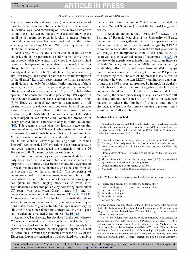
To establish the height of the body (item D1-32), one of
several methods may be used. With a continuous set of CT data,
virtual sections at arbitrary angles through the volume can be
calculated; then the distance between two points, such as both
ends of a bone, within such a plane can be measured. If the body
can be laid outstretched on the examination table, its full length
can even be determined from the CT data in one single
measurement. If this is not possible due to contractures, e.g. in
burnt victims, the body height can still be determined by adding
the measured lengths of a number of skeleton segments. Finally,
if the body is not complete, one of several anthropological
methods can be applied [25–28]. These are expressed as
formulas which have the lengths of several of the long bones of
the extremities as parameters [29,30]. Fig. 1 shows measure-
ments of a victim’s humerus, radius, femur, and tibia in
reformatted CT images.
The weight of a body (item D1-33) is best established by
weighing the body, but since body volume, which can be
measured from the data of a full body CT scan, is correlated
with body weight (Verma et al. [31], who measured body
volume with a water displacement technique, found the
correlation coefficient to be r = 0.9966 in a sample of soldiers),
it is possible to at least estimate the body weight using CT
data—this may prove helpful for control if doubt arises later
concerning the documented weight or if weighing has been
missed altogether.
In items Dl-34–D3-54, the forensic pathologist is asked for
physical descriptions of various parts of the body based on
external inspection. Among these, about 60% can, at least
ted tomography in disaster victim identification—Advantages and
nt.2006.08.004
M. Sidler et al. / Forensic Science International xxx (2006) xxx–xxx4
+ Models
FSI-5002; No of Pages 11
Fig. 1. Distance measurements within reformatted images that have been chosen to contain both end points of a selected bone; these measures are used for stature
estimation in a decomposed corpse. (a) Humerus, (b) radius, (c) femur and (d) tibia.
partially, be described using three-dimensional reconstructions
of soft tissues. A relatively small number of reconstructed
images can be used for many different items.
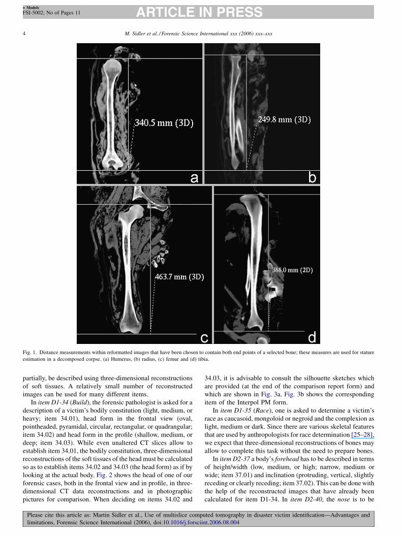
In item D1-34 (Build), the forensic pathologist is asked for a
description of a victim’s bodily constitution (light, medium, or
heavy; item 34.01), head form in the frontal view (oval,
pointheaded, pyramidal, circular, rectangular, or quadrangular;
item 34.02) and head form in the profile (shallow, medium, or
deep; item 34.03). While even unaltered CT slices allow to
establish item 34.01, the bodily constitution, three-dimensional
reconstructions of the soft tissues of the head must be calculated
so as to establish items 34.02 and 34.03 (the head form) as if by
looking at the actual body. Fig. 2 shows the head of one of our
forensic cases, both in the frontal view and in profile, in three-
dimensional CT data reconstructions and in photographic
pictures for comparison. When deciding on items 34.02 and
Please cite this article as: Martin Sidler et al., Use of multislice comp
limitations, Forensic Science International (2006), doi:10.1016/j.forscii
34.03, it is advisable to consult the silhouette sketches which
are provided (at the end of the comparison report form) and
which are shown in Fig. 3a. Fig. 3b shows the corresponding
item of the Interpol PM form.
In item D1-35 (Race), one is asked to determine a victim’s
race as caucasoid, mongoloid or negroid and the complexion as
light, medium or dark. Since there are various skeletal features
that are used by anthropologists for race determination [25–28],
we expect that three-dimensional reconstructions of bones may
allow to complete this task without the need to prepare bones.
In item D2-37 a body’s forehead has to be described in terms
of height/width (low, medium, or high; narrow, medium or
wide; item 37.01) and inclination (protruding, vertical, slightly
receding or clearly receding; item 37.02). This can be done with
the help of the reconstructed images that have already been
calculated for item D1-34. In item D2-40, the nose is to be
uted tomography in disaster victim identification—Advantages and
nt.2006.08.004
M. Sidler et al. / Forensic Science International xxx (2006) xxx–xxx 5
+ Models
FSI-5002; No of Pages 11
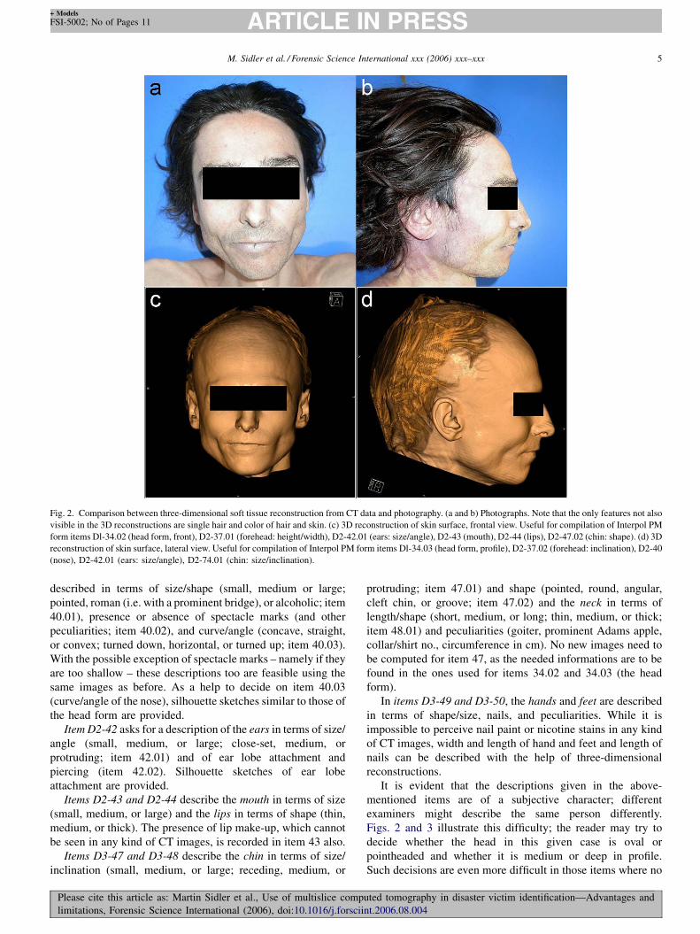
Fig. 2. Comparison between three-dimensional soft tissue reconstruction from CT data and photography. (a and b) Photographs. Note that the only features not also
visible in the 3D reconstructions are single hair and color of hair and skin. (c) 3D reconstruction of skin surface, frontal view. Useful for compilation of Interpol PM
form items Dl-34.02 (head form, front), D2-37.01 (forehead: height/width), D2-42.01 (ears: size/angle), D2-43 (mouth), D2-44 (lips), D2-47.02 (chin: shape). (d) 3D
reconstruction of skin surface, lateral view. Useful for compilation of Interpol PM form items Dl-34.03 (head form, profile), D2-37.02 (forehead: inclination), D2-40
(nose), D2-42.01 (ears: size/angle), D2-74.01 (chin: size/inclination).
described in terms of size/shape (small, medium or large;
pointed, roman (i.e. with a prominent bridge), or alcoholic; item
40.01), presence or absence of spectacle marks (and other
peculiarities; item 40.02), and curve/angle (concave, straight,
or convex; turned down, horizontal, or turned up; item 40.03).
With the possible exception of spectacle marks – namely if they
are too shallow – these descriptions too are feasible using the
same images as before. As a help to decide on item 40.03
(curve/angle of the nose), silhouette sketches similar to those of
the head form are provided.
Item D2-42 asks for a description of the ears in terms of size/
angle (small, medium, or large; close-set, medium, or
protruding; item 42.01) and of ear lobe attachment and
piercing (item 42.02). Silhouette sketches of ear lobe
attachment are provided.
Items D2-43 and D2-44 describe the mouth in terms of size
(small, medium, or large) and the lips in terms of shape (thin,
medium, or thick). The presence of lip make-up, which cannot
be seen in any kind of CT images, is recorded in item 43 also.
Items D3-47 and D3-48 describe the chin in terms of size/
inclination (small, medium, or large; receding, medium, or
Please cite this article as: Martin Sidler et al., Use of multislice compu
limitations, Forensic Science International (2006), doi:10.1016/j.forscii
protruding; item 47.01) and shape (pointed, round, angular,
cleft chin, or groove; item 47.02) and the neck in terms of
length/shape (short, medium, or long; thin, medium, or thick;
item 48.01) and peculiarities (goiter, prominent Adams apple,
collar/shirt no., circumference in cm). No new images need to
be computed for item 47, as the needed informations are to be
found in the ones used for items 34.02 and 34.03 (the head
form).
In items D3-49 and D3-50, the hands and feet are described
in terms of shape/size, nails, and peculiarities. While it is
impossible to perceive nail paint or nicotine stains in any kind
of CT images, width and length of hand and feet and length of
nails can be described with the help of three-dimensional
reconstructions.
It is evident that the descriptions given in the above-
mentioned items are of a subjective character; different
examiners might describe the same person differently.
Figs. 2 and 3 illustrate this difficulty; the reader may try to
decide whether the head in this given case is oval or
pointheaded and whether it is medium or deep in profile.
Such decisions are even more difficult in those items where no
ted tomography in disaster victim identification—Advantages and
nt.2006.08.004

M. Sidler et al. / Forensic Science International xxx (2006) xxx–xxx6
+ Models
FSI-5002; No of Pages 11
Fig. 3. Excerpt from the Interpol Comparison Report and PM form: (a) Silhouette sketch provided at the end of the comparison report. Use this to decide on an
individual’s head form. The reader may try to classify the individual in Fig. 2c and d and (b) item Dl-34 (build) of the Interpol PM form.
sketch is provided, such as nose or ear size. There is always the
risk that an examiner thinks those noses and ears to be of
medium size that are similar to his own, which can make
matching AM and PM forms difficult, as they are virtually
never compiled by the same person. It must be noted, though,
that these difficulties are the same whether one inspects an
actual dead body or three-dimensional CT reconstructions. The
advantage of the CT approach lies in documentation. Whereas
photographic documentation limits the views of a deceased to a
number of angles, from a CT data set reconstructed views from
different angles can be calculated in an infinite number, at any
time. If, e.g. no picture has been taken showing the ears’ angle,
what the examiner wrote down in item D2-42.01 can still be
controlled later if CT data of the head are available. It may also
be seen as an advantage that reconstructed images do not have
the same emotional impact on viewers as photographs.
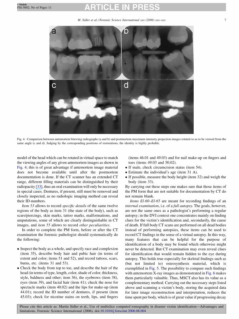
Using item D2-45, the forensic pathologist has to roughly
describe a victim’s teeth in terms of condition (natural,
untreated, treated, crowns, bridges, or implants; item 45.01),
gaps and missing teeth (item 45.02), and dentures (part, upper,
part, lower, full upper, full lower, ID-number; item 45.03). A
much more thorough listing of dental findings will be asked for
in section F of the form, which is to be filled in by a forensically
Please cite this article as: Martin Sidler et al., Use of multislice comp
limitations, Forensic Science International (2006), doi:10.1016/j.forscii
trained odontologist. Both tasks can be accomplished by
interpreting a so-called dental CT, i.e. a panoramic jaw
overview calculated from transversal CT slices [32], or a
maximum intensity projection (MIP) image of the cranial CT
data [18]. Since these visualization techniques allow a detailed
description of a victim’s dental status, we expect jaw resection
not necessary in most cases, and thus the brittle teeth of burned
victims, e.g. are not in danger of being destroyed further. The
images (or, for that matter, the cranial CT data they are derived
from), being of digital nature, can be sent to forensic
odontologists electronically; ideally, a majority of the involved
forensic odontologists would be able to do their valuable work
in their offices while only a small number would be needed at
the mortuary near the actual disaster site for those exceptional
cases where artifacts due to metal used in dental restorations
reduce the value of the CT images – a problem which will
become less relevant in the future as amalgamic restorations
become fewer nowadays – and for extracting teeth to be used
for age estimation (discussed later). Both visualization
techniques are also useful for comparison with antemortem
radiologic images. While the postmortem dental CT can be
compared to antemortem classic orthopantomographies (OPGs)
or bitewing radiographs, MIP provides a three-dimensional
uted tomography in disaster victim identification—Advantages and
nt.2006.08.004
M. Sidler et al. / Forensic Science International xxx (2006) xxx–xxx 7
+ Models
FSI-5002; No of Pages 11
Fig. 4. Comparison between antemortem bitewing radiographs (a and b) and postmortem maximum intensity projection images rotated so as to be viewed from the
same angle (c and d). Judging by the corresponding positions of restorations, the identity is highly probable.
model of the head which can be rotated in virtual space to match
the viewing angles of any given antemortem images as shown in
Fig. 4; this is of great advantage if antemortem image material
does not become available until after the postmortem
documentation is done. If the CT scanner has an extended CT
range, different filling materials can be distinguished by their
radiopacity [33], thus an oral examination will only be necessary
in special cases. Dentures, if present, still must be removed and
closely inspected, as no radiologic imaging method can reveal
their ID-numbers.
Item 53 allows to record specific details of the same twelve
regions of the body as item 31 (the state of the body), such as
scars/piercings, skin marks, tattoo marks, malformations, and
amputations, some of which are clearly distinguishable in CT
images, and item 55 allows to record other peculiarities.
In order to complete the PM form, before or after the CT
examination the forensic pathologist should systematically do
the following:
� I
nspect the body as a whole, and specify race and complexion(item 35), describe body hair and pubic hair (in terms of
extent and color; items 51 and 52), and record tattoos, scars,
burns, etc. (items 31 and 53).
� C
heck the body from top to toe, and describe the hair of thehead (in terms of type, length, color, shade of color, thickness,
style, baldness and other; item 36), the eyebrows (item 38),
eyes (item 39), and facial hair (item 41); check the nose for
spectacle marks (item 40.02) and the lips for make-up (item
44.01); record the ID number of dentures, if present (item
45.03); check for nicotine stains on teeth, lips, and fingers
Please cite this article as: Martin Sidler et al., Use of multislice compute
limitations, Forensic Science International (2006), doi:10.1016/j.forsciint.
(items 46.01 and 49.03) and for nail make-up on fingers and
toes (items 49.03 and 50.02).
� I
f male, check circumcision status (item 54).� E
stimate the individual’s age (item 31 A).� I
f possible, measure the body height (item 32) and weigh thebody (item 33).
By carrying out these steps one makes sure that those items of
the PM form that are not suitable for documentation by CT do
not remain blank.
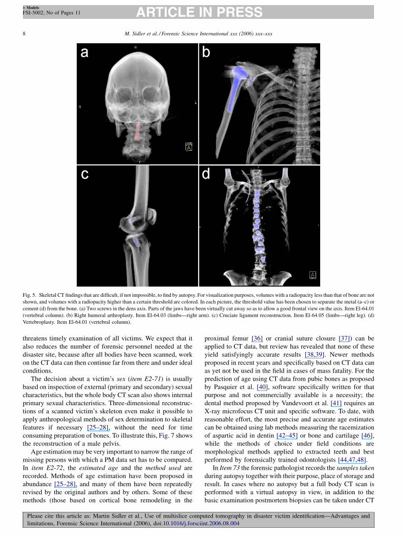
Items El-60–El-65 are meant for recording findings of an
internal examination, i.e. of a full autopsy. The goals, however,
are not the same ones as a pathologist’s performing a regular
autopsy; in the DVI context one concentrates mainly on finding
clues for the victim’s identification and, secondarily, the cause
of death. If full body CT scans are performed on all dead bodies
instead of performing autopsies, these items can be used to
record CT findings in the sense of a virtual autopsy. In this way,
many features that can be helpful for the purpose of
identification of a body may be found which otherwise might
never be detected. But CT examination may even reveal clues
for identification that would remain hidden to the eye during
autopsy. This holds true especially for skeletal findings such as
(but not limited to) osteosynthesis material, which is
exemplified in Fig. 5. The possibility to compare such findings
with antemortem X-ray images as demonstrated in Fig. 6 makes
them particularly valuable. Thus, MSCT also has its value as a
complementary method. Carrying out the necessary steps listed
above and scanning a victim’s body, storing the acquired data
for later image reconstruction and interpretation, reduces the
time spent per body, which is of great value if progressing decay
d tomography in disaster victim identification—Advantages and
2006.08.004
M. Sidler et al. / Forensic Science International xxx (2006) xxx–xxx8
+ Models
FSI-5002; No of Pages 11
Fig. 5. Skeletal CT findings that are difficult, if not impossible, to find by autopsy. For visualization purposes, volumes with a radiopacity less than that of bone are not
shown, and volumes with a radiopacity higher than a certain threshold are colored. In each picture, the threshold value has been chosen to separate the metal (a–c) or
cement (d) from the bone. (a) Two screws in the dens axis. Parts of the jaws have been virtually cut away so as to allow a good frontal view on the axis. Item El-64.01
(vertebral column). (b) Right humeral arthroplasty. Item El-64.03 (limbs—right arm). (c) Cruciate ligament reconstruction. Item El-64.05 (limbs—right leg). (d)
Vertebroplasty. Item El-64.01 (vertebral column).
threatens timely examination of all victims. We expect that it
also reduces the number of forensic personnel needed at the
disaster site, because after all bodies have been scanned, work
on the CT data can then continue far from there and under ideal
conditions.
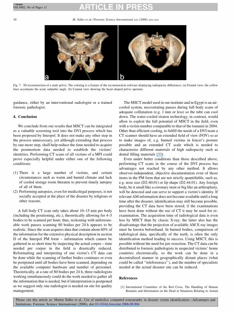
The decision about a victim’s sex (item E2-71) is usually
based on inspection of external (primary and secondary) sexual
characteristics, but the whole body CT scan also shows internal
primary sexual characteristics. Three-dimensional reconstruc-
tions of a scanned victim’s skeleton even make it possible to
apply anthropological methods of sex determination to skeletal
features if necessary [25–28], without the need for time
consuming preparation of bones. To illustrate this, Fig. 7 shows
the reconstruction of a male pelvis.
Age estimation may be very important to narrow the range of
missing persons with which a PM data set has to be compared.
In item E2-72, the estimated age and the method used are
recorded. Methods of age estimation have been proposed in
abundance [25–28], and many of them have been repeatedly
revised by the original authors and by others. Some of these
methods (those based on cortical bone remodeling in the
Please cite this article as: Martin Sidler et al., Use of multislice comp
limitations, Forensic Science International (2006), doi:10.1016/j.forscii
proximal femur [36] or cranial suture closure [37]) can be
applied to CT data, but review has revealed that none of these
yield satisfyingly accurate results [38,39]. Newer methods
proposed in recent years and specifically based on CT data can
as yet not be used in the field in cases of mass fatality. For the
prediction of age using CT data from pubic bones as proposed
by Pasquier et al. [40], software specifically written for that
purpose and not commercially available is a necessity; the
dental method proposed by Vandevoort et al. [41] requires an
X-ray microfocus CT unit and specific software. To date, with
reasonable effort, the most precise and accurate age estimates
can be obtained using lab methods measuring the racemization
of aspartic acid in dentin [42–45] or bone and cartilage [46],
while the methods of choice under field conditions are
morphological methods applied to extracted teeth and best
performed by forensically trained odontologists [44,47,48].
In Item 73 the forensic pathologist records the samples taken
during autopsy together with their purpose, place of storage and
result. In cases where no autopsy but a full body CT scan is
performed with a virtual autopsy in view, in addition to the
basic examination postmortem biopsies can be taken under CT
uted tomography in disaster victim identification—Advantages and
nt.2006.08.004

M. Sidler et al. / Forensic Science International xxx (2006) xxx–xxx 9
+ Models
FSI-5002; No of Pages 11
Please cite this article as: Martin Sidler et al., Use of multislice computed tomography in disaster victim identification—Advantages and
limitations, Forensic Science International (2006), doi:10.1016/j.forsciint.2006.08.004
Fig. 6. Comparison between antemortem X-ray images (a and d) and corresponding postmortem reformatted CT images (b, c and e). (a) Antemortem roentgenogram
showing osteosynthesis of right tibia by medullary nailing. (b) Postmortem reformatted CT image showing the canal of the more proximal of the two screws in a) after
removal of the osteosynthesis material. (c) Postmortem reformatted CT image showing the canals of the more distal screw and the marrow nail itself. Note also the
dislocation of the fibula ad latus and ad axim as in a). (d) Antemortem roentgenogram showing intramedullary minimal osteosynthesis of left humerus my means of a
helical wire [34,35]. (e) Postmortem reformatted CT image of a severely burnt body showing the same helix wire as in (d); due to the different configuration of the
shoulder joint, only the humerus is seen from the same angle. The finding of this intramedullary helix wire was crucial for the identification in this particular case. For
an explanation of the visualization technique see Fig. 5.
M. Sidler et al. / Forensic Science International xxx (2006) xxx–xxx10
+ Models
FSI-5002; No of Pages 11
Fig. 7. 3D reconstruction of a male pelvis. The coloring is a feature of the reconstruction software displaying radiopacity differences. (a) Frontal view; the yellow
lines accentuate the acute subpubic angle. (b) Cranial view showing the heart-shaped pelvic aperture.
guidance, either by an interventional radiologist or a trained
forensic pathologist.
4. Conclusion
We conclude from our results that MSCT can be integrated
as a valuable screening tool into the DVI process which has
been proposed by Interpol. It does not make any other step in
the process unnecessary, yet although extending that process
by one more step, shall help reduce the time needed to acquire
the postmortem data needed to establish the victims’
identities. Performing CT scans of all victims of a MFI could
prove especially helpful under either one of the following
conditions:
(1) T
Pl
lim
here is a large number of victims, and certain
circumstances such as warm and humid climate and lack
of cooled storage room threaten to prevent timely autopsy
of all of them.
(2) P
erforming autopsies, even for medicolegal purposes, is notsocially accepted at the place of the disaster by religious or
other reasons.
A full body CT scan only takes about 10–15 min per body
(including the positioning, etc.), theoretically allowing for 4–5
bodies to be scanned per hour; thus, reckoning with unforesee-
able work pauses scanning 80 bodies per 24 h appears to be
realistic. Since the scan acquires data that contain about 60% of
the information for the extensive physical description in section
D of the Interpol PM form – information which cannot be
gathered in so short time by inspecting the actual corpse – time
needed per corpse in the field is drastically reduced.
Reformatting and interpreting of one victim’s CT data can
be done while the scanning of further bodies continues or even
be postponed until all bodies have been scanned, depending on
the available computer hardware and number of personnel.
Theoretically, at a rate of 80 bodies per 24 h, three radiologists
working simultaneously could do the work needed to gather all
the information that is needed, but if interpretation is postponed
as we suggest only one radiologist is needed on site for quality
management.
ease cite this article as: Martin Sidler et al., Use of multislice comp
itations, Forensic Science International (2006), doi:10.1016/j.forscii
The MSCT model used in our institute and in Egypt is an air-
cooled system, necessitating pauses during full body scans of
adequate collimation (e.g. 1 mm or less) so the tube can cool
down. The water-cooled straton technology, in contrast, would
allow to exploit the full potential of MSCT in the field, even
with a victim number comparable to that of the tsunami in 2004.
Other than efficient cooling, to fulfill the needs of a DVI team a
CT scanner should have an extended field of view (FOV) so as
to make images of, e.g. burned victims in fencer’s posture
possible and an extended CT scale which is needed to
characterize different materials of high radiopacity such as
dental filling materials [33].
Even under better conditions than those described above,
performing CT scans in the course of the DVI process has
advantages not reached by any other method. It allows
observer-independent, objective documentation even of those
items in the PM form that are not strictly quantifiable, such as,
e.g. nose size (D2-40.01) or lip shape (D2-44.01). Any foreign
body, be it small like a coronary stent or big like an arthroplasty,
will be detected and can serve to support a victim’s identity. If
relevant AM information does not become available until a long
time after the disaster, identification may still become possible,
providing the CT data have been stored; if the examinations
have been done without the use of CT it may be used for re-
examination. The acquisition time of radiological data is even
less by MSCT than by classic X-ray; the latter also has the
disadvantage that the projection of available AM X-ray images
must be known beforehand. In burned bodies, comparison of
radiological data, specifically of the teeth, is often the only
identification method leading to success. Using MSCT, this is
possible without the need for jaw resection. The CT data can be
distributed to forensic pathologists in suspected victims’ home
countries electronically, so the work can be done in a
decentralized manner in geographically distant places (what
could be called ‘‘teleforensics’’), and the number of specialists
needed at the actual disaster site can be reduced.
References
[1] International Committee of the Red Cross, The Handling of Human
Remains and Information on the Dead in Situations Relating to Armed
uted tomography in disaster victim identification—Advantages and
nt.2006.08.004

M. Sidler et al. / Forensic Science International xxx (2006) xxx–xxx 11
+ Models
FSI-5002; No of Pages 11
Conflicts or Internal Violence and Involving Missing Persons http://
www.icrc.org/Web/eng/siteeng0.nsf/htmlall/5ZMJH5/$File/Inter-
pol_2004DVI_EN.pdf.
[2] International Criminal Police Organization, Disaster Victim Identification
Guide (1997) http://www.interpol.int/Public/DisasterVictim/guide/defaul-
t.asp.
[3] Pan American Health Organization, Management of Dead Bodies in
Disaster Situations, Washington, DC, 2004, pp. 139–140 http://www.pa-
ho.org/english/dd/ped/DeadBodiesBook.pdf.
[4] http://www.interpol.int/Public/DisasterVictim/Default.asp.
[5] V.O. McCarty, A.P. Sohn, R.S. Ritzlin, J.H. Gauthier, Scene investigation,
identification, and victim examination following the accident of Galaxy
203: disaster preplanning does work, J. Forensic Sci. 32 (1987) 983–987.
[6] J. Timperman, How some medicolegal aspects of the Zeebrugge Ferry
disaster apply to the investigation of mass disasters, Am. J. Forensic Med.
Pathol. 12 (1991) 286–290.
[7] J.M. Hutt, B. Ludes, B. Kaess, A. Tracqui, P. Mangin, Odontological
identification of the victims of flight AI.IT 5148 air disaster Lyon-
Strasbourg 20.01.1992, Int. J. Legal Med. 107 (1995) 275–279.
[8] H. Soomer, H. Ranta, A. Penttila, Identification of victims from the M/S
Estonia, Int. J. Legal Med. 114 (2001) 259–262.
[9] H.J. Meyer, The Kaprun cable car fire disaster—aspects of forensic
organisation following a mass fatality with 155 victims, Forensic Sci.
Int. 138 (2003) 1–7.
[10] P. Lunetta, H. Ranta, C. Cattaneo, A. Piccinini, R. Niskanen, A. Sajantila,
et al., International collaboration in mass disasters involving foreign
nationals within the EU: medico-legal investigation of Finnish victims
of the Milan Linate airport SAS SK 686 aircraft accident on 8 October
2001, Int. J. Legal Med. 117 (2003) 204–210.
[11] B.G. Brogdon, Forensic Radiology, CRC Press LLC, Boca Raton, Florida,
1998.
[12] M. Pfaffli, Postmortale Radiologische CT-Identifikation basierend auf
pramortalen klassischen Rontgenuntersuchungen, University of Bern,
Bern, Switzerland, 2003.
[13] W.D. Haglund, C.L. Fligner, Confirmation of human identification using
computerized tomography (CT), J. Forensic Sci. 38 (1993) 708–712.
[14] K.J. Reichs, Quantified comparison of frontal sinus patterns by means of
computed tomography, Forensic Sci. Int. 61 (1993) 141–168.
[15] D.R. Smith, K.G. Limbird, J.M. Hoffman, Identification of human skeletal
remains by comparison of bony details of the cranium using computerized
tomographic (CT) scans, J. Forensic Sci. 47 (2002) 937–939.
[16] T. Riepert, D. Ulmcke, U. Jendrysiak, C. Rittner, Computer-assisted
simulation of conventional roentgenograms from three-dimensional com-
puted tomography (CT) data–—an aid in the identification of unknown
corpses (FoXSIS), Forensic Sci. Int. 71 (1995) 199–204.
[17] T. Riepert, D. Ulmcke, F. Schweden, B. Nafe, Identification of unknown
dead bodies by X-ray image comparison of the skull using the X-ray
simulation program FoXSIS, Forensic Sci. Int. 117 (2001) 89–98.
[18] C. Jackowski, E. Aghayev, M. Sonnenschein, R. Dirnhofer, M. Thali,
Maximum Intensity Projection (MIP) of cranial computed tomography
(CCT) data for dental identification, Int. J. Legal Med. (2005) epub ahead
of print.
[19] http://www.medical.siemens.com.
[20] http://www.nationalgeographic.com.
[21] M. Thali, K. Yen, W. Schweitzer, P. Vock, Ch. Boesch, Ch. Ozdoba, et al.,
Virtopsy, a new imaging horizon in Forensic Pathology: virtual autopsy by
postmortem multislice computed tomography (MSCT) and magnetic
resonance imaging (MRI)—a feasibility study, J. Forensic Sci. 48
(2003) 386–403.
[22] http://www.virtopsy.com.
[23] M. Thali, K. Yen, W. Schweitzer, P. Vock, C. Ozdoba, R. Dirnhofer, Into
the deomposed body—forensic digital autopsy using multislice-computed
tomography, Forensic Sci. Int. 134 (2003) 109–114.
[24] M. Thali, K. Yen, T. Plattner, W. Schweitzer, P. Vock, Ch. Ozdoba, et al.,
Charred body: virtual autopsy with multi-slice computed tomography and
magnetic resonance imaging, J. Forensic Sci. 47 (2002) 1326–1331.
[25] W.M. Bass, Human Osteology: A Laboratory and Field Manual, fourth
ed., Missouri Archaeological Society, Columbia, 1995.
Please cite this article as: Martin Sidler et al., Use of multislice compu
limitations, Forensic Science International (2006), doi:10.1016/j.forscii
[26] W.D. Haglund, M.H. Sorg, Forensic Taphonomy: The Postmortem Fate of
Human Remains, CRC-Press, Boca Raton, Florida, 1996.
[27] K.J. Reichs, W.M. Bass, Forensic Osteology: Advances in the Identifica-
tion of Human Remains, second ed., Charles C. Thomas, Publisher Ltd.,
Springfield, Illinois, 1998.
[28] D.H. Ubelaker, Human Skeletal Remains: Excavation, Analysis,
Interpretation, third ed., Taraxacum, Washington, DC, 1999.
[29] G. Mall, M. Hubig, A. Buttner, J. Kuznik, R. Penning, M. Graw, Sex
determination and estimation of stature from the long bones of the arm,
Forensic Sci. Int. 117 (2001) 23–30.
[30] M.C. De Mendonca, Estimation of height from the length of long bones in
a Portuguese adult population, Am. J. Phys. Anthropol. 112 (2000) 39–48.
[31] S.S. Verma, H. Bharadwaj, T. Zachariah, S. Kishnani, M.R. Bhatia,
Prediction of body volume by a stepwise linear regression technique,
Eur. J. Appl. Physiol. Occup. Physiol. 52 (1983) 126–130.
[32] M. Thali, T. Markwalder, C. Jackowski, M. Sonnenschein, R. Dirnhofer,
Dental CT Imaging as a screening tool for dental profiling: advantages and
limitations, J. Forensic Sci. 51 (2006) 113–116.
[33] C. Jackowski, A. Lussi, M. Classens, T. Kilchoer, S. Bolliger, E.
Aghayev, et al., Extended CT scale overcomes restoration caused streak
artifacts for dental identification in CT–3D color encoded automatic
discrimination of dental restorations, J. Comput. Assist. Tomogr. 30
(2006) 510–513.
[34] H. Traxler, R. Surd, K.A. Laminger, A. Windisch, M.C. Sora, W. Firbas,
The treatment of subcapital humerus fracture with dynamic helix wire and
the risk of concommitant lesion of the axillary nerve, Clin. Anat. 14 (2001)
418–423.
[35] K. Laminger, Helical wire. US patent 6,174,312 (2001).
[36] M.F. Ericksen, Aging changes in thickness of the proximal femoral cortex,
Am. J. Phys. Anthropol. 59 (1982) 121–130.
[37] R.S. Meindl, C.O. Lovejoy, Ectocranial suture closure: a revised method
for the determination of skeletal age at death based on the lateral–anterior
sutures, Am. J. Phys. Anthropol. 68 (1985) 57–66.
[38] K.D. Gehring, H.T. Haffner, D. Weber, M. Graw, Investigations on the
reliability of determining an individual’s age from the proximal femur,
Homo 52 (2002) 214–220.
[39] I. Hershkovitz, B. Latimer, O. Dutour, L.M. Jellema, S. Wish-Baratz, C.
Rothschild, et al., Why do we fail in aging the skull from the sagittal
suture? Am. J. Phys. Anthropol. 103 (1997) 393–399.
[40] E. Pasquier, L. De Saint Martin Pernot, V. Burdin, C. Mounayer, C. Le
Rest, D. Colin, et al., Determination of age at death: assessment of an
algorithm of age prediction using numerical three-dimensional CT data
from pubic bones, Am. J. Phys. Anthropol. 108 (1999) 261–268.
[41] F.M. Vandevoort, L. Bergmans, J. Van Cleynenbreugel, D.J. Bielen, P.
Lambrechts, M. Wevers, et al., Age calculation using X-ray microfocus
computed tomographical scanning of teeth: a pilot study, J. Forensic Sci.
49 (2004) 787–790.
[42] T. Ogino, H. Ogino, B. Nagy, Application of aspartic acid racemization to
forensic odontology: postmortem designation of age at death, Forensic
Sci. Int. 29 (1985) 259–267.
[43] S. Ohtani, K. Yamamoto, Age estimation using the racemization of amino
acid in human dentin, J. Forensic Sci. 36 (1991) 792–800.
[44] H. Soomer, H. Ranta, M.J. Lincoln, A. Penttila, E. Leibur, Reliability and
validity of eight dental age estimation methods for adults, J. Forensic Sci.
48 (2003) 149–152.
[45] S. Ritz-Timme, C. Cattaneo, M.J. Collins, E.R. Waite, H.W. Schutz, H.J.
Kaatsch, et al., Age estimation: the state of the art in relation to the specific
demands of forensic practise, Int. J. Legal Med. 113 (2000) 129–136.
[46] S. Ohtani, Y. Matsushima, Y. Kobayashi, T. Yamamoto, Age estimation by
measuring the racemization of aspartic acid from total amino acid content
of several types of bone and rib cartilage: a preliminary account, J.
Forensic Sci. 47 (2002) 32–36.
[47] H. Lamendin, E. Baccino, J.F. Humbert, J.C. Tavernier, R.M. Nossintch-
ouk, A. Zerilli, A simple technique for age estimation in adult corpses: the
two criteria dental method, J. Forensic Sci. 37 (1992) 1373–1379.
[48] E. Baccino, D.H. Ubelaker, L.A. Hayek, A. Zerilli, Evaluation of seven
methods of estimating age at death from mature human skeletal remains, J.
Forensic Sci. 44 (1999) 931–936.
ted tomography in disaster victim identification—Advantages and
nt.2006.08.004
Related Documents


