Yonsei Med J 49(5):819 - 827, 2008 DOI 10.3349/ymj.2008.49.5.819 Yonsei Med J Vol. 49, No. 5, 2008 Purpose: Human embryonic stem cells (hESCs) can proliferate for a prolonged period and differentiate into cardiomyocytes in vitro. Recent studies used bone morphogenetic protein 2 (BMP2) to generate cardiomyocytes from hESCs, however, all those studies used early embryoid bodies (EBs) and did not retrieve cardiomyocytes with a high yield. In this study, we treated long-term cultured EBs with BMP2 in order to promote differentiation into cardiomyocytes from hESCs. Materials and Methods: hESC lines, including SNUhES3 and SNUhES4, were used in this study. Undifferentiated hESC colonies were detached to form EBs and cultured for up to 30 days. These long-term cultured EBs were differentiated into cardiomyocytes in serum-containing media. In our protocol, BMP2 was applied for 5 days after attachment of EBs. Cardiac specific markers, beating of differentiated cells and electron microscopic (EM) ultrastructures were evaluated and analyzed. Results: Compared to 10-day or 20-day EBs, 30-day EBs showed a higher expression level of cardiac specific markers, Nkx2.5 and -myosin heavy chain ( MHC). Treatment of BMP2 α α increased expression of cardiac troponin (cTn) I and -actinin α when evaluated at 20 days after attachment of 30-day EBs. Beating of differentiated cells was observed from 7 to 20 days after attachment. Moreover, EM findings demonstrated fine structures such as Z bands in these differentiated cardio- myocytes. These long-term cultured EBs yielded cardio- myocytes with an efficiency of as high as 73.6% when assessed by FACS. Conclusion: We demonstrated that the use of long-term cultured EBs may enhance differentiation into cardiomyocytes from hESCs when treated with BMP2. Key Words: Bone morphogenetic protein 2, cardiomyocytes, cell differentiation, embryoid bodies, embryonic stem cells, long-term INTRODUCTION Human embryonic stem cells, derived from inner cell mass (ICM) of preimplantation embryos, can proliferate for a prolonged period in vitro 1,2 and can differentiate into various cell types under suitable environment. Therefore, hESCs are con- sidered as a candidate cell source of cell-based therapies for heart diseases. Since 2001, there have been numerous studies of hESC-derived cardio- myocytes, 3-10 and these studies used spontaneous differentiation, 3,4,6,7,10 low-serum culture condition 8,9 and co-culture with endoderm-like cells. 5 However, practical methods using specific signaling mole- cules that are known to be efficient in differentia- tion into cardiomyocytes from hESCs are still limited. Development of uncommitted mesodermal pre- cardiac cells to early cardiomyocytes is regulated by stimulatory signals that are secreted from anterior primitive endoderm. 11 Bone morphoge- netic proteins (BMPs) signaling is main signaling pathway regulating the cardiomyogenesis. Among BMPs, BMP2 is known to play a crucial role in the induction of heart formation of vertebrate embryos. 12-14 In hESCs, BMP2 is known to be as an inducer for mesoderm or cardiac differentiation. Tomescot et al. showed that BMP2 treatment turned on Use of Long-term Cultured Embryoid Bodies May Enhance Cardiomyocyte Differentiation by BMP2 Yoon Young Kim, 1 Seung-Yup Ku, 1,2 Jiho Jang, 1 Sun Kyung Oh, 1,2 Hee Sun Kim, 1,2 Seok Hyun Kim, 1,2 Young Min Choi, 1,2 and Shin Yong Moon 1,2 1 Institute of Reproductive Medicine and Population, Medical Research Center, Seoul; 2 Department of Obstetrics and Gynecology, Seoul National University College of Medicine, Seoul, Korea. Received August 30, 2007 Accepted April 30, 2008 This research was supported by a grant (SC1150 and SC1120) from the Stem Cell Research Center of the 21st Century Frontier Research Program funded by the Ministry of Science and Technology, Republic of Korea. Reprint address: requests to Dr. Young Min Choi, Department of Obstetrics and Gynecology, Seoul National University College of Medicine, 28 Yeongeon-dong, Jongno-gu, Seoul 110-799, Korea. Tel: 82-2-2072-2385, Fax: 82-2-742-2028, E-mail: ymchoi@snu. ac.kr

Welcome message from author
This document is posted to help you gain knowledge. Please leave a comment to let me know what you think about it! Share it to your friends and learn new things together.
Transcript
Yonsei Med J 49(5):819 - 827, 2008
DOI 10.3349/ymj.2008.49.5.819
Yonsei Med J Vol. 49, No. 5, 2008
Purpose: Human embryonic stem cells (hESCs) can proliferatefor a prolonged period and differentiate into cardiomyocytesin vitro. Recent studies used bone morphogenetic protein 2(BMP2) to generate cardiomyocytes from hESCs, however, allthose studies used early embryoid bodies (EBs) and did notretrieve cardiomyocytes with a high yield. In this study, wetreated long-term cultured EBs with BMP2 in order to promotedifferentiation into cardiomyocytes from hESCs. Materials andMethods: hESC lines, including SNUhES3 and SNUhES4,were used in this study. Undifferentiated hESC colonies weredetached to form EBs and cultured for up to 30 days. Theselong-term cultured EBs were differentiated into cardiomyocytesin serum-containing media. In our protocol, BMP2 wasapplied for 5 days after attachment of EBs. Cardiac specificmarkers, beating of differentiated cells and electron microscopic(EM) ultrastructures were evaluated and analyzed. Results:Compared to 10-day or 20-day EBs, 30-day EBs showed ahigher expression level of cardiac specific markers, Nkx2.5and -myosin heavy chain ( MHC). Treatment of BMP2α αincreased expression of cardiac troponin (cTn) I and -actininαwhen evaluated at 20 days after attachment of 30-day EBs.Beating of differentiated cells was observed from 7 to 20 daysafter attachment. Moreover, EM findings demonstrated finestructures such as Z bands in these differentiated cardio-myocytes. These long-term cultured EBs yielded cardio-myocytes with an efficiency of as high as 73.6% whenassessed by FACS. Conclusion: We demonstrated that the useof long-term cultured EBs may enhance differentiation into
cardiomyocytes from hESCs when treated with BMP2.
Key Words: Bone morphogenetic protein 2, cardiomyocytes,cell differentiation, embryoid bodies, embryonic stem cells,long-term
INTRODUCTION
Human embryonic stem cells, derived from inner
cell mass (ICM) of preimplantation embryos, can
proliferate for a prolonged period in vitro1,2 and
can differentiate into various cell types under
suitable environment. Therefore, hESCs are con-
sidered as a candidate cell source of cell-based
therapies for heart diseases. Since 2001, there have
been numerous studies of hESC-derived cardio-
myocytes,3-10 and these studies used spontaneous
differentiation,3,4,6,7,10 low-serum culture condition8,9
and co-culture with endoderm-like cells.5 However,
practical methods using specific signaling mole-
cules that are known to be efficient in differentia-
tion into cardiomyocytes from hESCs are still
limited.
Development of uncommitted mesodermal pre-
cardiac cells to early cardiomyocytes is regulated
by stimulatory signals that are secreted from
anterior primitive endoderm.11 Bone morphoge-
netic proteins (BMPs) signaling is main signaling
pathway regulating the cardiomyogenesis. Among
BMPs, BMP2 is known to play a crucial role in the
induction of heart formation of vertebrate
embryos.12-14
In hESCs, BMP2 is known to be as an inducer
for mesoderm or cardiac differentiation. Tomescot
et al. showed that BMP2 treatment turned on
Use of Long-term Cultured Embryoid Bodies May EnhanceCardiomyocyte Differentiation by BMP2
Yoon Young Kim,1 Seung-Yup Ku,1,2 Jiho Jang,1 Sun Kyung Oh,1,2 Hee Sun Kim,1,2 Seok Hyun Kim,1,2
Young Min Choi,1,2 and Shin Yong Moon1,2
1Institute of Reproductive Medicine and Population, Medical Research Center, Seoul; 2Department of Obstetrics and Gynecology,
Seoul National University College of Medicine, Seoul, Korea.
Received August 30, 2007Accepted April 30, 2008
This research was supported by a grant (SC1150 and SC1120)from the Stem Cell Research Center of the 21st Century Frontier
Research Program funded by the Ministry of Science and
Technology, Republic of Korea.
Reprint address: requests to Dr. Young Min Choi, Department
of Obstetrics and Gynecology, Seoul National University College
of Medicine, 28 Yeongeon-dong, Jongno-gu, Seoul 110-799, Korea.Tel: 82-2-2072-2385, Fax: 82-2-742-2028, E-mail: ymchoi@snu.
ac.kr
Yoon Young Kim, et al.
Yonsei Med J Vol. 49, No. 5, 2008
expression of cardiac related genes in hESCs.15 In
addition, Pal et al. demonstrated that BMP2 with
low serum concentration could induce differen-
tiation into cardiomyocytes in many hESC lines.16
These studies used short-term cultured EBs for
differentiation into cardiomyocytes. However, the
yield of differentiation was reported to be around
30%.16
In this study, we used long-term cultured EBs
from hESC lines, SNUhES3 and SNUhES4, to
generate cardiomyocytes with BMP2 treatment in
order to enhance in vitro differentiation efficiency.
MATERIALS AND METHODS
Materials
Previously reported hESC lines, SNUhES32 and
SNUhES4,17 were used in this study. SNUhES3
and SNUhES4 have a normal karyotype (46, XY)
and express undifferentiated hESC-specific markers
such as alkaline phosphatase, Oct4, SSEA3, SSEA4,
Tra-1-60 and Tra-1-81.
Human embryonic stem cell culture
Human embryonic stem cells were cultured
according to previously described methods.18
Undifferentiated hESCs were maintained on
mitomycin C-(Sigma, St. Louis, MO, USA) treated
STO (ATCC, Manassas, VA, USA) feeder layer.
Undifferentiated hESC colonies were dissected
and replated onto a fresh feeder layer every 7
days. DMEM/F12 (Invitrogen, Carlsbad, CA,
USA), supplemented with 20% knockout serum
replacement (KO-SR; Invitrogen), 1% nonessential
amino acids (Invitrogen), 0.1 mM -mercaptoethaβ -
nol (Sigma), 0.4 ng/mL basic fibroblast growth
factor (bFGF; Invitrogen), 50 U/mL penicillin
(Invitrogen) and 50 g/mL streptomycin (Inviμ -
trogen), was used as hESC culture medium.
Embryoid body formation
To form EBs, undifferentiated hESC colonies
were cultured for 5 days. Day 5 hESC colonies
were incubated with 2 mg/mL collagenase type
IV (Invitrogen) for 30 minutes at 37°C to detach
colonies from dish bottom. Detached colonies
were transferred to bacterial dish and cultured for
10, 20, and 30 days in suspension. hESC culture
medium, excluding bFGF was used as EB medium.
Medium was changed every other day.
Differentiation into cardiomyocytes
After culture for 10, 20 and 30 days in suspen-
sion, EBs were transferred to gelatin pre-coated
tissue culture dishes. Knockout DMEM (Invitrogen),
supplemented with 20% fetal bovine serum (FBS;
HyClone, Logan, UT, USA), was used as a
differentiation medium for further differentiation,
and medium was changed every other day. Ten,
20 and 40 ng/mL BMP2 (R & D Systems, Minneapolis,
MN, USA) was treated for 5 days after plating to
promote differentiation.
Immunostaining
Cells were washed with phosphate-buffered
saline (PBS; Invitrogen) and fixed with 4%
paraformaldehyde (PFA; Sigma) for 15 minutes at
room temperature (RT). After washing with PBS,
cells were incubated overnight with 3% bovine
serum albumin (BSA; Sigma) solution, including
0.1% Triton X100 (Sigma), at 4°C to prevent non-
specific reaction. After washing with PBS, mouse
anti-human Nkx2.5 (R & D Systems), goat anti-
Nkx2.5 (Santa Cruz Biotechnology, Santa Cruz,
CA, USA), mouse anti-human cardiac Troponin I
(cTn I; Chemicon, Billerica, MA, USA), goat anti- -α
myosin heavy chain ( MHC; Santa Cruzα
Biotechnology), goat anti-human -actinin (Santaα
Cruz) and mouse anti-smooth muscle actin (SMA;
Chemicon) antibodies were added overnight at
4 C and then washed 3 times with PBST (PBS with˚
0.05% Tween 20). Each Alexa Fluor 488-labeled
donkey anti-mouse IgG, Alexa Fluor 488-labeled
donkey anti-goat IgG, Alexa Fluor 594-labeled
donkey anti-mouse IgG and Alexa Fluor 594-
labeled donkey anti-goat IgG (all from Molecular
Probes, Carlsbad, CA, USA) antibodies were applied
for 60 minutes at RT and washed three times with
PBST. 4'-6-diamidino-2-phenylindole (DAPI; Mole-
cular Probes) solution was added and incubated
for 30 minutes at RT for staining of nuclei. Stained
cells were analyzed using a confocal laser micro-
Cardiac Differentiation Using Long-term Cultured EB
Yonsei Med J Vol. 49, No. 5, 2008
scopy system (BioRad, Hercules, CA, USA).
Real-time quantitative PCR (Q-PCR)
Total RNA was isolated from EBs and
differentiated cells using RNeasy mini kit (Qiagen,
Valencia, CA, USA). cDNA was synthesized from
1 g of total RNA using Superscript II first-strandμ
synthesis system (Invitrogen). Quantitative PCR
was performed in Rotor-Gene 3000 (Corbett Life
Science, Sydney, Australia) using QuantiTect
SYBR green PCR kit (Qiagen). Primers used for
the reactions are listed in Table 1. CT was
calculated under default settings of Rotor-gene 6
program (Corbett Life Science). Relative gene
expression was normalized to GAPDH expression.
Transmission electron microscopy (TEM)
Cells were fixed with 2.5% glutaraldehyde
(Sigma) for 20 minutes at RT. Cells were then post-
fixed with 1% osmium tetroxide (OsO4; Sigma) in
0.04 M phosphate buffer for 10 minutes at 4°C,
supplemented with 0.14 M sucrose. Following
serial dehydration in ethanol and infiltration with
epoxy resin, cells were transferred to beam
capsules for polymerization. The capsules were
separated from the polymerized resin with a razor
blade. Embedded cells in hardened blocks were
observed under an optical microscope for ultra-
thin sectioning. Subsequently, ultra-thin sections
were obtained using an ultramicrotome (Sorvall
MT 6000). Cells were then observed under a
transmission electron microscope (JEM-1400; JEOL
Ltd.) at 50 kV.
FACS analysis
Cells were washed with PBS and treated with
0.25% trypsin-EDTA (Invitrogen) for 40 minutes at
37°C to dissociate them into single cells. After
washing with PBS, 3% BSA solution, including
0.05% Triton X100, was added for 20 minutes at
RT. Mouse anti-human Nkx2.5 (R & D Systems)
and goat anti- MHC (Santa Cruz Biotechnology)α
antibodies were applied for 60 minutes at RT and
then washed 3 times with PBST. Alexa Fluor
488-labeled donkey anti-goat IgG and Alexa Fluor
594-labeled donkey anti-mouse IgG (all from
Molecular Probes) were applied for 60 minutes at
RT and washed 3 times with PBST. Cells were
then analyzed using BD FACS Calibur SystemTM
(BD Sciences, San Jose, CA, USA).
RESULTS
Evaluation of pluripotent characteristics of
undifferentiated hESCs
Schematic process of differentiation into cardiac
cells is presented in Fig. 1. Briefly, EBs were
formed from hESCs at day 6, differentiated in
suspension for 30 days and attached for further
differentiation. From 7 days after attachment,
beating clusters started to appear and lasted for
as long as 30 days. We first examined the expres-
sions of alkaline phosphatase, Oct4, SSEA4, Tra-
1-60 and Tra-1-81 in undifferentiated hESCs. All
pluripotent hESC specific markers were highly
expressed in undifferentiated hESC colonies (Fig.
2). Expressions of cardiac specific markers were
Table 1. Primer Sequences and Conditions Used for RT-PCR and Real-time quantitative PCR
PCR GeneSequences
Forward Reverse
Real-time
quantitative
PCR
GAPDH GGCGTTCTCTTTGGAAAGGTGTTC GTACTCAGCGGCCAGCATCG
Brachyury TGCTTCCCTGAGACCCAGTT GATCACTTCTTTCCTTTGCATCAAG
Nkx2.5 TTGCGACGGGAGAGTTTGT GCCCGACGAGCTCAGTCCCAGTT
MHCɑ AGTGCTTCGTGCCCGATGAC TGCTGCAACACCTGTCCTC
RT, reverse transcription; PCR, polymerase chain reaction.
Yoon Young Kim, et al.
Yonsei Med J Vol. 49, No. 5, 2008
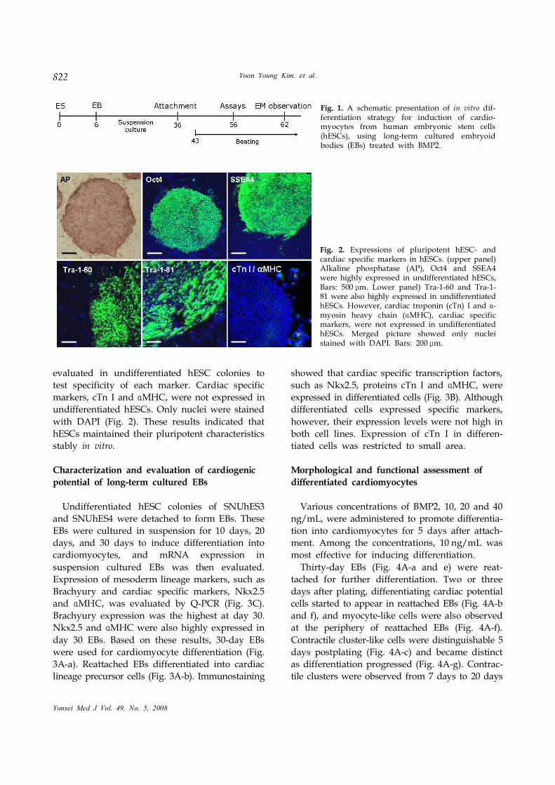
evaluated in undifferentiated hESC colonies to
test specificity of each marker. Cardiac specific
markers, cTn I and MHC, were not expressed inα
undifferentiated hESCs. Only nuclei were stained
with DAPI (Fig. 2). These results indicated that
hESCs maintained their pluripotent characteristics
stably in vitro.
Characterization and evaluation of cardiogenic
potential of long-term cultured EBs
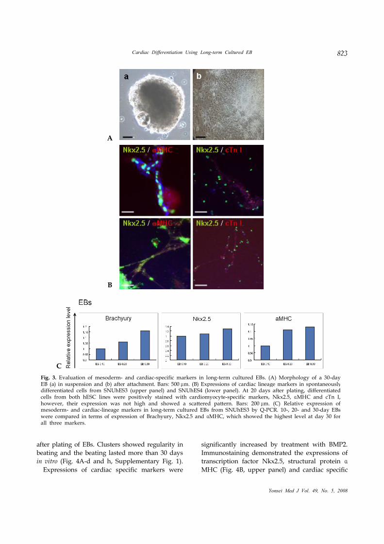
Undifferentiated hESC colonies of SNUhES3
and SNUhES4 were detached to form EBs. These
EBs were cultured in suspension for 10 days, 20
days, and 30 days to induce differentiation into
cardiomyocytes, and mRNA expression in
suspension cultured EBs was then evaluated.
Expression of mesoderm lineage markers, such as
Brachyury and cardiac specific markers, Nkx2.5
and MHC, was evaluated by Q-PCR (Fig. 3C).α
Brachyury expression was the highest at day 30.
Nkx2.5 and MHC were also highly expressed inα
day 30 EBs. Based on these results, 30-day EBs
were used for cardiomyocyte differentiation (Fig.
3A-a). Reattached EBs differentiated into cardiac
lineage precursor cells (Fig. 3A-b). Immunostaining
showed that cardiac specific transcription factors,
such as Nkx2.5, proteins cTn I and MHC, wereα
expressed in differentiated cells (Fig. 3B). Although
differentiated cells expressed specific markers,
however, their expression levels were not high in
both cell lines. Expression of cTn I in differen-
tiated cells was restricted to small area.
Morphological and functional assessment of
differentiated cardiomyocytes
Various concentrations of BMP2, 10, 20 and 40
ng/mL, were administered to promote differentia-
tion into cardiomyocytes for 5 days after attach-
ment. Among the concentrations, 10 ng/mL was
most effective for inducing differentiation.
Thirty-day EBs (Fig. 4A-a and e) were reat-
tached for further differentiation. Two or three
days after plating, differentiating cardiac potential
cells started to appear in reattached EBs (Fig. 4A-b
and f), and myocyte-like cells were also observed
at the periphery of reattached EBs (Fig. 4A-f).
Contractile cluster-like cells were distinguishable 5
days postplating (Fig. 4A-c) and became distinct
as differentiation progressed (Fig. 4A-g). Contrac-
tile clusters were observed from 7 days to 20 days
Fig. 1. A schematic presentation of in vitro dif-ferentiation strategy for induction of cardio-myocytes from human embryonic stem cells(hESCs), using long-term cultured embryoidbodies (EBs) treated with BMP2.
Fig. 2. Expressions of pluripotent hESC- andcardiac specific markers in hESCs. (upper panel)Alkaline phosphatase (AP), Oct4 and SSEA4were highly expressed in undifferentiated hESCs,Bars: 500 m.μ Lower panel) Tra-1-60 and Tra-1-81 were also highly expressed in undifferentiatedhESCs. However, cardiac troponin (cTn) I and -αmyosin heavy chain ( MHC), cardiac specificαmarkers, were not expressed in undifferentiatedhESCs. Merged picture showed only nucleistained with DAPI. Bars: 200 m.μ
Cardiac Differentiation Using Long-term Cultured EB
Yonsei Med J Vol. 49, No. 5, 2008
after plating of EBs. Clusters showed regularity in
beating and the beating lasted more than 30 days
in vitro (Fig. 4A-d and h, Supplementary Fig. 1).
Expressions of cardiac specific markers were
significantly increased by treatment with BMP2.
Immunostaining demonstrated the expressions of
transcription factor Nkx2.5, structural protein α
MHC (Fig. 4B, upper panel) and cardiac specific
Fig. 3. Evaluation of mesoderm- and cardiac-specific markers in long-term cultured EBs. (A) Morphology of a 30-dayEB (a) in suspension and (b) after attachment. Bars: 500 m. (B) Expressions of cardiac lineage markers in spontaneouslyμdifferentiated cells from SNUhES3 (upper panel) and SNUhES4 (lower panel). At 20 days after plating, differentiatedcells from both hESC lines were positively stained with cardiomyocyte-specific markers, Nkx2.5, MHC and cTn I,αhowever, their expression was not high and showed a scattered pattern. Bars: 200 m. (C) Relative expression ofμmesoderm- and cardiac-lineage markers in long-term cultured EBs from SNUhES3 by Q-PCR. 10-, 20- and 30-day EBswere compared in terms of expression of Brachyury, Nkx2.5 and MHC, which showed the highest level at day 30 forαall three markers.
A
B
C
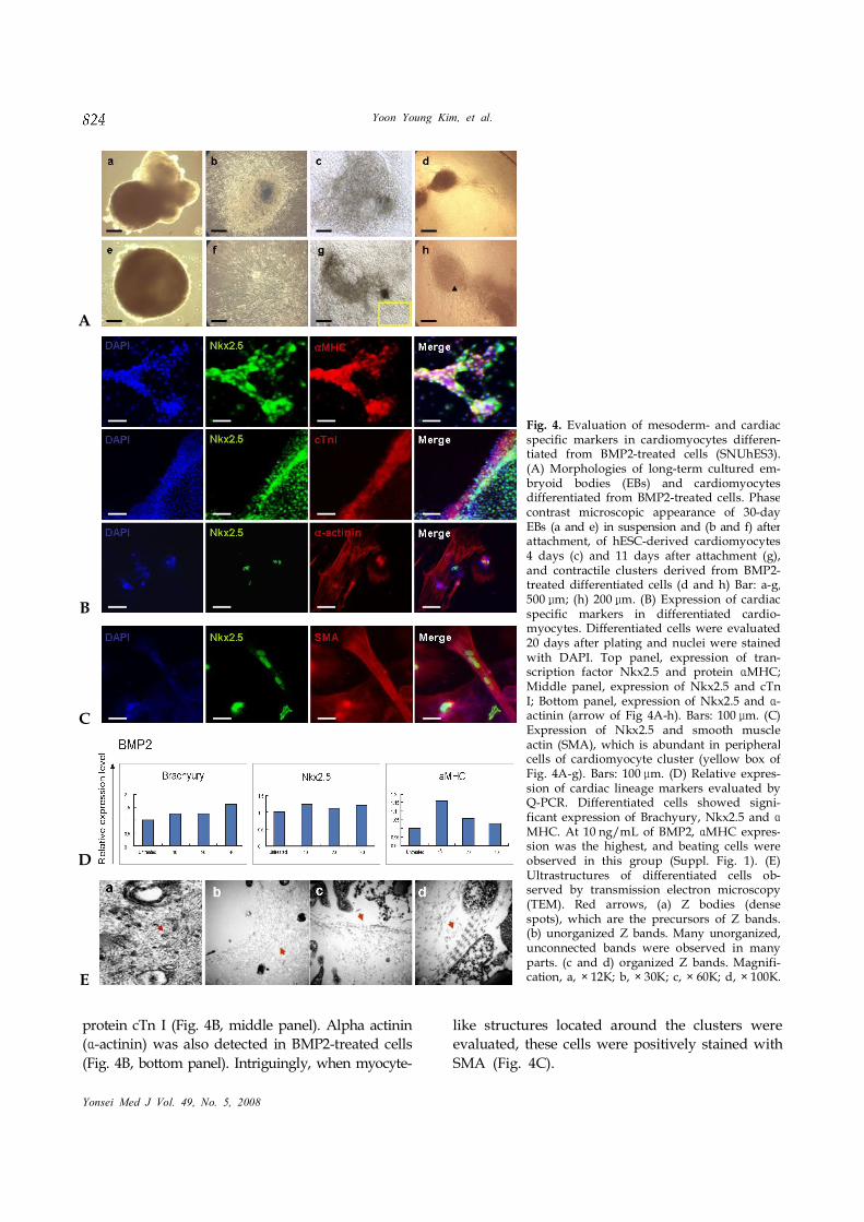
Fig. 4. Evaluation of mesoderm- and cardiacspecific markers in cardiomyocytes differen-tiated from BMP2-treated cells (SNUhES3).(A) Morphologies of long-term cultured em-bryoid bodies (EBs) and cardiomyocytesdifferentiated from BMP2-treated cells. Phasecontrast microscopic appearance of 30-dayEBs (a and e) in suspension and (b and f) afterattachment, of hESC-derived cardiomyocytes4 days (c) and 11 days after attachment (g),and contractile clusters derived from BMP2-treated differentiated cells (d and h) Bar: a-g,500 m; (h) 200μ m. (B) Expression of cardiacμspecific markers in differentiated cardio-myocytes. Differentiated cells were evaluated20 days after plating and nuclei were stainedwith DAPI. Top panel, expression of tran-scription factor Nkx2.5 and protein MHC;ɑMiddle panel, expression of Nkx2.5 and cTnI; Bottom panel, expression of Nkx2.5 and -ɑactinin (arrow of Fig 4A-h). Bars: 100 m. (C)μExpression of Nkx2.5 and smooth muscleactin (SMA), which is abundant in peripheralcells of cardiomyocyte cluster (yellow box ofFig. 4A-g). Bars: 100 m. (D) Relative expresμ -sion of cardiac lineage markers evaluated byQ-PCR. Differentiated cells showed signi-ficant expression of Brachyury, Nkx2.5 and αMHC. At 10 ng/mL of BMP2, MHC expresα -sion was the highest, and beating cells wereobserved in this group (Suppl. Fig. 1). (E)Ultrastructures of differentiated cells ob-served by transmission electron microscopy(TEM). Red arrows, (a) Z bodies (densespots), which are the precursors of Z bands.(b) unorganized Z bands. Many unorganized,unconnected bands were observed in manyparts. (c and d) organized Z bands. Magnifi-cation, a, × 12K; b, × 30K; c, × 60K; d, × 100K.
Yoon Young Kim, et al.
Yonsei Med J Vol. 49, No. 5, 2008
protein cTn I (Fig. 4B, middle panel). Alpha actinin
( -actinin) was also detected in BMP2-treated cellsα
(Fig. 4B, bottom panel). Intriguingly, when myocyte-
like structures located around the clusters were
evaluated, these cells were positively stained with
SMA (Fig. 4C).
A
B
C
D
E
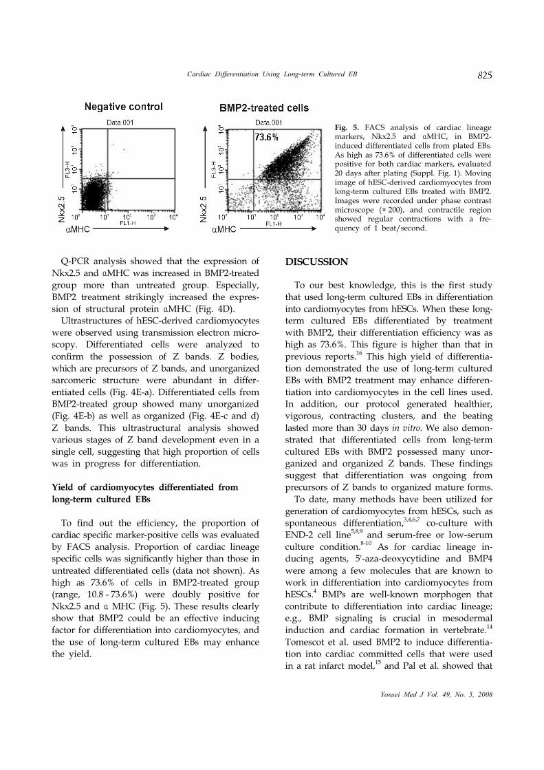
Fig. 5. FACS analysis of cardiac lineagemarkers, Nkx2.5 and MHC, in BMP2-αinduced differentiated cells from plated EBs.As high as 73.6% of differentiated cells werepositive for both cardiac markers, evaluated20 days after plating (Suppl. Fig. 1). Movingimage of hESC-derived cardiomyocytes fromlong-term cultured EBs treated with BMP2.Images were recorded under phase contrastmicroscope (× 200), and contractile regionshowed regular contractions with a fre-quency of 1 beat/second.
Cardiac Differentiation Using Long-term Cultured EB
Yonsei Med J Vol. 49, No. 5, 2008
Q-PCR analysis showed that the expression of
Nkx2.5 and MHC was increased in BMP2-treatedα
group more than untreated group. Especially,
BMP2 treatment strikingly increased the expres-
sion of structural protein MHC (Fig. 4D).α
Ultrastructures of hESC-derived cardiomyocytes
were observed using transmission electron micro-
scopy. Differentiated cells were analyzed to
confirm the possession of Z bands. Z bodies,
which are precursors of Z bands, and unorganized
sarcomeric structure were abundant in differ-
entiated cells (Fig. 4E-a). Differentiated cells from
BMP2-treated group showed many unorganized
(Fig. 4E-b) as well as organized (Fig. 4E-c and d)
Z bands. This ultrastructural analysis showed
various stages of Z band development even in a
single cell, suggesting that high proportion of cells
was in progress for differentiation.
Yield of cardiomyocytes differentiated from
long-term cultured EBs
To find out the efficiency, the proportion of
cardiac specific marker-positive cells was evaluated
by FACS analysis. Proportion of cardiac lineage
specific cells was significantly higher than those in
untreated differentiated cells (data not shown). As
high as 73.6% of cells in BMP2-treated group
(range, 10.8 - 73.6%) were doubly positive for
Nkx2.5 and MHC (Fig. 5). These results clearlyα
show that BMP2 could be an effective inducing
factor for differentiation into cardiomyocytes, and
the use of long-term cultured EBs may enhance
the yield.
DISCUSSION
To our best knowledge, this is the first study
that used long-term cultured EBs in differentiation
into cardiomyocytes from hESCs. When these long-
term cultured EBs differentiated by treatment
with BMP2, their differentiation efficiency was as
high as 73.6%. This figure is higher than that in
previous reports.16 This high yield of differentia-
tion demonstrated the use of long-term cultured
EBs with BMP2 treatment may enhance differen-
tiation into cardiomyocytes in the cell lines used.
In addition, our protocol generated healthier,
vigorous, contracting clusters, and the beating
lasted more than 30 days in vitro. We also demon-
strated that differentiated cells from long-term
cultured EBs with BMP2 possessed many unor-
ganized and organized Z bands. These findings
suggest that differentiation was ongoing from
precursors of Z bands to organized mature forms.
To date, many methods have been utilized for
generation of cardiomyocytes from hESCs, such as
spontaneous differentiation,3,4,6,7
co-culture with
END-2 cell line5,8,9 and serum-free or low-serum
culture condition.8-10
As for cardiac lineage in-
ducing agents, 5'-aza-deoxycytidine and BMP4
were among a few molecules that are known to
work in differentiation into cardiomyocytes from
hESCs.4 BMPs are well-known morphogen that
contribute to differentiation into cardiac lineage;
e.g., BMP signaling is crucial in mesodermal
induction and cardiac formation in vertebrate.14
Tomescot et al. used BMP2 to induce differentia-
tion into cardiac committed cells that were used
in a rat infarct model,15and Pal et al. showed that
Yoon Young Kim, et al.
Yonsei Med J Vol. 49, No. 5, 2008
BMP2 is another effective molecule for in vitro
differentiation into cardiomyocytes from hESCs in
low serum condition.16 However, these researchers
used short-term cultured EBs, and one of these
reports retrieved a yield of about 30%.16
In this study, we tried to generate cardiomyo-
cytes from our established hESC lines, SNUhES32
and SNUhES4.17 When treated with BMP2, both
cell lines showed comparable differentiation into
cardiomyocytes. To determine the optimal time
point of differentiation induction initiation, EBs
were cultured in suspension for 10 days, 20 days
and 30 days. Brachyury, which is known to be
expressed at precursor stage,19 and cardiac specific
markers, Nkx2.5 and MHC, were highly exα -
pressed in 30-day EBs. Accordingly, we were able
to obtain a high yield by using this stage's EBs.
Interestingly enough, each cluster among many
hESC-derived contractile clusters showed different
frequencies of contraction (23 - 62/minutes). In
addition, contractility of these hESC-derived
cardiac cells was found to be sensitive to tempera-
ture: contractility was significantly reduced when
exposed to lower temperature and beat rhythm
was recovered when temperature rose.
At the periphery of contractile regions, elongated
myocyte-like cells were observed. These cells were
connected to contractile clusters and contracted
when the cluster beat (Fig. 4A-c and g). These cells
were identified as smooth muscle cells. Smooth
muscle cells are derived from common progenitor
with cardiomyocytes and known to contribute to
formation of the mature heart.19
Our findings
suggested that differentiation from hESCs into
cardiomyocytes is accompanied by differentiation
of smooth muscle cells, which were abundant at
the peripheral region of clusters.
We used serum-containing media for the induc-
tion of differentiation. Reduced concentration of
serum did not effectively generate cardiomyocytes
in our cell lines (data not shown). Role of serum
in cardiomyocytes differentiation is still contro-
versial, although many studies adopted serum-
free or low-serum culture conditions in cardiac
differentiation.8,10
Therefore, the effects of serum
concentration should be the subject for further
studies.
In conclusion, we demonstrated that the use of
long-term cultured EBs with BMP2 treatment may
enhance differentiation into cardiomyocytes from
2 hESC lines, confirmed by increased expression
of cardiac specific markers, and observation of
contractile clusters and Z bands in differentiated
cells.
ACKNOWLEDGMENT
The authors thank Dr. Yong Bin Park for com-
ments about this manuscript.
REFERENCES
1. Thomson JA, Itskovitz-Eldor J, Shapiro SS, Waknitz
MA, Swiergiel JJ, Marshall VS, et al. Embryonic stem
cell lines derived from human blastocysts. Science
1998;282:1145-7.
2. Oh SK, Kim HS, Ahn HJ, Seol HW, Kim YY, Park YB,
et al. Derivation and characterization of new human
embryonic stem cell lines: SNUhES1, SNUhES2 and
SNUhES3. Stem Cells 2005;23:211-9.
3. Kehat I, Kenyagin-Karsenti D, Snir M, Segev H, Amit
M, Gepstein A, et al. Human embryonic stem cells can
differentiate into myocytes with structural and
functional properties of cardiomyocytes. J Clin Invest
2001;108:407-14.
4. Xu C, Police S, Rao N, Carpenter MK. Characterization
and enrichment of cardiomyocytes derived from
human embryonic stem cells. Circ Res 2002;91:501-8.
5. Mummery C, Ward-van Oostwaard D, Doevendans P,
Spijker R, van den Brink S, Hassink R, et al. Differen-
tiation of human embryonic stem cells to cardio-
myocytes: role of coculture with visceral endoderm-like
cells. Circulation 2003;107:2733-40.
6. He JQ, Ma Y, Lee Y, Thomson JA, Kamp TJ. Human
embryonic stem cells develop into multiple types of
cardiac myocytes: action potential characterization. Circ
Res 2003;93:32-9.
7. Kehat I, Khimovich L, Caspi O, Gepstein A, Shofti R,
Arbel G, et al. Electromechanical integration of cardio-
myocytes derived from human embryonic stem cells.
Nat Biotechnol 2004;22:1282-9.
8. Passier R, Oostwaard DW, Snapper J, Kloots J, Hassink
R, Kuijk E, et al. Increased cardiomyocyte differentia-
tion from human embryonic stem cells in serum-free
cultures. Stem Cells 2005;23:772-80.
9. Beqqali A, Kloots J, Ward-van Oostwaard D, Mummery
C, Passier R. Genome-wide transcriptional profiling of
human embryonic stem cells differentiating to
cardiomyocytes. Stem Cells 2006;24:1956-67.
10. Bettiol E, Sartiani L, Chicha L, Krause KH, Cerbai E,
Jaconi ME. Fetal bovine serum enables cardiac differen-
tiation of human embryonic stem cells. Differentiation
Cardiac Differentiation Using Long-term Cultured EB
Yonsei Med J Vol. 49, No. 5, 2008
2007;75:669-81.
11. Schlange T, Andrée B, Arnold HH, Brand T. BMP2 is
required for early heart development during a distinct
time period. Mech Dev 2000;91:259-70.
12. Yuasa S, Itabashi Y, Koshimizu U, Tanaka T, Sugimura
K, Kinoshita M, et al. Transient inhibition of BMP
signaling by Noggin induces cardiomyocyte differentia-
tion of mouse embryonic stem cells. Nature Biotechnol
2005;23:607-11.
13. Antin PB, Taylor RG, Yatskievych T. Precardiac
mesoderm is specified during gastrulation in quail. Dev
Dyn 1994;200:144-54.
14. Schultheiss TM, Burch JB, Lassar AB. A role for bone
morphogenetic proteins in the induction of cardiac
myogenesis. Genes Dev 1997;11:451-62.
15. Tomescot A, Leschik J, Bellamy V, Dubois G, Messas
E, Bruneval P, et al. Differentiation in vivo of cardiac
committed human embryonic stem cells in postmyo-
cardial infarcted rats. Stem Cells 2007;25:2200-5.
16. Pal R, Khanna A. Similar pattern in cardiac differentia-
tion of human embryonic stem cell lines, BG01V and
ReiCell hES1, under low serum concentration supple-
mented with bone morphogenetic protein-2. Differen-
tiation 2007;75:112-22.
17. Kwon YD, Oh SK, Kim HS, Ku SY, Kim SH, Choi YM,
et al. Cellular manipulation of human embryonic stem
cells by TAT-PDX1 protein transduction. Mol Ther
2005;12:28-32.
18. Oh SK, Kim HS, Park YB, Seol HW, Kim YY, Cho MS,
et al. Methods for expansion of human embryonic stem
cells. Stem Cells 2005;23:605-9.
19. Garry DJ, Olson EN. A common progenitor at the heart
of development. Cell 2006;127:1101-4.
Related Documents