Mol. Nutr. Food Res. 2014, 58, 1667–1684 1667 DOI 10.1002/mnfr.201400134 REVIEW Use of dietary phytochemicals to target inflammation, fibrosis, proliferation, and angiogenesis in uterine tissues: Promising options for prevention and treatment of uterine fibroids? Md Soriful Islam 1,2 , Most Mauluda Akhtar 3 , Andrea Ciavattini 4 , Stefano Raffaele Giannubilo 4 , Olga Protic 1 , Milijana Janjusevic 1 , Antonio Domenico Procopio 3 , James H. Segars 5 , Mario Castellucci 1∗ and Pasquapina Ciarmela 1,6 1 Department of Experimental and Clinical Medicine, Faculty of Medicine, Polytechnic University of Marche, Ancona, Italy 2 Biotechnology and Microbiology Laboratory, Department of Botany, University of Rajshahi, Rajshahi, Bangladesh 3 Department of Clinical and Molecular Sciences, Faculty of Medicine, Polytechnic University of Marche, Ancona, Italy 4 Department of Clinical Science, Polytechnic University of Marche, Ancona, Italy 5 Program in Reproductive and Adult Endocrinology, Eunice Kennedy Shriver National Institute of Child Health and Human Development, National Institutes of Health, Bethesda, MD, USA 6 Department of Information Engineering, Polytechnic University of Marche, Ancona, Italy Received: February 23, 2014 Revised: April 18, 2014 Accepted: April 22, 2014 Uterine leiomyomas (fibroids, myomas) are the most common benign tumors of female re- productive tract. They are highly prevalent, with 70–80% of women burdened by the end of their reproductive years. Fibroids are a leading cause of pelvic pain, abnormal vaginal bleed- ing, pressure on the bladder, miscarriage, and infertility. They are the leading indication for hysterectomy, and costs exceed 6 billion dollars annually in the United States. Unfortunately, no long-term medical treatments are available. Dysregulation of inflammatory processes are thought to be involved in the initiation of leiomyoma and extracellular matrix deposition, cell proliferation, and angiogenesis are the key cellular events implicated in leiomyoma growth. In modern pharmaceutical industries, dietary phytochemicals are used as source of new po- tential drugs for many kinds of tumors. Dietary phytochemicals may exert therapeutic effects by interfering with key cellular events of the tumorigenesis process. At present, a negligible number of phytochemicals have been tested as therapeutic agents against fibroids. In this con- text, our aim was to introduce some of the potential dietary phytochemicals that have shown anti-inflammatory, antiproliferative, antifibrotic, and antiangiogenic activities in different Correspondence: Dr. Pasquapina Ciarmela, Department of Exper- imental and Clinical Medicine, Faculty of Medicine, Polytechnic University of Marche, via Tronto 10/a, 60020 Ancona, Italy E-mail: [email protected] Fax: +39-0712206087 Abbreviations: AKT/PKB, protein kinase B; -SMA, -smooth muscle actin; CCR, chemokine (cc-motif) receptor; COX-2, cyclooxygenase-2; CTGF, connective tissue growth factor; CXCR, chemokine (cxc-motif) receptor; EA, ellagic acid; ECM, extra- cellular matrix; ECs, endothelial cells; EGCG, epigallocatechin gallate; EGF, epidermal growth factor; ERK, extracellular signal- regulated kinase; GnRHa, gonadotropin-releasing hormone ago- nist; HUVECs, human umbilical vein ECs; I3C, indole-3-carbinol; IGF, insulin-like growth factor; IB, nuclear factor of kappa light polypeptide gene enhancer in B-cells inhibitor alpha; iNOS, inducible nitric oxide synthase; MAPK, mitogen-activated protein kinase; MCP, monocyte chemoattractant protein; MIP, macrophage inflammatory protein; MMP, matrix metallopro- teinase; NADPH, nicotinamide adenine dinucleotide phosphate; NF-B, nuclear factor kappa light chain enhancer of activated B cells; NO, nitric oxide; NOX, NADPH oxidase; Nrf2, nuclear fac- tor erythroid 2 related factor 2; PDGF, platelet-derived growth factor; PDGFR, PDGF receptor; PGE, prostaglandin E; PI3K, phos- phoinositide 3-kinase; PPAR, peroxisome proliferator-activated receptor gamma; PSCs, pancreatic stellate cells; PCNA, prolifer- ating cell nuclear antigen; ROS, reactive oxygen species; SFN, sulforaphone; SMCs, smooth muscle cells; TGF-, transforming growth factor-; TNF-, tumor necrosis factor-alpha; UA, ursolic acid; VEGF, vascular endothelial growth factor; VEGFR, vascular endothelial growth factor receptor; VSMCs, vascular SMCs ∗ Additional corresponding author: Dr. Mario Castellucci, E-mail: [email protected] C 2014 WILEY-VCH Verlag GmbH & Co. KGaA, Weinheim www.mnf-journal.com

Welcome message from author
This document is posted to help you gain knowledge. Please leave a comment to let me know what you think about it! Share it to your friends and learn new things together.
Transcript

Mol. Nutr. Food Res. 2014, 58, 1667–1684 1667DOI 10.1002/mnfr.201400134
REVIEW
Use of dietary phytochemicals to target inflammation,
fibrosis, proliferation, and angiogenesis in uterine
tissues: Promising options for prevention and treatment
of uterine fibroids?
Md Soriful Islam1,2, Most Mauluda Akhtar3, Andrea Ciavattini4, Stefano Raffaele Giannubilo4,Olga Protic1, Milijana Janjusevic1, Antonio Domenico Procopio3, James H. Segars5,Mario Castellucci1∗ and Pasquapina Ciarmela1,6
1 Department of Experimental and Clinical Medicine, Faculty of Medicine, Polytechnic University of Marche,Ancona, Italy
2 Biotechnology and Microbiology Laboratory, Department of Botany, University of Rajshahi, Rajshahi, Bangladesh3 Department of Clinical and Molecular Sciences, Faculty of Medicine, Polytechnic University of Marche, Ancona,
Italy4 Department of Clinical Science, Polytechnic University of Marche, Ancona, Italy5 Program in Reproductive and Adult Endocrinology, Eunice Kennedy Shriver National Institute of Child Health and
Human Development, National Institutes of Health, Bethesda, MD, USA6 Department of Information Engineering, Polytechnic University of Marche, Ancona, Italy
Received: February 23, 2014Revised: April 18, 2014
Accepted: April 22, 2014
Uterine leiomyomas (fibroids, myomas) are the most common benign tumors of female re-productive tract. They are highly prevalent, with 70–80% of women burdened by the end oftheir reproductive years. Fibroids are a leading cause of pelvic pain, abnormal vaginal bleed-ing, pressure on the bladder, miscarriage, and infertility. They are the leading indication forhysterectomy, and costs exceed 6 billion dollars annually in the United States. Unfortunately,no long-term medical treatments are available. Dysregulation of inflammatory processes arethought to be involved in the initiation of leiomyoma and extracellular matrix deposition, cellproliferation, and angiogenesis are the key cellular events implicated in leiomyoma growth.In modern pharmaceutical industries, dietary phytochemicals are used as source of new po-tential drugs for many kinds of tumors. Dietary phytochemicals may exert therapeutic effectsby interfering with key cellular events of the tumorigenesis process. At present, a negligiblenumber of phytochemicals have been tested as therapeutic agents against fibroids. In this con-text, our aim was to introduce some of the potential dietary phytochemicals that have shownanti-inflammatory, antiproliferative, antifibrotic, and antiangiogenic activities in different
Correspondence: Dr. Pasquapina Ciarmela, Department of Exper-imental and Clinical Medicine, Faculty of Medicine, PolytechnicUniversity of Marche, via Tronto 10/a, 60020 Ancona, ItalyE-mail: [email protected]: +39-0712206087
Abbreviations: AKT/PKB, protein kinase B; �-SMA, �-smoothmuscle actin; CCR, chemokine (cc-motif) receptor; COX-2,cyclooxygenase-2; CTGF, connective tissue growth factor; CXCR,chemokine (cxc-motif) receptor; EA, ellagic acid; ECM, extra-cellular matrix; ECs, endothelial cells; EGCG, epigallocatechingallate; EGF, epidermal growth factor; ERK, extracellular signal-regulated kinase; GnRHa, gonadotropin-releasing hormone ago-nist; HUVECs, human umbilical vein ECs; I3C, indole-3-carbinol;IGF, insulin-like growth factor; I�B�, nuclear factor of kappalight polypeptide gene enhancer in B-cells inhibitor alpha;iNOS, inducible nitric oxide synthase; MAPK, mitogen-activated
protein kinase; MCP, monocyte chemoattractant protein; MIP,macrophage inflammatory protein; MMP, matrix metallopro-teinase; NADPH, nicotinamide adenine dinucleotide phosphate;NF-�B, nuclear factor kappa light chain enhancer of activated Bcells; NO, nitric oxide; NOX, NADPH oxidase; Nrf2, nuclear fac-tor erythroid 2 related factor 2; PDGF, platelet-derived growthfactor; PDGFR, PDGF receptor; PGE, prostaglandin E; PI3K, phos-phoinositide 3-kinase; PPAR�, peroxisome proliferator-activatedreceptor gamma; PSCs, pancreatic stellate cells; PCNA, prolifer-ating cell nuclear antigen; ROS, reactive oxygen species; SFN,sulforaphone; SMCs, smooth muscle cells; TGF-�, transforminggrowth factor-�; TNF-�, tumor necrosis factor-alpha; UA, ursolicacid; VEGF, vascular endothelial growth factor; VEGFR, vascularendothelial growth factor receptor; VSMCs, vascular SMCs∗Additional corresponding author: Dr. Mario Castellucci,E-mail: [email protected]
C© 2014 WILEY-VCH Verlag GmbH & Co. KGaA, Weinheim www.mnf-journal.com

1668 M. S. Islam et al. Mol. Nutr. Food Res. 2014, 58, 1667–1684
biological systems. This review could be useful to stimulate the evaluation of these phyto-chemicals as possible therapies for uterine fibroids.
Keywords:
Antifibrotic / Antiproliferative / Dietary phytochemicals / Inflammation / Uterinefibroid
1 Introduction
Uterine leiomyomas (fibroids or myomas) are common be-nign smooth muscle tumors of the uterus [1–3]. They arehighly prevalent, with 70–80% of women burdened by the endof their reproductive years [4]. Several studies have demon-strated that there are ethnic differences in fibroid burden[4–9]. African Americans have a higher (three times more)fibroid incidence [4, 5] and experience more severe symp-toms with larger and more numerous leiomyomas comparedwith white women [6, 9]. The common symptoms associatedwith uterine leiomyomas are irregular and/or heavy men-strual bleeding, pain in the pelvic region and the back, bulk-related symptoms (pressure on bladder and bowel as well asincrease in abdominal circumference), and subfertility [4,10].Uterine fibroids are the leading indication for hysterectomyin the United States [11], and fibroid associated costs $5.9–34.4 billion annually [12]. This complicated disease processalso exerts an enormous burden on health care resources inAustralia [13] and European countries [14].
Despite the widespread prevalence of the disease, thepathogenesis of leiomyomas is not well understood. An in-creasingly popular view is that uterine leiomyoma arise as aconsequence of a chronically active inflammatory immunesystem [15–17]. However, there is considerable evidence thatestrogens and progestogens promote tumor growth [18,19], asthe fibroids rarely appear before menarche and tend to regressafter menopause [20]. Besides growth factors, cytokines andchemokines may serve as mediators of sex steroids, and playan important role in the proliferation, fibrosis, and angiogen-esis processes that are ultimately involved in the formationand growth of uterine fibroids [1, 2, 10, 17, 21].
Gonadotropin-releasing hormone agonist (GnRHa) isonly medical therapy for leiomyoma treatment approved byUS Food and Drug Administration. GnRHa was developedon the basis of the induction of a hypoestrogenic state. Al-though this treatment temporally (up to 6 months) is effectiveas preoperative therapy to reduce fibroid size and symptoms[22, 23], the benefits of GnRHa are tempered by significantside effects resulting from hypoestrogenism (e.g., hot flashes,vaginal dryness, bone demineralization) [24–26]. In additionto GnRHa, several potential therapies such as mifepristone(antiprogestin) [27], and selective progesterone receptor mod-ulators such as asoprisnil [28], ulipristal acetate [29, 30], andproellex [31] have shown excellent therapeutic efficiency dur-ing the course of clinical trials. Additionally, aromatase in-hibitors have shown therapeutic efficacy for uterine fibroids,
but are not approved for that indication. Nonetheless, in com-parison with the burden of disease to society, medical treat-ments for leiomyoma are still very limited and no preventativetherapies have been developed.
Since ancient times, plants and plant-derived com-pounds have provided tremendous support in the traditionalmedicine systems, and have been used as source of new po-tential drugs in modern pharmaceutical industries. For exam-ple, from 1981 to 2010, natural products and their derivativeswere the source of 41% of new drugs and 79.8% of all ap-proved anticancer drugs [32]. In addition, the percentage ofdrugs from natural products without derivatives was greatlyincreased from 20.8% in 2009 to 50% in 2010 [32]. Recently,two systematic reviews assessed the efficacy of herbal prepa-rations for uterine fibroids [33, 34]. Meta-analyses demon-strated that Guizhi Fuling Formula plus mifepristone weremore effective than mifepristone alone in reducing the vol-ume of fibroids [33]. In addition, the Guizhi Fuling Formulasignificantly improved symptoms of dysmenorrhea eitherwhen it was used alone or in combination with mifepristone[33].
At present, only few dietary phytochemicals such as epigal-locatechin gallate (EGCG) [35,36], curcumin [37], isoliquiriti-genin [38], genistein [39], and resveratrol [40,41] (Table 1) havebeen studied in myometrium and fibroids. A large number ofphytochemicals remain to be tested for possible therapeuticeffects against uterine leiomyoma. In this review, we intro-duce some of the more promising phytochemicals (Figs. 1and 2) in the context of key features of leiomyoma develop-ment and growth: inflammation, fibrosis, cell proliferation,and angiogenesis.
2 Pathogenesis of uterine fibroids
2.1 Inflammatory mediators
Considerable evidence suggests that uterine leiomyoma de-velopment may be triggered, at least in part, by a chronicallyactive inflammatory immune system [15–17]. The concept ofinflammation actually fits into a theory of fibroid develop-ment based on an altered response to noxious stimuli; possi-bly tissue injury from extravasated menstrual blood into themyometrium, or hypoxia leading to altered tissue repair andfibrosis [15,16]. Among major cytokines, the expression of IL-1, IL-6, IL-11, IL-13, IL-15, IL-33, tumor necrosis factor (TNF)-�, granulocyte-macrophage colony-stimulating factor have
C© 2014 WILEY-VCH Verlag GmbH & Co. KGaA, Weinheim www.mnf-journal.com

Mol. Nutr. Food Res. 2014, 58, 1667–1684 1669
Table 1. Therapeutic effects of dietary phytochemicals on uterine fibroids
Dietary phytochemicals Dietary sources Therapeutic effects on uterine fibroids
Epigallocatechin gallate Green tea (Camelliasinensis)
(1) Inhibits the proliferation of leiomyoma cells [35,77].(2) Induces apoptosis in leiomyoma cells [35,77].(3) Reduces the volume and weight of tumors of female mice [77].(4) Reduces the incidence and size of spontaneously occurring
leiomyoma of the oviduct in Japanese quail [78].(5) Reduces uterine fibroid volume, fibroid-specific symptom severity,
induces significant improvement in health-related quality of life inpremenopausal women [36].
Curcumin Turmeric (Curcumalonga)
(1) Inhibits uterine leiomyoma cell proliferation [37].(2) Induces apoptosis in leiomyoma cells [37].(3) Inhibits fibronectin production in leiomyoma cells [37].(4) Acts as PPAR� ligand [80].
Isoliquiritigenin Licorice (Glycyrrhizauralensis), shallot(Allium ascalonicum),soybean (Glycine max)
(1) Induces the growth inhibition of leiomyoma cells [38].(2) Induces apoptosis in uterine leiomyoma cells [38].
Genistein Soybeans (G. max),lupine (Lupinus spp.),fava bean, (Vicia faba),kudzu (Puerarialobata), psoralea(Psoralea corylifolia)
(1) Stimulates leiomyoma cell proliferation at low concentration [83].(2) Inhibits leiomyoma and myometrial cell proliferation at high
concentration [83].(3) Increases caspase activity [83].(4) Induces apoptosis in myometrial and leiomyoma cells [83].(5) Downregulates activin A, Smad3, and other TGF-� pathway genes in
human uterine leiomyoma cells [84].(6) Reduces the incidence and size of spontaneously occurring
leiomyoma of the oviduct in the Japanese quail [86].Resveratrol More than 70 species of
plants, includingmulberries andpeanuts. Grapevines(Vitis vinifera) are themain sources.
(1) Inhibits proliferation of human uterine leiomyoma cells [40].(2) Induces apoptosis in human uterine leiomyoma cells [40,41].(3) Induces cell cycle arrest in human uterine leiomyoma cells [40].(4) Reduces collagen types I and III in human uterine leiomyoma cells
[40,41].
been implicated with their biological relevance to leiomyomapathophysiology [42–47]. The expression profiles of manychemokines and chemokine receptors have also been charac-terized in leiomyomas and matched myometrium. These in-clude monocyte chemoattractant protein (MCP)-1, IL-8, IL-8receptor type A, macrophage inflammatory protein (MIP)-1�,MIP-1�, RANTES, eotaxin, eotaxin-2, IL-8, chemokine (cc-motif) receptor (CCR) 1, CCR3, CCR5, chemokine (cxc-motif)receptor (CXCR) 1, and CXCR2 mRNA [48–50]. Furthermore,inflammatory mediators such as cyclooxygenase-2 (COX-2)[51] and nitric oxide (NO) [52] have been implicated in my-ometrial pathophysiology. The involvement of nuclear factorkappa light chain enhancer of activated B cells (NF-�B) depen-dent inflammatory pathway has been documented in leiomy-oma cells, as EGCG was reported to significantly decreasethe expression of NF-�B-dependent pathway genes such asproliferating cell nuclear antigen (PCNA), cyclin-dependentkinase 4, and B-cell lymphoma 2 as well as increase the ex-pression of the proapoptotic B-cell lymphoma 2 associatedX in a dose-dependent manner [35]. The above informationsupports the tenet that the inflammatory response may playan important role to initiate the development of uterine fi-broids. Therefore, anti-inflammatory agents could representpharmacological targets for fibroids.
2.2 Fibrosis
Fibrosis is a pathological feature of many chronic inflam-matory diseases. It is defined by the accumulation of excessextracellular matrix (ECM) components. Uterine leiomyomasare typically considered as a fibrotic disorder as they contain50% more ECM than the corresponding myometrium [53].The ECM of leiomyomas consists primarily of collagen, fi-bronectin, and proteoglycans [21,54–57]. The abnormal ECMstructure and orientation found in leiomyomas [21, 54], andalterations in ECM modifies mechanical stresses on residentcells, which leads to activation of internal mechanical signal-ing and may contribute to leiomyoma growth [58, 59]. Theinhibition of fibrosis is a big challenge to control this tu-mor; therefore, the development of novel antifibrotic agentscould represent a tractable approach for medical therapy.Two growth factors from the transforming growth factor-�(TGF-�) superfamily are known to be involved in the accu-mulation of ECM in leiomyoma. TGF-� increases fibronectinmRNA expression in both myometrial [60] and leiomyomacells [55, 60]. TGF-� also increases collagen 1A1 [60] andversican [57] mRNA expression in myometrial and leiomy-oma cells. Recently, our group demonstrated that activin-Aincreased fibronectin, collagen 1A1, and versican expression
C© 2014 WILEY-VCH Verlag GmbH & Co. KGaA, Weinheim www.mnf-journal.com

1670 M. S. Islam et al. Mol. Nutr. Food Res. 2014, 58, 1667–1684
Figure 1. Dietary phytochemicals not yet studied in uterine fibroids, their chemical structure, and food sources.
C© 2014 WILEY-VCH Verlag GmbH & Co. KGaA, Weinheim www.mnf-journal.com

Mol. Nutr. Food Res. 2014, 58, 1667–1684 1671
Figure 2. Regulation of majorcellular events, inflammation, fi-brosis, cell proliferation, and an-giogenesis, by dietary phyto-chemicals.
in leiomyoma cells [61]. Furthermore, platelet-derived growthfactor (PDGF) also reported to increase collagen �1 (I) in bothleiomyoma and myometrial cells [62]. The overproduced ECMitself may play a dynamic role in the metabolic processes lead-ing to tumor growth, by influencing cellular proliferation anddifferentiation and by serving as a repository for biologicallyactive growth factors, cytokines, chemokines, angiogenic andinflammatory response mediators, and proteases producedby tumor cells.
2.3 Cell proliferation
At least one mechanism responsible for leiomyomas under-going extensive enlargement is the increased rate of cell pro-liferation. Uterine cellular proliferation and differentiationare regulated by sex steroids, estrogen, and progesterone.Estrogen has traditionally been identified as the most im-portant sex steroid for fibroid growth; however, progesteroneseems to have the dominant steroidal influence on fibroids.This dominance is supported by the increased mitotic ratesin fibroids during the secretory phase of the menstrual cy-cle [63]. There is growing evidence that signaling pathwaysare directly activated by estrogen and progesterone receptors,and these pathways can also interact with growth factors, cy-tokines, and chemokine signaling systems to promote prolif-eration of leiomyomas. Several growth factors such as epider-
mal growth factor (EGF), heparin-binding EGF, insulin-likegrowth factor (IGF), and PDGF have been identified, whichare responsible for increasing myometrial and/or leiomyomacell proliferation by activating various signaling pathways [1].TGF-� exerts bimodal effects on cell proliferation and inducesproliferation of cells at low concentrations by stimulating au-tocrine PDGF secretion, whereas it induces the opposite ef-fect at higher concentrations via downregulation of the PDGFreceptor (PDGFR) and by direct growth inhibition [64, 65].Activin-A and myostatin have cytostatic effects in myome-trial cells, but they do not have an antiproliferative effect inleiomyoma cells [61]. Oxidative stress has been shown to be animportant player in uterine fibroids [66–68]. Fibroid cells arecharacterized by a unique nicotinamide adenine dinucleotidephosphate (NADPH) oxidase (NOX, a major source of su-peroxide and subsequent oxidative stress) profile. Expressionof NOX4 increased in fibroid compared to myometrial tis-sues and cells [66]. In addition, fibroid cells are reported tohave significantly lower antioxidant enzymes, superoxide dis-mutase, and catalase mRNA levels than normal myometrialcells [68]. Furthermore, NOX-derived reactive oxygen species(ROS) have been shown to be a critical component of themitogen-activated protein kinase (MAPK) pathway of EGFand PDGF signaling in leiomyoma smooth muscle cell (SMC)proliferation [67]. A recent study reported that adipocytescan enhance the proliferation of human leiomyoma cellsvia TNF-� proinflammatory cytokine [69]. Therefore, an
C© 2014 WILEY-VCH Verlag GmbH & Co. KGaA, Weinheim www.mnf-journal.com
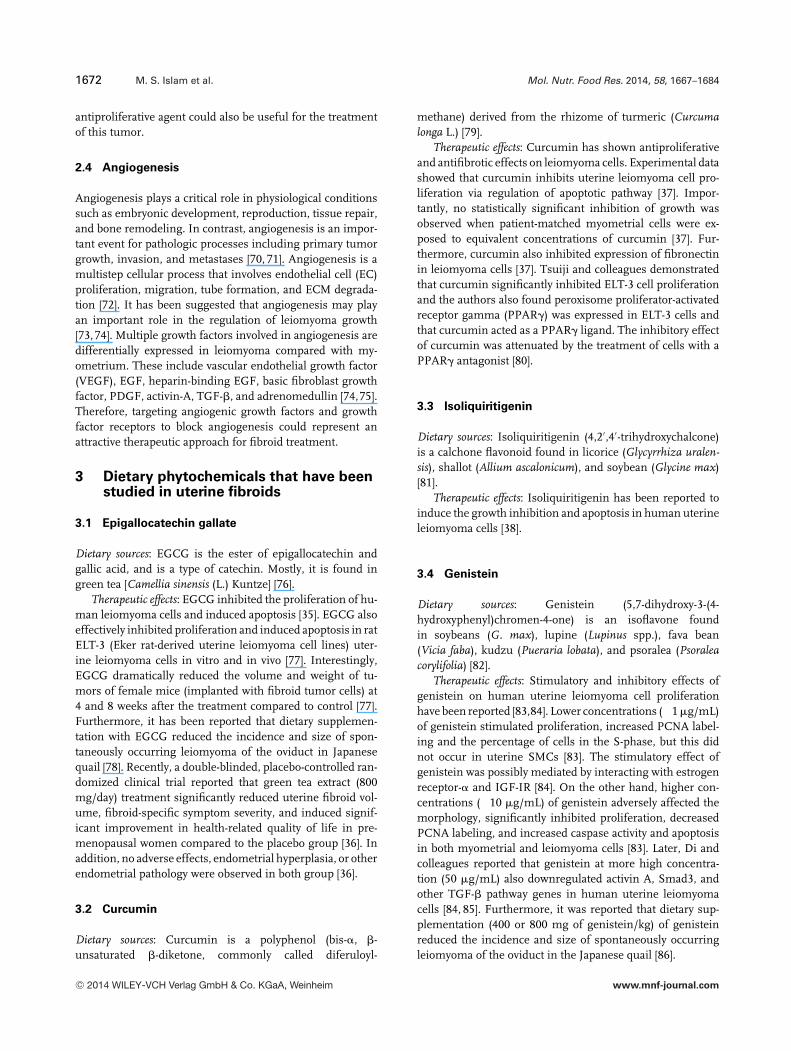
1672 M. S. Islam et al. Mol. Nutr. Food Res. 2014, 58, 1667–1684
antiproliferative agent could also be useful for the treatmentof this tumor.
2.4 Angiogenesis
Angiogenesis plays a critical role in physiological conditionssuch as embryonic development, reproduction, tissue repair,and bone remodeling. In contrast, angiogenesis is an impor-tant event for pathologic processes including primary tumorgrowth, invasion, and metastases [70, 71]. Angiogenesis is amultistep cellular process that involves endothelial cell (EC)proliferation, migration, tube formation, and ECM degrada-tion [72]. It has been suggested that angiogenesis may playan important role in the regulation of leiomyoma growth[73,74]. Multiple growth factors involved in angiogenesis aredifferentially expressed in leiomyoma compared with my-ometrium. These include vascular endothelial growth factor(VEGF), EGF, heparin-binding EGF, basic fibroblast growthfactor, PDGF, activin-A, TGF-�, and adrenomedullin [74,75].Therefore, targeting angiogenic growth factors and growthfactor receptors to block angiogenesis could represent anattractive therapeutic approach for fibroid treatment.
3 Dietary phytochemicals that have beenstudied in uterine fibroids
3.1 Epigallocatechin gallate
Dietary sources: EGCG is the ester of epigallocatechin andgallic acid, and is a type of catechin. Mostly, it is found ingreen tea [Camellia sinensis (L.) Kuntze] [76].
Therapeutic effects: EGCG inhibited the proliferation of hu-man leiomyoma cells and induced apoptosis [35]. EGCG alsoeffectively inhibited proliferation and induced apoptosis in ratELT-3 (Eker rat-derived uterine leiomyoma cell lines) uter-ine leiomyoma cells in vitro and in vivo [77]. Interestingly,EGCG dramatically reduced the volume and weight of tu-mors of female mice (implanted with fibroid tumor cells) at4 and 8 weeks after the treatment compared to control [77].Furthermore, it has been reported that dietary supplemen-tation with EGCG reduced the incidence and size of spon-taneously occurring leiomyoma of the oviduct in Japanesequail [78]. Recently, a double-blinded, placebo-controlled ran-domized clinical trial reported that green tea extract (800mg/day) treatment significantly reduced uterine fibroid vol-ume, fibroid-specific symptom severity, and induced signif-icant improvement in health-related quality of life in pre-menopausal women compared to the placebo group [36]. Inaddition, no adverse effects, endometrial hyperplasia, or otherendometrial pathology were observed in both group [36].
3.2 Curcumin
Dietary sources: Curcumin is a polyphenol (bis-�, �-unsaturated �-diketone, commonly called diferuloyl-
methane) derived from the rhizome of turmeric (Curcumalonga L.) [79].
Therapeutic effects: Curcumin has shown antiproliferativeand antifibrotic effects on leiomyoma cells. Experimental datashowed that curcumin inhibits uterine leiomyoma cell pro-liferation via regulation of apoptotic pathway [37]. Impor-tantly, no statistically significant inhibition of growth wasobserved when patient-matched myometrial cells were ex-posed to equivalent concentrations of curcumin [37]. Fur-thermore, curcumin also inhibited expression of fibronectinin leiomyoma cells [37]. Tsuiji and colleagues demonstratedthat curcumin significantly inhibited ELT-3 cell proliferationand the authors also found peroxisome proliferator-activatedreceptor gamma (PPAR�) was expressed in ELT-3 cells andthat curcumin acted as a PPAR� ligand. The inhibitory effectof curcumin was attenuated by the treatment of cells with aPPAR� antagonist [80].
3.3 Isoliquiritigenin
Dietary sources: Isoliquiritigenin (4,2′,4′-trihydroxychalcone)is a calchone flavonoid found in licorice (Glycyrrhiza uralen-sis), shallot (Allium ascalonicum), and soybean (Glycine max)[81].
Therapeutic effects: Isoliquiritigenin has been reported toinduce the growth inhibition and apoptosis in human uterineleiomyoma cells [38].
3.4 Genistein
Dietary sources: Genistein (5,7-dihydroxy-3-(4-hydroxyphenyl)chromen-4-one) is an isoflavone foundin soybeans (G. max), lupine (Lupinus spp.), fava bean(Vicia faba), kudzu (Pueraria lobata), and psoralea (Psoraleacorylifolia) [82].
Therapeutic effects: Stimulatory and inhibitory effects ofgenistein on human uterine leiomyoma cell proliferationhave been reported [83,84]. Lower concentrations (�1 �g/mL)of genistein stimulated proliferation, increased PCNA label-ing and the percentage of cells in the S-phase, but this didnot occur in uterine SMCs [83]. The stimulatory effect ofgenistein was possibly mediated by interacting with estrogenreceptor-� and IGF-IR [84]. On the other hand, higher con-centrations (�10 �g/mL) of genistein adversely affected themorphology, significantly inhibited proliferation, decreasedPCNA labeling, and increased caspase activity and apoptosisin both myometrial and leiomyoma cells [83]. Later, Di andcolleagues reported that genistein at more high concentra-tion (50 �g/mL) also downregulated activin A, Smad3, andother TGF-� pathway genes in human uterine leiomyomacells [84, 85]. Furthermore, it was reported that dietary sup-plementation (400 or 800 mg of genistein/kg) of genisteinreduced the incidence and size of spontaneously occurringleiomyoma of the oviduct in the Japanese quail [86].
C© 2014 WILEY-VCH Verlag GmbH & Co. KGaA, Weinheim www.mnf-journal.com

Mol. Nutr. Food Res. 2014, 58, 1667–1684 1673
3.5 Resveratrol
Dietary sources: Resveratrol (RVS; trans-3,4′,5-trihydro-xystilbene) is a polyphenolic phytoalexin produced in plants inresponse to environmental stress and infection by pathogenicmicroorganisms. It is found in more than 70 species of plants,including mulberries and peanuts. Grapevines (Vitis vinifera)are the main sources of resveratrol [87].
Therapeutic effects: Resveratrol has shown antiproliferativeand antifibrotic effects on leiomyoma cells. Experimental datashowed that resveratrol inhibits proliferation, induces apop-tosis and cell cycle arrest in human uterine leiomyoma cellsin vitro [40, 41]. In addition, resveratrol treatment reducedmRNA and protein expression of collagen types I and III in adose-dependent manner in human uterine leiomyoma cells[40, 41].
4 Dietary phytochemicals of possiblebenefit for uterine fibroids
4.1 Allicin
Dietary sources: Allicin (diallylthiosulphinate) is an organosul-fur compound obtained from garlic (Allium sativum L.), aspecies in the family Alliaceae [88].
Anti-inflammatory effect: Allicin has been shown to inhibitthe TNF-� induced expression of NO and H2O2 in the humanumbilical ECs [89]. Similarly, allicin inhibited spontaneousand TNF-� induced secretion of cytokines and chemokinesIL-1�, IL-8 from intestinal epithelial cells [90]. Allicin allevi-ated inflammatory injury in the spine, possibly via a reductionin secretion of inflammatory factors (IL-6, IL-8, and TNF-�)in a murine model of ankylosing spondylitis [91].
Antifibrotic effect: Allicin protected against cardiac hyper-trophy and fibrosis via attenuation of ROS-dependent signal-ing pathways [92], and through enhancement of Nrf2 antiox-idant signaling pathways [93]. Allicin also protected againstmyocardial fibrosis in streptozotocin-induced diabetic rats byblocking the expression of connective tissue growth factor(CTGF) and TGF-�1 protein [94].
Antiproliferative effect: Allicin has been reported to inducecaspase-mediated apoptosis in cervical cancer cells [95]. Al-licin also induced apoptosis in gastric cancer cells [96], murineT-lymphocytes [97], colon cancer cells via nuclear factor ery-throid 2 related factor 2 (Nrf2) [98], and in human glioblas-toma cells through an extracellular signal-regulated kinase(ERK) dependent pathway [99]. Growth inhibition of breastcancer cells by allicin was accompanied by accumulation ofcells in the G0/G1 and G2/M phases of the cell cycle [100].
Antiangiogenic effect: Allicin reduced angiogenesis in theaortic ring model as well as basic stages of vessel growth in-cluding ECs proliferation and tube formation. These effectswere accompanied by downregulation of intracellular actinpolymerization and protein kinase B (PKB/AKT) phosphory-lation [101].
4.2 Ellagic acid
Dietary sources: Ellagic acid (EA; 2,3,7,8-tetrahydroxy-chromeno[5,4,3-cde]chromene-5,10-dione) is a polyphenolcompound, found in many berries including strawberries,raspberries, cranberries, blackberries, pecans, pomegranates,walnuts, wolfberry, and grapes [102].
Anti-inflammatory effect: EA has been shown to downreg-ulate inflammatory mediators such as IL-1�, IL-6, TNF-�,and MCP-1 mRNA expression in diabetic mice [103]. Ad-ditionally, EA decreased COX-2, inducible nitric oxide syn-thase (iNOS), TNF-�, IL-6, and NF-�B expression in 1,2-dimethylhydrazine-induced colon carcinogenesis [104]. EAalso inhibited LPS-induced expression of enzymes COX-2,microsomal prostaglandin E (PGE) synthase-1, and cytoso-lic phospholipase A2� involved in the synthesis of PGE2in human monocytes [105]. In the chronic ulcerative colitismodel, EA reduced intestinal inflammation, and downregu-lated COX-2 and iNOS and blocked signaling pathways suchas p38 MAPK, NF-�B, and signal transducer and activator oftranscription 3 [106].
Antifibrotic effect: EA protected against car-bon tetrachloride-induced liver fibrosis [107] andischemia/reperfusion-induced gastric injury [108]. EAhas also been reported to block transformation of pancreaticstellate cells (PSCs) to an activated, myofibroblast-likephenotype. EA inhibited expression of �-smooth muscleactin (�-SMA) and collagen genes, and activation of AP-1and MAPKs [(ERK, c-Jun N-terminal kinase, and p38 MAPK]in PSCs [109]. Suzuki et al. also reported that EA atten-uated pancreatic fibrosis by decreasing collagen content,TGF-�1 expression, and the number of �-SMA-positive cells(activated PSCs) [110].
Antiproliferative effect: EA inhibited PDGF-BB-inducedPSCs proliferation [109] and proliferation of primary culturesof rat aortic SMCs [111]. The antiproliferative effect of EA wasalso mediated by the induction of cell cycle arrest and/or apop-tosis in many cancer cell types, including cervical carcinomacells [112], pancreatic cancer cells [113], ovarian carcinomacells [114], colon, breast, and prostatic cancer cells [115], andoral carcinoma cells [116]. EA inhibited bladder cancer cellproliferation via p38-MAPK and/or c-Jun medicated caspase-3 activation [117].
Antiangiogenic effect: EA inhibited VEGF-induced phos-phorylation of VEGF receptor (VEGFR)-2 in ECs as well asPDGF-induced phosphorylation of PDGFR in SMCs. EA alsoinhibited VEGF-induced migration of ECs as well as theirdifferentiation into capillary-like tubular structures and abol-ished PDGF-dependent SMCs migration [118]. EA exertedantiangiogenesis effects via a VEGFR-2 signaling pathway inbreast cancer [119]. EA inhibited a series of VEGF-induced an-giogenesis processes including proliferation, migration, andtube formation of ECs, and directly inhibited VEGFR-2 tyro-sine kinase activity and its downstream signaling pathways,including MAPK and phosphoinositide 3-kinase (PI3K)/AKTin ECs [119].
C© 2014 WILEY-VCH Verlag GmbH & Co. KGaA, Weinheim www.mnf-journal.com

1674 M. S. Islam et al. Mol. Nutr. Food Res. 2014, 58, 1667–1684
4.3 Indole-3-carbinol
Dietary sources: Indole-3-carbinol (I3C; 1H-indol-3-ylmethanol) is found in cruciferous vegetables such asbroccoli, cabbage, cauliflower, brussels sprouts, bok choy,collard greens, mustard greens, kale, Chinese cabbage,radishes, turnips, kohirabi, arugula, watercress, and daikon[120].
Anti-inflammatory effect: I3C has been shown to be an in-hibitor of NF-�B and nuclear factor of kappa light polypep-tide gene enhancer in B-cells inhibitor alpha (I�B�) kinaseactivation [121]. I3C also suppressed the production of proin-flammatory mediators including TNF-�, IL-1�, IL-6, IL-12,and NO, but increased IL-10 levels in LPS-activated dendriticcells [122]. In addition, I3C suppressed the production ofproinflammatory mediators (such as IL-6, IL-1�, TNF-�, IL-10, iNOS, and NO) in macrophages [123–125].
Antifibrotic effect: I3C inhibited hepatic stellate cells prolif-eration (with or without PDGF-BB stimulation) by blockingthe NADPH oxidase/ROS/p38 MAPK pathway. The expres-sion of �-SMA, levels of type I collagen, NOX activity, andROS were decreased by I3C in this cell type [126].
Antiproliferative effect: I3C inhibited PDGF-BB-inducedproliferation of vascular SMCs (VSMCs) by inducing an ar-rest of cells in both the G0/G1 and S phases [127]. I3C wasalso reported to suppress the proliferation of a wide variety oftumor cells, including breast [128], prostate [129], colon [130],lung [131], and leukemia [121] by inducing apoptosis and cellcycle arrest.
Antiangiogenic effect: I3C suppressed angiogenesis by in-hibiting tube formation and VEGF secretion in ECs [132] and,at least in part, via inactivation of ERK1/2 in human umbili-cal vein ECs (HUVECs) [133]. Antiangiogenic activity of I3Cin ECs stimulated with activated macrophages has also beenreported [134].
4.4 Lycopene
Dietary sources: Lycopene is a carotenoid compound natu-rally found in tomato, watermelon, papaya, pink guava, pinkgrapefruit, and apricots [135].
Anti-inflammatory effect: Lycopene attenuated LPS-induced TNF-� secretion in macrophages [136] and inhib-ited NF-�B-mediated IL-8 expression in cigarette smoke-stimulated macrophages [137]. Lycopene also inhibited proin-flammatory cytokines (MCP-1, IL-6), and activation Toll-likereceptor 4 and its downstream ERK and the NF-�B signalingpathway in HUVECs [138].
Antifibrotic effect: Lycopene inhibited bleomycin-inducedpulmonary fibrosis in rats [139], oral submucous [140], andliver fibrosis [141]. It improved cardiac function and myocar-dial fibrosis after acute myocardial infarction in rats via themodulation of p38 and matrix metalloproteinase (MMP)-9[142].
Antiproliferative effect: Lycopene has been found to inhibitproliferation of several types of cancer cells by modulatinggrowth factor mediated signaling pathways, inducing apop-tosis, and arresting cell cycle. Lycopene suppressed IGF-I-stimulated growth of mammary cancer cells [143]. Similarly,lycopene inhibited PDGF-BB-induced proliferation of SMCs,and markedly inhibited PDGF-BB-induced PDGFR-�, phos-pholipase C-�, and ERK1/2 phosphorylation in rat SMCs andprimary cultured aortic SMCs [144]. The antiproliferative ef-fect of lycopene in several cancer cells such as human hep-atoma Hep3B cells [145], breast and endometrial cancer cells[146], prostate carcinoma cells [147], and colon adenocarci-noma cells [148] are mediated by inducing cell cycle arrestand apoptosis.
Antiangiogenic effect: An inhibitory effect of lycopene onproangiogenic agents, VEGF and TNF-� in HUVEC and rataortic rings has been reported [149]. Lycopene may inhibitangiogenesis by inhibiting MMP-2 and the urokinase plas-minogen activator system through the inhibition of VEGFR2-mediated PI3K-AKT and ERK/p38 signaling pathways [150].High doses of lycopene reduced tumor growth in nude micexenotransplanted with the prostate carcinoma cells, partly bydecreasing the circulating levels of VEGF [151].
4.5 Quercetin
Dietary sources: Quercetin (3,3′,4′,5,7-pentahydroxyflavone) isa flavonol present in tea, lemon, tomato [152], onion leaves[153], and strawberries [154].
Anti-inflammatory effect: Quercetin attenuated TNF-induced inflammation in hepatic cells by inhibition ofthe NF-�B signaling pathway [155]. The inhibitory ac-tion of quercetin on the MIP-1�-induced inflammatory re-sponses of macrophages was mediated by downregulationof CCR1/CCR5, and inhibition of activation of c-Jun N-terminal kinase, p38 MAPK, and I�B kinase, as well as I�B�
degradation [156]. Quercetin also inhibited LPS-induced NO,PGE2, iNOS, COX-2, TNF-�, IL-1�, IL-6, and granulocyte-macrophage colony-stimulating factor mRNA and proteinexpression in macrophage cells [157]. Quercetin has beenshown to inhibit IL-1�-induced production of MMPs, COX-2, and PGE2 by rheumatoid synovial fibroblast [158], andIL-6 and IL-8 mRNA expression in Graves’ orbitopathy or-bital fibroblasts [159]. Quercetin was also effective in attenu-ating TNF-�-mediated inflammation and insulin resistancein primary human adipocytes. It attenuated TNF-�-inducedexpression of inflammatory genes, such as IL-6, IL-1�, IL-8,and MCP-1 and the secretion of IL-6, IL-8, and MCP-1 [160].
Antifibrotic effect: Quercetin possessed antifibrotic proper-ties in hepatic fibrosis [161], pulmonary fibrosis [162], kid-ney fibroblasts [163]. It suppressed TGF-�-induced collagenproduction in lung fibroblasts by quercetin-induced hemeoxygenase-1 [164]. Additionally, quercetin improved hepaticfibrosis through induction of hematopoietic stem cells apop-tosis and downregulation of profibrotic molecules such as
C© 2014 WILEY-VCH Verlag GmbH & Co. KGaA, Weinheim www.mnf-journal.com
Mol. Nutr. Food Res. 2014, 58, 1667–1684 1675
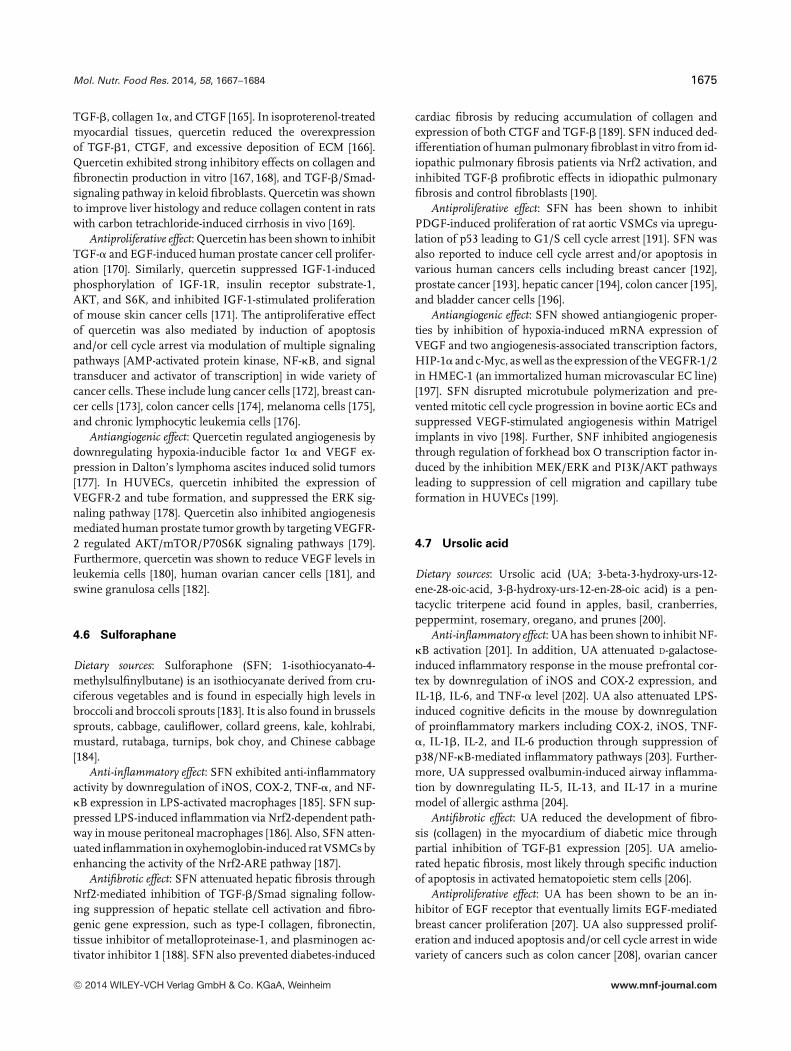
TGF-�, collagen 1�, and CTGF [165]. In isoproterenol-treatedmyocardial tissues, quercetin reduced the overexpressionof TGF-�1, CTGF, and excessive deposition of ECM [166].Quercetin exhibited strong inhibitory effects on collagen andfibronectin production in vitro [167, 168], and TGF-�/Smad-signaling pathway in keloid fibroblasts. Quercetin was shownto improve liver histology and reduce collagen content in ratswith carbon tetrachloride-induced cirrhosis in vivo [169].
Antiproliferative effect: Quercetin has been shown to inhibitTGF-� and EGF-induced human prostate cancer cell prolifer-ation [170]. Similarly, quercetin suppressed IGF-1-inducedphosphorylation of IGF-1R, insulin receptor substrate-1,AKT, and S6K, and inhibited IGF-1-stimulated proliferationof mouse skin cancer cells [171]. The antiproliferative effectof quercetin was also mediated by induction of apoptosisand/or cell cycle arrest via modulation of multiple signalingpathways [AMP-activated protein kinase, NF-�B, and signaltransducer and activator of transcription] in wide variety ofcancer cells. These include lung cancer cells [172], breast can-cer cells [173], colon cancer cells [174], melanoma cells [175],and chronic lymphocytic leukemia cells [176].
Antiangiogenic effect: Quercetin regulated angiogenesis bydownregulating hypoxia-inducible factor 1� and VEGF ex-pression in Dalton’s lymphoma ascites induced solid tumors[177]. In HUVECs, quercetin inhibited the expression ofVEGFR-2 and tube formation, and suppressed the ERK sig-naling pathway [178]. Quercetin also inhibited angiogenesismediated human prostate tumor growth by targeting VEGFR-2 regulated AKT/mTOR/P70S6K signaling pathways [179].Furthermore, quercetin was shown to reduce VEGF levels inleukemia cells [180], human ovarian cancer cells [181], andswine granulosa cells [182].
4.6 Sulforaphane
Dietary sources: Sulforaphone (SFN; 1-isothiocyanato-4-methylsulfinylbutane) is an isothiocyanate derived from cru-ciferous vegetables and is found in especially high levels inbroccoli and broccoli sprouts [183]. It is also found in brusselssprouts, cabbage, cauliflower, collard greens, kale, kohlrabi,mustard, rutabaga, turnips, bok choy, and Chinese cabbage[184].
Anti-inflammatory effect: SFN exhibited anti-inflammatoryactivity by downregulation of iNOS, COX-2, TNF-�, and NF-�B expression in LPS-activated macrophages [185]. SFN sup-pressed LPS-induced inflammation via Nrf2-dependent path-way in mouse peritoneal macrophages [186]. Also, SFN atten-uated inflammation in oxyhemoglobin-induced rat VSMCs byenhancing the activity of the Nrf2-ARE pathway [187].
Antifibrotic effect: SFN attenuated hepatic fibrosis throughNrf2-mediated inhibition of TGF-�/Smad signaling follow-ing suppression of hepatic stellate cell activation and fibro-genic gene expression, such as type-I collagen, fibronectin,tissue inhibitor of metalloproteinase-1, and plasminogen ac-tivator inhibitor 1 [188]. SFN also prevented diabetes-induced
cardiac fibrosis by reducing accumulation of collagen andexpression of both CTGF and TGF-� [189]. SFN induced ded-ifferentiation of human pulmonary fibroblast in vitro from id-iopathic pulmonary fibrosis patients via Nrf2 activation, andinhibited TGF-� profibrotic effects in idiopathic pulmonaryfibrosis and control fibroblasts [190].
Antiproliferative effect: SFN has been shown to inhibitPDGF-induced proliferation of rat aortic VSMCs via upregu-lation of p53 leading to G1/S cell cycle arrest [191]. SFN wasalso reported to induce cell cycle arrest and/or apoptosis invarious human cancers cells including breast cancer [192],prostate cancer [193], hepatic cancer [194], colon cancer [195],and bladder cancer cells [196].
Antiangiogenic effect: SFN showed antiangiogenic proper-ties by inhibition of hypoxia-induced mRNA expression ofVEGF and two angiogenesis-associated transcription factors,HIP-1� and c-Myc, as well as the expression of the VEGFR-1/2in HMEC-1 (an immortalized human microvascular EC line)[197]. SFN disrupted microtubule polymerization and pre-vented mitotic cell cycle progression in bovine aortic ECs andsuppressed VEGF-stimulated angiogenesis within Matrigelimplants in vivo [198]. Further, SNF inhibited angiogenesisthrough regulation of forkhead box O transcription factor in-duced by the inhibition MEK/ERK and PI3K/AKT pathwaysleading to suppression of cell migration and capillary tubeformation in HUVECs [199].
4.7 Ursolic acid
Dietary sources: Ursolic acid (UA; 3-beta-3-hydroxy-urs-12-ene-28-oic-acid, 3-�-hydroxy-urs-12-en-28-oic acid) is a pen-tacyclic triterpene acid found in apples, basil, cranberries,peppermint, rosemary, oregano, and prunes [200].
Anti-inflammatory effect: UA has been shown to inhibit NF-�B activation [201]. In addition, UA attenuated D-galactose-induced inflammatory response in the mouse prefrontal cor-tex by downregulation of iNOS and COX-2 expression, andIL-1�, IL-6, and TNF-� level [202]. UA also attenuated LPS-induced cognitive deficits in the mouse by downregulationof proinflammatory markers including COX-2, iNOS, TNF-�, IL-1�, IL-2, and IL-6 production through suppression ofp38/NF-�B-mediated inflammatory pathways [203]. Further-more, UA suppressed ovalbumin-induced airway inflamma-tion by downregulating IL-5, IL-13, and IL-17 in a murinemodel of allergic asthma [204].
Antifibrotic effect: UA reduced the development of fibro-sis (collagen) in the myocardium of diabetic mice throughpartial inhibition of TGF-�1 expression [205]. UA amelio-rated hepatic fibrosis, most likely through specific inductionof apoptosis in activated hematopoietic stem cells [206].
Antiproliferative effect: UA has been shown to be an in-hibitor of EGF receptor that eventually limits EGF-mediatedbreast cancer proliferation [207]. UA also suppressed prolif-eration and induced apoptosis and/or cell cycle arrest in widevariety of cancers such as colon cancer [208], ovarian cancer
C© 2014 WILEY-VCH Verlag GmbH & Co. KGaA, Weinheim www.mnf-journal.com

1676 M. S. Islam et al. Mol. Nutr. Food Res. 2014, 58, 1667–1684
[209], prostate cancer [210], nonsmall cell lung cancer [211],gastric cancer, liver cancer [212], cervical cancer [213], pan-creatic cancer [214], and bladder cancer [215], through modu-lating multiple signaling pathways (AKT/ERK, COX-2/PGE2,p300/NF-�B/CAMP response element-binding protein 2, andcytochrome c).
Antiangiogenic effect: UA inhibited key steps of angiogen-esis in vitro, including EC proliferation, migration, and dif-ferentiation [216]. UA also inhibited tumor angiogenesis bythe downregulation of VEGF in an Ehrlich ascites carcinomatumor [217], VEGF-A and basic fibroblast growth factor incolorectal cancer [218], and VEGF in melanoma cells [219].
5 Concluding remarks
Uterine fibroids are extremely common benign tumors, andthe condition exacts a significant morbidity on the health ofwomen. Unfortunately, few medical treatments are availablefor this condition. In this context, dietary phytochemicalscould play an important role in the new drug developmentfor leiomyoma treatment. Currently, only EGCG [35], cur-cumin [37], isoliquiritigenin [38], genistein [39], and resvera-trol [40,41] (Table 1) have been tested for therapeutic efficacyfor fibroids. Among these, EGCG has shown excellent effi-ciency to reduce leiomyoma cells proliferation in vitro [35],and to reduce the volume and weight of tumors of femalemice (implanted with fibroid tumor cells in vivo [77]). Adouble-blinded, placebo-controlled randomized clinical trialhas shown that green tea extract treatment significantly re-duced uterine fibroid volume, fibroid-specific symptom sever-ity, and induced significant improvement in health-relatedquality of life in premenopausal women compared to theplacebo group [36]. In addition, in a case-control study of Ital-ian women, it was shown that the risk of uterine leiomyomawas inversely associated with the intake of green vegetablesand fruit [220]. Furthermore, in a prospective cohort study,investigators found that a high intake of fruit, particularlycitrus fruit, was inversely associated with uterine fibroidsrisk among black women [221]. Therefore, considering thekey characteristics of leiomyoma development and growth,inflammation, fibrosis, proliferation, and angiogenesis, ouraim was to introduce some promising dietary phytochemicals(allicin, EA, I3C, lycopene, quercetin, sulforaphane, and UA)(Fig. 1) that have already shown multiple therapeutic effects indifferent biological conditions. Throughout this manuscript,we introduced and emphasized the enormous potential of di-etary phytochemicals as possible effective therapeutic agentsthrough (i) inhibition of inflammatory mediators; (ii) inhi-bition of fibrosis by decreasing ECM deposition, profibroticgrowth factors expression, inactivation of activated cell types(responsible for myofibroblastic transformation); (iii) inhibi-tion of cell proliferation via the activation of the apoptoticpathway and cell cycle arrest, as well as through inhibition ofgrowth factors and/or their receptors; and (iv) inhibition of an-giogenesis by reducing angiogenic growth factors/receptors
and angiogenesis-related transcription factors. Based on theavailable evidence, these compounds modulate and regulatethe key biological processes involved in leiomyoma devel-opment and growth (Fig. 2). Alone, these phytochemicalsare promising, but it is also possible that in combinationthe therapeutic effects could be additive and the magnitudeof the effect could translate to a significant clinical therapy.Lastly, further study of these promising compounds mightlead to development of strategies to prevent the condition inwomen at risk for this extremely common, but debilitatingdisease.
In this review, dietary phytochemicals have been shownas tumor-fighting weapons. However, greater attention isneeded to clarify the following important issues. (i) Poorpotency and bioavailability of dietary phytochemicals cre-ates challenges to scientists. However, introducing syntheticanalogs of dietary phytochemicals could be a solution forthese potency and bioavailability limitations. For example, thepotency of synthetic curcumin analog EF24 was shown to beapproximately tenfold greater than that of natural curcumin[222]. (ii) Instability of dietary phytochemicals is often asso-ciated with pH and/or enzyme-mediated degradation in theupper gut. These can be overcome by formulation approachessuch as enteric coating. A major stability factor that is oftenoverlooked is the effect of microbiota in the gastrointestinaltract. The stability of a drug to the microbiota is clinicallyrelevant as drug metabolism can render a drug pharmacolog-ically active, inactive, or toxic. An important example of thesignificance of metabolism was seen in Japan in 1993 whensorivudine, a promising antiviral drug was introduced into theJapanese market. This was later discovered to be transformedby gut microbiota into (E)-5-(2-bromovinyl)uracil, which caninhibit the metabolism of the anticancer drug 5-fluorouracilleading to toxic levels of this drug [223]. (iii) Although moststudies have suggested that dietary phytochemicals kill tumorcells selectively, phytochemicals may have similar effect onnormal cells as well [224]. (iv) In many cases, the chemopre-ventive effects of dietary phytochemicals in cultured cells ortissues are only achievable at supraphysiological concentra-tions. Such concentrations might not be attained when thephytochemicals are administered as part of diet. (v) The ef-ficacy of most dietary phytochemicals has been tested onlyin preclinical conditions, either in vitro or in vivo. However,the beneficial effects of dietary phytochemicals in humansare largely unknown. Based on the above facts, it is clear thatthere is huge need to better understand the efficacy of dietaryphytochemicals in the prevention and treatment of uterinefibroids. Future studies should focus on careful and accuratecharacterization of dietary phytochemicals, better elucidationof the molecular mechanisms involved in their actions, de-termination of their efficacy by in vivo studies using properanimal models of uterine fibroids, and demonstration of theirsafety and effectiveness in clinical trials.
This work was supported by a grant from the “FondazioneCassa di Risparmio di Fabriano e Cupramontana” (to M.C. and
C© 2014 WILEY-VCH Verlag GmbH & Co. KGaA, Weinheim www.mnf-journal.com

Mol. Nutr. Food Res. 2014, 58, 1667–1684 1677
P.C.) and by Italian Ministry of the University and Research(PRIN 2010–2011, No. 20102CHST5_007, to S.R.G.).
The authors have declared no conflict of interest.
6 References
[1] Ciarmela, P., Islam, M. S., Reis, F. M., Gray, P. C. et al.,Growth factors and myometrium: biological effects in uter-ine fibroid and possible clinical implications. Hum. Reprod.Update 2011, 17, 772–790.
[2] Walker, C. L., Stewart, E. A., Uterine fibroids: the elephantin the room. Science 2005, 308, 1589–1592.
[3] Islam, M. S., Protic, O., Toti, P., Giannubilo, S. R. et al., Uter-ine leiomyoma: available medical treatments and new pos-sible therapeutic options. J. Clin. Endocrinol. Metab. 2013,98, 921–934.
[4] Day Baird, D., Dunson, D. B., Hill, M. C., Cousins, D. et al.,High cumulative incidence of uterine leiomyoma in blackand white women: ultrasound evidence. Am. J. Obstet.Gynecol. 2003, 188, 100–107.
[5] Marshall, L. M., Spiegelman, D., Barbieri, R. L., Goldman,M. B. et al., Variation in the incidence of uterine leiomyomaamong premenopausal women by age and race. Obstet.Gynecol. 1997, 90, 967–973.
[6] Kjerulff, K. H., Langenberg, P., Seidman, J. D., Stolley, P. D.et al., Uterine leiomyomas. Racial differences in severity,symptoms and age at diagnosis. J. Reprod. Med. 1996, 41,483–490.
[7] Moore, A. B., Flake, G. P., Swartz, C. D., Heartwell, G. et al.,Association of race, age and body mass index with grosspathology of uterine fibroids. J. Reprod. Med. 2008, 53, 90–96.
[8] Peddada, S. D., Laughlin, S. K., Miner, K., Guyon, J.-P. et al.,Growth of uterine leiomyomata among premenopausalblack and white women. Proc. Natl. Acad. Sci. USA 2008,105, 19887–19892.
[9] Weiss, G., Noorhasan, D., Schott, L. L., Powell, L. et al.,Racial differences in women who have a hysterectomy forbenign conditions. Women’s Health Issues 2009, 19, 202–210.
[10] Stewart, E. A., Uterine fibroids. Lancet 2001, 357, 293–298.
[11] Farquhar, C. M., Steiner, C. A., Hysterectomy rates in theUnited States 1990–1997. Obstet. Gynecol. 2002, 99, 229–234.
[12] Cardozo, E. R., Clark, A. D., Banks, N. K., Henne, M. B.et al., The estimated annual cost of uterine leiomyomata inthe United States. Am. J. Obstet. Gynecol. 2012, 206(211),e211–e219.
[13] Treloar, S. A., Do, K.-A., O’Connor, V. M., O’Connor, D. T.et al., Predictors of hysterectomy: an Australian study. Am.J. Obstet. Gynecol. 1999, 180, 945–954.
[14] Downes, E., Sikirica, V., Gilabert-Estelles, J., Bolge, S. C.et al., The burden of uterine fibroids in five European coun-tries. Eur. J. Obstet. Gynecol. Reprod. Biol. 2010, 152, 96–102.
[15] Wegienka, G., Are uterine leiomyoma a consequence of achronically inflammatory immune system? Med. Hypothe-ses 2012, 79, 226–231.
[16] Leppert, P., Fouany, M., Segars, J. H., in: Segars, J. H. (Ed.)Understanding Uterine Fibroids—Fibroids, John Wiley &Sons, Ltd, Oxford 2013, pp. 1–10.
[17] Chegini, N., Proinflammatory and profibrotic mediators:principal effectors of leiomyoma development as a fibroticdisorder. Semin. Reprod. Med. 2010, 28, 180–203.
[18] Marsh, E. E., Bulun, S. E., Steroid hormones and leiomy-omas. Obstet. Gynecol. Clin. North Am. 2006, 33, 59–67.
[19] Rein, M. S., Barbieri, R. L., Friedman, A. J., Progesterone: acritical role in the pathogenesis of uterine myomas. Am. J.Obstet. Gynecol. 1995, 172, 14–18.
[20] Englund, K., Blanck, A., Gustavsson, I., Lundkvist, U. et al.,Sex steroid receptors in human myometrium and fibroids:changes during the menstrual cycle and gonadotropin-releasing hormone treatment. J. Clin. Endocrinol. Metab.1998, 83, 4092–4096.
[21] Islam, M. S., Protic, O., Stortoni, P., Grechi, G. et al., Complexnetworks of multiple factors in the pathogenesis of uterineleiomyoma. Fertil. Steril. 2013, 100, 178–193.
[22] Friedman, A. J., Rein, M. S., Harrison-Atlas, D., Garfield, J.M. et al., A randomized, placebo-controlled, double-blindstudy evaluating leuprolide acetate depot treatment beforemyomectomy. Fertil. Steril. 1989, 52, 728–733.
[23] Andreyko, J. L., Blumenfeld, Z., Marshall, L. A., Monroe,S. E. et al., Use of an agonistic analog of gonadotropin-releasing hormone (nafarelin) to treat leiomyomas: assess-ment by magnetic resonance imaging. Am. J. Obstet. Gy-necol. 1988, 158, 903–910.
[24] Leather, A. T., Studd, J. W. W., Watson, N. R., Holland, E. F.N., The prevention of bone loss in young women treatedwith GnRH analogues with “add-back” estrogen therapy.Obstet. Gynecol. 1993, 81, 104–107.
[25] Friedman, A. J., Hoffman, D. I., Comite, F., Browneller, R. W.et al., Treatment of leiomyomata uteri with leuprolide ac-etate depot: a double-blind, placebo-controlled, multicen-ter study. The Leuprolide Study Group. Obstet. Gynecol.1991, 77, 720–725.
[26] Stovall, T. G., Muneyyirci-Delale, O., Summitt, R. L., Jr.,Scialli, A. R., GnRH agonist and iron versus placebo andiron in the anemic patient before surgery for leiomyomas:a randomized controlled trial. The Leuprolide Study Group.Obstet. Gynecol. 1995, 86, 65–71.
[27] Carbonell Esteve, J. L., Acosta, R., Heredia, B., Perez, Y.et al., Mifepristone for the treatment of uterine leiomyomas:a randomized controlled trial. Obstet. Gynecol. 2008, 112,1029–1036.
[28] Chwalisz, K., Larsen, L., Mattia-Goldberg, C., Edmonds,A. et al., A randomized, controlled trial of asoprisnil,a novel selective progesterone receptor modulator, inwomen with uterine leiomyomata. Fertil. Steril. 2007, 87,1399–1412.
[29] Donnez, J., Tomaszewski, J., Vazquez, F., Bouchard, P.et al., Ulipristal acetate versus leuprolide acetate for uterinefibroids. N. Engl. J. Med. 2012, 366, 421–432.
C© 2014 WILEY-VCH Verlag GmbH & Co. KGaA, Weinheim www.mnf-journal.com

1678 M. S. Islam et al. Mol. Nutr. Food Res. 2014, 58, 1667–1684
[30] Donnez, J., Tatarchuk, T. F., Bouchard, P., Puscasiu, L. et al.,Ulipristal acetate versus placebo for fibroid treatment be-fore surgery. N. Engl. J. Med. 2012, 366, 409–420.
[31] Wiehle, R. D., Goldberg, J., Brodniewicz, T., Jarus-Dziedzic,K. et al., Effects of a new progesterone receptor modulator,CDB-4124, on fibroid size and uterine bleeding. US Obstet.Gynecol. 2008, 3, 17–20.
[32] Newman, D. J., Cragg, G. M., Natural products as sourcesof new drugs over the 30 years from 1981 to 2010. J. Nat.Prod. 2012, 75, 311–335.
[33] Chen, N.-N., Han, M., Yang, H., Yang, G.-Y. et al., Chineseherbal medicine Guizhi Fuling Formula for treatment ofuterine fibroids: a systematic review of randomised clin-ical trials. BMC Complement. Altern. Med. 2014, 14, 2.
[34] Liu, J. P., Yang, H., Xia, Y., Cardini, F., Herbal preparationsfor uterine fibroids. Cochrane Database Syst. Rev. 2009, 4,CD005292.
[35] Zhang, D., Al-Hendy, M., Richard-Davis, G., Montgomery-Rice, V. et al., Antiproliferative and proapoptotic effects ofepigallocatechin gallate on human leiomyoma cells. Fertil.Steril. 2010, 94, 1887–1893.
[36] Roshdy, E., Rajaratnam, V., Maitra, S., Sabry, M. et al., Treat-ment of symptomatic uterine fibroids with green tea ex-tract: a pilot randomized controlled clinical study. Int. J.Women’s Health 2013, 5, 477–486.
[37] Malik, M., Mendoza, M., Payson, M., Catherino, W. H., Cur-cumin, a nutritional supplement with antineoplastic activ-ity, enhances leiomyoma cell apoptosis and decreases fi-bronectin expression. Fertil. Steril. 2009, 91, 2177–2184.
[38] Kim, D., Ramachandran, S., Baek, S., Kwon, S. H. et al., In-duction of growth inhibition and apoptosis in human uter-ine leiomyoma cells by isoliquiritigenin. Reprod. Sci. 2008,15, 552.
[39] Shushan, A., Ben-Bassat, H., Mishani, E., Laufer, N. et al.,Inhibition of leiomyoma cell proliferation in vitro by genis-tein and the protein tyrosine kinase inhibitor TKS050. Fertil.Steril. 2007, 87, 127–135.
[40] Catherino, W. H., Parrott, E., Segars, J., Proceedings fromthe National Institute of Child Health and Human Develop-ment Conference on the Uterine Fibroid Research UpdateWorkshop. Fertil. Steril. 2011, 95, 9–12.
[41] Christman, G. M., Marsh, C. A., Campbell, E. J., in: Segars,J. H. (Ed.) Counseling the Patient with Uterine Fibroids—Fibroids, John Wiley & Sons, Oxford 2012, pp. 134–144.
[42] Hatthachote, P., Gillespie, J. I., Complex interactions be-tween sex steroids and cytokines in the human pregnantmyometrium: evidence for an autocrine signaling systemat term. Endocrinology 1999, 140, 2533–2540.
[43] Litovkin, K. V., Domenyuk, V. P., Bubnov, V. V., Zaporozhan,V. N., Interleukin-6-174G/C polymorphism in breast cancerand uterine leiomyoma patients: a population-based casecontrol study. Exp. Oncol. 2007, 29, 295–298.
[44] Luo, X., Ding, L., Xu, J., Chegini, N., Gene expression profil-ing of leiomyoma and myometrial smooth muscle cells inresponse to transforming growth factor-beta. Endocrinol-ogy 2005, 146, 1097–1118.
[45] Kurachi, O., Matsuo, H., Samoto, T., Maruo, T., Tumor necro-sis factor-� expression in human uterine leiomyoma andits down-regulation by progesterone. J. Clin. Endocrinol.Metab. 2001, 86, 2275–2280.
[46] Chegini, N., Tang, X. M., Ma, C., Regulation of trans-forming growth factor-beta1 expression by granulocytemacrophage-colony-stimulating factor in leiomyoma andmyometrial smooth muscle cells. J. Clin. Endocrinol.Metab. 1999, 84, 4138–4143.
[47] Santulli, P., Even, M., Chouzenoux, S., Millischer, A.-E. et al.,Profibrotic interleukin-33 is correlated with uterine leiomy-oma tumour burden. Hum. Reprod. 2013, 28, 2126–2133.
[48] Sozen, I., Olive, D. L., Arici, A., Expression and hor-monal regulation of monocyte chemotactic protein-1 in my-ometrium and leiomyomata. Fertil. Steril. 1998, 69, 1095–1102.
[49] Senturk, L. M., Sozen, I., Gutierrez, L., Arici, A., Interleukin8 production and interleukin 8 receptor expression in hu-man myometrium and leiomyoma. Am. J. Obstet. Gynecol.2001, 184, 559–566.
[50] Syssoev, K. A., Kulagina, N. V., Chukhlovin, A. B., Moro-zova, E. B. et al., Expression of mRNA for chemokines andchemokine receptors in tissues of the myometrium anduterine leiomyoma. Bull. Exp. Biol. Med. 2008, 145, 84–89.
[51] Cesen-Cummings, K., Houston, K. D., Copland, J. A., Moor-man, V. J. et al., Uterine leiomyomas express myome-trial contractile-associated proteins involved in pregnancy-related hormone signaling. J. Soc. Gynecol. Investig. 2003,10, 11–20.
[52] Khorram, O., Garthwaite, M., Magness, R. R., Endome-trial and myometrial expression of nitric oxide synthaseisoforms in pre-and postmenopausal women. J. Clin. En-docrinol. Metab. 1999, 84, 2226–2232.
[53] Fujita, M., Histological and biochemical studies of collagenin human uterine leiomyomas. Hokkaido Igaku Zasshi 1985,60, 602–615.
[54] Leppert, P. C., Baginski, T., Prupas, C., Catherino, W. H. et al.,Comparative ultrastructure of collagen fibrils in uterineleiomyomas and normal myometrium. Fertil. Steril. 2004,82(Suppl 3), 1182–1187.
[55] Arici, A., Sozen, I., Transforming growth factor-beta3 is ex-pressed at high levels in leiomyoma where it stimulatesfibronectin expression and cell proliferation. Fertil. Steril.2000, 73, 1006–1011.
[56] Stewart, E. A., Friedman, A. J., Peck, K., Nowak, R. A., Rel-ative overexpression of collagen type I and collagen typeIII messenger ribonucleic acids by uterine leiomyomas dur-ing the proliferative phase of the menstrual cycle. J. Clin.Endocrinol. Metab. 1994, 79, 900–906.
[57] Norian, J. M., Malik, M., Parker, C. Y., Joseph, D. et al., Trans-forming growth factor beta3 regulates the versican vari-ants in the extracellular matrix-rich uterine leiomyomas.Reprod. Sci. 2009, 16, 1153–1164.
[58] Rogers, R., Norian, J., Malik, M., Christman, G. et al., Me-chanical homeostasis is altered in uterine leiomyoma. Am.J. Obstet. Gynecol. 2008, 198, 474.e471–474.e411.
C© 2014 WILEY-VCH Verlag GmbH & Co. KGaA, Weinheim www.mnf-journal.com

Mol. Nutr. Food Res. 2014, 58, 1667–1684 1679
[59] Norian, J. M., Owen, C. M., Taboas, J., Korecki, C. et al.,Characterization of tissue biomechanics and mechanicalsignaling in uterine leiomyoma. Matrix Biol. 2012, 31, 57–65.
[60] Joseph, D. S., Malik, M., Nurudeen, S., Catherino, W. H.,Myometrial cells undergo fibrotic transformation under theinfluence of transforming growth factor beta-3. Fertil. Steril.2010, 93, 1500–1508.
[61] Islam, M. S., Catherino, W. H., Protic, O., Janjusevic,M. et al., Role of activin-A and myostatin and theirsignaling pathway in human myometrial and leiomy-oma cell function. J. Clin. Endocrinol. Metab. 2014, 99,775–785.
[62] Liang, M., Wang, H., Zhang, Y., Lu, S., Wang, Z., Expres-sion and functional analysis of platelet-derived growth fac-tor in uterine leiomyomata. Cancer Biol. Ther. 2006, 5,28–33.
[63] Kawaguchi, K., Fujii, S., Konishi, I., Nanbu, Y. et al., Mitoticactivity in uterine leiomyomas during the menstrual cycle.Am. J. Obstet. Gynecol. 1989, 160, 637–641.
[64] Wolanska, M., Bankowski, E., Transforming growth fac-tor beta and platelet-derived growth factor in human my-ometrium and in uterine leiomyomas at various stages oftumour growth. Eur. J. Obstet. Gynecol. Reprod. Biol. 2007,130, 238–244.
[65] Arici, A., Sozen, I., Expression, menstrual cycle-dependentactivation, and bimodal mitogenic effect of transforminggrowth factor-beta1 in human myometrium and leiomy-oma. Am. J. Obstet. Gynecol. 2003, 188, 76–83.
[66] Fletcher, N. M., Saed, M. G., Abuanzeh, S., Abu-Soud, H.M. et al., Nicotinamide adenine dinucleotide phosphate ox-idase is differentially regulated in normal myometrium ver-sus leiomyoma. Reprod. Sci. 2014 [Epub ahead of print].
[67] Mesquita, F. S., Dyer, S. N., Heinrich, D. A., Bulun, S. E. et al.,Reactive oxygen species mediate mitogenic growth factorsignaling pathways in human leiomyoma smooth musclecells. Biol. Reprod. 2010, 82, 341–351.
[68] Fletcher, N. M., Saed, M. G., Abu-Soud, H. M., Al-Hendy,A. et al., Uterine fibroids are characterized by an impairedantioxidant cellular system: potential role of hypoxia inthe pathophysiology of uterine fibroids. J. Assist. Reprod.Genet. 2014, 30, 969–974.
[69] Nair, S., Al-Hendy, A., Adipocytes enhance the prolifera-tion of human leiomyoma cells via TNF-� proinflammatorycytokine. Reprod. Sci. 2011, 18, 1186–1192.
[70] Carmeliet, P., Angiogenesis in health and disease. Nat. Med.2003, 9, 653–660.
[71] Imhof, B. A., Aurrand-Lions, M., Angiogenesis and inflam-mation face off. Nat. Med. 2006, 12, 171–172.
[72] Chung, A. S., Lee, J., Ferrara, N., Targeting the tumourvasculature: insights from physiological angiogenesis. Nat.Rev. Cancer 2010, 10, 505–514.
[73] Fleischer, R., Weston, G. C., Vollenhoven, B. J., Rogers, P. A.W., Pathophysiology of fibroid disease: angiogenesis andregulation of smooth muscle proliferation. Best Pract. Res.Clin. Obstet. Gynaecol. 2008, 22, 603–614.
[74] Tal, R., Segars, J. H., The role of angiogenic factors in fi-broid pathogenesis: potential implications for future ther-apy. Hum. Reprod. Update 2013, 20, 194–216.
[75] Ciarmela, P., Carrarelli, P., Islam, M. S., Janjusevic, M. et al.,Effect of ulipristal acetate on activin A expression andfunctions in myometrial and leiomyoma cells. Reprod. Sci.2014.
[76] Singh, B. N., Shankar, S., Srivastava, R. K., Green tea cat-echin, epigallocatechin-3-gallate (EGCG): mechanisms, per-spectives and clinical applications. Biochem. Pharmacol.2011, 82, 1807–1821.
[77] Zhang, D., Al-Hendy, M., Richard-Davis, G., Montgomery-Rice, V. et al., Green tea extract inhibits proliferation ofuterine leiomyoma cells in vitro and in nude mice. Am. J.Obstet. Gynecol. 2010, 202, 289.e281–289.e289.
[78] Ozercan, I. H., Sahin, N., Akdemir, F., Onderci, M. et al.,Chemoprevention of fibroid tumors by [-]-epigallocatechin-3-gallate in quail. Nutr. Res. 2008, 28, 92–97.
[79] Wilken, R., Veena, M. S., Wang, M. B., Srivatsan, E. S., Cur-cumin: a review of anti-cancer properties and therapeuticactivity in head and neck squamous cell carcinoma. Mol.Cancer 2011, 10, 12.
[80] Tsuiji, K., Takeda, T., Li, B., Wakabayashi, A. et al., Inhibitoryeffect of curcumin on uterine leiomyoma cell proliferation.Gynecol. Endocrinol. 2011, 27, 512–517.
[81] Cuendet, M., Guo, J., Luo, Y., Chen, S. et al., Cancer chemo-preventive activity and metabolism of isoliquiritigenin, acompound found in licorice. Cancer Prev. Res. 2010, 3, 221–232.
[82] Kaufman, P. B., Duke, J. A., Brielmann, H., Boik, J. et al., Acomparative survey of leguminous plants as sources of theisoflavones, genistein and daidzein: implications for humannutrition and health. J. Altern. Complement. Med. 1997, 3,7–12.
[83] Moore, A. B., Castro, L., Yu, L., Zheng, X. et al., Stimula-tory and inhibitory effects of genistein on human uterineleiomyoma cell proliferation are influenced by the concen-tration. Hum. Reprod. 2007, 22, 2623–2631.
[84] Di, X., Yu, L., Moore, A. B., Castro, L. et al., A low concen-tration of genistein induces estrogen receptor-alpha andinsulin-like growth factor-I receptor interactions and prolif-eration in uterine leiomyoma cells. Hum. Reprod. 2008, 23,1873–1883.
[85] Di, X., Andrews, D. M. K., Tucker, C. J., Yu, L. et al., Ahigh concentration of genistein down-regulates activin A,Smad3 and other TGF-� pathway genes in human uterineleiomyoma cells. Exp. Mol. Med. 2012, 44, 281–292.
[86] Sahin, K., Akdemir, F., Tuzcu, M., Sahin, N. et al., Genisteinsuppresses spontaneous oviduct tumorigenesis in Quail.Nutr. Cancer 2009, 61, 799–806.
[87] Labinskyy, N., Csiszar, A., Veress, G., Stef, G. et al., Vascu-lar dysfunction in aging: potential effects of resveratrol, ananti-inflammatory phytoestrogen. Curr. Med. Chem. 2006,13, 989.
[88] Block, E., The chemistry of garlic and onions. Sci. Am. 1985,252, 114–119.
C© 2014 WILEY-VCH Verlag GmbH & Co. KGaA, Weinheim www.mnf-journal.com

1680 M. S. Islam et al. Mol. Nutr. Food Res. 2014, 58, 1667–1684
[89] Mo, S.-J., Son, E.-W., Rhee, D.-K., Pyo, S., Modulation ofTNF-�-induced ICAM-1 expression, NO and H2O2 produc-tion by alginate, allicin and ascorbic acid in human en-dothelial cells. Arch. Pharm. Res. 2003, 26, 244–251.
[90] Lang, A., Lahav, M., Sakhnini, E., Barshack, I. et al., Allicininhibits spontaneous and TNF-alpha induced secretion ofproinflammatory cytokines and chemokines from intestinalepithelial cells. Clin. Nutr. 2004, 23, 1199–1208.
[91] Gu, X., Wu, H., Fu, P., Allicin attenuates inflammationand suppresses HLA-B27 protein expression in ankylos-ing spondylitis mice. J. Biomed. Biotechnol. 2013, 2013,171573.
[92] Liu, C., Cao, F., Tang, Q.-Z., Yan, L. et al., Allicin protectsagainst cardiac hypertrophy and fibrosis via attenuatingreactive oxygen species-dependent signaling pathways. J.Nutr. Biochem. 2010, 21, 1238–1250.
[93] Li, X.-H., Li, C.-Y., Xiang, Z.-G., Hu, J.-J. et al., Allicin amelio-rates cardiac hypertrophy and fibrosis through enhancingof Nrf2 antioxidant signaling pathways. Cardiovasc. DrugsTher. 2012, 26, 457–465.
[94] Liu, Y., Qi, H., Wang, Y., Wu, M. et al., Allicin protects againstmyocardial apoptosis and fibrosis in streptozotocin-induced diabetic rats. Phytomedicine 2012, 19, 693–698.
[95] Oommen, S., Anto, R. J., Srinivas, G., Karunagaran, D., Al-licin (from garlic) induces caspase-mediated apoptosis incancer cells. Eur. J. Pharmacol. 2004, 485, 97–103.
[96] Zhang, W., Ha, M., Gong, Y., Xu, Y. et al., Allicin inducesapoptosis in gastric cancer cells through activation of bothextrinsic and intrinsic pathways. Oncol. Rep. 2010, 24,1585–1592.
[97] Wang, Z., Liu, Z., Cao, Z., Li, L., Allicin induces apoptosis inEL-4 cells in vitro by activation of expression of caspase-3 and-12 and up-regulation of the ratio of Bax/Bcl-2. Nat.Prod. Res. 2012, 26, 1033–1037.
[98] Bat-Chen, W., Golan, T., Peri, I., Ludmer, Z. et al., Allicinpurified from fresh garlic cloves induces apoptosis in coloncancer cells via Nrf2. Nutr. Cancer 2010, 62, 947–957.
[99] Cha, J. H., Choi, Y. J., Cha, S. H., Choi, C. H. et al., Allicininhibits cell growth and induces apoptosis in U87MG hu-man glioblastoma cells through an ERK-dependent path-way. Oncol. Rep. 2012, 28, 41–48.
[100] Hirsch, K., Danilenko, M., Giat, J., Miron, T. et al., Effectof purified allicin, the major ingredient of freshly crushedgarlic, on cancer cell proliferation. Nutr. Cancer 2000, 38,245–254.
[101] Sela, U., Brill, A., Kalchenko, V., Dashevsky, O. et al., Allicininhibits blood vessel growth and downregulates Akt phos-phorylation and actin polymerization. Nutr. Cancer 2008,60, 412–420.
[102] Vattem, D. A., Shetty, K., Biological functionality of ellagicacid: a review. J. Food Biochem. 2005, 29, 234–266.
[103] Chao, P. C., Hsu, C. C., Yin, M. C., Anti-inflammatory andanti-coagulatory activities of caffeic acid and ellagic acid incardiac tissue of diabetic mice. Nutr. Metab. 2009, 6, 33.
[104] Umesalma, S., Sudhandiran, G., Differential inhibitory ef-fects of the polyphenol ellagic acid on inflammatory me-
diators NF-kappaB, iNOS, COX-2, TNF-alpha, IL-6 in 1,2-dimethylhydrazine-induced rat colon carcinogenesis. BasicClin. Pharmacol. Toxicol. 2010, 107, 650–655.
[105] Karlsson, S., Nanberg, E., Fjaeraa, C., Wijkander, J., Ellagicacid inhibits lipopolysaccharide-induced expression of en-zymes involved in the synthesis of prostaglandin E2 in hu-man monocytes. Br. J. Nutr. 2010, 103, 1102.
[106] Marın, M., Marıa Giner, R., Rıos, J.-L., Carmen Recio, M.,Intestinal anti-inflammatory activity of ellagic acid in theacute and chronic dextrane sulfate sodium models of micecolitis. J. Ethnopharmacol. 2013, 150, 925–934.
[107] Thresiamma, K. C., Kuttan, R., Inhibition of liver fibrosisby ellagic acid. Indian J. Physiol. Pharmacol. 1996, 40,363–366.
[108] Iino, T., Tashima, K., Umeda, M., Ogawa, Y. et al., Effectof ellagic acid on gastric damage induced in ischemic ratstomachs following ammonia or reperfusion. Life Sci. 2002,70, 1139–1150.
[109] Masamune, A., Satoh, M., Kikuta, K., Suzuki, N. et al., Ellagicacid blocks activation of pancreatic stellate cells. Biochem.Pharmacol. 2005, 70, 869–878.
[110] Suzuki, N., Masamune, A., Kikuta, K., Watanabe, T. et al.,Ellagic acid inhibits pancreatic fibrosis in male WistarBonn/Kobori rats. Dig. Dis. Sci. 2009, 54, 802–810.
[111] Rani P. U., Kesavan, R., Ganugula, R., Kumar, P. U. et al.,Ellagic acid inhibits PDGF-BB-induced vascular smoothmuscle cell proliferation and prevents atheroma formationin streptozotocin-induced diabetic rats. J. Nutr. Biochem.2013, 24, 1830–1839.
[112] Narayanan, B. A., Geoffroy, O., Willingham, M. C., Re, G.G. et al., p53/p21 (WAF1/CIP1) expression and its possiblerole in G1 arrest and apoptosis in ellagic acid treated cancercells. Cancer Lett. 1999, 136, 215–221.
[113] Edderkaoui, M., Lugea, A., Hui, H., Eibl, G. et al., Ellagic acidand embelin affect key cellular components of pancreaticadenocarcinoma, cancer, stellate cells. Nutr. Cancer 2013,65, 1232–1244.
[114] Chung, Y.-C., Lu, L.-C., Tsai, M.-H., Chen, Y.-J. et al., Theinhibitory effect of ellagic acid on cell growth of ovariancarcinoma cells. Evid. Based Complement. Alternat. Med.2013, 2013, 306705.
[115] Losso, J. N., Bansode, R. R., Trappey Ii, A., Bawadi, H. A.et al., In vitro anti-proliferative activities of ellagic acid. J.Nutr. Biochem. 2004, 15, 672–678.
[116] Weisburg, J. H., Schuck, A. G., Reiss, S. E., Wolf, B. J. et al.,Ellagic acid, a dietary polyphenol, selectively cytotoxic toHSC-2 oral carcinoma cells. Anticancer Res. 2013, 33, 1829–1836.
[117] Qiu, Z., Zhou, B., Jin, L., Yu, H. et al., In vitro antioxidantand antiproliferative effects of ellagic acid and its colonicmetabolite, urolithins, on human bladder cancer T24 cells.Food Chem. Toxicol. 2013, 59, 428–437.
[118] Labrecque, L., Lamy, S., Chapus, A. l., Mihoubi, S. et al.,Combined inhibition of PDGF and VEGF receptors by ellagicacid, a dietary-derived phenolic compound. Carcinogenesis2005, 26, 821–826.
C© 2014 WILEY-VCH Verlag GmbH & Co. KGaA, Weinheim www.mnf-journal.com

Mol. Nutr. Food Res. 2014, 58, 1667–1684 1681
[119] Wang, N., Wang, Z.-Y., Mo, S.-L., Loo, T. Y. et al., Ellagic acid,a phenolic compound, exerts anti-angiogenesis effects viaVEGFR-2 signaling pathway in breast cancer. Breast CancerRes. Treat. 2012, 134, 943–955.
[120] Aggarwal, B. B., Ichikawa, H., Molecular targets and an-ticancer potential of indole-3-carbinol and its derivatives.Cell Cycle 2005, 4, 1201–1215.
[121] Takada, Y., Andreeff, M., Aggarwal, B. B., Indole-3-carbinolsuppresses NF-�B and I�B� kinase activation, causing inhi-bition of expression of NF-�B-regulated antiapoptotic andmetastatic gene products and enhancement of apoptosisin myeloid and leukemia cells. Blood 2005, 106, 641–649.
[122] Benson, J. M., Shepherd, D. M., Dietary ligands of the arylhydrocarbon receptor induce anti-inflammatory and im-munoregulatory effects on murine dendritic cells. Toxicol.Sci. 2013, 124, 327–338.
[123] Chen, Y.-H., Dai, H.-J., Chang, H.-P., Suppression of in-ducible nitric oxide production by indole and isothio-cyanate derivatives from Brassica plants in stimulatedmacrophages. Planta Med. 2003, 69, 696–700.
[124] Tsai, J.-T., Liu, H.-C., Chen, Y.-H., Suppression of in-flammatory mediators by cruciferous vegetable-derivedindole-3-carbinol and phenylethyl isothiocyanate inlipopolysaccharide-activated macrophages. Mediators In-flamm. 2010, 2010, 293642.
[125] Jiang, J., Kang, T. B., Shim, D. W., Oh, N. H. et al.,Indole-3-carbinol inhibits LPS-induced inflammatory re-sponse by blocking TRIF-dependent signaling pathway inmacrophages. Food Chem. Toxicol. 2013, 57, 256–261.
[126] Ping, J., Li, J.-t., Liao, Z.-x., Shang, L. et al., Indole-3-carbinol inhibits hepatic stellate cells proliferation by block-ing NADPH oxidase/reactive oxygen species/p38 MAPKpathway. Eur. J. Pharmacol. 2011, 650, 656–662.
[127] Guan, H., Chen, C., Zhu, L., Cui, C. et al., Indole-3-carbinolblocks platelet-derived growth factor-stimulated vascularsmooth muscle cell function and reduces neointima for-mation in vivo. J. Nutr. Biochem. 2012, 24, 62–69.
[128] Marconett, C. N., Singhal, A. K., Sundar, S. N., Firestone, G.L., Indole-3-carbinol disrupts estrogen receptor-alpha de-pendent expression of insulin-like growth factor-1 receptorand insulin receptor substrate-1 and proliferation of humanbreast cancer cells. Mol. Cell. Endocrinol. 2012, 363, 74–84.
[129] Zhang, J., Hsu, B. A., Jocelyn, C., Kinseth, B. A. et al.,Indole 3 carbinol induces a G1 cell cycle arrest and in-hibits prostate specific antigen production in human LNCaPprostate carcinoma cells. Cancer 2003, 98, 2511–2520.
[130] Suzui, M., Inamine, M., Kaneshiro, T., Morioka, T. et al.,Indole-3-carbinol inhibits the growth of human colon carci-noma cells but enhances the tumor multiplicity and volumeof azoxymethane-induced rat colon carcinogenesis. Int. J.Oncol. 2005, 27, 1391–1399.
[131] Choi, H.-S., Cho, M.-C., Lee, H. G., Yoon, D.-Y., Indole-3-carbinol induces apoptosis through p53 and activation ofcaspase-8 pathway in lung cancer A549 cells. Food Chem.Toxicol. 2010, 48, 883–890.
[132] Wu, H.-T., Lin, S.-H., Chen, Y.-H., Inhibition of cell prolif-eration and in vitro markers of angiogenesis by indole-3-
carbinol, a major indole metabolite present in cruciferousvegetables. J. Agric. Food Chem. 2005, 53, 5164–5169.
[133] Kunimasa, K., Kobayashi, T., Kaji, K., Ohta, T., An-tiangiogenic effects of indole-3-carbinol and 3, 3’-diindolylmethane are associated with their differential reg-ulation of ERK1/2 and Akt in tube-forming HUVEC. J. Nutr.2010, 140, 1–6.
[134] Wang, M.-L., Shih, C.-K., Chang, H.-P., Chen, Y.-H., Antian-giogenic activity of indole-3-carbinol in endothelial cellsstimulated with activated macrophages. Food Chem. 2012,134, 811–820.
[135] Holzapfel, N. P., Holzapfel, B. M., Champ, S., Feldthusen, J.et al., The potential role of lycopene for the prevention andtherapy of prostate cancer: from molecular mechanisms toclinical evidence. Int. J. Mol. Sci. 2013, 14, 14620–14646.
[136] Marcotorchino, J., Romier, B., Gouranton, E., Riollet, C.et al., Lycopene attenuates LPS-induced TNF-� secretion inmacrophages and inflammatory markers in adipocytes ex-posed to macrophage-conditioned media. Mol. Nutr. FoodRes. 2010, 56, 725–732.
[137] Simone, R. E., Russo, M., Catalano, A., Monego, G. et al.,Lycopene inhibits NF-kB-mediated IL-8 expression andchanges redox and PPAR� signalling in cigarette smoke–stimulated macrophages. PLoS One 2011, 6, e19652.
[138] Wang, Y., Gao, Y., Yu, W., Jiang, Z. et al., Lycopene protectsagainst LPS-induced proinflammatory cytokine cascade inHUVECs. Pharmazie 2013, 68, 681–684.
[139] Zhou, C., Han, W., Zhang, P., Cai, M. et al., Lycopene fromtomatoes partially alleviates the bleomycin-induced exper-imental pulmonary fibrosis in rats. Nutr. Res. 2008, 28, 122–130.
[140] Kumar, A., Bagewadi, A., Keluskar, V., Singh, M., Efficacy oflycopene in the management of oral submucous fibrosis.Oral Surg. Oral Med. Oral Pathol. Oral Radiol. Endod. 2007,103, 207–213.
[141] Kitade, Y., Watanabe, S., Masaki, T., Nishioka, M. et al., Inhi-bition of liver fibrosis in LEC rats by a carotenoid, lycopene,or a herbal medicine, Sho-saiko-to. Hepatol. Res. 2002, 22,196–205.
[142] Palozza, P., E Simone, R., Catalano, A., Saraceni, F.et al., Modulation of MMP-9 pathway by lycopene inmacrophages and fibroblasts exposed to cigarette smoke.Inflamm. Allergy Drug Targets 2012, 11, 36–47.
[143] Karas, M., Amir, H., Fishman, D., Danilenko, M. et al., Ly-copene interferes with cell cycle progression and insulin-like growth factor I signaling in mammary cancer cells.Nutr. Cancer 2000, 36, 101–111.
[144] Lo, H.-M., Hung, C.-F., Tseng, Y.-L., Chen, B.-H. et al., Ly-copene binds PDGF-BB and inhibits PDGF-BB-induced in-tracellular signaling transduction pathway in rat smoothmuscle cells. Biochem. Pharmacol. 2007, 74, 54–63.
[145] Park, Y. O., Hwang, E. S., Moon, T. W., The effect of lycopeneon cell growth and oxidative DNA damage of Hep3B humanhepatoma cells. Biofactors 2005, 23, 129–139.
[146] Nahum, A., Hirsch, K., Danilenko, M., Watts, C. K. et al., Ly-copene inhibition of cell cycle progression in breast andendometrial cancer cells is associated with reduction in
C© 2014 WILEY-VCH Verlag GmbH & Co. KGaA, Weinheim www.mnf-journal.com

1682 M. S. Islam et al. Mol. Nutr. Food Res. 2014, 58, 1667–1684
cyclin D levels and retention of p27 (Kip1) in the cyclinE-cdk2 complexes. Oncogene 2001, 20, 3428–3436.
[147] Palozza, P., Colangelo, M., Simone, R., Catalano, A. et al.,Lycopene induces cell growth inhibition by altering meval-onate pathway and Ras signaling in cancer cell lines. Car-cinogenesis 2010, 31, 1813–1821.
[148] Palozza, P., Serini, S., Boninsegna, A., Bellovino, D. et al.,The growth-inhibitory effects of tomatoes digested in vitroin colon adenocarcinoma cells occur through down regu-lation of cyclin D1, Bcl-2 and Bcl-xL. Br. J. Nutr. 2007, 98,789–795.
[149] Elgass, S., Cooper, A., Chopra, M., Lycopene inhibits angio-genesis in human umbilical vein endothelial cells and rataortic rings. Br. J. Nutr. 2012, 108, 431–439.
[150] Chen, M.-L., Lin, Y.-H., Yang, C.-M., Hu, M.-L., Lycopene in-hibits angiogenesis both in vitro and in vivo by inhibit-ing MMP-2/uPA system through VEGFR2-mediated PI3K-Akt and ERK/p38 signaling pathways. Mol. Nutr. Food Res.2012, 56, 889–899.
[151] Yang, C.-M., Yen, Y.-T., Huang, C.-S., Hu, M.-L., Growthinhibitory efficacy of lycopene and �-carotene againstandrogen-independent prostate tumor cells xenografted innude mice. Mol. Nutr. Food Res. 2011, 55, 606–612.
[152] Hertog, M. G. L., Hollman, P. C. H., Van de Putte, B., Contentof potentially anticarcinogenic flavonoids of tea infusions,wines, fruit juices. J. Agric. Food Chem. 1993, 41, 1242–1246.
[153] Miean, K. H., Mohamed, S., Flavonoid (myricetin, quercetin,kaempferol, luteolin, apigenin) content of edible tropicalplants. J. Agric. Food Chem. 2001, 49, 3106–3112.
[154] Seeram, N. P., Lee, R., Scheuller, H. S., Heber, D., Iden-tification of phenolic compounds in strawberries by liq-uid chromatography electrospray ionization mass spec-troscopy. Food Chem. 2006, 97, 1–11.
[155] Granado-Serrano, A. B., Martin, M. A., Bravo, L., Goya, L.et al., Quercetin attenuates TNF-induced inflammation inhepatic cells by inhibiting the NF-kB pathway. Nutr. Cancer2012, 64, 588–598.
[156] Noh, H.-J., Kim, C.-S., Kang, J.-H., Park, J.-Y. et al.,Quercetin suppresses MIP-1�-induced adipose inflamma-tion by downregulating its receptors CCR1/CCR5 and in-hibiting inflammatory signaling. J. Med. Food 2013, 17,550–557.
[157] Endale, M., Park, S.-C., Kim, S., Kim, S.-H. et al., Quercetindisrupts tyrosine-phosphorylated phosphatidylinositol 3-kinase and myeloid differentiation factor-88 association,inhibits MAPK/AP-1 and IKK/NF-�B-induced inflammatorymediators production in RAW 264.7 cells. Immunobiology2013, 218, 1452–1467.
[158] Sung, M.-S., Lee, E.-G., Jeon, H.-S., Chae, H.-J. et al.,Quercetin inhibits IL-1�-induced proliferation and produc-tion of MMPs, COX-2, PGE2 by rheumatoid synovial fibrob-last. Inflammation 2012, 35, 1585–1594.
[159] Yoon, J. S., Lee, H. J., Choi, S. H., Chang, E.-J. et al.,Quercetin inhibits IL-1�-induced inflammation, hyaluronanproduction and adipogenesis in orbital fibroblasts fromGraves’ orbitopathy. PLoS One 2011, 6, e26261.
[160] Chuang, C.-C., Martinez, K., Xie, G., Kennedy, A. et al.,Quercetin is equally or more effective than resveratrol in at-tenuating tumor necrosis factor-� mediated inflammationand insulin resistance in primary human adipocytes. Am.J. Clin. Nutr. 2011, 92, 1511–1521.
[161] Lee, E. S., Lee, H. E., Shin, J. Y. et al., The flavonoid quercetininhibits dimethylnitrosamine-induced liver damage in rats.J. Pharm. Pharmacol. 2003, 55, 1169–1174.
[162] Baowen, Q., Yulin, Z., Xin, W., Wenjing, X. et al., A furtherinvestigation concerning correlation between anti-fibroticeffect of liposomal quercetin and inflammatory cytokinesin pulmonary fibrosis. Eur. J. Pharmacol. 2010, 642, 134–139.
[163] Hu, Q., Noor, M., Wong, Y. F., Hylands, P. J. et al., In vitro anti-fibrotic activities of herbal compounds and herbs. Nephrol.Dial. Transplant. 2009, 24, 3033–3041.
[164] Nakamura, T., Matsushima, M., Hayashi, Y., Shibasaki,M. et al., Attenuation of transforming growth factor-beta-stimulated collagen production in fibroblasts by quercetin-induced heme oxygenase-1. Am. J. Respir. Cell Mol. Biol.2011, 44, 614–620.
[165] Hernandez-Ortega, L. D., Alcantar-Dıaz, B. E., Ruiz-Corro, L.A., Sandoval-Rodriguez, A. et al., Quercetin improves hep-atic fibrosis reducing hepatic stellate cells and regulatingpro-fibrogenic/anti-fibrogenic molecules balance. J. Gas-troenterol. Hepatol. 2012, 27, 1865–1872.
[166] Li, M., Jiang, Y., Jing, W., Sun, B. et al., Quercetinprovides greater cardioprotective effect than its glyco-side derivative rutin on isoproterenol-induced cardiac fi-brosis in the rat. Can. J. Physiol. Pharmacol. 2013, 91,951–959.
[167] Phan, T. T., See, P., Tran, E., Nguyen, T. T. T. et al., Sup-pression of insulin-like growth factor signalling pathwayand collagen expression in keloid-derived fibroblasts byquercetin: its therapeutic potential use in the treatmentand/or prevention of keloids. Br. J. Dermatol. 2003, 148,544–552.
[168] Phan, T. T., Lim, I. J., Sun, L., Chan, S. Y. et al., Quercetin in-hibits fibronectin production by keloid-derived fibroblasts.Implication for the treatment of excessive scars. J. Derma-tol. Sci. 2003, 33, 192–194.
[169] Pavanato, A., Tunon, M. J., Sanchez-Campos, S., Marroni,C. A. et al., Effects of quercetin on liver damage in rats withcarbon tetrachloride-induced cirrhosis. Dig. Dis. Sci. 2003,48, 824–829.
[170] Huynh, H., Nguyen, T. T. T., Chan, E., Tran, E., Inhi-bition of ErbB-2 and ErbB-3 expression by quercetinprevents transforming growth factor alpha (TGF-I±)-and epidermal growth factor (EGF)-induced human PC-3prostate cancer cell proliferation. Int. J. Oncol. 2003, 23,821–829.
[171] Jung, M., Bu, S. Y., Tak, K.-H., Park, J.-E. et al., Anticarcino-genic effect of quercetin by inhibition of insulin-like growthfactor (IGF)-1 signaling in mouse skin cancer. Nutr. Res.Pract. 2013, 7, 439–445.
[172] Yang, J.-H., Hsia, T.-C., Kuo, H.-M., Chao, P.-D. L. et al., Inhi-bition of lung cancer cell growth by quercetin glucuronides
C© 2014 WILEY-VCH Verlag GmbH & Co. KGaA, Weinheim www.mnf-journal.com

Mol. Nutr. Food Res. 2014, 58, 1667–1684 1683
via G2/M arrest and induction of apoptosis. Drug Metab.Dispos. 2006, 34, 296–304.
[173] Choi, J.-A., Kim, J.-Y., Lee, J.-Y., Kang, C.-M. et al., Inductionof cell cycle arrest and apoptosis in human breast cancercells by quercetin. Int. J. Oncol. 2001, 19, 837–844.
[174] Kim, H.-J., Kim, S.-K., Kim, B.-S., Lee, S.-H. et al., Apoptoticeffect of quercetin on HT-29 colon cancer cells via the AMPKsignaling pathway. J. Agric. Food Chem. 2010, 58, 8643–8650.
[175] Cao, H.-H., Tse, A. K.-W., Kwan, H.-Y., Yu, H. et al., Quercetinexerts anti-melanoma activities and inhibits STAT3 signal-ing. Biochem. Pharmacol. 2013, 87, 424–434.
[176] Gokbulut, A. A., Apohan, E., Baran, Y., Resveratrol andquercetin-induced apoptosis of human 232B4 chronic lym-phocytic leukemia cells by activation of caspase-3 and cellcycle arrest. Hematology 2013, 18, 144–150.
[177] Anand, K., Asthana, P., Kumar, A., Ambasta, R. K. et al.,Quercetin mediated reduction of angiogenic markers andchaperones in DLA-induced solid tumours. Asian Pac. J.Cancer Prev. 2011, 12, 2829–2835.
[178] Zhao, D., Qin, C., Fan, X., Li, Y. et al., Inhibitory effects ofquercetin on angiogenesis in larval zebrafish and humanumbilical vein endothelial cells. Eur. J. Pharmacol. 2014,723, 360–367.
[179] Pratheeshkumar, P., Budhraja, A., Son, Y.-O., Wang, X.et al., Quercetin inhibits angiogenesis mediated humanprostate tumor growth by targeting VEGFR-2 regulatedAKT/mTOR/P70S6K signaling pathways. PLoS One 2012, 7,e47516.
[180] Zhong, L., Chen, F. Y., Wang, H. R., Ten, Y. et al., Effects ofquercetin on morphology and VEGF secretion of leukemiacells NB4 in vitro]. Zhonghua Zhong Liu Za Zhi 2006, 28, 25.
[181] Luo, H., Jiang, B.-H., King, S. M., Chen, Y. C., Inhibition ofcell growth and VEGF expression in ovarian cancer cells byflavonoids. Nutr. Cancer 2008, 60, 800–809.
[182] Santini, S. E., Basini, G., Bussolati, S., Grasselli, F., Thephytoestrogen quercetin impairs steroidogenesis and an-giogenesis in swine granulosa cells in vitro. J. Biomed.Biotechnol. 2009, 2009, 419891.
[183] Zhang, Y., Talalay, P., Cho, C.-G., Posner, G. H., A majorinducer of anticarcinogenic protective enzymes from broc-coli: isolation and elucidation of structure. Proc. Natl. Acad.Sci. USA 1992, 89, 2399–2403.
[184] Kristal, A. R., Lampe, J. W., Brassica vegetables andprostate cancer risk: a review of the epidemiological evi-dence. Nutr. Cancer 2002, 42, 1–9.
[185] Heiss, E., Herhaus, C., Klimo, K., Bartsch, H. et al., Nuclearfactor �B is a molecular target for sulforaphane-mediatedanti-inflammatory mechanisms. J. Biol. Chem. 2001, 276,32008–32015.
[186] Lin, W., Wu, R. T., Wu, T., Khor, T.-O. et al., Sulforaphanesuppressed LPS-induced inflammation in mouse peritonealmacrophages through Nrf2 dependent pathway. Biochem.Pharmacol. 2008, 76, 967–973.
[187] Zhao, X. D., Zhou, Y. T., Lu, X. J., Sulforaphane enhances theactivity of the Nrf2-ARE pathway and attenuates inflamma-
tion in OxyHb-induced rat vascular smooth muscle cells.Inflamm. Res. 2013, 62, 857–863.
[188] Challa, A. A., Vukmirovic, M., Blackmon, J., Stefanovic, B.,Withaferin-a reduces type I collagen expression in vitro andinhibits development of myocardial fibrosis in vivo. PLoSOne 2012, 7, e42989.
[189] Bai, Y., Cui, W., Xin, Y., Miao, X. et al., Prevention by sul-foraphane of diabetic cardiomyopathy is associated withup-regulation of Nrf2 expression and transcription activa-tion. J. Mol. Cell. Cardiol. 2013, 57, 82–95.
[190] Artaud-Macari, E., Goven, D., Brayer, S., Hamimi, A. et al.,Nuclear factor erythroid 2-related factor 2 nuclear translo-cation induces myofibroblastic dedifferentiation in idio-pathic pulmonary fibrosis. Antioxid. Redox Signal. 2013,18, 66–79.
[191] Yoo, S.-H., Lim, Y., Kim, S.-J., Yoo, K.-D. et al., Sulforaphaneinhibits PDGF-induced proliferation of rat aortic vascularsmooth muscle cell by up-regulation of p53 leading to G1/Scell cycle arrest. Vasc. Pharmacol. 2013, 59, 44–51.
[192] Kanematsu, S., Uehara, N., Miki, H., Yoshizawa, K. et al., Au-tophagy inhibition enhances sulforaphane-induced apop-tosis in human breast cancer cells. Anticancer Res. 2010,30, 3381–3390.
[193] Clarke, J. D., Hsu, A., Yu, Z., Dashwood, R. H. et al., Differ-ential effects of sulforaphane on histone deacetylases, cellcycle arrest and apoptosis in normal prostate cells versushyperplastic and cancerous prostate cells. Mol. Nutr. FoodRes. 2011, 55, 999–1009.
[194] Jeon, Y. K., Yoo, D. R., Jang, Y. H., Jang, S. Y. et al., Sul-foraphane induces apoptosis in human hepatic cancer cellsthrough inhibition of 6-phosphofructo-2-kinase/fructose-2,6-biphosphatase4, mediated by hypoxia inducible factor-1-dependent pathway. Biochim. Biophys. Acta 2011, 1814,1340–1348.
[195] Wang, M., Chen, S., Wang, S., Sun, D. et al., Effectsof phytochemicals sulforaphane on uridine diphosphate-glucuronosyltransferase expression as well as cell-cyclearrest and apoptosis in human colon cancer Caco-2 cells.Chin. J. Physiol. 2012, 55, 134–144.
[196] Park, H. S., Han, M. H., Kim, G.-Y., Moon, S.-K. et al., Sul-foraphane induces reactive oxygen species-mediated mi-totic arrest and subsequent apoptosis in human bladdercancer 5637 cells. Food Chem. Toxicol. 2014, 64, 157–165.
[197] Bertl, E., Bartsch, H., Gerhuser, C., Inhibition of angiogen-esis and endothelial cell functions are novel sulforaphane-mediated mechanisms in chemoprevention. Mol. CancerTher. 2006, 5, 575–585.
[198] Jackson, S. J. T., Singletary, K. W., Venema, R. C., Sul-foraphane suppresses angiogenesis and disrupts endothe-lial mitotic progression and microtubule polymerization.Vasc. Pharmacol. 2007, 46, 77–84.
[199] Davis, R., Singh, K. P., Kurzrock, R., Shankar, S., Sul-foraphane inhibits angiogenesis through activation ofFOXO transcription factors. Oncol. Rep. 2009, 22, 1473–1478.
[200] Checker, R., Sandur, S. K., Sharma, D., Patwardhan, R. S.et al., Potent anti-inflammatory activity of ursolic acid, a
C© 2014 WILEY-VCH Verlag GmbH & Co. KGaA, Weinheim www.mnf-journal.com

1684 M. S. Islam et al. Mol. Nutr. Food Res. 2014, 58, 1667–1684
triterpenoid antioxidant, is mediated through suppressionof NF-kB, AP-1 and NF-AT. PLoS One 2012, 7, e31318.
[201] Shishodia, S., Majumdar, S., Banerjee, S., Aggarwal, B. B.,Ursolic acid inhibits nuclear factor-kappaB activation in-duced by carcinogenic agents through suppression of Ikap-paBalpha kinase and p65 phosphorylation: correlation withdown-regulation of cyclooxygenase 2, matrix metallopro-teinase. Cancer Res. 2003, 63, 4375–4383.
[202] Lu, J., Wu, D.-M., Zheng, Y.-l., Hu, B. et al., Urso-lic acid attenuates D-galactose-induced inflammatory re-sponse in mouse prefrontal cortex through inhibitingAGEs/RAGE/NF-kB pathway activation. Cereb. Cortex 2010,20, 2540–2548.
[203] Wang, Y.-J., Lu, J., Wu, D.-M., Zheng, Z.-H. et al., Ursolic acidattenuates lipopolysaccharide-induced cognitive deficits inmouse brain through suppressing p38/NF-kB mediated in-flammatory pathways. Neurobiol. Learn. Mem. 2011, 96,156–165.
[204] Kim, S.-H., Hong, J.-H., Lee, Y.-C., Ursolic acid, a potentialPPARg agonist, suppresses ovalbumin-induced airway in-flammation and Penh by down-regulating IL-5, IL-13, IL-17in a mouse model of allergic asthma. Eur. J. Pharmacol.2013, 701, 131–143.
[205] Yang, J. J., Gong, Y., Shi, J., Qi, M. Y., [Study on the effect ofursolic acid (UA) on the myocardial fibrosis of experimentaldiabetic mice]. Zhongguo Ying Yong Sheng Li Xue Za Zhi2013, 29, 353–356.
[206] Wang, X., Ikejima, K., Kon, K., Arai, K. et al., Ursolic acidameliorates hepatic fibrosis in the rat by specific inductionof apoptosis in hepatic stellate cells. J. Hepatol. 2011, 55,379–387.
[207] Sathya, S., Sudhagar, S., Sarathkumar, B., Lakshmi, B. S.,EGFR inhibition by pentacyclic triterpenes exhibit cell cycleand growth arrest in breast cancer cells. Life Sci. 2013, 95,53–62.
[208] Wang, J., Liu, L., Qiu, H., Zhang, X. et al., Ursolic acid simul-taneously targets multiple signaling pathways to suppressproliferation and induce apoptosis in colon cancer cells.PLoS One 2013, 8, e63872.
[209] Wang, X., Li, L., Wang, B., Xiang, J., Effects of ursolic acidon the proliferation and apoptosis of human ovarian cancercells. J. Huazhong Univ. Sci. Technolog. Med. Sci. 2009, 29,761–764.
[210] Zhang, Y., Kong, C., Zeng, Y., Wang, L. et al., Ursolic acidinduces PC 3 cell apoptosis via activation of JNK and in-hibition of Akt pathways in vitro. Mol. Carcinog. 2010, 49,374–385.
[211] Hsu, Y.-L., Kuo, P.-L., Lin, C.-C., Proliferative inhibition, cell-cycle dysregulation, induction of apoptosis by ursolic acidin human non-small cell lung cancer A549 cells. Life Sci.2004, 75, 2303–2316.
[212] Wang, X., Zhang, F., Yang, L., Mei, Y. et al., Ursolic acid in-hibits proliferation and induces apoptosis of cancer cells invitro and in vivo. J. Biomed. Biotechnol. 2011, 2011, 419343.
[213] Yim, E.-K., Lee, M.-J., Lee, K.-H., Um, S.-J. et al., Antiprolif-erative and antiviral mechanisms of ursolic acid and dex-amethasone in cervical carcinoma cell lines. Int. J. Gynecol.Cancer 2006, 16, 2023–2031.
[214] Chadalapaka, G., Jutooru, I., McAlees, A., Stefanac, T. et al.,Structure-dependent inhibition of bladder and pancreaticcancer cell growth by 2-substituted glycyrrhetinic and ur-solic acid derivatives. Bioorg. Med. Chem. Lett. 2008, 18,2633–2639.
[215] Gai, L., Cai, N., Wang, L., Xu, X. et al., Ursolic acid inducesapoptosis via Akt/NF-�B signaling suppression in T24 hu-man bladder cancer cells. Mol. Med. Rep. 2013, 7, 1673–1677.
[216] Cardenas, C., Quesada, A. R., Medina, M. A., Effects ofursolic acid on different steps of the angiogenic process.Biochem. Biophys. Res. Commun. 2004, 320, 402–408.
[217] Saraswati, S., Agrawal, S. S., Alhaider, A. A., Ursolic acid in-hibits tumor angiogenesis and induces apoptosis throughmitochondrial-dependent pathway in Ehrlich ascites carci-noma tumor. Chem. Biol. Interact. 2013, 206, 153–165.
[218] Lin, J., Chen, Y., Wei, L., Hong, Z. et al., Ursolic acid in-hibits colorectal cancer angiogenesis through suppressionof multiple signaling pathways. Int. J. Oncol. 2013, 43,1666–1674.
[219] Kanjoormana, M., Kuttan, G., Antiangiogenic activity of ur-solic acid. Integr. Cancer Ther. 2010, 9, 224–235.
[220] Chiaffarino, F., Parazzini, F., La Vecchia, C., Chatenoud, L.et al., Diet and uterine myomas. Obstet. Gynecol. 1999, 94,395–398.
[221] Wise, L. A., Radin, R. G., Palmer, J. R., Kumanyika, S. K.et al., Intake of fruit, vegetables, carotenoids in relation torisk of uterine leiomyomata. Am. J. Clin. Nutr. 2011, 94,1620–1631.
[222] Kasinski, A. L., Du, Y., Thomas, S. L., Zhao, J. et al., Inhibitionof IkappaB kinase-nuclear factor-kappaB signaling path-way by 3,5-bis(2-flurobenzylidene) piperidin-4-one (EF24),a novel monoketone analog of curcumin. Mol. Pharmacol.2008, 74, 654–661.
[223] Okuda, H., Ogura, K., Kato, A., Takubo, H. et al., A possi-ble mechanism of eighteen patient deaths caused by in-teractions of sorivudine, a new antiviral drug, with oral 5-fluorouracil prodrugs. J. Pharmacol. Exp. Ther. 1998, 287,791–799.
[224] Gupta, S. C., Kim, J. H., Prasad, S., Aggarwal, B. B., Reg-ulation of survival, proliferation, invasion, angiogenesis,metastasis of tumor cells through modulation of inflam-matory pathways by nutraceuticals. Cancer Metastasis Rev.2010, 29, 405–434.
C© 2014 WILEY-VCH Verlag GmbH & Co. KGaA, Weinheim www.mnf-journal.com
Related Documents