Use of ab Initio Calculations To Predict the Biological Potency of Carboxylesterase Inhibitors Craig E. Wheelock, ² Michael E. Colvin, ‡ Ippei Uemura, § Marilyn M. Olmstead, | James R. Sanborn, ² Yoshiaki Nakagawa, § A. Daniel Jones, ⊥ and Bruce D. Hammock* ,² Department of Entomology and Cancer Research Center, University of California, Davis, California 95616, Biology and Biotechnology Research Program, Mailstop L-448, Lawrence Livermore National Laboratory, Livermore, California 94550, Division of Applied Life Sciences, Graduate School of Agriculture, Kyoto University, Kyoto 606-8502, Japan, Department of Chemistry, University of California, Davis, California 95616, and Department of Chemistry, The Pennsylvania State University, University Park, Pennsylvania 16802 Received February 12, 2002 Carboxylesterases are important enzymes responsible for the hydrolysis and metabolism of numerous pharmaceuticals and xenobiotics. These enzymes are potently inhibited by trifluo- romethyl ketone containing (TFK) inhibitors. We demonstrated that the ketone hydration state was affected by the surrounding chemical moieties and was related to inhibitor potency, with inhibitors that favored the gem-diol conformation exhibiting greater potency. Ab initio calculations were performed to determine the energy of hydration of the ketone, and the values were correlated with esterase inhibition data for a series of carboxylesterase inhibitors. This system was examined in three different mammalian models (human liver microsomes, murine liver microsomes, and commercial porcine liver esterase) and in an insect enzyme preparation (juvenile hormone esterase). In all cases, the extent of ketone hydration was strongly correlated with biological potency. Our results showed a very strong correlation with the extent of hydration, accounting for 94% of activity for human liver microsome esterase inhibition (p < 0.01). The atomic charge on the carbon atom of the carbonyl group in the TFK also strongly correlated with inhibitor potency, accounting for 94% of inhibition activity in human liver microsomes (p < 0.01). In addition, we provide crystallographic evidence of intramolecular hydrogen bonding in sulfur-containing inhibitors and relate these data to gem-diol formation. This study provides insight into the mechanism of carboxylesterase inhibition and raises the possibility that inhibitors that too strongly favor the gem-diol configuration have decreased potency due to low rate of ketone formation. Introduction Carboxylesterases (EC 3.1.1.1) are a group of enzymes in the R/ hydrolase family that are important in the hydrolysis and subsequent metabolism of numerous therapeutic agents and xenobiotics. 1,2 They have ex- tremely broad substrate selectivity, which makes their nomenclature somewhat confusing, and they are often collectively referred to as “esterases”. 3 Carboxylesteras- es are of clinical importance because of their high abundance, ability to hydrolyze numerous pharmaceu- ticals, and location in a diverse array of tissues, includ- ing liver, blood plasma, lung, small intestine, brain, stomach, spleen, heart, testis, prostate, pancreas, colon, macrophages, and monocytes (refs 2 and 4 and refer- ences therein). They hydrolyze a wide array of drugs and xenobiotics, including the -blockers flestolol 5 and esmolol, 6 the Ca 2+ -activated K + channel blocker cetiedil, 7 cocaine, and heroin 4,8 and are important in the trans- esterification of meperidine (Demerol) and methyl- phenidate (Ritalin). 9 The hydrolytic activity of carboxyl- esterases has been employed to design prodrugs such as the anti-HIV drug glycovir, 10 L-DOPA ester anti- Parkinsonian agents, 11 the blood cholesterol drug lov- astatin, 12 and the chemotherapeutic agent CPT-11. 13-16 Carboxylesterase activity has also been used to reduce the gastrotoxicity of ibuprofen by masking the free carboxylic acid with an ester group that is hydrolyzed in vivo. 17 Carboxylesterases may play a role in protect- ing the male reproductive system against xenobiotics 18,19 and have been demonstrated to inhibit invasion of the malaria sporozite Plasmodium falciparum in human hepatocytes. 20 In addition, carboxylesterases are key enzymes in the metabolism and detoxification of pyre- throid, 21 organophosphate, 22 and carbamate 23 insecti- cides. The pharmacokinetic behavior of ester-containing compounds is highly influenced by esterlytic activity, with many prodrugs and soft drugs relying on carboxy- lesterase activity for activation and deactivation, re- spectively. 3,24,25 One difficulty encountered in the acti- vation of these compounds is effecting the controlled release of active agent over time, since this is dependent on endogenous enzyme activity. For example, flestolol has a half-life of ∼5-10 min in human plasma and its primary route of metabolism is via esterases. It is estimated that only 5% of the active drug reaches the target site. 26 From a therapeutic standpoint, it could be * To whom correspondence should be addressed. Phone: 530-752- 7519. Fax: 530-752-1537. E-mail: [email protected]. ² Department of Entomology and Cancer Research Center, Univer- sity of California, Davis. ‡ Lawrence Livermore National Laboratory. § Kyoto University. | Department of Chemistry, University of California, Davis. ⊥ The Pennsylvania State University. 5576 J. Med. Chem. 2002, 45, 5576-5593 10.1021/jm020072w CCC: $22.00 © 2002 American Chemical Society Published on Web 11/07/2002

Welcome message from author
This document is posted to help you gain knowledge. Please leave a comment to let me know what you think about it! Share it to your friends and learn new things together.
Transcript
Use of ab Initio Calculations To Predict the Biological Potency ofCarboxylesterase Inhibitors
Craig E. Wheelock,† Michael E. Colvin,‡ Ippei Uemura,§ Marilyn M. Olmstead,| James R. Sanborn,†Yoshiaki Nakagawa,§ A. Daniel Jones,⊥ and Bruce D. Hammock*,†
Department of Entomology and Cancer Research Center, University of California, Davis, California 95616,Biology and Biotechnology Research Program, Mailstop L-448, Lawrence Livermore National Laboratory,Livermore, California 94550, Division of Applied Life Sciences, Graduate School of Agriculture, Kyoto University,Kyoto 606-8502, Japan, Department of Chemistry, University of California, Davis, California 95616,and Department of Chemistry, The Pennsylvania State University, University Park, Pennsylvania 16802
Received February 12, 2002
Carboxylesterases are important enzymes responsible for the hydrolysis and metabolism ofnumerous pharmaceuticals and xenobiotics. These enzymes are potently inhibited by trifluo-romethyl ketone containing (TFK) inhibitors. We demonstrated that the ketone hydration statewas affected by the surrounding chemical moieties and was related to inhibitor potency, withinhibitors that favored the gem-diol conformation exhibiting greater potency. Ab initiocalculations were performed to determine the energy of hydration of the ketone, and the valueswere correlated with esterase inhibition data for a series of carboxylesterase inhibitors. Thissystem was examined in three different mammalian models (human liver microsomes, murineliver microsomes, and commercial porcine liver esterase) and in an insect enzyme preparation(juvenile hormone esterase). In all cases, the extent of ketone hydration was strongly correlatedwith biological potency. Our results showed a very strong correlation with the extent ofhydration, accounting for 94% of activity for human liver microsome esterase inhibition (p <0.01). The atomic charge on the carbon atom of the carbonyl group in the TFK also stronglycorrelated with inhibitor potency, accounting for 94% of inhibition activity in human livermicrosomes (p < 0.01). In addition, we provide crystallographic evidence of intramolecularhydrogen bonding in sulfur-containing inhibitors and relate these data to gem-diol formation.This study provides insight into the mechanism of carboxylesterase inhibition and raises thepossibility that inhibitors that too strongly favor the gem-diol configuration have decreasedpotency due to low rate of ketone formation.
Introduction
Carboxylesterases (EC 3.1.1.1) are a group of enzymesin the R/â hydrolase family that are important in thehydrolysis and subsequent metabolism of numeroustherapeutic agents and xenobiotics.1,2 They have ex-tremely broad substrate selectivity, which makes theirnomenclature somewhat confusing, and they are oftencollectively referred to as “esterases”.3 Carboxylesteras-es are of clinical importance because of their highabundance, ability to hydrolyze numerous pharmaceu-ticals, and location in a diverse array of tissues, includ-ing liver, blood plasma, lung, small intestine, brain,stomach, spleen, heart, testis, prostate, pancreas, colon,macrophages, and monocytes (refs 2 and 4 and refer-ences therein). They hydrolyze a wide array of drugsand xenobiotics, including the â-blockers flestolol5 andesmolol,6 the Ca2+-activated K+ channel blocker cetiedil,7cocaine, and heroin4,8 and are important in the trans-esterification of meperidine (Demerol) and methyl-phenidate (Ritalin).9 The hydrolytic activity of carboxyl-
esterases has been employed to design prodrugs suchas the anti-HIV drug glycovir,10 L-DOPA ester anti-Parkinsonian agents,11 the blood cholesterol drug lov-astatin,12 and the chemotherapeutic agent CPT-11.13-16
Carboxylesterase activity has also been used to reducethe gastrotoxicity of ibuprofen by masking the freecarboxylic acid with an ester group that is hydrolyzedin vivo.17 Carboxylesterases may play a role in protect-ing the male reproductive system against xenobiotics18,19
and have been demonstrated to inhibit invasion of themalaria sporozite Plasmodium falciparum in humanhepatocytes.20 In addition, carboxylesterases are keyenzymes in the metabolism and detoxification of pyre-throid,21 organophosphate,22 and carbamate23 insecti-cides.
The pharmacokinetic behavior of ester-containingcompounds is highly influenced by esterlytic activity,with many prodrugs and soft drugs relying on carboxy-lesterase activity for activation and deactivation, re-spectively.3,24,25 One difficulty encountered in the acti-vation of these compounds is effecting the controlledrelease of active agent over time, since this is dependenton endogenous enzyme activity. For example, flestololhas a half-life of ∼5-10 min in human plasma and itsprimary route of metabolism is via esterases. It isestimated that only 5% of the active drug reaches thetarget site.26 From a therapeutic standpoint, it could be
* To whom correspondence should be addressed. Phone: 530-752-7519. Fax: 530-752-1537. E-mail: [email protected].
† Department of Entomology and Cancer Research Center, Univer-sity of California, Davis.
‡ Lawrence Livermore National Laboratory.§ Kyoto University.| Department of Chemistry, University of California, Davis.⊥ The Pennsylvania State University.
5576 J. Med. Chem. 2002, 45, 5576-5593
10.1021/jm020072w CCC: $22.00 © 2002 American Chemical SocietyPublished on Web 11/07/2002
useful to have selective enzyme inhibitors to regulateenzyme activity and subsequent prodrug activation anddrug residence time. Alternatively, unanticipated es-terase inhibition could dramatically alter the potencyof pharmaceuticals. In addition, inhibiting a metabolicpathway can be an effective method to study themetabolism and fate of a compound.
The most potent inhibitors of carboxylesterases foundto date are a group of compounds collectively known astrifluoromethyl ketones (TFKs), consisting of a trifluo-romethyl group placed R to a ketone (Figure 1).27 Thetrifluoroacyl chemical moiety is very efficient at inhibit-ing enzymes whose catalytic mechanisms involve anucleophilic attack by a catalytic residue in the enzymeactive site.28 There are several examples of importantenzymes that have been successfully inhibited by com-pounds containing this moiety including acetylcho-linesterase,29 HIV-1 protease,30 human leukocyte31 andneutrophil elastase,32 fatty acid amide hydrolase,33 andcarboxylesterase.34 The extreme potency of these com-pounds is due to the polarization of the carbonyl by thetrifluoromethyl group, thereby greatly increasing theelectrophilicity of the carbonyl carbon and subsequentlyincreasing its susceptibility to nucleophilic attack.35 Thepotency of TFK-containing compounds has also beenattributed to their being transition state analogue (TSA)inhibitors.36 Upon binding to the enzyme, these inhibi-tors form a tetrahedral complex that mimics the transi-tion state of the enzyme-substrate complex.37,38 Sincethere is no cleavable chemical bond, the enzyme isinhibited. The large number of TFK-containing carboxy-lesterase inhibitors reported in the literature have beenextensively analyzed via quantitative structure-activityrelationship (QSAR) techniques.35,39 The resulting equa-tions contained multiple parameters to accurately de-scribe the biological potency of these compounds. Themajority of the developed equations relied on molarrefractivity or lipophilicity as the major descriptors anddid not examine electronic effects in detail. However, ithas also been shown that increasing the degree offluorination generally increases inhibitor potency.35
Brodbeck et al.40 reported the first use of the TFKmoiety in enzyme inhibition for acetylcholinesterase in1979, and Hammock and co-workers showed in 1982that TFK-containing inhibitors were potent on othercarboxylesterases.36 Successive modifications of thisstructure involved substituting a sulfur atom â to thecarbonyl36,41 and various degrees of unsaturation at theR position42 as well as alkyl substitutions R and γ tothe carbonyl43 (Figure 1). These alterations had a largeeffect on inhibitor strength, with the sulfur-containinginhibitors exhibiting the greatest potency. However, themechanism for the increased potency of the sulfurcompounds was not well understood. There have beenseveral hypotheses set forth,34,35,44 but none has ad-
equately accounted for inhibitor activity. We thereforedesigned this study to further refine the structure-activity relationship of the TFK-containing carboxy-lesterase inhibitors.
In TFK-containing inhibitors, the polarization of thecarbonyl shifts the equilibrium toward the gem-diol form(Figure 1).44 Linderman et al. demonstrated that abinitio calculations could be used to estimate the elec-trophilicity of the carbonyl of the TFK moiety, whichwas demonstrated to be correlated with biological activ-ity.45,46 We hypothesized that the activity of carboxyl-esterase inhibitors might be predicted based on theirdegree of ketone hydration. Inhibitors that electronicallyfavor the tetrahedral sp3-hybridized geometry of thehydrated state (gem-diol), versus the trigonal planar sp2-hybridized carbonyl, should also favor this conformationin the inhibited enzyme. Therefore, moieties that sup-port the tetrahedral geometry should stabilize theenzyme-bound inhibitor complex, thus increasing thestrength of the inhibitor bond and resulting in a morepotent inhibitor. To quantitatively examine this effect,it was necessary to determine the ketone/gem-diolequilibrium for a series of inhibitors with varyingsubstituents surrounding the ketone moiety. Anothertheory for the activity of TFK inhibitors predicts that ahydroxy group in the gem-diol forms an intramolecularhydrogen bond with groups in the â position (Figure 1).It is hypothesized that this bond stabilizes both the gem-diol form of the inhibitor and the enzyme-bound inhibi-tor complex, leading to increased inhibitor potency. Totest this hypothesis, we estimated the potential forformation of the intramolecular hydrogen bonds anddetermined if the bond strengths correlated with inhibi-tor potency.
We predicted the degree of hydration of inhibitors bycalculating the electronic energy (∆E) and the Gibb’sfree energy (∆G, given by ∆G ) ∆H - T∆S) for theketone/gem-diol equilibrium reaction. The magnitudeand sign of the energy term determines the relativeconcentration of ketone/gem-diol at equilibrium. Itwould be difficult to accurately measure the ketone/gem-diol equilibrium, and the result would depend on thephysical environment in which the experiments wereperformed (i.e., gas, aqueous, or protein microenviron-ment). We therefore used ab initio quantum chemicalcalculations to determine the energies of ketone hydra-tion to form the gem-diol. These calculations wereperformed in both gas phase (dielectric constant ε ) 1)and aqueous phase environments (ε ) 78.34) to deter-mine the effects of the physical environment on theequilibrium state. We correlated these modeling datawith the biological potency of the inhibitors to determinethe relationship between inhibitor potency and degreeof ketone hydration. We were then able to synthesizeinhibitors of varying inhibition potency based on hydra-tion energy calculations. The results of this studyallowed us to accurately predict inhibitor potency beforeundertaking time-consuming syntheses and enabled usto further evaluate the use of ab initio calculations as atool to predict biological activity. In addition, results ofthis study have provided evidence explaining the mech-anism of the increased potency of sulfur-containingcarboxylesterase inhibitors.
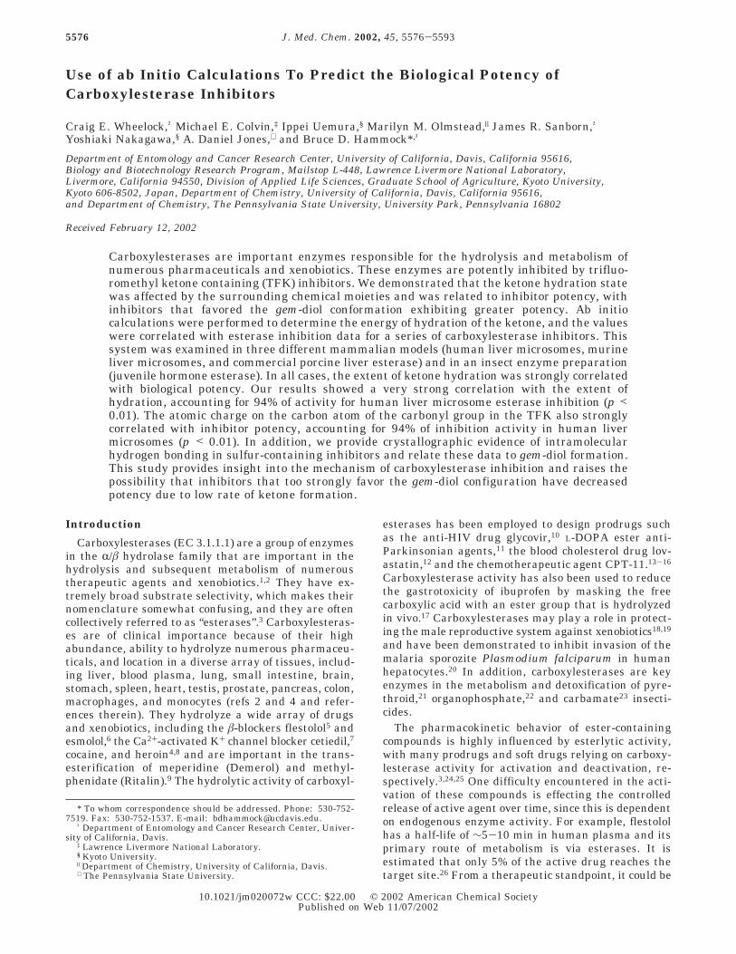
Figure 1. Hydration reaction of trifluoromethyl ketone fromthe ketone to the gem-diol. Greek letters indicate carbonpositions relative to the carbonyl (gem-diol) referred to in thetext.
Carboxylesterase Inhibitors Journal of Medicinal Chemistry, 2002, Vol. 45, No. 25 5577

Overview of Computational Chemical Methods
A wide variety of chemical simulation methods havebeen developed, ranging from empirical ball-and-springtype molecular mechanics models to ab initio (firstprinciples) quantum chemical methods that calculateapproximate solutions to the exact quantum mechanicalequations describing the electrons and nuclei. Typically,the choice of methods involves tradeoffs among ac-curacy, size of the chemical system, and computationalcost. There is a hierarchy of different quantum chemicalmethods involving increasingly accurate mathematicaldescriptions of the electronic wave function, the math-ematical description of the distribution of electronsaround the nuclei of a molecule.47 Over the past 2decades, a class of methods called density functionaltheory (DFT) has been developed that includes empiricalparametrizations of the electron-electron interactionsand often provides accuracy comparable to that of theearlier high-level quantum chemical methods but witha much lower computational cost. The DFT methods areusually denoted by the empirical electron-electron“functional” employed. Two widely used DFT functionalsare the Becke three-parameter hybrid exchange func-tional (B3) and the Lee-Yang-Parr (LYP) gradient-corrected electron correlation functional49 that havebeen widely demonstrated to yield accurate chemicalstructures and reaction energies when used with suf-ficient basis sets for most molecules.47
An important limitation of the quantum chemicalmethods described above is that they describe only anisolated (usually described as “gas-phase”) molecule andtherefore do not include the chemical environment, suchas solvent molecules and counterions. Explicitly includ-ing the surrounding molecules is usually too computa-tionally costly for quantum chemistry methods; how-ever, several methods have been developed within thequantum chemistry approach for including the effectsof solvent interactions. Typically these methods modelthe solvent as a continuous medium that polarizes inresponse to the quantum chemically derived charges.Although there are situations where explicit inclusionof the solvent is necessary, these so-called polarizablecontinuum models (PCM) have proven to be reasonablyaccurate in predicting solvent-phase chemical propertiesincluding total solvation energies and acid constants.50,51
One version of PCM, the conductor-like screening sol-vation model COSMO,52 models the surrounding solventby means of polarization charges distributed on thesolvent-exposed surface of the molecule. Although struc-tural optimizations can be performed using these PCMsolvent models, earlier studies have shown that for mostmolecules reoptimizing in the solvent does not signifi-cantly affect the solution-phase energies.51
A method related to these polarizable continuummodels but including a more realistic representation ofthe polar solvent is the Langevin dipole method ofWarshel (ChemSol),53 which models the solvent as alarge set of polarizable dipoles on a fixed three-dimensional grid.54 This approach has recently beenparametrized for use with ab initio derived solutecharges and shown to yield solvation energies forneutral and ionic molecules comparable to or better thanPCM methods and has been extensively applied to theproblem of phosphate hydrolysis.53 Where the appropri-
ate parameters were available (the ChemSol methoddoes not have the appropriate parameters for theselenium atom), we also used the Langevin dipolemethod to calculate the effects of aqueous solvation onthe hydration reaction energies of the compounds.
In addition to molecular structures and energies,another frequently calculated value from the electronicwave function is the effective atomic charges, the netelectronic and nuclear charge on each atom. Historically,Mulliken population analysis has been widely used, butthe derived atomic charges have proved to be extremelysensitive to the molecular configuration and wavefunction. Another method, natural atomic populationanalysis (NPA), has been developed that overcomesmost of the limitations of the earlier method55 and yieldsatomic charges that validate many qualitative chemicalconcepts.
Methods
We were particularly interested in the role of thesulfur atom in inhibitor potency because several re-searchers have reported the ability of this atom to conferenhanced inhibition potency for esterase inhibition.34-36,44
We therefore weighted our selection of compound struc-tures for study toward sulfur-containing inhibitors(Tables 1 and 2). The energy of hydration calculationswere performed in a number of different environmentsin order to determine the optimal parameters for thecorrelation (Tables 3 and 4). A total of five differentenergies were examined: the electronic energy in thegas phase (∆Egas), the energy in the aqueous phase(∆Eaq), the free energy in the gas phase (∆Ggas), and thefree energy in the aqueous phase using two differentcalculation methods, the PCM model (∆Gaq, COSMO)and the Langevin dipole model (∆Gaq, ChemSol). Thesignificance of the correlation values was then comparedfor all enzymes for each energy value. These terms arefurther defined in the Supporting Information.
Quantum Chemical Calculations. The molecularstructures of the ketone and hydrated (gem-diol) formsof each inhibitor were built using AMPAC.56 The alkanechains were built in the fully extended all-trans con-formation, consistent with recent experimental data foraqueous phase saturated alkanes.57 All structures wereoptimized using DFT with the B3LYP functional usinga 6-31G* basis set. The free energies (298 K) of thesestructures were determined from the total B3LYP/6-31G* electronic energies of the optimized molecularstructures combined with vibrational, thermal, andentropic terms calculated from the B3LYP/6-31G* har-monic vibrational frequencies using standard formu-las.58 The atomic charges were predicted using NPAfrom the B3LYP/6-31G* wave function at the B3LYP/6-31G* optimized geometries. Aqueous phase solvationfree energies were calculated using either the COSMOor ChemSol solvation model coupled to the B3LYP/6-31G* wave function at the gas phase optimized geom-etries, based on previous theoretical studies showingthat reoptimizing the gas phase structures within thePCM solvent models has very little effect on theresults.51
All quantum chemical calculations were performedusing Guassian 98, revision A.7.59 The Langevin dipolecalculations were performed using ChemSol version
5578 Journal of Medicinal Chemistry, 2002, Vol. 45, No. 25 Wheelock et al.

2.1.60 All calculations were run on Compaq AlphaServer4100 model 5/533 quadprocessor workstations with a533 MHz EV5.6 CPU running OSF, version 5.1.
Hydrogen Bond Strength Calculations. The in-tramolecular hydrogen bond strengths for the thioetherand sulfone containing compounds were estimated usingcalculations on model gem-diol compounds (see Resultsand Discussion). These compounds were fully optimizedas described above, and single-point solution-phasecalculations where calculated using the COSMO orChemSol models with dielectric constants that mimicthree different solvents: water (ε ) 78.39), methanol (ε) 32.63), and benzene (ε ) 2.247). These structures werethen reoptimized with inclusion of internal constraintson the rotations of the gem-diol hydroxyl groups toprohibit hydrogen bonding to the thioether sulfur atomor the sulfone oxygen atoms. The intramolecular hy-drogen bond energies were then estimated as thedifference in electronic energies between the constrainedand unconstrained structures.
Results and DiscussionTrifluoromethyl ketones are potent inhibitors of car-
boxylesterases. However, modulation of inhibitor po-tency may be desirable for different goals. Ab initiocalculations were performed to test the hypothesis thatthe hydration state of the inhibitory ketone function isa critical component of carboxylesterase inhibition. Aninitial model was developed with juvenile hormoneesterase (JHE) from the cabbage looper Trichoplusia niand then applied to three mammalian systems. Thisenzyme was chosen to develop the correlative model
Table 1. Structure and Calculated log P Data for SynthesizedCompounds
a All compounds have a hexyl (CH3(CH2)5) moiety as the Rgroup. b Calculated log P (ClogP) values were determined usingthe ClogP program of Leo.64 c ClogP determinations were alsoperformed for the hydrated ketone (gem-diol). d The measuredlog P value was 3.09 ( 0.02 (n ) 3). e The measured log P valuewas 3.83 ( 0.04 (n ) 3).
Table 2. Structure and Calculated log P Data for TheoreticalCompounds
a All compounds have a hexyl (CH3(CH2)5) moiety as the Rgroup. b Calculated log P (ClogP) values were determined usingthe ClogP program of Leo.64 c ClogP determinations were alsoperformed for the hydrated ketone (gem-diol).
Table 3. Calculated Hydration Energies for SynthesizedCompounds
no. ∆Egasa ∆Ggas
b ∆Eaqc
∆Gaq(COSMO) d
∆Gaq(ChemSol) e
carbonylchargef
1 -4.72 10.01 -5.05 9.69 15.0 0.5942 -12.99 1.58 -12.17 2.40 6.3 0.5233 -13.15 2.00 -12.65 2.51 7.7 0.4994 -7.17 7.50 -6.67 8.01 14.5 0.4445 -9.16 3.81 -10.14 2.82 9.5 0.4166 -19.51 -2.60 -16.74 0.17 4.2 0.4847 -17.42 -2.23 -14.98 0.21 2.9 0.4798 -12.30 3.78 -9.58 6.50 12.3 0.5829 -8.73 5.95 -5.69 8.99 12.2 0.573
10 -19.77 -4.46 -17.80 -2.49 0.4 0.48211 -11.11 4.69 -8.75 7.05 11.2 0.57612 -25.06 -10.25 -21.69 -6.88 -0.4 0.46713 -15.05 1.38 -11.06 5.37 10.9 0.567
a Energy of hydration in the gas phase (kcal/mol). b Free energyof hydration in the gas phase (kcal/mol). c Energy of hydration inthe aqueous phase using the COSMO solvation model (kcal/mol).52
d Free energy of hydration in the aqueous phase using COSMOsolvation model (kcal/mol).52 e Free energy of hydration in theaqueous phase using the Langevin dipole (ChemSol) solvationmodel (kcal/mol).53 f Calculated atomic charge on the carbonylcarbon (eV).
Carboxylesterase Inhibitors Journal of Medicinal Chemistry, 2002, Vol. 45, No. 25 5579

given that a single predominant isozyme in T. nihemolymph is responsible for the hydrolysis of juvenilehormone,61,62 thus reducing the potential for the effectsof multiple isozymes with different inhibition profiles.Additionally, there is a large body of data available forJHE inhibition by TFK-containing compounds.34,41,42,63
This study was initiated by mining data for a seriesof six compounds (1-6) from the available JHE litera-ture.35,36 The initial results showed a positive correlationbetween the hydration state of the ketone and inhibitorpotency (data not shown). Compounds were then de-signed to explore the effects of potential functionalgroups surrounding the ketone moiety (Tables 1 and 2,compounds 7-22). We used our initial regression equa-tions for JHE to predict the biological potency of thesenew compounds in an attempt to predict the most potentinhibitors.
There have been a number of published studies onthe potency of TFK inhibitors35,39,42,43 that have identi-fied lipophilicity as a key parameter in determininginhibitor potency. For many esterases, the most potentinhibitors have large log P values (>3). Therefore, toreduce the hydrophobic variance, the inhibitor alkylchain was fixed at six carbons. In addition, the calcu-lated energies of hydration were evaluated for covari-ance with lipophilicity. The log P values for both theketone and gem-diol forms of the inhibitors were deter-mined using Leo’s ClogP program (Tables 1 and 2).64
To test the ability of this program to estimate partitioncoefficients for these compounds, we measured log Pvalues for compounds 6 and 8. Work by Thomas et al.reported that it was not possible to directly measureoctanol/water partitioning for these hydrated ketonecompounds because of hemiacetal formation.65 Thepartition coefficient was therefore determined in cyclo-hexane and calculated for octanol using a solventregression equation. Results indicated that predictedvalues were within 1 order of magnitude of measuredvalues (Table 1). This observed trend was in closeagreement with that of Thomas and co-workers.65 It isimportant to remember that any equilibrium measuredrepresents both compound partitioning and possibleshifts in hydration, thereby affecting the final value.Additionally, even the “experimental” log P had to be
estimated using a regression equation and is thereforenot a directly measured value.
We subsequently examined the correlations betweenthe calculated log P values and biological potency forboth the ketone and gem-diol forms of the inhibitors andfound little correlation (R2 ) 0.16 for the ketone andR2 ) 0.03 for the gem-diol). After verifying that lipo-philicity did not account for our observed trend, weproceeded with the study and selected a subset of ourmodel compounds for synthesis to validate the correla-tion model (compounds 6-13). Together, this group ofcompounds rationally extended the structure of thecarboxylesterase inhibitors and addressed our two goalsfor this project: to develop a model for carboxylesteraseinhibition that accounted for varying moieties aroundthe carbonyl group and to determine the role of sulfurin increased inhibition potency.
These eight compounds were assayed for inhibitionactivity against three mammalian enzyme systemsand JHE (Tables 5 and 6). Following observation ofIC50 data, it was determined that compound 12 hada significantly different bimolecular rate constant (ki)than compounds 6-11 (see Kinetic Analysis). Thisvariation in kinetic performance made it inappropriate
Table 4. Calculated Hydration Energies for TheoreticalCompounds
no. ∆Egasa ∆Ggas
b ∆Eaqc
∆Gaq(COSMO)d
∆Gaq(ChemSol)e
carbonylchargef
14 -17.35 -2.16 -15.05 0.14 g 0.44915 -24.40 -9.47 -21.55 -6.61 g 0.44916 -21.12 -5.71 -17.61 -2.20 g 0.46517 -11.06 4.04 -8.88 6.22 g 0.56118 -14.01 1.27 -10.31 4.97 g 0.55819 -12.05 3.10 -9.48 5.67 g 0.57320 -20.60 -5.42 -16.62 -1.45 -1.0 0.49121 -18.61 -3.53 -19.29 -4.21 0.4 0.44222 -9.29 5.47 -6.94 7.82 12.6 0.556
a Energy of hydration in the gas phase (kcal/mol). b Free energyof hydration in the gas phase (kcal/mol). c Energy of hydration inthe aqueous phase using the COSMO solvation model (kcal/mol).52
d Free energy of hydration in the aqueous phase using COSMOsolvation model (kcal/mol).52 e Free energy of hydration in theaqueous phase using the Langevin dipole (ChemSol) solvationmodel (kcal/mol).53 fCalculated atomic charge on the carbonylcarbon (eV). g The value was not determined because the ChemSolmethod lacks parameters for the selenium atom.
Table 5. Inhibition of Carboxylesterase Activity bySynthesized Compoundsa
no.human
-log IC50
murine-log IC50
porcine-log IC50
JHEb
-log IC50
1 2.07c 0.77c 1.17c 2.15d
2 5.30c 4.35c 4.61c 5.00d
3 5.36c 4.42c 4.67c 5.17d
4 3.03c 1.83c 2.19c 4.10e
5 3.80c 2.69c 3.02c 6.40e
6 8.16 ( 0.03 7.90 ( 0.04 8.08 ( 0.02 7.51 ( 0.017 7.33 ( 0.01 6.35 ( 0.01 6.17 ( 0.01 6.83 ( 0.028 4.55 ( 0.09 3.02 ( 0.12 3.01 ( 0.09 2.86 ( 0.079 3.98 ( 0.03 3.32 ( 0.04 3.96 ( 0.06 3.29 ( 0.04
10 7.32 ( 0.01 6.92 ( 0.06 7.36 ( 0.08 7.08 ( 0.0111 4.26 ( 0.03 3.37 ( 0.05 3.59 ( 0.02 3.31 ( 0.1212 6.51 ( 0.02 6.35 ( 0.01 6.77 ( 0.03 5.85 ( 0.03
a IC50 is the concentration of inhibitor at which 50% of enzymeactivity was inhibited. Data for compound 13 are excluded becauseof instability. bJuvenile hormone esterase (JHE) from the cabbagelooper Trichoplusia ni. c Data estimated from linear regression foreach enzyme using ∆Egas regression values (see Table 7). d Datafrom Hammock et al.36 e Data from Szekacs et al.35
Table 6. Predicted Inhibition of Carboxylesterase Activity byTheoretical Compoundsa
no.humanb
-log IC50
murineb
-log IC50
porcineb
-log IC50
JHEb,c
-log IC50
12d 10.00 9.59 9.62 9.3913d 6.10 5.25 5.46 4.2914 7.00 6.24 6.42 6.4615 9.75 9.30 9.34 9.2716 8.47 7.88 7.98 7.4417 4.54 3.52 3.81 3.9318 5.69 4.80 5.03 4.4519 4.93 3.95 4.22 4.1620 8.26 7.65 7.77 7.1321 7.49 6.79 6.94 8.2822 3.85 2.75 3.07 3.26a IC50 is the concentration of inhibitor at which 50% of enzyme
activity was inhibited. b Data estimated from linear regression foreach enzyme using ∆Egas regression values (see Table 7). c Juvenilehormone esterase (JHE) from the cabbage looper Trichoplusia ni.Data estimated from linear regression for each enzyme using∆Gaq(COSMO) regression values (see Table 7). d The predicted IC50values for compounds 12 and 13 are shown here because they differgreatly from experimental values.
5580 Journal of Medicinal Chemistry, 2002, Vol. 45, No. 25 Wheelock et al.

to include compound 12 in the correlation analyses. Inaddition, it was found that compound 13 was unstableunder the assay conditions and quickly disproportion-ated into a number of products, making accuratemeasurement of the IC50 value impossible. All correla-tion analyses were therefore performed with compounds6-11 for the mammalian enzymes and compounds1-11 for JHE.
Comparison of Energy Values. All four esterasesystems examined in this study provided similar results.A notable trend was that all models had a bias towardthe prediction of strong inhibitors, with the absoluteerror increasing with decreasing inhibitor potency. Wecorrelated the measured inhibitory potencies of thesecompounds with energy to hydrate the ketone to formthe corresponding gem-diol. We used five differentmathematical models to estimate different forms of thehydration energies in these correlations: the gas phaseelectronic energies (total energy at 0 K excludingvibrational zero-point energy) and free energies, denoted∆Egas and ∆Ggas; the aqueous phase electronic and freeenergies using the COSMO solvent model, denoted ∆Eaqand ∆Gaq(COSMO); and the aqueous phase free energiesusing the Langevin dipole solvation model, denoted∆Gaq(ChemSol). Table 7 shows the strongest linearcorrelations found between the inhibitory potencies andthe hydration energies for each enzyme preparation (thecorrelations for all hydration energies are shown inTable 18 in Supporting Information). The positivecorrelations between energy of hydration and biologicalpotency were upheld for all four enzyme preparationswith all five energies. The ∆Egas values provided the bestcorrelations in all three mammalian enzyme systemsbut were only slightly better than the ∆Ggas values. TheJHE system was quite different in that ∆Egas valuesprovided the poorest correlations and ∆Gaq(COSMO)provided the best. It was expected that the free energyvalues would provide better correlations than the elec-tronic energies because of the inclusion of entropiceffects. However, it is unlikely that the small variationsbetween the R2 values for the electronic and freeenergies are statistically significant. Inclusion of aque-ous solvation effects on the hydration energy by theCOSMO and ChemSol models did not improve thecorrelations. The ChemSol model gave the poorestcorrelation. This model is in principle more realisticthan the COSMO model, given that water molecules aremodeled as discrete dipoles surrounding the molecule.However, it is a new method that has not been fullyparametrized for sulfur-containing groups,53 which couldexplain its somewhat poorer correlation values. AllChemSol free energy values still correlated stronglywith inhibitor potency. The calculated energies allcorrelated very strongly with each other as well, with
all correlation coefficients greater than or equal to 0.92(Table 8). Given the high interrelation coefficients, itappears that any of these models are appropriate forestimating the hydration energy and subsequent bio-logical potency of carboxylesterase inhibitors.
Juvenile Hormone Esterase Activity. JHE cor-relations were initially performed with 11 compoundsincluding the 5 compounds that were collected fromliterature sources. However, on the basis of analysis,compound 5 was excluded from the correlation. Thiscompound contains a triple bond in the position R tothe carbonyl and has a geometry radically different fromthe geometries of the other inhibitors used for thiscorrelation.42 The different geometry could interferewith the mode of binding employed by the other inhibi-tors in this series, which all contain a bent form ofgeometry at this point (either tetrahedral sp3 or trigonalplanar sp2) as opposed to linear (sp). The exclusion ofcompound 5 had a very significant effect on the correla-tion values in the gas phase, with the R2 value for ∆Egasincreasing from 0.63 (P(F) ) 0.24) to 0.82 (P(F) ) 0.38),whereas the ∆Gaq(COSMO) value only increased slightlyfrom 0.85 (P(F) ) 0.41) to 0.87 (P(F) ) 0.42). Theobserved trend showed that aqueous models providedimproved regression values over gaseous, and freeenergy correlations were tighter than electronic cor-relations. Compound 5 had essentially no effect on theR2 value for the aqueous free energy calculations,showing that these values were superior at describinginhibitor activity. In this system, these results suggestthat the effects of the unsaturation are better modeledin an aqueous environment. The JHE data correlatedhighly with the mammalian data (Table 9), with allcorrelation values greater than or equal to 0.97.
Mammalian Carboxylesterase Inhibition. Thehuman liver microsome, mouse liver microsome, andcommercial porcine esterase preparations used for car-boxylesterase activity analysis were crude preparationslikely consisting of multiple isozymes. The porcine
Table 7. Regression and Correlation Analysisa
enzyme energy parameter equation R2 rsb P(F) c p value
humand ∆Egase y ) -0.39x + 0.23 0.94 0.98 0.47 <0.01
murined ∆Egase y ) -0.43x - 1.28 0.90 0.98 0.45 <0.01
porcined ∆Egase y ) -0.42x - 0.78 0.84 0.98 0.42 <0.01
JHEf ∆Gaq(COSMO) g y ) -0.42x + 6.52 0.87 0.94 0.42 <0.01a All correlation and regression values for all energy parameters are given in Supporting Information (Table 18). b The Spearman-
Rank correlation coefficient. c F test probability value. d n ) 6 (compounds 6-11). e Energy of hydration in the gas phase (kcal/mol).f Juvenile hormone esterase, n ) 10 (compounds 1-11, excluding compound 5). g Free energy of hydration in the aqueous phase usingCOSMO solvation model (kcal/mol).52
Table 8. Correlation Values between Calculated HydrationEnergies
∆Egasa ∆Ggas
b ∆Eaqc
∆Gaq(COSMO)d
∆Gaq(ChemSol)e
∆Egas 1.00 0.99 0.97 0.93 0.92∆Ggas 1.00 0.97 0.96 0.95∆Eaq 1.00 0.99 0.95∆Gaq(COSMO) 1.00 0.96∆Gaq(ChemSol) 1.00
a Energy of hydration in the gas phase (kcal/mol). b Free energyof hydration in the gas phase (kcal/mol). c Energy of hydration inthe aqueous phase using the COSMO solvation model (kcal/mol).52
d Free energy of hydration in the aqueous phase using COSMOsolvation model (kcal/mol).52 e Free energy of hydration in theaqueous phase using the Langevin dipole (ChemSol) solvationmodel (kcal/mol).53
Carboxylesterase Inhibitors Journal of Medicinal Chemistry, 2002, Vol. 45, No. 25 5581

esterase preparation undergoes a one-step acetoneprecipitation purification but still contains multipleesterases.34,41 It is expected that the individual isozymesrespond differently to chemical inhibition, resulting invarying inhibition profiles. In addition, each isozymewould be expected to have a different substrate profile.The substrate p-nitrophenyl acetate (PNPA) has beenshown to be very nonspecific and is hydrolyzed by abroad range of esterases2 and was therefore used forall mammalian carboxylesterase assays.
All three mammalian esterase systems examined inthis study provided similar results. The positive cor-relations between energy of hydration and biologicalpotency were upheld for all three enzyme preparationswith all five energy parameters (Table 7; and Table 18in Supporting Information). In addition, the inhibitionvalues were highly correlated among all four enzymesystems (including JHE), with all correlation valuesgreater than or equal to 0.95 (Table 9). One of the aimsof this project was to examine the role of the sulfur atomin esterase inhibition. The mammalian esterase assayswere conducted with six different inhibitors, four ofwhich contained a sulfur atom. Hammock et al. initiallyhypothesized that the increased potency of the â-thio-ether compounds, such as 6, over the trifluoromethylketones, such as 3, was due to the sulfur mimicking theelectronic properties of the 2,3-double bond of juvenilehormone.36 Although this observation holds for all JHEsso far examined, the higher potency of the thioether-containing compounds on mammalian enzymes whosesubstrates lack R,â-unsaturated esters does not supportapplication of this hypothesis to esterases in general.All three mammalian enzymes had significant correla-tions between inhibitor potency and all energy ofhydration calculations at the p < 0.01 level (except forsome porcine esterase values). The effects of sulfurtherefore appear to be adequately described by itscontribution to ketone hydration. However, furtheranalysis showed that in all mammalian systems theinhibition of sulfur-containing inhibitors was oftenunderestimated, indicating the existence of additionaleffects not accounted for by contributions to ketonehydration.
One possibility is that the sulfur atom could formassociations with amino acid residues in the enzymethat enhance its binding. Morgan and McAdon showedthat sulfur is capable of forming interactions witharomatic residues, where interaction is defined as“separated by the sum of their van der Waals’ radii, orso close that no other atom can intervene”.66 They foundthat the maximal allowable separation between the twoatoms is 5.0 Å and reported that sulfur-aromaticinteractions are energetically favorable. It is thereforefeasible that the sulfur atom in these inhibitors couldbe forming sulfur-aromatic interactions with residuesin the active site, which would account for the additionalactivity of the thioether-containing inhibitors unex-
plained by the hydration energies. However, the activityof both sulfone and thioether inhibitors was underpre-dicted in all of the enzyme systems examined, not justthe thioether-containing compounds. It is probable thatthe sulfone moiety is not capable of forming the sametype of sulfur-aromatic interaction as the thioether.Therefore, the additional effects of sulfur are probablyexerted by a different mechanism, such as bindingeffects that are not solely explained by the hydrationextent of the ketone moiety. However, these effectsappear to be minor compared to the effects of sulfurupon ketone hydration.
We only optimized one parameter in these studies,ketone hydration, and extensive work has been con-ducted previously to maximize esterase inhibition pa-rameters with TFK inhibitors. Therefore, this study isuseful for understanding the mechanism of carboxyl-esterase inhibition and provides information on animportant parameter for QSAR studies, but it does notstand alone as an independent model. For example,previous studies have shown that the hydrophobicalkane tail is very important for activity, and we haveused that optimized parameter for all compounds in thisstudy35,42,43 In addition, there are other models thatcorrelate physical constants with esterase activity.Buchwald and Bodor reported that steric effects sur-rounding the ester moiety were correlated with esterhydrolysis rates,24 with the inaccessible solid anglearound the sp2 oxygen (Ωh
Od) of the ester carbonylhaving the most significant correlation with the rate ofmetabolism.3 It is possible that the steric environmentsurrounding the carbonyl oxygen of the TFK inhibitorsalso correlates with inhibition potency, and furtherresearch could examine these effects. We also appliedour model to electric eel acetylcholinesterase inhibitionusing data from Szekacs et al.35 and found no correlationbetween ketone hydration state and enzyme inhibition(see Supporting Information for a graphical representa-tion of those results). It therefore appears that thismodel exhibits some selectivity for carboxylesterases.However, it is necessary to verify this observation withother enzyme systems.
Human Carboxylesterase Activity. The correla-tions between the five energy values and human livercarboxylesterase inhibition were the strongest of allmammalian systems examined. The electronic energy(∆Egas) had the strongest correlation with an R2 valueof 0.94 (p < 0.01; Figure 2). The aqueous energies (bothelectronic and Gibbs) also provided good correlations.
Table 9. Correlation Values for Enzyme Inhibitiona
human murine porcine JHE
human 1.00 0.98 0.95 0.98murine 1.00 0.99 0.99porcine 1.00 0.97JHE 1.00a Correlation analysis was performed for compounds 6-12.
Figure 2. Correlation between inhibition of human livermicrosome carboxylesterase activity (the concentration ofinhibitor that inhibits 50% of enzyme activity, IC50) and thegas phase electronic energy of hydration (∆Egas) of the ketonemoiety in the enzyme inhibitor (p < 0.01). All assays wereperformed in triplicate, and procedures are explained in thetext.
5582 Journal of Medicinal Chemistry, 2002, Vol. 45, No. 25 Wheelock et al.
The percent error for the predicted potency of allinhibitors was less than 10%, and only the potency ofcompound 6 was significantly underpredicted. Thisobservation supports previously reported results thatthe potency of the TFK-containing thioether is greaterthan predicted by its structure.41 However, the observedIC50 is only 2-fold more potent than the predicted value(see Supporting Information for a list of all predictedvalues). Interestingly, compound 8, a thioether-contain-ing compound that has a methyl group in place of thetrifluoromethyl group, was overpredicted by the model.It therefore appears that the increase in potency ob-served with the thioether functions in tandem with thetrifluoromethyl group. This effect could be furtherexamined by synthesizing sulfur-containing inhibitorsof varying degrees of fluorination on the methyl groupR to the ketone.
Murine Carboxylesterase Activity. The resultswere very similar to the human microsomal data, with∆Egas giving the best correlation to inhibitor potencywith an R2 of 0.90 (p < 0.01). All compounds except 8and 9 predicted IC50 values with less than 10% error.The mouse had a prediction pattern very similar to thatof the human preparation, with compound 6 beingunderpredicted and 8 being overpredicted (35% error).Compound 7 was well predicted, and compound 9 wasunderpredicted (24% error). These observations do notsupport the theory that there is some component of thesulfone moiety that interferes with binding. The bindingseems to be more a function of the trifluoromethyl groupthan the sulfone. One possibility is that the trifluoro-methyl group itself is interacting with residues in theenzyme active site. To examine those possible effectswould require advanced docking analysis using a TFK-containing inhibitor. A crystal structure for rabbitcarboxylesterase has recently been published67 andcould be used to examine the potential interactions ofthe trifluoromethyl group or the sulfone moiety versusthe thioether. In addition, there is a homology model ofJHE that could be used to probe these effects andcompare variations in mammalian versus insect car-boxylesterases.68 These results demonstrate that theesterlytic activity in human and murine systems seemsto function with a similar mechanism. This observationprovides further justification for the application ofmurine data to human systems when studying carboxy-lesterase activity and mechanism of action.
Porcine Carboxylesterase Activity. The porcineesterase assays were conducted with a commercialpreparation, thus serving as a benchmark enzyme forcomparisons with other research groups. The resultsparalleled those observed for the other mammaliansystems but gave the lowest correlations for any of themammalian enzyme systems. The correlations generallyoverestimated inhibitor potency, with four inhibitorspredicted to be more potent than observed. This trendwas observed for all five calculated energies. The resultsfor compounds 8 and 9 were similar in both the porcineand murine preparations with both showing a very highlevel of deviation from the value predicted from thecalculated hydration energies, 44% and 28%, respec-tively. It therefore appears that the inhibitory potenciesof these two compounds are not well-predicted by theirhydration energy. The biological data for porcine es-
terase showed that the inhibitors were consistently lesspotent than for the other mammalian systems. Theresults for this system were more similar to the JHEdata than to the results of other mammalian systems,with a correlation value of 0.97 versus 0.95 for thehuman microsomal enzyme (Table 9). It therefore ap-pears that caution should be used in applying theresults for porcine esterase inhibition studies to othermammalian systems, including humans, since they donot appear to be directly correlated.
Kinetic Analysis. Compound 12 was predicted to bethe most potent carboxylesterase inhibitor based on theenergy calculations with a -log IC50 value for humanliver microsomal esterase inhibition of 10.00 (Table 6).However, the observed -log IC50 was 6.51 (Table 5),showing almost 4 orders of magnitude difference ininhibition potency. At first, it was assumed that thisinhibitor was an exception to the observed correlationbetween biological potency and energy of hydration.However, the mechanism of esterase inhibition is de-pendent on several processes (Figure 3). It is assumedthat the ketone is the active form of the inhibitor, notthe gem-diol.44 Hence, if the compound is hydrated infree solution, then it is necessary for it to first dehydrateto the ketone in order to inhibit the enzyme. The rateof this reaction is described by kH in Figure 3. It hasbeen shown that fluorinated compounds in aqueoussolutions are hydrated between 97% and 100%, whereasthe hydration of nonfluorinated ketones is well below1%.69 This large degree of ketone hydration was furtherdemonstrated by Scott and Zuman who measuredvalues of k-H for aqueous solutions of R,R,R-trifluoro-acetophenone in the range 0.0036-0.018.70 These hy-dration levels are qualitatively consistent with thedifferent predicted hydration energies calculated in thisstudy.
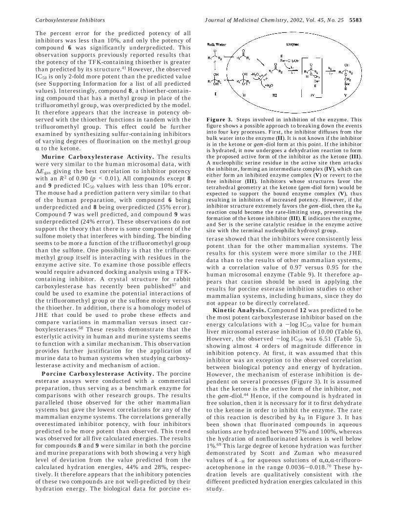
Figure 3. Steps involved in inhibition of the enzyme. Thisfigure shows a possible approach to breaking down the eventsinto four key processes. First, the inhibitor diffuses from thebulk water into the enzyme (II). It is not known if the inhibitoris in the ketone or gem-diol form at this point. If the inhibitoris hydrated, it now undergoes a dehydration reaction to formthe proposed active form of the inhibitor as the ketone (III).A nucleophilic serine residue in the active site then attacksthe inhibitor, forming an intermediate complex (IV), which caneither form an inhibited enzyme complex (V) or revert to thefree inhibitor (III). Inhibitors whose structures favor thetetrahedral geometry at the ketone (gem-diol form) would beexpected to support the bound enzyme complex (V), thusresulting in inhibitors of increased potency. However, if theinhibitor structure extremely favors the gem-diol, then the kH
reaction could become the rate-limiting step, preventing theformation of the ketone inhibitor (III). E indicates the enzyme,and Ser is the serine catalytic residue in the enzyme activesite with the terminal nucleophilic hydroxyl group.
Carboxylesterase Inhibitors Journal of Medicinal Chemistry, 2002, Vol. 45, No. 25 5583
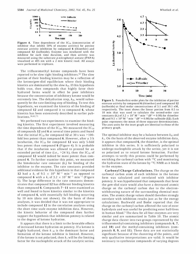
The trifluoromethyl ketone compounds have beenreported to be slow tight binding inhibitors.62 The slowportion of their binding kinetics may be a reflection ofthe ketone/gem-diol equilibrium where their bindingkinetics are dominated by the slow kH. If this hypothesisholds true, then compounds that highly favor theirhydrated forms would in effect be poor inhibitorsbecause the concentration of inhibitory ketone would beextremely low. The dehydration step, kH, would subse-quently be the rate-limiting step of binding. To test thishypothesis, we examined the kinetics of the binding ofcompound 12 and compared it to compound 6, whosekinetics has been extensively described in earlier pub-lications.36,62,71
We performed two experiments to examine the bind-ing kinetics. The first experiment involved measuringthe time dependence of the IC50. We measured the IC50of compounds 12 and 6 at several time points and foundthat the initial IC50 for compound 12 at 30 s was >100-fold less potent than compound 6. However, after a 3 hincubation, the IC50 for compound 12 was only 5-foldless potent than compound 6 (Figure 4). It is probablethat if the incubation was allowed to proceed for anextended period of time (i.e., >24 h), then the IC50 ofcompound 12 would indeed be more potent than com-pound 6. To further examine this point, we measuredthe bimolecular rate constant (ki) for binding of theinhibitor to the enzyme. The rate constants providedadditional evidence for this hypothesis in that compound12 had a ki of 9.5 × 10⁵ M-1 min-1 as opposed tocompound 6 with a ki of 3.2 × 10⁷ M-1 min-1 (Figure5). The large difference in the rate constants demon-strates that compound 12 has different binding kineticsthan compound 6. Compounds 7-11 were examined aswell and found to have kinetics similar to the kineticsof compound 6, with essentially full binding occurringby 10 min (data not shown). On the basis of theseanalyses, it was decided that it was not appropriate toinclude compound 12 in the correlation analyses usingthe short time scale enzyme inhibition data. However,the kinetic behavior of the compound does furthersupport the hypothesis that inhibition potency is relatedto the degree of ketone hydration.
It appears that there is a limit to the favorable effectsof increased ketone hydration on potency. If a ketone ishighly hydrated, then k-H is the dominant factor andformation of the ketone inhibitor is limited. However,if the ketone is not polarized, then k1 will be the limitingfactor for the nucleophilic attack of the catalytic serine.
The optimal inhibitor may be a balance between kH andk1. On the basis of the observed enzyme inhibition data,it appears that compound 6, the thioether, is the optimalinhibitor in this series. It is sufficiently polarized toundergo nucleophilic attack by the serine, yet it is notso polarized as to retard ketone formation. Furtherattempts to verify this process could be performed byenriching the carbonyl carbon with 13C and monitoringthe hydration state of the ketone by 13C NMR as it bindsto the enzyme.
Carbonyl Charge Calculations. The charge on thecarbonyl carbon atom of each inhibitor in the ketoneform was calculated and correlated with inhibitorpotency. It was hypothesized that compounds that favorthe gem-diol state would also have a decreased atomiccharge on the carbonyl carbon due to the electron-withdrawing nature of the surrounding chemical moi-eties. The atomic charge values should therefore stronglycorrelate with inhibition results just as for the energycalculations. Buchwald and Bodor reported that thecharge on the carbonyl carbon affected the rate of esterhydrolysis and was useful for predicting ester half-livesin human blood.3 The data for all four enzymes are verysimilar and are summarized in Table 10. The atomiccharge data cluster into two groups represented by thetrifluoromethyl-containing inhibitors (compounds 6, 7,and 10) and the methyl-containing inhibitors (com-pounds 8, 9, and 11). These data are not statisticallysignificant because of the clustering of the points, andonly qualitative interpretations are valid. It would benecessary to synthesize compounds of varying degrees
Figure 4. Time dependence of the IC50 (concentration ofinhibitor that inhibit 50% of enzyme activity) for porcineesterase activity inhibition by compound 6 (thioether) andcompound 12 (sulfoxide). Enzyme was incubated with theinhibitor for each time duration, and then activity wasmeasured using the substrate p-nitrophenyl acetate (PNPA)visualized at 405 nm with a 2 min kinetic read. All assayswere performed in triplicate.
Figure 5. Pseudo-first-order plots for the inhibition of porcineesterase activity by compound 6 (thioether) and compound 12(sulfoxide) at final molar concentrations of 3.1 and 39.1 nM,respectively. The inset shows the linear portion from 0.5 to10 min that was used to calculate the second-order rateconstants (ki) of 3.2 × 10⁷ M-1 min-1 (R2 ) 0.99) for thioether(6) and 9.5 × 10⁵ M-1 min-1 (R2 ) 0.98) for sulfoxide (12). Eachpoint represents the mean of three separate determinations.The axes units for the inset graph are identical to those of theprimary graph.
5584 Journal of Medicinal Chemistry, 2002, Vol. 45, No. 25 Wheelock et al.

of fluorination in order to map out the region betweenthe two clusters. We found that the carbonyl carboncharges correlated well with the calculated energies,with free energy values giving the strongest correlation(for ∆Gaq, R2 ) 0.93; for ∆Gaq(COSMO), R2 ) 0.91; for∆Gaq(ChemSol), R2 ) 0.95) and electronic energiesproviding slightly weaker correlations (for ∆Eaq, R2 )0.86; for ∆Egas, R2 ) 0.88). The more thermodynamicallyrelevant free energy term had a stronger correlationwith carbonyl carbon charge in both the gas andaqueous states, indicating that entropic effects are alsoimportant for this relationship. The correlation resultsare graphically displayed in the Supporting Information.
The inhibition of human liver esterlytic activitycorrelated most strongly with atomic charge on thecarbonyl carbon, giving an R2 of 0.94 (Figure 6), withthe murine enzyme preparation essentially the samewith an R2 of 0.93. The porcine enzyme did not correlateas well but still had a relatively strong correlation (R2
) 0.89). JHE gave the most interesting response out ofthe four enzymes studied. The initial correlation wasperformed with compounds 1-11 and gave a poorregression with an R2 of 0.58. There were two mainoutliers, compounds 4 and 5, which are both unsatur-ated compounds. The estimated charge on the inhibitorcarbonyl carbon was much lower than would be pre-dicted by the biological activity. It therefore appearsthat the effects of unsaturation upon carbonyl hydrationare less than calculations would predict. This observa-tion is particularly interesting in light of the fact thatthe alkene compound (4) was shown to be a more potentinhibitor than its energy calculations predicted. Thisobservation was attributed to the unsaturation mimick-ing the unsaturation in juvenile hormone, the naturalsubstrate for JHE. However, even with this additionalfactor, the alkene inhibitor potency is poorly predictedby atomic charge. Compounds 4 and 5 therefore behavedsimilarly, even though 5 was not designed as a mimicof juvenile hormone. These results demonstrate that
there could be additional effects influencing the degreeof ketone hydration for unsaturated systems or alter-natively the unsaturation is interacting with otherresidues in the active site. However, the calculationsgive an excellent estimation of the activity for saturatedsystems (compounds 6-11).
Intramolecular Hydrogen Bond Studies. A num-ber of researchers have theorized that the presence ofan intramolecular hydrogen bond between the hydratedketone (gem-diol) and the sulfur atom â to the ketonecould play a role in inhibitor potency through thestabilization of the gem-diol or the tetrahedral enzymeinhibitor complex (Figure 7).34,35,42 We therefore evalu-ated the role, if any, of this hydrogen bonding ininhibition potency. Olmstead et al. had previouslyshown that an internal hydrogen bond existed in thecrystal structure of the carboxylesterase inhibitor 1,1,1-trifluoro-5-phenyl-4-thiapentane-2,2-diol.72 We thereforeknew that the bond existed in thioether-containingcompounds in the crystalline state. In a previous project,we had synthesized a sulfone analogue of the thioethercompound that was also a crystalline solid.34 We ob-tained a crystal structure of this compound to examineif the intramolecular hydrogen bond was also presentin the sulfone analogue. A comparison of the twocompounds, the thioether and the sulfone, enabled usto examine the potential role of the intramolecularhydrogen bond in inhibitor potency.
The compound 1,1,1-trifluoro-3-(octane-1-sulfonyl)-propane-2,2-diol (26) was selected for crystal structuredetermination. It had crystals that were finer and more“needlelike” than 1,1,1-trifluoro-3-(hexane-1-sulfonyl)-
Table 10. Regression and Correlation Analyses for CalculatedAtomic Charge on Inhibitor Carbonyl Carbon
enzyme equation R2 rsa P(F) b p value
humanc y ) 34.46x - 24.17 0.94 0.97 0.47 <0.01murinec y ) 39.47x - 26.04 0.93 0.97 0.47 <0.01porcinec y ) 38.38x - 25.68 0.89 0.95 0.45 <0.01JHEd y ) 23.71x - 17.06 0.58 0.76 0.20 >0.05JHEe y ) 40.11x - 26.16 0.94 0.97 0.47 <0.01
a The Spearman-Rank correlation coefficient. b F test prob-ability value. c n ) 6 (compounds 6-11). d n ) 11 (compounds1-11). e n ) 9 (compounds 1-11, except compounds 4 and 5).
Figure 6. Correlation between inhibition of human livermicrosome carboxylesterase activity (the concentration ofinhibitor that inhibits 50% of enzyme activity, IC50) and thecalculated partial atomic charge of the ketone carbon atom ofthe enzyme inhibitor (p < 0.01). All assays were performed intriplicate as described in the text.
Figure 7. (A) Crystal structure of 1,1,1-trifluoro-5-phenyl-4-thiapentane-2,2-diol. The structure shows the presence of a2.37 Å intramolecular hydrogen bond between the sulfur atomand a hydroxy group of the gem-diol. Data were taken fromOlmstead et al.72 (Cambridge Crystallographic Data Centredeposition number “FORFUN”). (B) Crystal structure of 1,1,1-trifluoro-3-(octane-1-sulfonyl)‚propane-2,2-diol (26). The struc-ture shows the presence of a 2.34 Å intramolecular hydrogenbond between an oxygen atom of the sulfone moiety and ahydroxy group of the gem-diol (Cambridge CrystallographicData Centre deposition number “CCDC 178195”). See Sup-porting Information for the crystallographic coordinates.
Carboxylesterase Inhibitors Journal of Medicinal Chemistry, 2002, Vol. 45, No. 25 5585
propane-2,2-diol (7) and were readily crystallized froma 50:50 mixture of n-butyl chloride and hexanes. Crys-tals of 26 crystallized with two molecules in the asym-metric unit. The two molecules differed in the confor-mation of the terminal methyl group of the octyl chain,giving torsion angles of -68° and 179° for molecules 1and 2, respectively. Each molecule had one intramo-lecular hydrogen bond that is depicted for molecule 1(Figure 7B). In addition, there is intermolecular hydro-gen bonding between each molecule and its inversion-related pair. The crystal packing of the molecules placeshydrophilic and hydrophobic ends in proximity (seeSupporting Information for a full listing of the atomiccoordinates for all atoms in the assigned structure of26).
We had thus demonstrated that the intramolecularhydrogen bond existed in both the sulfone and thioethercompounds (in the crystalline state). We hypothesizedthat the gem-diol would be capable of forming a strongerhydrogen bond with the oxygen of the sulfone than withthe sulfur of the thioether. Oxygen is well-known toform stronger hydrogen bonds than sulfur.73 However,some evidence has shown that sulfur is capable offorming relatively strong hydrogen bonds in certainenvironments.73 Another factor was that the sulfonecompound formed a six-membered ring whereas thethioether compound formed a five-membered ring. Wetherefore had multiple effects occurring, and it wasuncertain which effect would have the dominant role.The biological data for inhibition of carboxylesteraseactivity in human microsomes showed that the thioethercompound (6) was more potent than the sulfone com-pound (7). This trend was consistent in the other threeesterase systems examined in this study (Table 5).These observations were also supported by the ab initioenergy calculations.
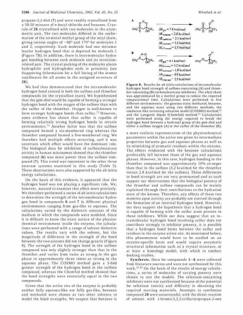
On the basis of this evidence, it appeared that thehydrogen bond was not playing a significant role. We,however, wanted to examine this effect more precisely.We therefore performed a series of ab initio calculationsto determine the strength of the intramolecular hydro-gen bond in compounds 6 and 7 in different physicalenvironments ranging from gas-like to aqueous. Thecalculations varied by the dielectric constant of themedium in which the compounds were modeled. Sinceit is difficult to know the exact nature of the physico-chemical environment inside the enzyme, the calcula-tions were performed with a range of solvent dielectricvalues. The results vary with the solvent, but themagnitude of difference in the strength of the bondbetween the two systems did not change greatly (Figure8). The strength of the hydrogen bond in the sulfonecompound was only slightly stronger than that in thethioether and varies from twice as strong in the gasphase to approximately three times as strong in theaqueous phase. The COSMO method gave a 3-foldgreater strength of the hydrogen bond in the sulfonecompound, whereas the ChemSol method showed thatthe bond strengths were essentially equal in the twocompounds.
Given that the active site of the enzyme is probablyneither fully aqueous-like nor fully gas-like, benzeneand methanol were chosen as two other solvents tomodel the bond strengths. We suspect that benzene is
a more realistic representation of the physicochemicalparameters within the active site given its intermediateproperties between gas and aqueous phases as well asits mimicking of aromatic residues within the enzyme.The effects evidenced with the benzene calculationspredictably fall between those of the gas and aqueousphases. However, in this case, hydrogen bonding in thethioether compound was approximately 10% strongerthan that in the sulfone (3.2 kcal/mol for the thioetherversus 2.8 kcal/mol for the sulfone). These differencesin bond strength are not very pronounced and as suchsupport our observations that the biological potency ofthe thioether and sulfone compounds can be mainlyexplained through their contributions to the hydrationstate of the ketone. Therefore, any effects of these twomoieties upon activity are probably not exerted throughthe formation of an internal hydrogen bond. However,our data support the hypothesis that a hydrogen bondis capable of forming with the sulfur atom present inthese inhibitors. While our data suggest that an in-tramolecular hydrogen bond involving sulfur does notcontribute strongly to inhibitor potency, it is possiblethat a hydrogen bond forms between the sulfur andresidues in the enzyme active site. As mentioned before,this phenomenon would have to be studied on anenzyme-specific basis and would require enzymaticstructural information such as a crystal structure, orat least a homology model, with which to conductdocking studies.
Synthesis. Data for compounds 1-6 were collectedfrom literature sources and were not synthesized for thiswork.35,36 On the basis of the results of energy calcula-tions, a series of molecules of varying potency werechosen to test the models. The selenium-containinginhibitors were not synthesized because of the potentialfor selenium toxicity and difficulty in obtaining therequired starting materials. Attempts to synthesizecompound 20 were unsuccessful, with the direct reactionof amines with 1-bromo-3,3,3-trifluroropropan-2-one
Figure 8. Results for ab initio calculations of intramolecularhydrogen bond strength of sulfone-containing (A) and thioet-her-containing (B) carboxylesterase inhibitors. The alkyl chainwas approximated by a methyl group to reduce the requiredcomputational time. Calculations were performed in fivedifferent environments: the gaseous state; methanol, benzene,and the aqueous state using two different methods; theconductor-like screening solvation model (COSMO) method;52
and the Langevin dipole (ChemSol) method.53 Calculationswere performed using the energy required to break thehydrogen bond between a hydroxyl group of the gem-diol andeither a sulfone oxygen (A) or the sulfur of the thioether (B).
5586 Journal of Medicinal Chemistry, 2002, Vol. 45, No. 25 Wheelock et al.

resulting in cyclic products. The synthesis of compound10 proved to be difficult, and several different methodswere attempted before the successful one reported inthis study. Initially, hexanol was directly treated with1-bromo-3,3,3-trifluroropropan-2-one, using methods ofJohnstone and Rose.74 A number of catalysts were thenattempted including ZnI2 and AgClO4 as well as forma-tion of the alkylate anion using NaH and CaH2. ADMSO/KOH system was also attempted, all withoutsuccess. The same series of reactions was also used inan attempt to synthesize compound 11 using chloroac-etone instead of 1-bromo-3,3,3-trifluroropropan-2-one,again with no success. The syntheses of 10 and 11 werefinally achieved by oxidizing hexyloxyethanol to thealdehyde (23), followed by methylation (24) and oxida-tion of the resultant alcohol to the ketone for 11 ortrifluoromethylation to form 25 and oxidation of thealcohol to the ketone for 10 (Scheme 1).
The synthesis of 10 always resulted in formation ofthe gem-diol compound as evidenced by the broad OHsinglet by 1H NMR (D2O exchangeable). Several unsuc-cessful attempts were made to dehydrate the compound,including use of MgSO4, CaCl2, and molecular sieves inCHCl3. This tendency of electron-deficient carbonyls tohydrate has been reported earlier with the sulfurderivates of these compounds.34-36
A number of obstacles were experienced in thesynthesis of compound 12. The sulfoxide-selective oxida-tion was difficult to achieve because it was importantto ensure that no sulfone was synthesized as a byprod-uct. Attempts to use H2O2 as the oxidizing agentaccording to methods of Drabowicz and Mikolajczyk75
were unsuccessful and resulted in multiple byproducts.The use of m-chloroperoxybenzoic acid (m-CPBA) hasbeen extensively referenced for oxidation reactions,including the formation of sulfoxides and sulfones76 (andreferences therein). We found that it was extremelydifficult to effect the selective conversion of thioetherto sulfoxide using published procedures.77 Syntheses
always resulted in some sulfone production. It wastherefore necessary to use a smaller amount of oxidizingagent than predicted. The maximum acceptable amountwas 0.5 mmol of m-CPBA per 1.0 mmol of thioether (6).Additionally, it was found that compound 12 was notthermally stable. The compound decomposed into thestarting thioether and free thiol, as well as disulfide,when examined by GC/EI-MS (injection port at 250 °C).Analysis by LC/ESI-MS was used to confirm the struc-ture of 12, and time course analyses showed that thecompound was completely stable in both ethanolic andaqueous solutions with no measurable degradation afterseveral days. While the dehydration product dominatedthe spectra, the molecular ion was detectable.
The same problems as with compound 12 wereexperienced with the synthesis of compound 13. It waspossible to increase the molar equivalents of m-CPBAto 0.85 mmol of m-CPBA per 1.0 mmol of thioether (8),resulting in selective sulfoxide formation. However,compound 13 was not stable and quickly degradedunder ambient conditions. The rate of degradation wasenhanced greatly in nucleophilic solvents such as metha-nol, ethanol, acetonitrile, and water. Interestingly, thecompound was stable by GC/EI-MS analysis but quicklydegraded under LC/ESI-MS analysis or in aqueoussystems (see Supporting Information for additional dataon the synthesis of compounds 12 and 13 and agraphical representation of the dependence of sulfuroxidation on the molar amount of m-CPBA).
Conclusion
For a set of esterase inhibitors chosen to minimizevariance in lipophilicity and to maximize variance inelectronic characteristics of the reactive carbonyl, wewere able to correlate biological potency with both thecharge on the carbonyl carbon and the hydration energyof the ketone. These results provide insight into themechanism of esterase inhibition by polarized ketone-containing inhibitors. This evidence supports earliersuggestions that inhibitors that favor tetrahedral ge-ometry (i.e., hydrated ketones) exert their increasedpotency by favoring the tetrahedral geometry of theinhibitor-bound enzyme complex (a transition-stateanalogue). However, data indicate that there is thepotential to have an inhibitor that is “too hydrated”, andwe provide evidence that the highly hydrated inhibitorsare very slow tight binders because of the decreasedconcentration of the ketone (which we assume to be theactive form of the inhibitor). It is still unclear whichspecies, the gem-diol or the ketone, is the active inhibi-tor. If the ketone is the inhibitor, which most studiesseem to suggest, then the key step in enzyme inhibitionbecomes the dehydration to the ketone. These resultsalso demonstrate that the enhanced role of sulfur inesterase inhibition can be largely attributed to its effectsupon ketone hydration. Additionally, our results indi-cate that intramolecular hydrogen bonding between thegem-diol of the hydrated ketone and the sulfur atomdoes not account for the increased potency of sulfur-containing inhibitors. Results showed that in this casemurine liver microsome esterlytic activity is an ap-propriate model for human liver microsome esterlyticactivity, with the two systems exhibiting similar inhibi-tion profiles and correlations with inhibitor parameters.
Scheme 1. Synthesis of Ether-ContainingCarboxylesterase Inhibitors (DMP Is the Dess-MartinPeriodinane)
Carboxylesterase Inhibitors Journal of Medicinal Chemistry, 2002, Vol. 45, No. 25 5587
However, a porcine system did not correlate nearly aswell with either the murine or human systems and hadresponses that were more similar to the insect enzymeJHE. The mammalian models consistently providedbetter correlations in the gas phase, and the JHE modelhad slightly better correlations in the aqueous phase.However, the differences were small enough to prohibitspecific comments regarding potential differences withinthe physicochemical parameters among these enzymes.In summary, we have employed quantum chemicalcalculations to elucidate a mechanism for the inhibitionof esterases. Results from this study should be usefulin designing esterase-activated prodrugs and soft drugsas well as in understanding the mechanism of action ofcarboxylesterases.
Experimental SectionCAUTION! The Dess-Martin periodinane has been reported
to be explosive upon impact or at temperatures above 200 °C.Proper care should be used in the handling and storage of thiscompound.
General. All work was conducted under an inert atmo-sphere (either N2 or Ar) in oven-dried glassware. Reactionsolvents were dehydrated when necessary in accordance withstandard protocols. Reaction progress and product purity wereassessed on 10 cm F254 silica thin-layer chromatography (TLC)plates (250 µm thickness, EM Science; Gibbstown, NJ) visual-ized with either phosphomolybdic acid and heating or 2,4-dinitrophenylhydrazine. Purity was initially assayed by thenumber of spots responding to the visualization agents andthe lack of UV-sensitive spots at 254 nm, confirming theremoval of UV-active impurities. Flash column chromatogra-phy was performed on silica gel (Merck Kieselgel 60). Yieldsfor all reactions were not optimized. Unless stated otherwise,all materials were obtained from commercial suppliers andwere used without further purification.
Structural characterization and purity were provided by 1HNMR, 13C NMR, 19F NMR (Mercury 300, Varian; Palo Alto,CA), GC/EI-MS, LC/ESI-MS, HPLC, and LC/TOF-MS. For GCanalysis, samples were analyzed on an HP 6890 GC (AgilentTechnologies; Engelwood, CO) equipped with a 30 m DB-17MScolumn (J&W Scientific; Folsom, CA) with a 0.25 mm internaldiameter and a 0.25 µm film thickness with a He carrier gasat a flow rate of 0.8 mL/min. The injector temperature was250 °C, and the initial column temperature was 50 °C and washeld for 5.00 min and then ramped at 15 °C/min to 320 °Cand held for 2.00 min. The GC was interfaced with an HP 5973mass spectrometer that was run in full-scan mode from 50 to550 m/z with a quadrupole temperature of 186 °C and a sourcetemperature of 240 °C. Electron ionization (EI) fragmentationpatterns supported all reported structures. Mass spectra aswell as total ion chromatograms (TIC) are provided in Sup-porting Information. LC/ESI-MS analysis was performed witha Micromass Quattro Ultima (Manchester, U.K.) mass spec-trometer in negative mode coupled to a Waters 2790 liquidchromatograph (Milford, MA). The mass spectrometer conevoltage was set at 50 V with a capillary voltage of 3.0 kV, usinga scan range of 50-300 m/z and a 3 s scan time. All ESI massspectral data supported the proposed structures.
Compound purity was assessed by HPLC using two differentsystems. Method A used a Spherisorb ODS 5 µm 4.6 × 250mm C-18 column (Waters) and a Waters 600 solvent deliverysystem with an HP 1100 variable-wavelength UV detector(Agilent Technologies). The system was run at a flow rate of1 mL/min with a linear gradient of 100% solvent A (20% ACNand 80% H2O) to 100% solvent B (100% ACN) in 80 min andthen 100% B for 20 min. Data collection was performed withWaters Millennium32 chromatography manager. Method Bemployed a Brownlee Spheri-5 5 µm 4.6 × 220 mm cyanopropylcolumn and a Waters 680 gradient controller equipped with aWaters 510 gradient solvent delivery system with an HP 1050variable-wavelength detector. The system was run a flow rate
of 1 mL/min with a linear gradient of 100% solvent A (H2O)to 50% solvent B (50% MeOH and 50% H2O) in 80 min andthen 50% B for 20 min. Both detection systems were run at205 nm and revealed a single UV absorbing peak. Analysis at254 nm resulted in no detectable impurities. These data areprovided in Table 17A in Supporting Information.
High-resolution mass spectrometry (HRMS) was performedon an LCT orthogonal time-of-flight mass spectrometer (TOF/MS, Micromass UK Limited; Manchester, U.K.). The massspectrometer was operated with MassLynx 3.5 software. Thespectrometer was equipped with a Z-flow electrospray ioniza-tion source, and sodium trifluoroacetate was used for externalmultipoint calibration.78 Mass accuracy was typically betterthan 10 ppm, and resolution was typically better than 4500full width at half-maximum (fwhm). The system was operatedat a flow rate of 25 µL/min. Data were acquired in eithernegative (Table 17B in Supporting Information) or positivemode (Table 17C in Supporting Information) depending on thecompound, with the following parameters: capillary 2880 V,sample cone 30 V, RF lens 250 V, extraction cone 10 V,acceleration 180 V, pusher cycle time 50 s-1, ion energy 30 V,and reflectron 1794 V. The analog-digital converter wasoperated at 3.6 GHz frequency. The reported spectra representthe averages of 20-40 consecutive spectra. MassLynx softwarewas used for all data processing and mass measurementdeterminations.
All compounds were >97% purity as evidenced by TLC, GC/EI-MS, HPLC, NMR, and a sharp melting point when ap-propriate (see Table 17A in Supporting Information forcomplete purity assessment of all compounds). Melting pointdata were collected with a Thomas-Hoover apparatus (A. H.Thomas Co.; Philadelphia, PA) and are uncorrected. Due tothe hygroscopic nature of these compounds, it was not possibleto obtain accurate elemental analyses for purity assessment.This observation has been reported by other authors forcompounds of similar structure.29,34
Hexyloxyethanal (23). A solution of oxalyl chloride (85mmol, 7.4 mL) in CH2Cl2 (150 mL) was cooled to -70 °C in adry ice/EtOH bath (the internal temperature was kept below-60 °C throughout the reaction and subsequent workup). Asolution of DMSO (179 mmol, 12.7 mL) in CH2Cl2 (20 mL) wasthen added dropwise. After gas evolution had completelysubsided (1 h), 2-hexyloxyethanol (66 mmol, 9.60 g) in CH2Cl2
(10 mL) was added to the mixture. After 30 min, triethylamine(180 mmol, 25 mL) was added dropwise, and the resultantwhite suspension was stirred for an additional 30 min. Thecooling bath was then removed, and the internal temperaturewas raised to 0 °C. Ice-cold 3 N aqueous HCl (200 mL) waspoured slowly into the reaction mixture with vigorous stirringto give a clear two-phase solution. The phases were separated,and the water phase was extracted with CH2Cl2 (50 mL). Thecombined organic extract was washed with 1 N aqueous HCl(2 × 100 mL), saturated aqueous NaHCO3 (100 mL), and brine(100 mL). This solution was dried over MgSO4, filtered, andstripped under reduced pressure. The residual yellow oil waspurified by distillation (bp 50 °C, 3 mmHg) to provide 23 as acolorless oil (49 mmol, 7.03 g, 74% yield). 1H NMR (CDCl3) δ(ppm) 0.90 (t, J ) 6.8 Hz, 3H), 1.3-1.4 (m, 6H), 1.6-1.7 (m,2H), 3.53 (t, J ) 6.6 Hz, 2H), 4.06 (s, 2H), 9.74 (s, 1H); 13CNMR δ (ppm) 14.0, 22.6, 25.7, 29.5, 31.6, 72.3, 76.3, 201.2;single spot by TLC (hexanes/EtOAc, 80:20); GC/EI-MS m/z[M]+ ) 144.1 (<1%), [M - CHO]+ ) 115.1 (33%), [M - C3H7O]+
) 101.1 (4%), [M - C2H3O2]+ ) 85.1 (58%); LC/ESI-MS m/zcalculated for [M] ) 144.1, observed [M - H]- ) 143.1.
1-Hexyloxypropan-2-ol (24). Compound 23 (21 mmol, 3.00g) in THF (5 mL) was added to a 0.92 M solution of methyl-magnesium bromide in THF (28 mmol, 30 mL) dropwise at 0°C. After the mixture was stirred for 30 min, 1 N aqueous HCl(40 mL) was slowly added with vigorous stirring. The bulk ofthe THF was then removed by rotary evaporation. Theresultant two-phase mixture was extracted with Et2O (3 × 25mL), and the combined extract was washed with saturatedaqueous NaHCO3 (50 mL) and brine (50 mL). This solutionwas dried over MgSO4 and filtered, and the solvent was
5588 Journal of Medicinal Chemistry, 2002, Vol. 45, No. 25 Wheelock et al.

stripped under reduced pressure. The residual oil was purifiedby distillation (bp 57 °C, 1 mmHg) to afford 24 as a colorlessoil (14 mmol, 2.32 g, 70% yield). 1H NMR (CDCl3) δ (ppm) 0.89(m, 3H), 1.14 (d, J ) 6.4 Hz, 3H), 1.30 (m, 6H), 1.58 (m, 2H),2.4 (broad s, 1H), 3.20 (dd, J ) 9.4, 8.3 Hz, 1H), 3.41 (dd, J )9.4, 3.1 Hz, 1H), 3.47 (m, 2H), 3.96 (ddq, J ) 3.1, 8.3, 6.4 Hz,1H); 13C NMR (CDCl3) δ (ppm) 14.0, 18.6, 22.6, 25.8, 29.6, 31.6,66.4, 71.4, 76.2; single spot by TLC (hexanes/EtOAc, 80:20);GC/EI-MS m/z [M]+ ) 160.3 (<1%), [M - CH3]+ ) 145.1 (2%),[M - C3H7O]+ ) 101.1 (3%), [M - C3H6O2]+ ) 85.1 (45%); LC/ESI-MS m/z calculated for [M] ) 160.1, observed [M - H]- )159.1.
1-Hexyloxypropan-2-one (11). The Dess-Martin perio-dinane (DMP) was prepared according to standard methods79,80
and added (28 mmol, 5.82 g) to a solution of compound 24 (12mmol, 2.00 g) in CH2Cl2 (30 mL) at room temperature, andthe solution was stirred overnight. The resultant cloudysolution was diluted with Et2O (150 mL), washed with 10%w/v aqueous Na2S2O3 (100 mL, saturated with NaHCO3), andbrine (100 mL), and dried over MgSO4. After the solution wasfiltered and concentrated, the residual oil was purified bydistillation (bp 53 °C, 1 mmHg) to give 11 as a colorless oil(8.6 mmol, 1.36 g, 69% yield). 1H NMR (CDCl3) δ (ppm) 0.89(t, J ) 6.9 Hz, 3H), 1.3-1.4 (m, 6H), 1.6-1.7 (m, 2H), 2.16 (s,3H), 3.48 (t, J ) 6.6 Hz, 2H), 4.02 (s, 2H); 13C NMR (CDCl3) δ(ppm) 14.0, 22.6, 25.7, 26.3, 29.5, 31.7, 72.0, 76.4, 207.5; singlespot by TLC (hexanes/EtOAc, 80:20); GC/EI-MS m/z [M]+ )158.1 (<1%), [M - C2H3O]+ ) 115.1 (4%), [M - C3H5O2]+ )85.1 (76%); LC/ ESI-MS m/z calculated for [M] ) 158.1,observed [M - H]+ ) 159.1; TOF HRMS calculated for [M +H]+ ) 159.1385, observed 159.1407.
1,1,1-Trifluoro-3-hexyloxypropan-2-ol (25). A catalyticamount (∼20 mg) of cesium fluoride was added to a solutionof 24 (20 mmol, 2.95 g) and (trifluoromethyl)trimethylsilane(21 mmol, 3.10 g) in THF (35 mL) according to the methods ofSingh et al.81 After the evolution of heat had subsided (∼1 h),aqueous 3 N HCl (50 mL) was added and the mixture wasvigorously stirred for 1 h. After the THF was removed byrotary evaporation, the aqueous residue was extracted withEt2O (2 × 70 mL). The combined extracts were washed withbrine (100 mL) and dried over MgSO4. The resulting solutionwas filtered and stripped under reduced pressure to give anoil that was purified by flash column chromatography (hexane/EtOAc,12:1) to afford 25 as a colorless oil (16 mmol, 3.50 g,80% yield). 1H NMR (CDCl3) δ (ppm) 0.89 (t, J ) 6.9 Hz, 3H),1.3-1.4 (m, 6H), 1.5-1.6 (m, 2H), 2.9 (broad s, 1H, D2Oexchangeable), 3.51 (t, J ) 6.6 Hz, 2H), 3.61 (dd, J ) 6.1, 10.1Hz, 1H), 3.68 (ddq, J ) 10.1, 3.8, 0.8 Hz, 1H), 4.11 (m, 1H);13C NMR (CDCl3) δ (ppm) 14.0, 22.6, 25.6, 29.4, 31.6, 67.9,69.3 (qCCF, J ) 30 Hz), 72.1, 124.2 (qCF, J ) 292 Hz); 19F NMR(CDCl3) δ (ppm) -78.24 (d, J ) 6.0); single spot by TLC(hexanes/EtOAc, 80:20); GC/EI-MS m/z [M]+ ) 214.2 (<1%),[M - C6H130]+ ) 113.0 (25%), [M - C3H4O2F3]+ ) 85.1 (95%),[M - C8H17O2]+ ) 69.0 (25%); LC/ ESI-MS m/z calculated for[M] ) 214.1, observed [M - H]- ) 213.1.
1,1,1-Trifluoro-3-hexyloxypropane-2,2-diol (10). TheDMP, synthesized according to standard methods,79,80 wasadded (7 mmol, 2.97 g) to a solution of compound 25 (5 mmol,1.00 g) in CH2Cl2 (16 mL) at room temperature, and thesolution was stirred overnight. The resultant cloudy solutionwas diluted with Et2O (100 mL), washed with 10% w/v aqueousNa2S2O3 (100 mL, saturated with NaHCO3) and brine (100mL), and dried over MgSO4. After the solution was filteredand concentrated, the residual oil was purified by flash columnchromatography (hexane/EtOAc, 4:1) to give 10 as a colorlessoil (4.2 mmol, 0.90 g, 85% yield). 1H NMR (CDCl3) δ (ppm)0.89 (t, J ) 6.8 Hz, 3H), 1.3-1.4 (m, 6H), 1.6-1.7 (m, 2H), 3.6(m, 3.3H), 3.79 (broad s, 1.6H, exchangeable by D2O); 13C NMR(CDCl3) δ (ppm) 14.0, 22.5, 25.5, 29.4, 31.5, 69.6, 71.4 (qCCF, J) 105 Hz), 72.8, 122.5 (qCF, J ) 285 Hz); 19F NMR (CDCl3) δ(ppm) -78.34 (s, 32%), -85.73 (s, 68%); single spot by TLC(hexanes/EtOAc, 80:20); GC/EI-MS m/z [M - H2O]+ ) 212.1(<1%), [M - H2O - C2OF3]+ ) 115.1 (22%), [M - H2O -C3H2O2F3]+ ) 85.1 (75%), [M - H2O - C8H15O2]+ ) 69.0 (30%);
LC/ ESI-MS m/z calculated for [M - H2O]- ) 212.1, observed[M - H2O-H]- ) 211.1; TOF HRMS calculated for [M - H2O- H]- ) 211.0946, observed 211.0945.
1-Hexylsulfanylpropan-2-one (8). 1-Hexanethiol (82.5mmol, 11.7 mL) was added to a stirring mixture of potassiumcarbonate (11.4 g) in CCl4 (20 mL). Chloroacetone (75 mmol,6 mL) was added dropwise over 10 min, followed by thecatalytic addition of 1,8-diazabicyclo[5.4.0]undec-7-ene (DBU).The reaction was allowed to proceed overnight under a gentlestream of N2. The reaction mixture was quenched with Et2O(20 mL) and washed with saturated aqueous NaHCO3 (3 ×50 mL). The organic fraction was dried over MgSO4 andfiltered, and the solvent was stripped under reduced pressure.The mixture was purified by distillation (bp 59 °C, 0.5 mmHg)to give a colorless oil (46.2 mmol, 8.1 g, 56% yield). 1H NMR(CDCl3) δ 0.86 (t, J ) 6.7 Hz, 3H), 1.26 (m, 4H), 1.34 (m, 2H),1.54 (m, J ) 7.6 Hz, 2H), 2.28 (s, 3H), 2.46 (t, J ) 7.1 Hz, 2H),3.18 (s, 2H); 13C NMR (CDCl3) δ 14.26, 22.76, 27.87, 28.65,29.16, 31.58, 32.34, 42.23, 204.30; single spot by TLC (hexanes/EtOAc, 80:20); GC/EI-MS m/z [M]+ ) 174.1 (24%), [M -C2H3O]+ ) 131.1 (14%), [M - C3H5O]+ ) 117.1 (15%); LC/ ESI-MS ions not observed in positive or negative mode; TOF HRMScalculated for [M + H]+ ) 175.1156, observed 175.1113.
1-(Hexane-1-sulfonyl)propan-2-one (9). Compound 8 (4.4mmol, 0.77 g) was added to a stirring solution of CHCl3 (50mL), followed by m-chloroperoxybenzoic acid (70-75%, 11.6mmol, 2.7 g). After 30 min, the reaction was worked upaccording to methods of Wheelock et al.34 It was not necessaryto further purify the final product, which had an overall yieldof 77%. 1H NMR (CDCl3) δ 0.92 (t, J ) 6.5 Hz, 3H), 1.32 (m,4H), 1.45 (m, 2H), 1.84 (q, J ) 6.5 Hz, 2H), 2.44 (s, 3H), 3.11(t, J ) 7.3, 2H), 4.03 (s, 3H); 13C NMR (CDCl3) δ 13.87, 21.75,22.23, 27.90, 31.08, 32.02, 53.41, 63.63, 197.40; single spot byTLC (hexanes/EtOAc, 80:20); GC/EI-MS m/z [M - C3H5O]+ )149.1 (21%), [M - C3H5O2]+ ) 131.1 (2%), [M - C3H5O3S]+ )85.1 (23%); LC/ESI-MS m/z calculated for [M] ) 206.1,observed [M - H]- ) 205.1; TOF HRMS calculated for [M -H]- ) 205.0899, observed 205.0913.
1-(Hexane-1-sulfinyl)propan-2-one (13). m-CPBA (70-75%, 0.43 mmol, 100 mg) was added to 8 (0.50 mmol, 87 mg)with stirring in CH2Cl2 (50 mL) at 0 °C. The reaction wasallowed to proceed for 1 h, and the mixture was brought up toroom temperature using adapted methods of Ando et al.77 Themixture was then washed with saturated NaHCO3 (3 × 50mL) and brine (50 mL), dried with MgSO4, filtered, andstripped under reduced pressure. The crude mixture wasrecrystallized from hexanes to give white needlelike crystalsof 13 (0.41 mmol, 77 mg, 81% yield). Mp ) 55-57 °C; 1H NMR(CDCl3) δ 0.92 (t, J ) 6.2 Hz, 3H), 1.28 (m, 4H), 1.41 (m, 2H),1.72 (m, 2H), 2.31 (s, 3H), 2.73 (m, 2H), 3.72 (dd, J ) 13.5,29.9 Hz, 2H); 13C NMR (CDCl3) δ 14.16, 22.56, 22.72, 28.57,31.51, 32.73, 52.89, 63.06, 200.61; single spot by TLC (hexanes/EtOAc, 80:20); GC/EI-MS m/z [M + H]+ ) 191.1 (1%), [M + H- O]+ ) 173.1 (84%), [M - C3H5O]+ ) 133.1 (27%), [M -C3H5O2S]+ ) 85.1 (19%); not stable by LC/ ESI-MS analysis.
1,1,1-Trifluoro-3-hexylsulfanylpropan-2-one (6). Com-pound 6 was synthesized according to the methods of Wheelocket al. and is not further reported here.34 1H NMR (CDCl3) δ0.87 (t, J ) 6.7 Hz, 3H), 1.28 (m, 4H), 1.37 (m, 2H), 1.57 (m,2H), 2.50 (t, J ) 7.3 Hz, 2H), 3.48 (s, 2H); 13C NMR (CDCl3) δ13.94, 22.46, 28.27, 28.49, 31.25, 31.92, 34.76, 115.51 (qCCF, J) 35 Hz), 185.01 (qCF, J ) 292 Hz); 19F NMR (CDCl3) δ -76.30(s); single spot by TLC (chloroform/ethanol, 95:5); GC/EI-MSm/z [M]+ ) 228.1 (34%), [M - C2OF3]+ ) 131.1 (67%), [M -C3H2OF3]+ ) 117.1 (100%); LC/ ESI-MS m/z calculated for [M]-
) 228.1, observed [M - H]- ) 227.1; TOF HRMS calculatedfor [M - H]- ) 227.0718, observed 227.0708.
1,1,1-Trifluoro-3-(hexane-1-sulfonyl)propane-2,2-diol(7). Compound 7 was synthesized according to methods ofWheelock et al.34 with a minor adaptation. Compound 6 wasoxidized with m-CPBA for 1 h instead of the 48 h reported.This length of time was sufficient for full oxidation to thesulfone in >99% conversion. The remaining procedure wasperformed as previously published. 1H NMR (CDCl3) δ 0.89
Carboxylesterase Inhibitors Journal of Medicinal Chemistry, 2002, Vol. 45, No. 25 5589

(t, J ) 6.7 Hz, 3H), 1.31 (m, 4H), 1.42 (m, 2H), 1.85 (m, 2H),3.33 (m, 2H), 3.39 (s, 2H), 5.23 (broad s, 1.6H, D2O exchange-able); 13C NMR (CDCl3) δ 14.13, 21.87, 22.50, 28.14, 31.31,52.77, 56.40, 92.01 (qCCF, J ) 34 Hz), 121.78 (qCF, J ) 287 Hz);19F NMR (CDCl3) δ -79.26, (s, 3%), -87.17 (s, 97%); singlespot by TLC (chloroform/ethanol, 95:5); GC/EI-MS m/z [M -H2O]+ ) 260.1 (<1%), [M - H2O - C3H2OF3]+ ) 149.1 (16%),[M - H2O - C6H13O2S]+ ) 111.0 (23%); LC/ ESI-MS m/zcalculated for [M - H2O]- ) 260.1, observed [M - H2O - H]-
) 259.1; TOF HRMS calculated for [M - H2O - H]- )259.0616, observed 259.0597.
1,1,1-Trifluoro-3-(hexane-1-sulfinyl)propan-2-one (12).m-CPBA (70-75%, 0.75 mmol, 175 mg) was dissolved in CH2-Cl2 (10 mL) and was washed with saturated NaHCO3 (50 mL)to remove the m-chlorobenzoic acid degradation product. Thissolution was then added dropwise over 20 min to a solution of6 (1.5 mmol, 344 mg) and NaHCO3 (∼2.4 mmol, 200 mg) inCH2Cl2 (50 mL) at 0 °C. The reaction was allowed to proceedfor 1 h and then brought up to room temperature and workedup as for 13 to give a viscous yellow liquid. Approximately 1-2mL of hexanes was added, which initiated precipitation of awhite solid. This solid was recrystallized from a mixture ofhexanes and n-butyl chloride (50:50) to give a white powderysolid (0.44 mmol, 90 mg, 29% yield). Mp ) 79-80 °C; 1H NMR(CDCl3) δ 0.91 (t, J ) 7.0 Hz, 3H), 1.33 (m, 4H), 1.47 (m, 2H),1.78 (m, 2H), 2.88 (m, 2H), 3.03 (m, 2H), 4.88 (broad s, 0.8 H,D2O exchangeable), 6.35 (broad s, 0.8 H, D2O exchangeable);13C NMR (CDCl3) δ 14.18, 22.42, 22.58, 28.53, 31.47, 50.36,53.31; 19F NMR (CDCl3) δ -79.56 (s, 4%), -87.63 (s, 96%);single spot by TLC (chloroform/ethanol, 95:5); compound wasnot thermally stable but was detected among various degrada-tion products by GC/EI-MS m/z [M]+ ) 244.1 (<1%), [M - H- O]+ ) 227.1 (29%), [M - CF3]+ ) 175.1 (2%), [M - C3H2-OF3]+ ) 133.1 (8%); LC/ ESI-MS m/z calculated for [M - H2O]-
) 244.1, observed [M - H2O - H]- ) 243.1.1,1,1-Trifluoro-3-(octane-1-sulfonyl)propane-2,2-diol
(26). Compound 26 (Figure 7B) was synthesized using thesame procedures as for compound 7. The final product wasrecrystallized from n-butyl chloride to give white needlelikecrystals in 94% yield. Mp ) 87-88 °C; 1H NMR (CDCl3) δ 0.88(t, J ) 6.45 Hz, 3H), 1.29 (broad m, 10H), 1.87 (m, 2H), 3.33(m, 2H), 3.39 (s, 2H), 4.79 (broad s, 1.5H, D2O exchangeable);13C NMR (CDCl3) δ 13.96, 21.72, 22.53, 28.24, 28.41, 28.88,31.64, 52.57, 56.27, 91.88 (qCCF, J ) 33 Hz), 121.78 (qCF, J )286 Hz); 19F NMR (CDCl3) δ -79.21 (s, 3%), -87.29 (s, 97%);single spot by TLC (chloroform/ethanol, 95:5); GC/EI-MS m/z[M - H2O]+ ) 288.1 (<1%), [M - H2O - C3H2OF3]+ ) 177.1(28%), [M - H2O - C8H17O2S]+ ) 111.0 (10%); LC/ ESI-MSm/z calculated for [M - H2O]- ) 288.1, observed [M - H2O -H]- ) 287.1.
X-ray Structure Determination of 1,1,1-Trifluoro-3-(octane-1-sulfonyl)propane-2,2-diol (26). A colorless plateof dimensions 0.80 mm × 0.16 mm × 0.02 mm was mountedin the 130 K nitrogen cold stream provided by a Siemens LT-2low-temperature apparatus on the goniometer head of aSiemens P4 diffractometer. Diffraction data were collected withnickel-filtered Cu KR radiation supplied by a Siemens rotatinganode to a maximum 2Θ of 112°. A total of 4528 reflectionswere collected in the range +h, +k, (l, of which 3888 wereunique (R(int))0.032) and 2888 were observed (I > 2σ(I)). Thestructure was solved by direct methods (SHELXS-97FN1, ver-sion 5.10, Bruker Analytical X-ray Instruments, Inc.; Madison,WI) and refined by full-matrix least-squares on F2 (SHELXL-97FN1). All non-hydrogen atoms were refined with anisotropicthermal parameters. Hydrogen atoms were added by geometryand refined using a riding model except for the hydroxylhydrogens, which were located on a difference map and refinedwith a constrained O-H distance of 0.84(1) Å. The maximumand minimum peaks in the final difference Fourier mapcorresponded to 0.50 and -0.31 e Å-3. The refinement con-verged with a wR2 value of 0.124 using all data and an R1value of 0.045 for observed data using 361 parameters, 4restraints, GOF ) 1.04. Crystal Data: C11H21F3O4S, M )
306.34, monoclinic, P21/c, a ) 31.750(5) Å, b ) 5.3684(8) Å, c) 18.409(3) Å, â ) 106.583(14)°, Z ) 8.
Enzyme Assays. Juvenile Hormone Esterase (JHE)Activity. JHE activity assays were conducted by the methodsof Sparks and Hammock61 as described in Wheelock et al.34
Briefly, Trichoplusia ni larvae were harvested on day 2 of thefifth instar, and hemolymph was collected and diluted 1:1 with0.1 M sodium phosphate buffer (pH 7.4) containing ∼0.5 mmolof phenylthiourea. The diluted enzyme had a specific activityof 1.0 nmol of juvenile hormone III min-1 mg-1 protein andwas frozen at -80 °C. JHE activity was measured with apartition assay based on the hydrolysis of [3H] juvenilehormone III ([3H] JH III). Hemolymph was diluted 300-foldin sodium phosphate buffer (pH 7.4, 0.1 M, 0.1 mg/mL bovineserum albumin) and incubated with inhibitor stock dilutionsfor 10 min at 30 °C. All inhibitor dilutions were prepared inethanol, and total ethanol concentration in the assay neverexceeded 2% of the total volume. The substrate [3H] JH IIIwas then added for a final concentration of 5 µM and incubatedfor 15 min at 30 °C. Following incubation, 50 µL of a MeOH/water/ammonium hydroxide solution (10:9:1) was added. Themixture was extracted with 250 µL of isooctane, and a 50 µLaliquot of the aqueous phase was counted by a Wallac 1409liquid scintillation counter (Wallac; Turku, Finland).
Mammalian Carboxylesterase Activity. Carboxylesteraseactivity assays were conducted according to the methods ofWheelock et al.34 and are only briefly described here. Assayswere performed in 96-well microtiter styrene flat-bottom plates(Dynex Technologies, Inc.; Chantilly, VA) at 30 °C usingp-nitrophenyl acetate (PNPA) as the substrate. Inhibitor stocksolutions were prepared in ethanol and used at concentrationsthat never exceeded more than 1% of the total assay volume.Total assay volume was 200 µL in sodium phosphate buffer(pH 7.4, 0.1 M). Enzyme was incubated with the inhibitor for5 min unless otherwise noted, and then the substrate wasadded for a final concentration of 0.5 mM followed by opticaldensity analysis at 405 nm on a microplate reader (Spectramax200, Molecular Devices; Sunnyvale, CA). Protein concentra-tions were determined using the Pierce BCA protein assay(Pierce; Rockford, IL) with bovine serum albumin as thestandard.
Human Carboxylesterase Activity. Human liver mi-crosomes were purchased from Gentest (Woburn, MA; lot no.17, catalog no. 452161). Microsomes were prepared from 16individuals (10 males, 6 females) and were partially character-ized for P450 activity and pathogenicity. Donor informationfor each individual as well as P450 activity and pathogenicitydata can be obtained directly from Gentest (http://www-.gentest.com/index.html) using the above-supplied catalog andlot numbers. Microsomes were solubilized as described for themurine liver microsomes and stored at -80 °C until use.Activity assays were performed as described above using 3.1µg of protein per well.
Murine Carboxylesterase Activity. Murine liver mi-crosomal carboxylesterase activities were performed withsolubilized microsomes prepared by methods adapted fromliterature procedures.34,39,82 Sixteen week-old male Swiss-Webster mice were purchased from Charles River BreedingLaboratory (Hollister, CA) and were 30-35 g upon receipt.Mice were housed in HEPA-filtered racks for 7 d before useand were fed and watered ab libitum with a light cycle of 12h of light and 12 h of darkness. Animal care procedures wereapproved by the Animal Use and Care Committee at theUniversity of California, Davis. After 1 week, mice weresacrificed by carbon dioxide asphyxiation and livers wereimmediately excised, rinsed in a 1.15% KCl solution (w/v),homogenized on ice with a Polytron homogenizer (BrinkmanInstruments; Westbury, NY), and centrifuged at 10000g for20 min at 4 °C. Supernatant fractions were further centrifugedat 100000g for 60 min. The resulting pellet was resuspendedand recentrifuged at 100000g for an additional 60 min. Thispellet was then resuspended in sodium phosphate buffer (pH7.4, 0.1 M) for a final protein concentration of 10.9 mg/mL andfrozen at -80 °C. Microsomes were then solubilized with 1%
5590 Journal of Medicinal Chemistry, 2002, Vol. 45, No. 25 Wheelock et al.

n-octyl-â-D-glucopyranoside (Sigma Chemical Co.) and dilutedwith additional sodium phosphate buffer to give a finalconcentration of 0.42 mg/mL. Solubilized microsomes werethen dialyzed against sodium phosphate buffer for 48 h at 4°C to remove the detergent. Activity assays were performedas described above using 0.88 µg of total protein per well.
Porcine Carboxylesterase Activity. Porcine carboxy-lesterase (EC 3.1.1.1) was purchased from Sigma Chemical Co.(catalog no. E-3019). Working solutions of enzyme wereprepared at 0.02 mg of protein/mL in sodium phosphate buffer(pH 7.4, 0.1 M) for an activity of 0.3 units/mL (one unit willhydrolyze 1.0 µmol of butyrate to butyric acid and ethanol atpH 8.0 at 25 °C). Assays were conducted as described aboveusing 0.2 µg of protein per well.
Enzyme Inhibition Measurement. The concentration ofinhibitor required to reduce enzyme activity by 50% (IC50) wasdetermined using the same method for all enzymes.34 Aminimum of five data points, each point resulting from at leastthree individual analyses, were used to determine the IC50.Inhibitor concentrations were adjusted such that the linearregion of the curve had at least two points above the IC50 andtwo points below.
Partition Coefficient Measurements. Partition coef-ficients were determined according to the methods of Leo etal.64 as described in Thomas et al.65 Briefly, the targetcompound was dissolved in cyclohexane (0.1 M) and gentlyshaken with an equal volume of water for 12 h at roomtemperature. The quantity of compound in the aqueous phasewas quantified by GC/EI-MS under the conditions describedabove. A solvent regression equation was used to calculate thepartitioning into octanol,
where KO/W represents the partition coefficients for the octanol/water system and KC/W represents those for the cyclohexane/water system.
Statistical Analysis. Statistical analyses were performedusing the data analysis module of Microsoft Excel (Microsoft;Redmond, WA). Spearman’s coefficients were taken fromSiegel.83
Acknowledgment. The authors thankfully acknowl-edge Dr. Christophe Morisseau and Dr. John Newmanfrom the Department of Entomology at the Universityof California for many useful discussions. We thank Dr.Takaho Watanabe for performing the HRMS studiesand Dexter Morin for assistance with the HPLC analy-ses. C.E.W. was supported by a UC TSR&TP GraduateFellowship, a UC TSR&TP Lead Campus Program inEcotoxicology Fellowship, and a University of CaliforniaSystemwide Biotechnology Research Program Fellow-ship in Structure Assisted Drug Discovery (Grant 2001-7). NMR spectra were acquired with the assistance ofthe UC Davis NMR Facility; the NMR spectrometerutilized was funded by NSF Grant 97244. This work wasin part performed under the auspices of the U.S.Department of Energy by the University of California,Lawrence Livermore National Laboratory under Con-tract W-7405-Eng 48. This project was supported in partby NIEHS Grant R37 ES02710, NIEHS SuperfundGrant P42 ES04699, USDA Competitive Research Grant2001-35302-09919, and NIEHS Center for Environmen-tal Health Sciences Grant P30 ES 05707.
Supporting Information Available: Crystallographiccoordinates for 1,1,1-trifluoro-3-(octane-1-sulfonyl)propane-2,2-diol (26) and additional data that include calculated quantumchemical data for all compounds, correlations between carbonylcarbon charge and calculated energies, correlations betweenacetylcholinesterase activity and calculated energies, predicted
IC50 values for inhibitors synthesized in this study, purity andGC/EI-MS data for synthesized compounds, and a graphicalrepresentation of the dependence of sulfur oxidation on molarquantity of m-CPBA. This material is available free of chargevia the Internet at http://pubs.acs.org.
References(1) Ollis, D. L.; Cheah, E.; Cygler, M.; Dijkstra, B.; Frolow, F.; et
al. The R/â hydrolase fold. Protein Eng. 1992, 5, 197-211.(2) Satoh, T.; Hosokawa, M. The mammalian carboxylesterases:
From molecules to functions. Annu. Rev. Pharmacol. Toxicol.1998, 38, 257-288.
(3) Buchwald, P.; Bodor, N. Structure-based estimation of enzymatichydrolysis rates and its application in computer-aided ret-rometabolic drug design. Pharmazie 2000, 55, 210-217.
(4) Pindel, E. V.; Kedishvili, N. Y.; Abraham, T. L.; Brzezinski, M.R.; Zhang, J.; et al. Purification and cloning of a broad substratespecificity human liver carboxylesterase that catalyzes thehydrolysis of cocaine and heroin. J. Biol. Chem. 1997, 272,14769-14775.
(5) Stampfli, H. F.; Quon, C. Y. Polymorphic metabolism of flestololand other ester containing compounds by a carboxylesterase inNew Zealand white rabbit blood and cornea. Res. Commun. Mol.Pathol. Pharmacol. 1995, 88, 87-97.
(6) Yang, H. S.; Wu, W. M.; Bodor, N. Soft drugs. XX. Design,synthesis, and evaluation of ultra-short acting beta-blockers.Pharm. Res. 1995, 12, 329-336.
(7) Roxburgh, C. J.; Ganellin, C. R.; Athmani, S.; Bisi, A.; Quaglia,W.; et al. Synthesis and structure-activity relationships ofCetiedil analogues as blockers of the Ca2+-activated K+ perme-ability of erythrocytes. J. Med. Chem. 2001, 44, 3244-3253.
(8) Brzezinski, M. R.; Spink, B. J.; Dean, R. A.; Berkman, C. E.;Cashman, J. R.; et al. Human liver carboxylesterase hCE-1:binding specificity for cocaine, heroin, and their metabolites andanalogs. Drug Metab. Dispos. 1997, 25, 1089-1096.
(9) Bourland, J. A.; Martin, D. K.; Mayersohn, M. Carboxylesterase-mediated transesterification of meperidine (Demerol) and me-thylphenidate (Ritalin) in the presence of [2H6] ethanol: pre-liminary in vitro findings using a rat liver preparation. J. Pharm.Sci. 1997, 86, 1494-1496.
(10) Cook, C. S.; Karabatsos, P. J.; Schoenhard, G. L.; Karim, A.Species dependent esterase activities for hydrolysis of an anti-HIV prodrug glycovir and bioavailability of active SC-48334.Pharm. Res. 1995, 12, 1158-1164.
(11) Van de Waterbeemd, H.; Testa, B.; Marrel, C.; Cooper, D. R.;Jenner, P.; et al. Multivariate statistical analysis of L-DOPAesters as potential anti-Parkinsonian prodrugs. Drug Des.Delivery 1987, 2, 135-143.
(12) Tang, B. K.; Kalow, W. Variable activation of lovastatin byhydrolytic enzymes in human plasma and liver. J. Clin. Phar-macol. 1995, 47, 449-451.
(13) Senter, P. D.; Marquardt, H.; Thomas, B. A.; Hammock, B. D.;Frank, I. S.; et al. The role of rat serum carboxylesterase in theactivation of paclitaxel and camptothecin prodrugs. Cancer Res.1996, 56, 1471-1474.
(14) Khanna, R.; Morton, C. L.; Danks, M. K.; Potter, P. M. Proficientmetabolism of irinotecan by a human intestinal carboxylesterase.Cancer Res. 2000, 60, 4725-4728.
(15) Senter, P. D.; Beam, K. S.; Mixan, B.; Wahl, A. F. Identificationand activities of human carboxylesterases for the activation ofCPT-11, a clinically approved anticancer drug. BioconjugateChem. 2001, 12, 1074-1080.
(16) Weirdl, M.; Morton, C. L.; Weeks, J. K.; Danks, M. K.; Harris,L. C.; et al. Sensitization of human tumor cells to CPT-11 viaadenoviral-mediated delivery of a rabbit liver carboxylesterase.Cancer Res. 2001, 61, 5078-5082.
(17) Lolli, M. L.; Cena, C.; Medana, C.; Lazzarato, L.; Morini, G.; etal. A new class of ibuprofen derivatives with reduced gastrotox-icity. J. Med. Chem. 2001, 44, 3463-3468.
(18) Jewell, W. T.; Miller, M. G. Identification of a carboxylesteraseas the major protein bound by molinate. Toxicol. Appl. Phar-macol. 1998, 149, 226-234.
(19) Mikhailov, A. T.; Torrado, M. Carboxylesterase overexpressionin the male reproductive tract: a universal safeguading mech-anism? Reprod., Fertil. Dev. 1999, 11, 133-145.
(20) van Pelt, J. F.; Moshage, H. J.; Depla, E.; Verhave, J. P.; Yap,S. H. Identification of human liver carboxylesterase as one ofthe proteins involved in Plasmodium falciparum malaria sporo-zoite invasion in primary cultures of human hepatocytes. J.Hepatol. 1997, 27, 688-698.
(21) Casida, J. E.; Gammon, D. W.; Glickman, A. H.; Lawrence, L.J. Mechanisms of selective action of pyrethroid insecticides.Annu. Rev. Pharmacol. Toxicol. 1983, 23, 413-438.
(22) Wallace, T. J.; Ghosh, S.; Grogan, W. M. Molecular cloning andexpression of rat lung carboxylesterase and its potential role inthe detoxification of organophosphorus compounds. Am. J.Respir. Cell Mol. Biol. 1999, 20, 1201-1208.
log KO/W ) 0.941 log KC/W + 0.69
Carboxylesterase Inhibitors Journal of Medicinal Chemistry, 2002, Vol. 45, No. 25 5591

(23) Gupta, R. C.; Dettbarn, W. D. Role of carboxylesterases in theprevention and potentiation of N-methylcarbamate toxicity.Chem. Biol. Interact. 1993, 87, 295-303.
(24) Buchwald, P.; Bodor, N. Quantitative structure-metabolismrelationships: Steric and nonsteric effects in the enzymatichydrolysis of noncongener carboxylic esters. J. Med. Chem. 1999,42, 5160-5168.
(25) Bodor, N.; Buchwald, P. Soft drug design: General principlesand recent applications. Med. Res. Rev. 2000, 20, 58-101.
(26) Molinoff, P. B.; Ruddon, R. W. Goodman & Gilman’s thepharmacological basis of therapeutics, 9th ed.; Hardman, J. G.,Limbird, L. E., Eds.; McGraw-Hill: New York, 1996; p 1905.
(27) Quinn, D. M. Esterases of the R/â hydrolase fold family.Biotransformation; Elsevier Science: Oxford, 1997; pp 243-264.
(28) Quinn, D. M. Ester hydrolysis. Enzymes, Enzyme Mechanisms,Proteins, and Aspects of NO Chemistry; Elsevier Science: Oxford,1999; pp 101-137.
(29) Nair, H. K.; Lee, K.; Quinn, D. M. m-(N,N,N-Trimethylammo-nio)trifluoroacetophenone: A femtomolar inhibitor of acetylcho-linesterase. J. Am. Chem. Soc. 1993, 115, 9939-9941.
(30) Amour, A.; Reboud-Ravaux, M.; de Rosny, E.; Abouabdellah, A.;Begue, J. P.; et al. Stereoselective synthesis of peptidyl trifluo-romethyl alcohols and ketones: Inhibitory potency againsthuman leucocyte elastase, cathepsin G, porcine pancreaticelastase and HIV-1 protease. J. Pharm. Pharmacol. 1998, 50,593-600.
(31) Veale, C. A.; Bernstein, P. R.; Bohnert, C. M.; Brown, F. J.;Bryant, C.; et al. Orally active trifluoromethyl ketone inhibitorsof human leukocyte elastase. J. Med. Chem. 1997, 40, 3173-3181.
(32) Edwards, P. D.; Andisik, D. W.; Bryant, C. A.; Ewing, B.; Gomes,B.; et al. Discovery and biological activity or orally active peptidyltrifluoromethyl ketone inhibitors of human neutrophil elastase.J. Med. Chem. 1997, 40, 1876-1885.
(33) Boger, D. L.; Sato, H.; Lerner, A. E.; Austin, B. J.; Patterson, J.E.; et al. Trifluoromethyl ketone inhibitors of fatty acid amidehydrolase: A probe of structural and conformational featurescontributing to inhibition. Bioorg. Med. Chem. Lett. 1999, 9,265-270.
(34) Wheelock, C. E.; Severson, T. F.; Hammock, B. D. Synthesis ofnew carboxylesterase inhibitors and evaluation of potency andwater solubility. Chem. Res. Toxicol. 2001, 14, 1563-1572.
(35) Szekacs, A.; Bordas, B.; Hammock, B. D. Transition state analogenzyme inhibitors: Structure-activity relationships of trifluo-romethyl ketones. Rational Approaches to Structure, Activity,and Ecotoxicology of Agrochemicals; CRC Press: Boca Raton,FL, 1992; pp 219-249.
(36) Hammock, B. D.; Wing, K. D.; McLaughlin, J.; Lovell, V. M.;Sparks, T. C. Trifluoromethylketones as possible transition stateanalog inhibitors of juvenile hormone esterase. Pestic. Biochem.Physiol. 1982, 17, 76-88.
(37) Wolfenden, R. Transition state analogues for enzyme catalysis.Nature 1969, 223, 704-705.
(38) Lienhard, G. E. Enzymatic catalysis and transition-state theory.Science 1973, 180, 149-154.
(39) Huang, T. L.; Shiotsuki, T.; Uematsu, T.; Borhan, B.; Li, Q. X.;et al. Structure-activity relationships for substrates and inhibi-tors of mammalian liver microsomal carboxylesterases. Pharm.Res. 1996, 13, 1495-1500.
(40) Brodbeck, U.; Schweikert, K.; Gentinetta, R.; Rottenberg, M.Fluorinated aldehydes and ketones acting as quasi-substrateinhibitors of acetylcholinesterase. Biochim. Biophys. Acta 1979,567, 357-369.
(41) Ashour, M. B. A.; Hammock, B. D. Substituted trifluoroketonesas potent, selective inhibitors of mammalian carboxylesterases.Biochem. Pharmacol. 1987, 36, 1869-1879.
(42) Linderman, R. J.; Upchurch, L.; Lonikar, M.; Venkatesh, K.; Roe,R. M. Inhibition of insect juvenile hormone esterase by R,â-unsaturated and R-acetylenic trifluoromethyl ketones. Pestic.Biochem. Physiol. 1989, 35, 291-299.
(43) Linderman, R. J.; Leazer, J.; Venkatesh, K.; Roe, R. M. Theinhibition of insect juvenile hormone esterase by trifluorometh-ylketones: steric parameters at the active site. Pestic. Biochem.Physiol. 1987, 29, 266-277.
(44) Roe, R. M.; Anspaugh, D. D.; Venkatesh, K.; Linderman, R. J.;Graves, D. M. A novel geminal diol as a highly specific and stablein vivo inhibitor of insect juvenile hormone esterase. Arch. InsectBiochem. Physiol. 1997, 36, 165-179.
(45) Linderman, R. J.; Jamois, E. A. A semi-empirical and ab initioanalysis of fluoroketones as reactive electrophiles. J. FluorineChem. 1991, 53, 79-91.
(46) Linderman, R. J.; Tennyson, S. D.; Schultz, D. A. Comparativestability of fluoroketone hemi-thio acetals, ketals and hydrates.Tetrahedron Lett. 1994, 35, 6437-3440.
(47) Jensen, F. Introduction to Computational Chemistry; John Wileyand Sons: New York, 1999.
(48) Becke, A. D. Density-functional thermochemistry. III. The roleof exact exchange. J. Chem. Phys. 1993, 98, 5648-5652.
(49) Lee, C.; Yang, W.; Parr, R. G. Development of the Colle-Salvetticorrelation-energy formula into a functional of the electrondensity. Phys. Rev. B 1988, 37, 785-789.
(50) Schuurmann, G.; Cossi, M.; Barone, V.; Tomasi, J. Prediction ofthe pKa of carboxylic acids using the ab initio continuum-solvation model PCM-UAHF. J. Phys. Chem. A 1998, 102, 6706-6712.
(51) Tran, N. L.; Colvin, M. E. The prediction of biochemical aciddissociation constants using first principles quantum chemicalsimulations. J. Mol. Struct.: THEOCHEM 2000, 532, 127-137.
(52) Barone, V.; Cossi, M. Quantum calculation of molecular energiesand energy gradients in solution by a conductor solvent model.J. Phys. Chem. 1998, 102, 1995-2001.
(53) Florian, J.; Warshel, A. Langevin dipoles model for ab initiocalculations of chemical processes in solution: Parametrizationand application to hydration free energies of neutral and ionicsolutes and conformational analysis in aqueous solution. J. Phys.Chem. B 1997, 101, 5583-5595.
(54) Luzhkov, V.; Warshel, A. Microscopic models for quantum-mechanical calculations of chemical processes in solutionssLD/AMPAC and SCAAS/AMPAC calculations of solvation energies.J. Comput. Chem. 1992, 13, 199-213.
(55) Reed, A. E.; Weinstock, R. B.; Weinhold, F. Natural populationanalysis. J. Chem. Phys. 1985, 83, 735-746.
(56) AMPAC, version 6.55; Semichem Inc.: Kansas City, MO, 1999.(57) Tolls, J.; van Dijk, J.; Verbruggen, E. J. M.; Hermens, J. L. M.;
Loeprecht, B.; et al. Aqueous solubility-molecular size relation-ships: A mechanistic case study using C-10- to C-19-alkanes.J. Phys. Chem. A 2002, 106, 2760-2765.
(58) Hout, R. F., Jr.; Levi, B. A.; Hehre, W. J. Effect of electroncorrelation on theoretical vibrational frequenies. J. Comput.Chem. 1982, 3, 234-250.
(59) Frisch, M. J.; Trucks, G. W.; Schlegel, H. B.; Scuseria, G. E.;Robb, M. A.; Cheeseman, J. R.; Zakrzewski, V. G.; Montgomery,J. A., Jr.; Stratmann, R. E.; Burant, J. C.; Dapprich, S.; Millam,J. M.; Daniels, A. D.; Kudin, K. N.; Strain, M. C.; Farkas, O.;Tomasi, J.; Barone, V.; Cossi, M.; Cammi, R.; Mennucci, B.;Pomelli, C.; Adamo, C.; Clifford, S.; Ochterski, J.; Petersson, G.A.; Ayala, P. Y.; Cui, Q.; Morokuma, K.; Malick, D. K.; Rabuck,A. D.; Raghavachari, K.; Foresman, J. B.; Cioslowski, J.; Ortiz,J. V.; Stefanov, B. B.; Liu, G.; Liashenko, A.; Piskorz, P.;Komaromi, I.; Gomperts, R.; Martin, R. L.; Fox, D. J.; Keith, T.;Al-Laham, M. A.; Peng, C. Y.; Nanayakkara, A.; Gonzalez, C.;Challacombe, M.; Gill, P. M. W.; Johnson, B. G.; Chen, W.; Wong,M. W.; Andres, J. L.; Head-Gordon, M.; Replogle, E. S.; Pople,J. A. Gaussian 98, revision A.7; Gaussian, Inc.: Pittsburgh, PA,1998.
(60) Florian, J.; Warshel, A. ChemSol, version 2.1; University ofSouthern California: Los Angeles, CA, 1998.
(61) Sparks, T.; Hammock, B. Induction and regulation of juvenilehormone esterases during the last larval instar of the cabbagelooper, Trichoplusia Ni. J. Insect Physiol. 1979, 25, 551-560.
(62) Abdel-Aal, Y. A. I.; Hammock, B. D. Transition state analogs asligands for affinity purification of juvenile hormone esterase.Science 1986, 233, 1073-1075.
(63) Shiotsuki, T.; Huang, T. L.; Hammock, B. D. Affinity purificationof mouse liver carboxylesterases with trifluoromethyl ketonesas ligands. J. Pestic. Sci. 1994, 19, 321-324.
(64) Leo, A. J. Calculating log Poct from structures. Chem. Rev. 1993,93, 1281-1306.
(65) Thomas, T. C.; Szekacs, A.; Rojas, S.; Hammock, B. D.; Wilson,B. W.; et al. Characterization of neuropathy target esterase usingtrifluoromethyl ketones. Biochem. Pharmacol. 1987, 40, 2587-2596.
(66) Morgan, R. S.; McAdon, J. M. Predictor for sulfur-aromaticinteractions in globular proteins. Int. J. Pept. Protein Res. 1980,15, 177-180.
(67) Bencharit, S.; Morton, C. L.; Howard-Williams, E. L.; Danks,M. K.; Potter, P. M.; et al. Structural insights into CPT-11activation by mammalian carboxylesterases. Nat. Struct. Biol.2002, 9, 337-342.
(68) Thomas, B. A.; Church, W. B.; Lane, T. R.; Hammock, B. D.Homology model of juvenile hormone esterase from the crop pest,Heliothis virescens. Proteins 1999, 34, 184-196.
(69) Guthrie, J. P. Carbonyl addition reactions: Factors affecting thehydrate-hemiacetal and hemiacetal equilibrium constants. Can.J. Chem. 1975, 53, 898-906.
(70) Scott, W. J.; Zuman, P. Hydration-dehydration constants ofR,R,R-trifluoroacetophenone by spectral and electrochemicalmethods. J. Chem. Soc., Faraday Trans. 1 1976, 1192-1200.
(71) Abdel-Aal, Y. A. I.; Hammock, B. D. Apparent multiple catalyticsites involved in the ester hydrolysis of juvenile hormones bythe hemolymph and by an affinity-purified esterase from Mand-uca sexta Johannson (Lepidoptera: Sphingidae). Arch. Biochem.Biophys. 1985, 243, 206-219.
(72) Olmstead, M. M.; Musker, W. K.; Hammock, B. D. Structure ofthe hydrate form of a â-thiotrifluoromethyl ketone, a potentesterase inhibitor. Acta Crystallogr. 1987, C43, 1726-1728.
5592 Journal of Medicinal Chemistry, 2002, Vol. 45, No. 25 Wheelock et al.
(73) Rablen, P. R.; Lockman, J. W.; Jorgensen, W. L. Ab initio studyof hydrogen-bonded complexes of small organic molecules withwater. J. Phys. Chem. A 1998, 102, 3782-3797.
(74) Johnstone, R. A. W.; Rose, M. E. A rapid, simple, and mildprocedure for alkylation of phenols, alcohols, amides, and acids.Tetrahedron 1979, 35, 2169-2173.
(75) Drabowicz, J.; Mikolajczyk, M. An improved method for oxidationof sulfides to sulfoxides with hydrogen peroixde in methanol.Synth. Commun. 1981, 11, 1025-1030.
(76) March, J. Advanced Organic Chemistry: Reactions, Mechanisms,and Structure, 4th ed.; John Wiley & Sons: New York, 1992; p1495.
(77) Ando, W.; Hanyu, Y.; Kumamoto, Y.; Takata, T. Synthesis ofdiisopropylidenethiirane (thiiranoradialene) and its reactions.Tetrahedron 1986, 42, 1989-1994.
(78) Moini, M.; Jones, B. L.; Rogers, R. M.; Jiang, L. F. Sodiumtrifluoroacetate as a tune/calibration compound for positive- andnegative-ion electrospray ionization mass spectrometry in themass range of 100-4000 Da. J. Am. Soc. Mass Spectrom. 1998,9, 977-980.
(79) Dess, D. B.; Martin, J. C. Readily accessible 12-I-5 oxidant forthe conversion of primary and secondary alcohols to aldehydesand ketones. J. Org. Chem. 1983, 48, 4155-4156.
(80) Ireland, R. E.; Liu, L. An improved procedure for the preparationof the Dess-Martin periodinane. J. Org. Chem. 1993, 58, 2899.
(81) Singh, R. P.; Cao, G.; Kirchmeier, R. L.; Shreeve, J. M. Cesiumfluoride catalyzed trifluoromethylation of esters, aldehydes, andketones with (trifluoromethyl)trimethylsilane. J. Org. Chem.1999, 64, 2873-2876.
(82) Huang, T. L.; Villalobos, S. A.; Hammock, B. D. Effect ofhepatotoxic doses of paracetamol and carbon tetrachloride onthe serum and hepatic carboxylesterase activity in mice. J.Pharm. Pharmacol. 1993, 45, 458-465.
(83) Siegel, S. Nonparametric statistics for the behavioral sciences;McGraw-Hill Book Company: New York, 1956; p 312.
JM020072W
Carboxylesterase Inhibitors Journal of Medicinal Chemistry, 2002, Vol. 45, No. 25 5593
Related Documents