Original Article 409 Vol. 20, No. 6, 2006 Annals of Nuclear Medicine Vol. 20, No. 6, 409–416, 2006 ORIGINAL ARTICLE Received February 3, 2006, revision accepted May 10, 2006. For reprint contact: Tsutomu Zeniya, Ph.D., Department of Investigative Radiology, Advanced Medical Engineering Cen- ter, National Cardiovascular Center Research Institute, 5–7–1 Fujishiro-dai, Suita, Osaka 565–8565, JAPAN. E-mail: [email protected] Use of a compact pixellated gamma camera for small animal pinhole SPECT imaging Tsutomu ZENIYA,* Hiroshi WATABE,* Toshiyuki AOI,* Kyeong Min KIM,** Noboru TERAMOTO,* Takeshi TAKENO,* Yoichiro OHTA,* Takuya HAYASHI,* Hiroyuki MASHINO,*** Toshihiro OTA,*** Seiichi YAMAMOTO**** and Hidehiro IIDA* *Department of Investigative Radiology, Advanced Medical Engineering Center, National Cardiovascular Center Research Institute **Nuclear Medicine Laboratory, Radiological and Medical Sciences Research Center, Korea Institute Radiological and Medical Sciences ***Molecular Imaging Laboratory, Inc. ****Department of Electrical Engineering, Kobe City College of Technology Objectives: Pinhole SPECT which permits in vivo high resolution 3D imaging of physiological functions in small animals facilitates objective assessment of pharmaceutical development and regenerative therapy in pre-clinical trials. For handiness and mobility, the miniature size of the SPECT system is useful. We developed a small animal SPECT system based on a compact high- resolution gamma camera fitted to a pinhole collimator and an object-rotating unit. This study was aimed at evaluating the basic performance of the detection system and the feasibility of small animal SPECT imaging. Methods: The gamma camera consists of a 22 × 22 pixellated scintillator array of 1.8 mm × 1.8 mm × 5 mm NaI(Tl) crystals with 0.2-mm gap between the crystals coupled to a 2″ flat panel position-sensitive photomultiplier tube (Hamamatsu H8500) with 64 channels. The active imaging region of the camera was 43.8 mm × 43.8 mm. Data acquisition is controlled by a personal computer (Microsoft Windows) through the camera controller. Projection data over 360° for SPECT images are obtained by synchronizing with the rotating unit. The knife-edge pinhole collimators made of tungsten are attached on the camera and have 0.5-mm and 1.0-mm apertures. The basic performance of the detection system was evaluated with 99m Tc and 201 Tl solutions. Energy resolution, system spatial resolution and linearity of count rate were measured. Rat myocardial perfusion SPECT scans were sequentially performed following intravenous injection of 201 TlCl. Projection data were reconstructed using a previously validated pinhole 3D-OSEM method. Results: The energy resolution at 140 keV was 14.8% using a point source. The system spatial resolutions were 2.8-mm FWHM and 2.5-mm FWHM for 99m Tc and 201 Tl line sources, respec- tively, at 30-mm source distance (magnification factor of 1.3) using a 1.0-mm pinhole. The linearity between the activity and count rate was good up to 10 kcps. In a rat study, the left ventricular walls were clearly visible in all scans. Conclusions: We developed a compact SPECT system using compact gamma camera for small animals and evaluated basic physical performances. The present system may be of use for quantitation of biological functions such as myocardial blood flow in small animals. Key words: SPECT, pinhole collimator, compact pixellated gamma camera, small animal INTRODUCTION SMALL ANIMAL PET (Positron Emission Tomography) or SPECT (Single Photon Emission Computed Tomogra- phy) which permits in vivo high resolution three-dimen- sional (3D) imaging of physiological functions in small

Welcome message from author
This document is posted to help you gain knowledge. Please leave a comment to let me know what you think about it! Share it to your friends and learn new things together.
Transcript

Original Article 409Vol. 20, No. 6, 2006
Annals of Nuclear Medicine Vol. 20, No. 6, 409–416, 2006
ORIGINAL ARTICLE
Received February 3, 2006, revision accepted May 10, 2006.For reprint contact: Tsutomu Zeniya, Ph.D., Department of
Investigative Radiology, Advanced Medical Engineering Cen-ter, National Cardiovascular Center Research Institute, 5–7–1Fujishiro-dai, Suita, Osaka 565–8565, JAPAN.
E-mail: [email protected]
Use of a compact pixellated gamma camera for small animalpinhole SPECT imaging
Tsutomu ZENIYA,* Hiroshi WATABE,* Toshiyuki AOI,* Kyeong Min KIM,** Noboru TERAMOTO,*Takeshi TAKENO,* Yoichiro OHTA,* Takuya HAYASHI,* Hiroyuki MASHINO,***
Toshihiro OTA,*** Seiichi YAMAMOTO**** and Hidehiro IIDA*
*Department of Investigative Radiology, Advanced Medical Engineering Center, National Cardiovascular Center Research Institute**Nuclear Medicine Laboratory, Radiological and Medical Sciences Research Center,
Korea Institute Radiological and Medical Sciences***Molecular Imaging Laboratory, Inc.
****Department of Electrical Engineering, Kobe City College of Technology
Objectives: Pinhole SPECT which permits in vivo high resolution 3D imaging of physiologicalfunctions in small animals facilitates objective assessment of pharmaceutical development andregenerative therapy in pre-clinical trials. For handiness and mobility, the miniature size of theSPECT system is useful. We developed a small animal SPECT system based on a compact high-resolution gamma camera fitted to a pinhole collimator and an object-rotating unit. This study wasaimed at evaluating the basic performance of the detection system and the feasibility of small animalSPECT imaging. Methods: The gamma camera consists of a 22 × 22 pixellated scintillator arrayof 1.8 mm × 1.8 mm × 5 mm NaI(Tl) crystals with 0.2-mm gap between the crystals coupled to a2″ flat panel position-sensitive photomultiplier tube (Hamamatsu H8500) with 64 channels. Theactive imaging region of the camera was 43.8 mm × 43.8 mm. Data acquisition is controlled by apersonal computer (Microsoft Windows) through the camera controller. Projection data over 360°for SPECT images are obtained by synchronizing with the rotating unit. The knife-edge pinholecollimators made of tungsten are attached on the camera and have 0.5-mm and 1.0-mm apertures.The basic performance of the detection system was evaluated with 99mTc and 201Tl solutions. Energyresolution, system spatial resolution and linearity of count rate were measured. Rat myocardialperfusion SPECT scans were sequentially performed following intravenous injection of 201TlCl.Projection data were reconstructed using a previously validated pinhole 3D-OSEM method.Results: The energy resolution at 140 keV was 14.8% using a point source. The system spatialresolutions were 2.8-mm FWHM and 2.5-mm FWHM for 99mTc and 201Tl line sources, respec-tively, at 30-mm source distance (magnification factor of 1.3) using a 1.0-mm pinhole. The linearitybetween the activity and count rate was good up to 10 kcps. In a rat study, the left ventricular wallswere clearly visible in all scans. Conclusions: We developed a compact SPECT system usingcompact gamma camera for small animals and evaluated basic physical performances. The presentsystem may be of use for quantitation of biological functions such as myocardial blood flow in smallanimals.
Key words: SPECT, pinhole collimator, compact pixellated gamma camera, small animal
INTRODUCTION
SMALL ANIMAL PET (Positron Emission Tomography) orSPECT (Single Photon Emission Computed Tomogra-phy) which permits in vivo high resolution three-dimen-sional (3D) imaging of physiological functions in small
Annals of Nuclear Medicine410 Tsutomu Zeniya, Hiroshi Watabe, Toshiyuki Aoi, et al
laboratory animals, facilitates objective assessment ofpharmaceutical development and regenerative therapy inpre-clinical trials.1–6 Small animal PET has been widelyused due to high spatial resolution approaching 1 mm.7–9
SPECT can also offer high-resolution images by attach-ing a pinhole collimator with a large magnification factor,when the object is placed close to the pinhole.10–13 Spatialresolution is improved particularly when a small diameterpinhole is employed.14–17
However, a conventional pinhole SPECT has two ma-jor limitations. One is its poor sensitivity as comparedwith small animal PET. The sensitivity of pinhole SPECTis in the order of 1/100–1/1,000 of that of small animalPET, depending on the pinhole diameter, but can beimproved by positioning the pinhole collimator close tothe object, or by using multiple-detector systems or mul-tiple-pinhole systems.14,15,17,19,20 Another limitation isthe non-uniformity of spatial resolution in the recon-structed 3D images. In pinhole SPECT, the spatial resolu-tion is axially blurred with increased distance from themidplane. This non-uniformity of spatial resolution canbe improved by complete data acquisition as demon-strated in our earlier study.18
Besides high spatial resolution, the SPECT systemhas several advantages over PET as its operation issimple, and it does not require an on-site radiochemistrylaboratory or a cyclotron for producing radiopharma-ceuticals. Pinhole SPECT systems are often composedof clinically used SPECT cameras with pinhole col-limator.3–6,10–15,18,20 However, the clinically used SPECTcameras are inappropriate for small animal imaging,largely due to a lack of manufacturing precision. Alsothey are not readily accessible to most animal researchlaboratories.22
To overcome these drawbacks, several dedicated smallanimal pinhole SPECT systems using compact high-resolution gamma cameras have been already devel-oped.23–25 They used 5″ position-sensitive photomulti-plier tube (PSPMT) which had the camera active imageregion of around 100 mm × 100 mm. In this study, we haveemployed more a compact pixellated gamma camera withactive image region of 43.8 mm × 43.8 mm squarecoupled to 2″ PSPMT and have developed a compactpinhole SPECT system dedicated to small animal imag-ing. This study was aimed at evaluating the basic physicalperformances and the feasibility of small animal imagingin this compact SPECT system.
MATERIALS AND METHODS
Detection system descriptionThe gamma camera consists of a 22 × 22 pixellatedscintillator array of 1.8 mm × 1.8 mm × 5 mm NaI(Tl)crystals with 0.2-mm white epoxy gap of diffuse, opaquereflective material between the crystals (Fig. 1 (a)) opti-cally coupled to a 2″ flat PSPMT (Hamamatsu H8500)
with 64 channel anodes (Fig. 1 (b)). The scintillator arrayhas a 0.5-mm aluminum window and 2-mm glass windowon gamma-ray input and light output sides, respectively.The active imaging region of the camera was 43.8 mm ×43.8 mm square.
Figure 2 shows a schematic diagram of a positioncalculation circuit. Analog outputs from 64 PSPMTsthrough preamplifiers are weighted in proportion to coor-dinates and are summed in X+, X−, Y+ and Y− directions.After applying gated integration and analog-to-digitalconversion for these encoded four analog outputs, theposition is obtained by calculating the center of the gravityfrom the four signals, and is assigned to either pixel in 256× 256 matrix as a raw image. Also an energy spectrumwith 32 channels is collected for each pixel. And then, byaddress translation using look-up-table (LUT), the rawimage of 256 × 256 × 32 matrix is converted to 22 × 22 ×1 image matrix according to the number of scintillatorsand energy window described below. The counts withinthe region divided by 22 × 22-matrix grid are summed.The positions of horizontal and vertical lines of the gridare alterable by the interactive tool (Fig. 3) on a personalcomputer (Windows 2000 (Microsoft)) (PC). Thus, allevents are assigned to a 1.8 mm × 1.8 mm crystal in theimage matrix. On the other hand, the energy window is setas above and below channel widths from a photopeak
a b
Fig. 1 (a) Photograph of pixellated NaI scintillator array. (b)Photograph of 2-inch flat panel position-sensitive photomulti-plier tube.
Fig. 2 Schematic diagram of position calculation circuit.
Original Article 411Vol. 20, No. 6, 2006
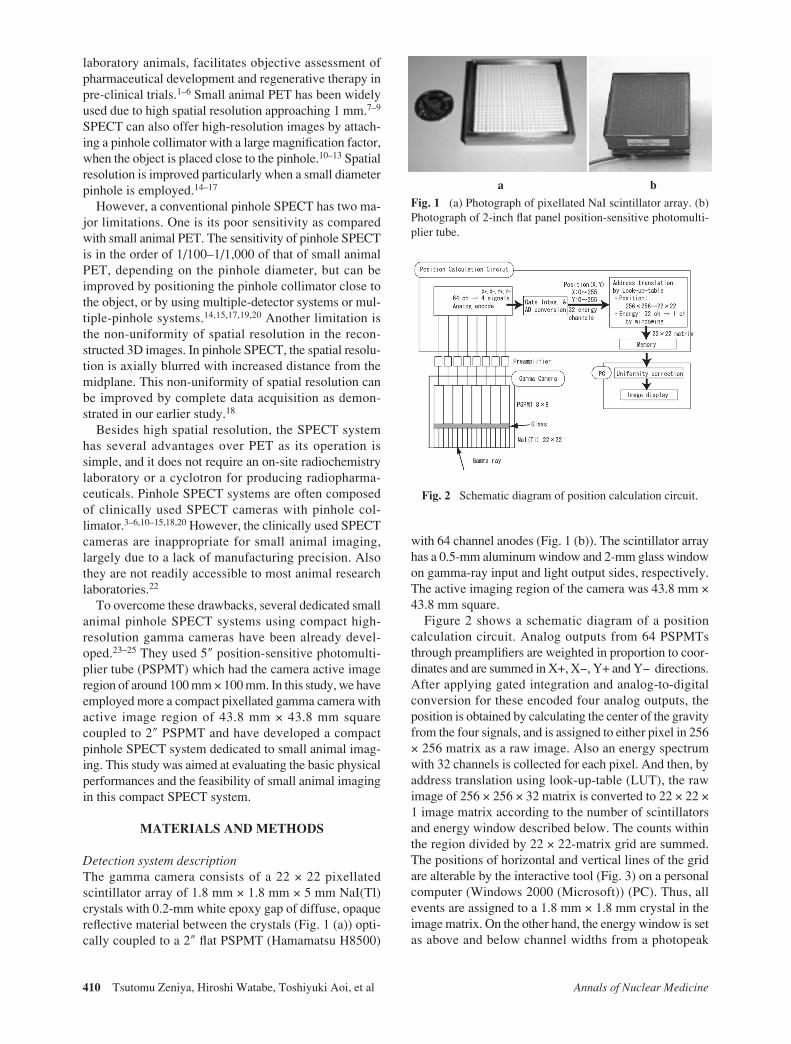
channel searched in each pixel. We assume the use of tworadioisotopes of 99mTc and 201Tl. The main photopeaksare 140 keV and 70 keV for 99mTc and 201Tl, respectively.Here, the camera gain for 201Tl was set about twice asmuch as that for 99mTc. The energy width with eachchannel corresponds to approximately 10 keV and 5 keVfor 99mTc and 201Tl, respectively. The energy window wasactually set at five channels, namely, approximately 36%for both 99mTc and 201Tl and centered on the photopeakchannel searched. Finally, the converted 22 × 22-pixelimage is stored on memory and then is transferred to thePC. The PC can perform several tasks such as correctinguniformity, displaying and analyzing images. Figure 3shows flood raw image by irradiating with a 99mTc pointsource. Separation between pixels was well performedexcept for pixels in columns and rows at the edge. Theeffective image field-of-view (FOV) that does not enclosethe edge pixels was 20 × 20 pixels (or 39.8 mm × 39.8mm). The resulting image is used as correction matrix tocorrect for non-uniformity in sensitivity over camera’sFOV.
Fig. 3 Interactive tool on PC for making address translationtable. The flood raw image was obtained by irradiating with a99mTc point source. The separation between pixels was wellperformed.
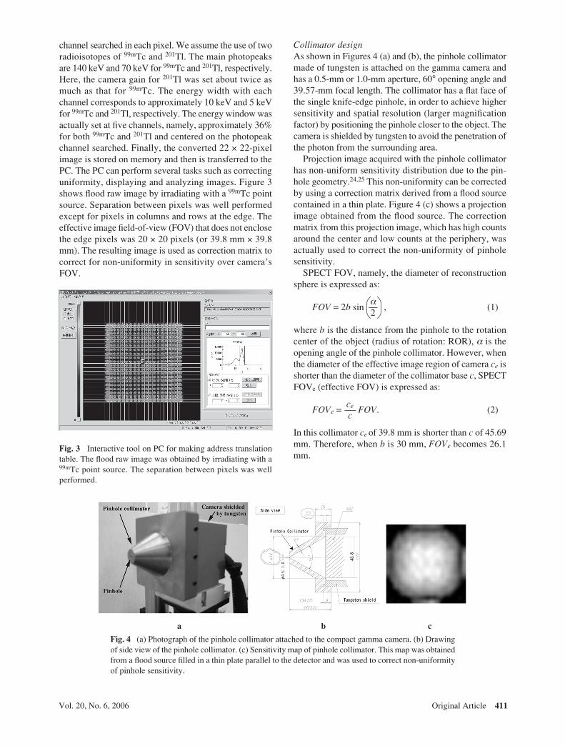
Fig. 4 (a) Photograph of the pinhole collimator attached to the compact gamma camera. (b) Drawingof side view of the pinhole collimator. (c) Sensitivity map of pinhole collimator. This map was obtainedfrom a flood source filled in a thin plate parallel to the detector and was used to correct non-uniformityof pinhole sensitivity.
a b c
Collimator designAs shown in Figures 4 (a) and (b), the pinhole collimatormade of tungsten is attached on the gamma camera andhas a 0.5-mm or 1.0-mm aperture, 60° opening angle and39.57-mm focal length. The collimator has a flat face ofthe single knife-edge pinhole, in order to achieve highersensitivity and spatial resolution (larger magnificationfactor) by positioning the pinhole closer to the object. Thecamera is shielded by tungsten to avoid the penetration ofthe photon from the surrounding area.
Projection image acquired with the pinhole collimatorhas non-uniform sensitivity distribution due to the pin-hole geometry.24,25 This non-uniformity can be correctedby using a correction matrix derived from a flood sourcecontained in a thin plate. Figure 4 (c) shows a projectionimage obtained from the flood source. The correctionmatrix from this projection image, which has high countsaround the center and low counts at the periphery, wasactually used to correct the non-uniformity of pinholesensitivity.
SPECT FOV, namely, the diameter of reconstructionsphere is expressed as:
FOV = 2b sinα
, (1)2
where b is the distance from the pinhole to the rotationcenter of the object (radius of rotation: ROR), α is theopening angle of the pinhole collimator. However, whenthe diameter of the effective image region of camera ce isshorter than the diameter of the collimator base c, SPECTFOVe (effective FOV) is expressed as:
FOVe = ce FOV. (2)c
In this collimator ce of 39.8 mm is shorter than c of 45.69mm. Therefore, when b is 30 mm, FOVe becomes 26.1mm.

Annals of Nuclear Medicine412 Tsutomu Zeniya, Hiroshi Watabe, Toshiyuki Aoi, et al
SPECT imaging systemFigure 5 shows the pinhole SPECT acquisition systemusing a compact gamma camera. Data acquisition iscontrolled by the PC through the camera controller. Therotation of the object stage is synchronized to step andshoot acquisition of the SPECT camera.
Basic system performancesThe performances of the detection system such as theenergy resolution, system spatial resolution, sensitivityand linearity of the count rate were examined with 99mTc(140 keV) and 201Tl (70 keV) sources.
(1) Energy resolution: Energy resolution was meas-ured by uniform irradiation with a 2.18 MBq 99mTc pointsource placed at 2 m distant from the camera without thecollimator for 12 hours and is defined for each crystal’senergy spectrum as full width at half maximum (FWHM)of the photopeak divided by its amplitude. The energyresolution was obtained from an energy spectrum for onecrystal near the center of the camera.
(2) System spatial resolution: The FWHMs of the linespread functions (LSFs) were measured in planar imageusing 99mTc and 201Tl line sources with 1.14-mm innerdiameter placed at 30 mm distant from the 1-mm pinhole.The magnification factor was 1.32. The LSFs of linesources were computed by deconvoluting with rectangu-lar function of 1.14-mm width. The spatial resolutionswere defined as the FWHM of Gaussian function obtainedfrom this deconvolution.
Fig. 5 Pinhole SPECT acquisition system using compactgamma camera. This system consists of compact gamma camerawith pinhole collimator, camera controller, PC, object rotatingstage and stage controller.
Fig. 6 (a) Energy spectrum obtained from this detection system. The energy resolution was 14.8%FWHM at 140 keV. (b) Planar image profile of 1.14-mm 99mTc line source. The system spatial resolutionobtained from the LSF was 2.8-mm FWHM. (c) On-axis sensitivities for 99mTc or 201Tl as a function ofdistance from the pinhole for 0.5-mm or 1.0-mm diameters. (d) Relationship between the source activityand the count rate measured by following decay of 99mTc source.

Original Article 413Vol. 20, No. 6, 2006
(3) Sensitivity: The system sensitivity on the centralaxis was measured using a small cylindrical phantom of0.1 ml at eight points in the range from 20 to 80-mmdistances with 0.5- or 1.0-mm pinholes and 99mTc or 201Tlsources.
(4) Linearity of count rate: The counts per 10 min weresequentially measured by following decay of the 99mTcsource. The cylindrical phantom made of glass with 24.3-mm outer diameter and 21.8-mm inner diameter was filledwith uniform 99mTc solution. The center of the phantomwas positioned at 30 mm distant from the 1-mm pinhole.Consequently, the relationship between activity and de-tected counts was examined.
Flood phantom SPECT studyA flood phantom SPECT study was performed to evaluatethe uniformity of the reconstruction images. The phantomused in this study was the same one as the cylindricalphantom used to evaluate the linearity of count rate, andwas filled with uniform 99mTc solution. The pinholecollimator with 1-mm diameter was used. The ROR was30 mm. This resulted in a magnification factor of 1.32.Projection data of 120 views were acquired over 360°using step and shoot acquisition; 10 sec/step, 3° incre-ments. Decay correction and the above-mentioned pin-hole sensitivity correction were applied for projectiondata before reconstruction. The projection data werereconstructed using our previously validated pinhole 3D-OSEM method employing a 3D voxel-driven projector in
both back- and forward-projections with eight subsets andtwo iterations. The corrections for attenuation, scatter andpenetration were not done.
Animal SPECT studyRat myocardial perfusion SPECT scans were sequentiallyperformed. A male rat weighing 220 g was anesthetizedwith sodium pentobarbital and held vertically on theobject rotating stage, and then was scanned after intrave-nous 2-min administration of 6.21 MBq/1.5 ml 201TlClinto the tail vein. The pinhole of 1 mm was used. The RORwas 30 mm. Scans for 10 min were sequentially per-formed four times using 360° step and shoot acquisition;5 sec/step, 3° increments. Like the flood phantom study,projection data were reconstructed using our pinhole 3D-OSEM method with eight subsets and two iterations.Corrections for attenuation, scatter, penetration and pin-hole sensitivity were not performed.
RESULTS
Basic system performances(1) Energy resolution: Figure 6 (a) shows a sample
energy spectrum from one crystal near the center of thedetector block. The energy resolution was 20.8-keV(14.8%) FWHM at 140 keV.
(2) System spatial resolution: Figure 6 (b) shows aplanar image profile of the 99mTc line source. The spatialresolutions were 2.8-mm FWHM and 2.5-mm FWHM for99mTc and 201Tl, respectively.
(3) Sensitivity: Figure 6 (c) shows system sensitivityon the central axis as a function of distance from thepinhole for 99mTc with 0.5- and 1.0-mm pinholes and for201Tl with 1.0-mm pinhole. The sensitivity of 201Tl wasslightly smaller than that of 99mTc. In the case of 99mTc, thesensitivities at a pinhole-source distance of 30 mm were27.0 and 53.0 cps/MBq with 0.5-mm and 1.0-mm pin-holes, respectively. In the case of 201Tl, the sensitivity atthe same distance was 49.1 cps/MBq with the 1.0-mmpinhole.
Fig. 7 SPECT images of uniform cylindrical phantom. (a)–(c)are without pinhole sensitivity correction. (d)–(f) are with thecorrection. (a) and (d) are transverse images. (b) and (e) arecoronal images. (c) and (f) are sagittal images. The profiles in thex and y directions were attached to the transverse images of (a)and (d).
Fig. 8 Short- and long-axial images of rat myocardial perfusionobtained by sequential SPECT scans. The left ventricular wallsand cavities were clearly visible in all of four frames obtained for40 min.

Annals of Nuclear Medicine414 Tsutomu Zeniya, Hiroshi Watabe, Toshiyuki Aoi, et al
b
f
(4) Linearity of count rate: Figure 6 (d) shows therelationship between the source activity and the countrate. The linearity was good up to 10 kcps and theregression line was y = 0.0588x + 0.009 (r2 = 0.9999).However, the ratio of the count rate to the source activitygradually decreased over 10 kcps.
Flood phantom SPECT studyFigure 7 shows SPECT images of uniform cylindricalphantom. The image reconstructed with pinhole geo-metrical sensitivity correction was almost uniform, whilethe image without the correction had high counts aroundthe center and low counts at the periphery.
Animal SPECT studyFigure 8 shows sequential SPECT images of a rat myocar-dial perfusion in four frames obtained for 40 min. The leftventricular walls and cavities were clearly visible in allframes.
DISCUSSION
We have developed a compact SPECT system using acompact pixellated gamma camera for small animals andsucceeded in sequential SPECT imaging of rat myocar-dial perfusion. In this system we employed 2″ PSPMTrather than 5″ PSPMT which was used by other investiga-tors21–23 because the use of 2″ PSPMT allows one toconstruct a more inexpensive, compact and lighter sys-tem.
The energy resolution of 14.8% FWHM in this camerawas worse than that of approximately 10% FWHM inclinical SPECT gamma camera. So, the profile of thephotopeak in the energy spectrum was as broad as the 36%energy window used. McElroy et al. reported22 that intheir pinhole system, scatter fraction did not contribute asignificant amount to images (about 5%) for mouse sized2.5-cm diameter cylinder when the usual 20% energywindow was used in their system with a 11.4% energyresolution. However, the scatter fractions were about 15%and 20% for rat sized 3.8-cm and 5.05-cm diametercylinders. Further study is needed to evaluate the contri-bution of scatter photons and develop proper scattercorrection technique26 for our system.
The measured system spatial resolutions were 2.8-mmFWHM and 2.5-mm FWHM for 99mTc and 201Tl, respec-tively. Here, the theoretical system spatial resolution fora pinhole collimated gamma camera R0 is given by
R0 ≅ Ri + de , (3)
where f is the distance between the pinhole and thedetector (focal length), b is ROR, Ri is the intrinsic cameraresolution, and de is the effective pinhole diameter ex-pressed as:
f + b
f
de ≈ d d + tan , (4)
where d is the actual pinhole diameter, µ is the linearattenuation coefficient of the collimator material, and α isthe opening angle of the pinhole collimator.22,27 Thetheoretical spatial resolutions in this experimental condi-tion are 2.5 mm and 2.3 mm for 99mTc and 201Tl, respec-tively. The spatial resolution of 99mTc is larger than that of201Tl due to its higher energy (µ ≈ 4.098 mm−1 for 99mTcand µ ≈ 20.870 mm−1 for 201Tl) and more penetrationphotons, which appeared in the experimental results.However, the measured spatial resolutions are slightlyworse than the theoretical ones. As one of the reasons forthe difference, the theoretical calculation assumes a doubleknife-edge pinhole collimator, while our pinhole collima-tor is single knife-edge. The number of penetration pho-tons in single knife-edge is larger than that in doubleknife-edge. Therefore, the actual pinhole diameter forsingle knife-edge is larger than that for double knife-edge.By accounting for the difference of the knife-edge of thecollimator, the measured spatial resolutions largely agreewith the theoretical ones. Weber et al. obtained rat myo-cardial images at a spatial resolution of 2.8-mm FWHM.11
The spatial resolution measured in our system is almostequal to that measured in their system.
However, the spatial resolution obtained in our systemmight be unsatisfactory for mouse imaging. Resolutioncan be improved by using a smaller diameter pinhole, butthis will decrease sensitivity in return for improvement ofresolution. Decreasing the crystal size or enlarging thedetection area of the camera can improve resolutionwithout decreasing sensitivity. Resolution is usually de-graded by non-zero diameter and edge penetration of apinhole. Alternatively, this degraded resolution can berecovered by incorporating the realistic pinhole modelinto reconstruction software.28,29 This approach does notrequire any modification of hardware and is applicable toour system.
From Figure 6 (c), positioning the pinhole closer to theobject is important for improvement of sensitivity inpinhole SPECT. In that respect, the flat face of a single-knife edge collimator is advantageous. However, caremust be taken of the effect of penetration for thin, singleknife-edge collimator. The sensitivity of 201Tl was slightlysmaller than that of 99mTc. This is considered that thenumber of penetrations for 201Tl was less than that for99mTc due to lower energy of 201Tl compared to 99mTc. Infuture we need to evaluate the effect of the penetration forboth 99mTc and 201Tl in single-knife edge.
A good linearity of the count rate up to 10 kcps wasshown in this study, although the ratio of the count rate tothe source activity gradually decreased over 10 kcps. Thischaracteristic of the count rate allows the present compactgamma camera system to be applied for rat myocardial
2µ
α2
2 2

Original Article 415Vol. 20, No. 6, 2006
SPECT imaging with 201Tl because the mean count ratewas 0.52 kcps during SPECT data acquisition in this ratstudy. This upper limitation of 10 kcps is due to thetransfer speed of the electric circuit which can be im-proved by replacement with faster electronics. The presentsystem also produced homogeneous reconstructed im-ages of the cylindrical phantom as shown in Figure 7. Thehomogeneity of images reconstructed from the floodphantom is important for quantitative analysis of physi-ological function.
In the animal SPECT study, rat myocardial tomo-graphic images were sequentially obtained. The SPECTimages could clearly visualize the rat myocardium andcardiac cavity. It is anticipated that these images could besignificantly improved if image gate is employed. Thetime-dependent change of regional tissue radioactivityconcentration obtained from such sequential tomographicimages can be applied for kinetic analysis using a com-partment model to estimate the regional myocardial bloodflow.30 The results for phantom and animal studies sup-port the feasibility of our system for quantitative assess-ments of regional myocardial blood flow on rat. Furtherstudy is needed to quantify myocardial blood flow bypinhole SPECT. Deloar et al. suggested that physicalfactors such as penetration and scatter are considered.31
Also, Wang et al. reported that both of attenuation correc-tion (AC) and scatter compensation (SC) are important toimprove quantitative accuracy because the values of re-constructed images were underestimated by 15% withoutAC and overestimated by 9% with only AC, while thequantitative accuracy was below 3% with both AC andSC.32
If this compact camera is combined with a rotatingapparatus, the camera will rotate around the animal laiddown. So, we can observe physiological function of smallanimals in more natural conditions, against especiallyacquiring complete data set with two orbits,18 than whenthe animals are held vertically like in this study. Themisalignment of a center of rotation (COR) might be aproblem, if the camera is rotated. It causes serious arti-facts in the reconstructed image.13 However, our compactcamera is sufficiently light to avoid the misalignment ofthe COR compared to clinical gamma camera. In the nearfuture, we will construct a small animal pinhole SPECTsystem, which permits acquisition of complete data bytwo-circular orbit, using a compact gamma camera.
CONCLUSION
We have developed a compact SPECT system using acompact pixellated gamma camera for small animals. Thecamera with an active detection area of 43.8 mm × 43.8mm was equipped with a pinhole collimator. We evalu-ated the basic physical performances and succeeded insequential SPECT imaging of rat myocardial perfusion.The present system may be of use for quantitation of
biological functions such as myocardial blood flow insmall animals.
ACKNOWLEDGMENT
This study was financially supported by the Budget for NuclearResearch of the Ministry of Education, Culture, Sports, Scienceand Technology, based on screening and counseling by theAtomic Energy Commission, Japan.
REFERENCES
1. Meikle SR, Eberl S, Iida H. Instrumentation and methodol-ogy for quantitative pre-clinical imaging studies. CurrPharm Des 2001; 7 (18): 1945–1966.
2. Chatziioannou AF. PET scanners dedicated to molecularimaging of small animal models. Mol Imaging Biol 2002; 4(1): 47–63.
3. Hirai T, Nohara R, Ogoh S, Chen LG, Kataoka K, Li XH, etal. Serial evaluation of fatty acid metabolism in rats withmyocardial infarction by pinhole SPECT. J Nucl Cardiol2001; 8 (4): 472–481.
4. Scherfler C, Donnemiller E, Schocke M, Dierkes K,Decristoforo C, Oberladstätter M, et al. Evaluation of stri-atal dopamine transporter function in rats by in vivo β-[123I]CIT pinhole SPECT. NeuroImage 2002; 17: 128–141.
5. Acton PD, Choi SR, Plössl K, Kung HF. Quantification ofdopamine transporters in the mouse brain using ultra-highresolution single-photon emission tomography. Eur J NuclMed 2002; 29 (5): 691–698.
6. Aoi T, Watabe H, Deloar HM, Ogawa M, Teramoto N,Kudomi N, et al. Absolute quantitation of regional myocar-dial blood flow of rats using dynamic pinhole SPECT. InConference Record of 2002 IEEE Nuclear Science Sympo-sium and Medical Imaging Conference (CD-ROM), 2003:M11-185.
7. Jeavons AP, Chandler RA, Dettmar CAR. A 3D HIDAC-PET camera with sub-millimetre resolution for imagingsmall animals. IEEE Trans Nucl Sci 1999; 46 (3): 468–473.
8. Seidel J, Vaquero JJ, Green MV. Resolution uniformity andsensitivity of the NIH ATLAS small animal PET scanner:comparison to simulated LSO scanners without depth-of-interaction capability. IEEE Trans Nucl Sci 2003; 50 (5):1347–1350.
9. Tai YC, Chatziioannou AF, Yang Y, Silverman RW,Meadors K, Siegel S, et al. MicroPET II: design, develop-ment and initial performance of an improved microPETscanner for small-animal imaging. Phys Med Biol 2003; 48:1519–1537.
10. Jaszczak RJ, Li J, Wang H, Zalutsky MR, Coleman RE.Pinhole collimation for ultra-high-resolution, small-field-of-view SPECT. Phys Med Biol 1994; 39: 425–437.
11. Weber DA, Ivanovic M, Franceschi D, Strand SE, ErlandssonK, Franceschi M, et al. Pinhole SPECT: an approach to invivo high resolution SPECT imaging in small laboratoryanimals. J Nucl Med 1994; 35 (2): 342–348.
12. Ishizu K, Mukai T, Yonekura Y, Pagani M, Fujita T, MagataY, et al. Ultra-high resolution SPECT system using fourpinhole collimators for small animal studies. J Nucl Med1995; 36 (12): 2282–2287.

Annals of Nuclear Medicine416 Tsutomu Zeniya, Hiroshi Watabe, Toshiyuki Aoi, et al
13. Ogawa K, Kawade T, Nakamura K, Kubo A, Ichihara T.Ultra high resolution pinhole SPECT for small animalstudy. IEEE Trans Nucl Sci 1998; 45 (6): 3122–3126.
14. Moore SC, Zimmerman RE, Mahmood A, Mellen R, LimCB. A triple-detector, multiple-pinhole system for SPECTimaging rodents. [Abstract] J Nucl Med 2004; 45 (suppl):97–98.
15. Beekman FJ, van der Have F, Vastenhouw B, van derLinden AJ, van Rijk PP, Burbach JP, et al. U-SPECT-I: anovel system for submillimeter-resolution tomography withradiolabeled molecules in mice. J Nucl Med 2005; 46 (7):1194–1200.
16. Sun M, Izaguirre EW, Funk T, Hwang AB, Carver J,Thompson S, et al. A CdZnTe-based high-resolutionmicroSPECT system. [Abstract] J Nucl Med 2005; 46(suppl 2): p170.
17. Lackas C, Hoppin JW, Schramm NU. Performance analysisof a submillimeter-resolution multi-pinhole SPECT small-animal imaging system. [Abstract] J Nucl Med 2005; 46(suppl 2): p171.
18. Zeniya T, Watabe H, Aoi T, Kim KM, Teramoto N, HayashiT, et al. A new reconstruction strategy for image improve-ment in pinhole SPECT. Eur J Nucl Med Mol Imaging 2004;31 (8): 1166–1172.
19. Liu Z, Kastis GA, Stevenson GD, Barrett HH, Furenlid LR,Kupinski MA, et al. Quantitative analysis of acute myocar-dial infarct in rat hearts with ischemia-reperfusion using ahigh-resolution stationary SPECT system. J Nucl Med2002; 43 (7): 933–939.
20. Schramm NU, Ebel G, Engeland U, Schurrat T, Béhé M,Behr TM. High-resolution SPECT using multipinhole col-limation. IEEE Trans Nucl Sci 2003; 50 (3): 315–320.
21. Schramm N, Wirrwar A, Sonnenberg F, Halling H. Com-pact high resolution detector for small animal SPECT. IEEETrans Nucl Sci 2000; 47 (3): 1163–1167.
22. McElroy DP, MacDonald LR, Beekman FJ, Wang Y, Patt
BE, Iwanczyk JS, et al. Performance evaluation of A-SPECT: a high resolution desktop pinhole SPECT systemfor imaging small animals. IEEE Trans Nucl Sci 2002; 49(5): 2139–2147.
23. Wojcik R, Goode AR, Smith MF, Beller GA, Ellman PI,Majewski S, et al. Dedicated small field of view SPECTsystem based on a 5″ PSPMT and crystal scintillator arrayfor high resolution small animal cardiac imaging. In Con-ference Record of 2003 IEEE Nuclear Science Symposiumand Medical Imaging Conference (CD-ROM), 2004: M3-43.
24. Smith MF, Jaszczak RJ. The effect of gamma ray penetra-tion on angle-dependent sensitivity for pinhole collimationin nuclear medicine. Med Phys 1997; 24 (11): 1701–1709.
25. Metzler SD, Bowsher JE, Smith MF, Jaszczak RJ. Analyti-cal determination of pinhole collimator sensitivity withpenetration. IEEE Trans Med Imag 2001; 20 (8): 730–741.
26. Deloar HM, Watabe H, Kim KM, Aoi T, Kunieda E, FujiiH, et al. Optimization of the width of the photopeak energywindow in the TDCS technique for scatter correction inquantitative SPECT. IEEE Trans Nucl Sci 2004; 51 (3):625–630.
27. Anger HO. Radioisotope cameras. In: Instrumentation innuclear medicine, Hine GJ (ed), New York; Academic,1967: 485–552.
28. Bequé D, Vanhove C, Andreyev A, Nuyts J, Defrise M.Correction for impact camera motion and resolution recov-ery in pinhole SPECT. In Conference Record of 2004 IEEENuclear Science Symposium and Medical Imaging Confer-ence (CD-ROM), 2005: M2-173.
29. Cao Z, Bal G, Acton PD. Pinhole SPECT reconstructionwith resolution recovery. [Abstract] J Nucl Med 2005; 45(suppl 2): 109–110.
30. Iida H, Eberl S. Quantitative assessment of regional myo-cardial blood flow with thallium-201 and SPECT. J NuclCardiol 1998; 5: 313–331.
Related Documents