Pathology Specimens for Urology Residents Dr Prashant Bansal

Welcome message from author
This document is posted to help you gain knowledge. Please leave a comment to let me know what you think about it! Share it to your friends and learn new things together.
Transcript

Pathology Specimens for Urology Residents
Dr Prashant Bansal

SPECIMEN NO 1


• Bilateral nephrectomy specimens.
• The external surface of both kidneys have a
bosselated appearance which is produced by
numerous cysts of varying sizes and
replacing the entire renal parenchyma.
• Cysts are thin walled and translucent and
filled with clear fluid.
• Adult (autosomal dominant) polycystic
kidney disease
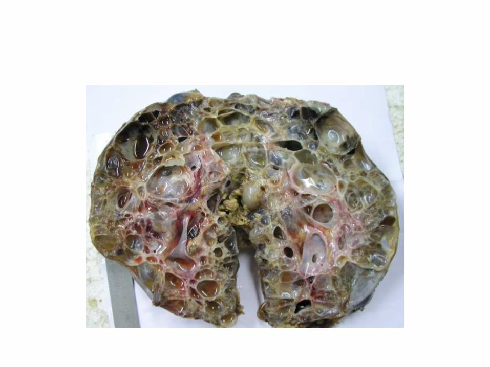
• Cut open specimen of kidney. • The kidney is enlarged and replaced by numerous cysts
of varying sizes arising at all levels in the cortex and medulla.
• The cysts are unilocular and are filled with clear serous to hemorrhagic fluid.
• They range from a few millimeters to several centimeters.
• Normal renal parenchyma is not identified. • No mass or papillary lesions seen. • ADPKD

CASE NO 2


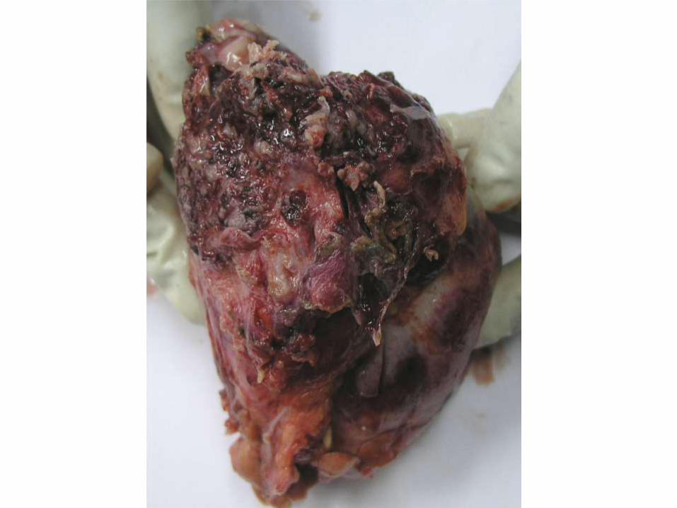
• A solitary poorly circumscribed solid grey white tumour is seen involving the upper calyceal system and infiltrating into the renal parenchyma.
• Foci of haemorrhage are identified in the pelvic mucosa.
• The rest of the renal parenchyma appears unremarkable.
• Urothelial carcinoma

CASE NO 4

• Hydatid Cyst Kidney

CASE NO 5
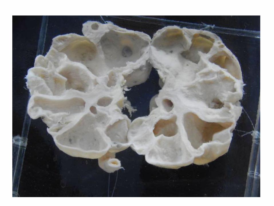
• Grossly hydronephrotic kidney with thinned out parenchyma

CASE NO 6
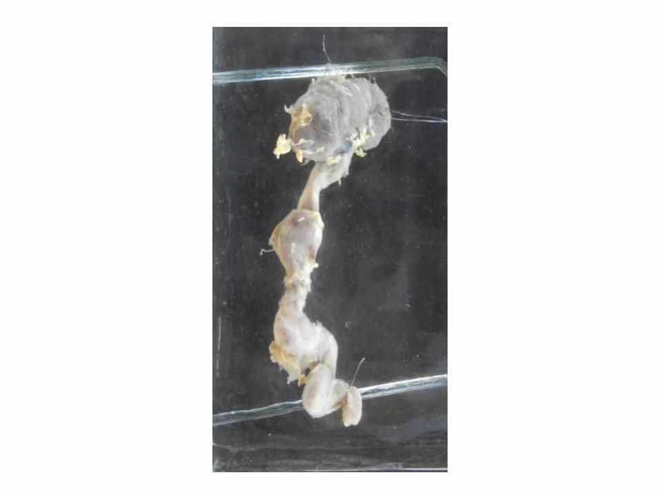

• Nephroureterectomy specimen
• Small sized kidney
• Grossly dilated and tortuous ureter
• s/o reflux nephropathy
CASE NO 7

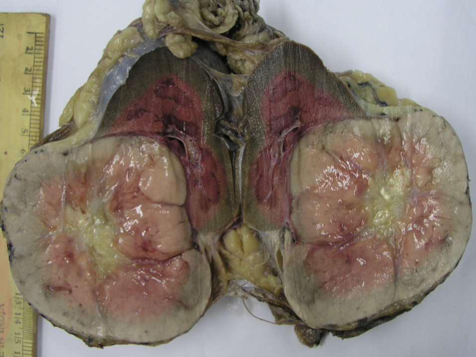
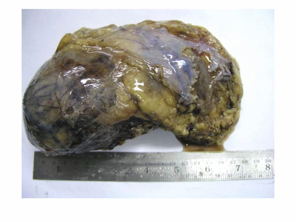
• Nephrectomy specimen
• Upper pole mass
• S/o RCC

CASE NO 9


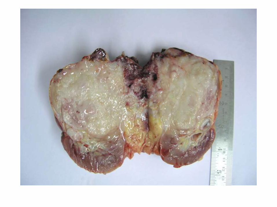
• Solitary well circumscribed exophytic tumour arising from the mid pole of kidney.
• Tumour is tan in colour, homogenous with a central scar.
• Rest of the renal parenchyma shows no significant pathology

Case 4


• Solitary well circumscribed tumour in the upper pole of kidney.
• Tumour is mahogany brown and homogenous.
• No cystic, necrotic or haemorrhagic areas seen.
• Rest of the renal parenchyma shows no significant pathology
• HPE was Chromophobe RCC (not to be commented in exam – JUST say RCC)

Case 6


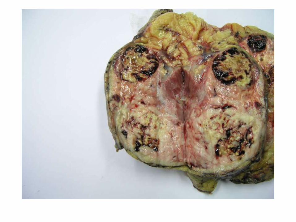
• Cut open specimen of kidney with dilated pelvicalyceal system.
• The calyceal mucosa has plaques of golden yellow tissue which focally invades the renal parenchyma and extends into the perirenal fat.
• Xanthogranulomatous pyelonephritis

Case 7


Case 9
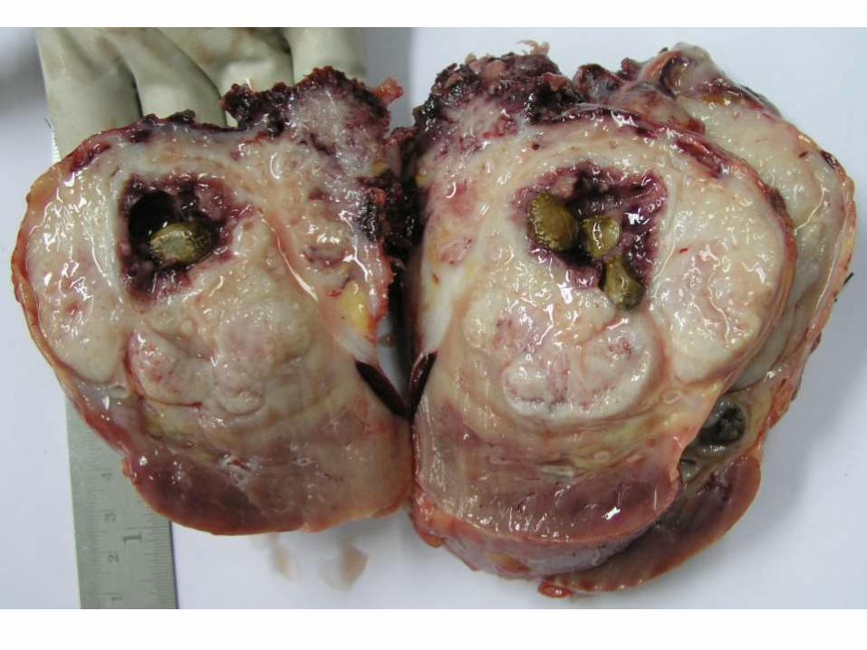

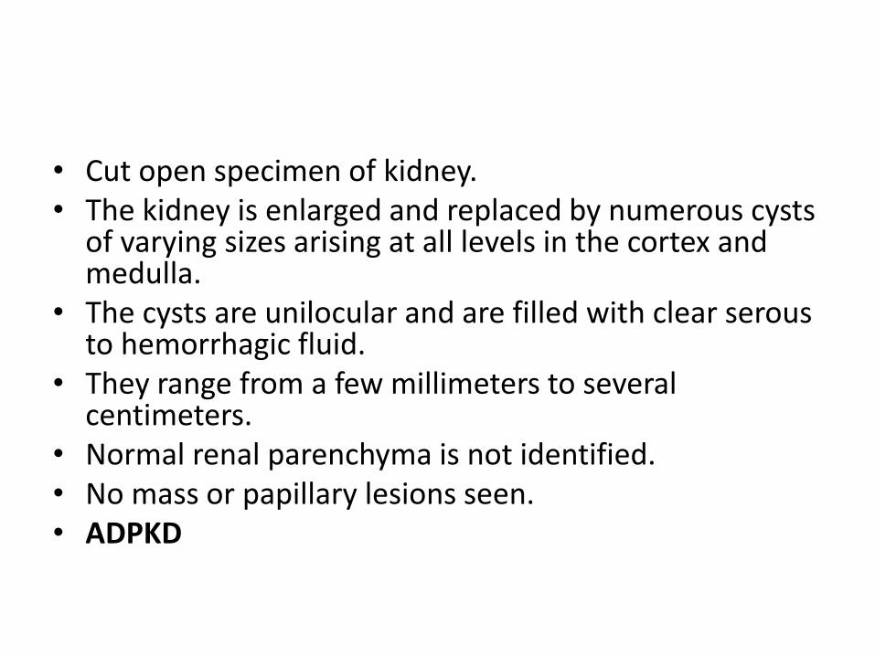
• Specimen of kidney.
• There is a solid white tumour involving the upper pole of the kidney.
• The tumour extends beyond the renal capsule into the perirenal fat.
• Calculi are embedded within the tumour.
• Squamous cell carcinoma with nephrolithiasis, kidney


Case 10


Case 11


• Renal tumour arising from the lower pole of the kidney. • Tumour is well circumscribed, solid with areas of necrosis. • The ipsilateral adrenal gland shows a tumour with the
same gross appearance as the renal tumour.• The renal parenchyma between the renal tumour and the
adrenal tumour is unremarkable.• The makes the adrenal mass a metastatic lesion and not an
extension from the primary tumour.• Also included in this image is the contra lateral adrenal
gland which appears grossly enlarged, suggesting metastatic involvement.
• (Clear cell RCC with bilateral adrenal metastasis)
Related Documents