OBSTUCTIVE UROPATHY OBSTUCTIVE UROPATHY CSBR.Prasad, MD.,

Welcome message from author
This document is posted to help you gain knowledge. Please leave a comment to let me know what you think about it! Share it to your friends and learn new things together.
Transcript

OBSTUCTIVE UROPATHYOBSTUCTIVE UROPATHY
CSBR.Prasad, MD.,
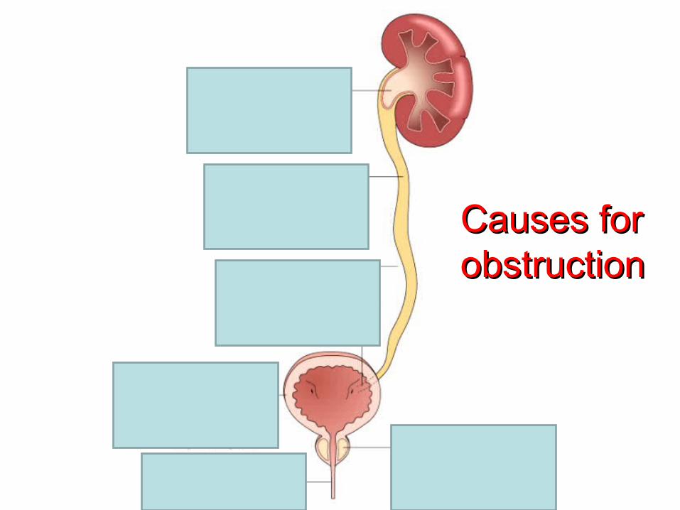
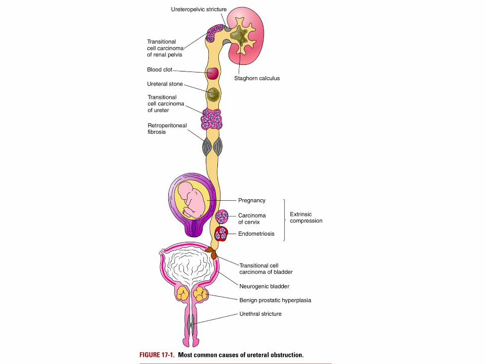
Causes for Causes for obstructionobstruction
Urolithiasis
• World wide distribution
• 2 % of population
• M:F 2:1
• Peak age 2nd to 3rd decade.
Types
1. Calcium stones
2. Mixed stones ( struvite)
3. Uric acid stones
4. Cystine stones

Prevalence of various types of Renal stones% of all stones
Ca.Oxalate and PhosphateCa.Oxalate and Phosphate 7070
Idiopathic hypercalciuria (50%)
Hypercalciuria & hypercalcemia (10%)
Hyperoxaluria (5%)
Enteric (4.5%)
Primary (0.5%)
Hyperuricosuria (20%)
Hypocitraturia
No known metabolic abnormality (15-20%)
Magnesium Ammonium Phosphate ((STRUVITE)) 15-20
Uric acidUric acid 5-105-10
Associated with hyperuricemia
Associated with hyperuricosuria
Idiopathic (50% of uric acid stones)
Cystine 1-2
Other or unknownOther or unknown +5+5

Calcium stones
• Most common 75%
• Pure stones of Ca oxalate 50%
• Pure stones of Ca phosphate 06%
• Mixture of Ca oxalate & Ca phosphate 45%
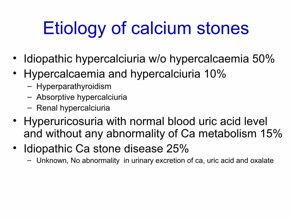
Etiology of calcium stones
• Idiopathic hypercalciuria w/o hypercalcaemia 50%• Hypercalcaemia and hypercalciuria 10%
– Hyperparathyroidism– Absorptive hypercalciuria– Renal hypercalciuria
• Hyperuricosuria with normal blood uric acid level and without any abnormality of Ca metabolism 15%
• Idiopathic Ca stone disease 25%– Unknown, No abnormality in urinary excretion of ca, uric acid and oxalate

Pathogenesis
• Imbalance b/n the degree of supersaturation of ions forming the stone and concentration of inhibition in urine
• Nidus – crystals of Ca oxalate, Ca PO4 precipitate in tubular lining around some fragment of debris in tubules
• The stone grow, deposition of more crystals at nidus

Factors contributing stone formation
• Urinary alkaline pH
• Decreased urinary volume
• Increased excretion of oxalate and uric acid

Morphology
• Small less than 1cm
• Ovoid, hard SPIKY surface
• Dark brown due to blood

Nephrolithiasis A large stone impacted in the renal pelvis
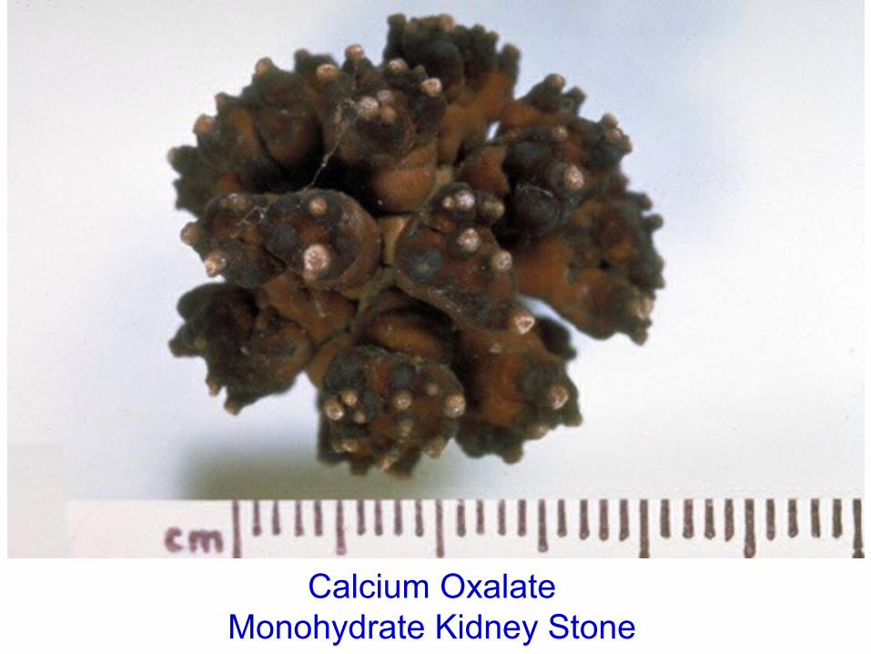
Calcium Oxalate Monohydrate Kidney Stone

Mixed stones (Struvite stones)15 %
• Magnesium phosphate
• Ammonium phosphate STRUVITE• Calcium phosphate
Triple phosphate stones

Struvite stones
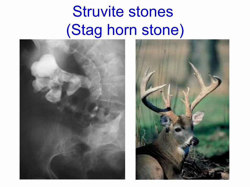
Struvite stones (Stag horn stone)

Etiology of Struvite stones
• Infection of UT with urea splitting bacteria
• Proteus, Klebsiella, Enterobacter
• Infection induced stones

Morphology struvitie stones
• Yellow - white or grey
• Soft, friable, irregular in shape
• Stag horn stone: large solitary stone that takes the shape of renal pelvis


Uric acid stones. 6%- etiology
• Hyperuricaemia, hyperuricosuria• Primary/Secondary gout (due to myeloproliferative dis)• Leukemia on chemotherapy• Administration of uricosuric drugs (Salicylates, Probenicid)• Other factors acid pH less than 6 low urinary volume
High nucleic acid turnover

Pathogenesis of uric acid stones
• Solubility of uric acid at pH 7 is 200 mg/dl
• at pH 5 is 15 mg/dl
• Urine becomes acidic, solubility UA decreases
• Prepecipitation of uric acid crystals favours uric acid stones.

Uric acid stones - 6%
• Radiolucent X-ray• But visible on US or CT
Radiolucent stonesUric acidXanthineTriamtereneDihydroxyadenine
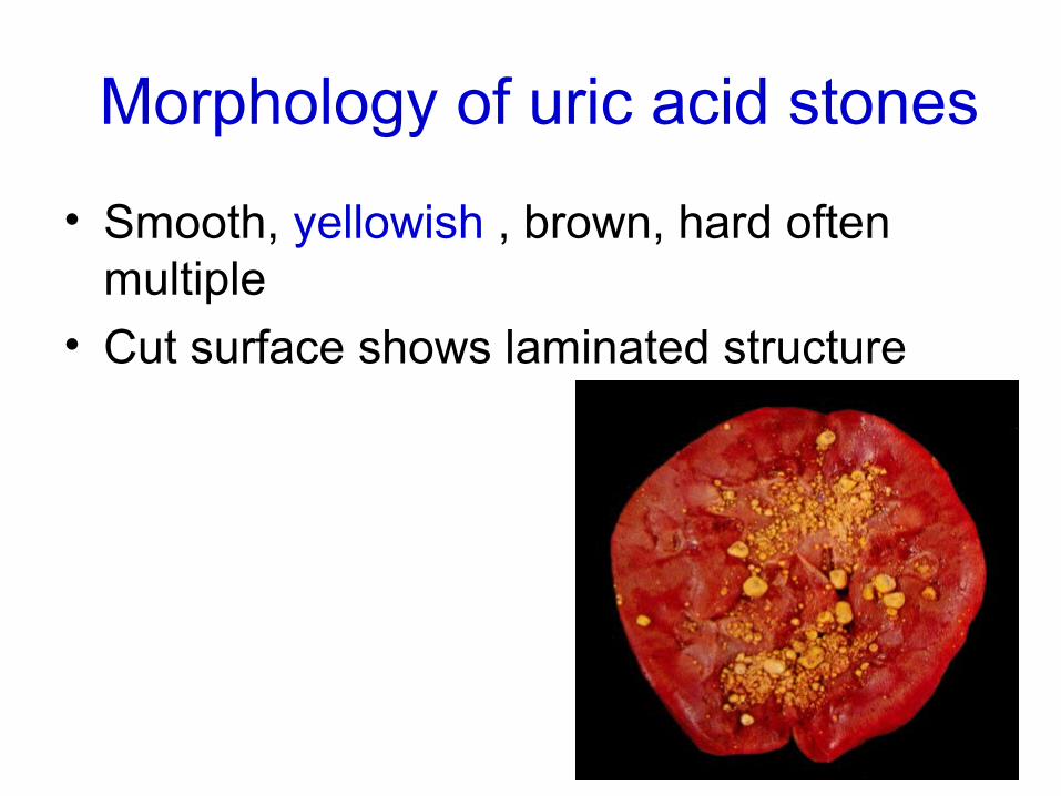
Morphology of uric acid stones
• Smooth, yellowish , brown, hard often multiple
• Cut surface shows laminated structure

Cystine stones 2 %etiology
Cystinuria
Genetically determined
Defect in transport of cystine across
CM/renal tubules, mucosa
Pathogenesis of cystine stones
• Cystine is least soluble among all aminoacids
• Under excess cystineuria- concretion and stone formation
Morphology of cystine stones
• Small round, smooth
• Multiple, yellow, waxy

Other stones less than 2 %
• Inherited xanthene metabolism
• Xanthinuria
• Xanthene stones

UROLITHIASIS
Deficiency of inhibitors of crystal formation
•Pyrophosphate
•Diphosphonate
•Citrate
•Glycosaminoglycans
•Osteopontin
•Nephrocalcin

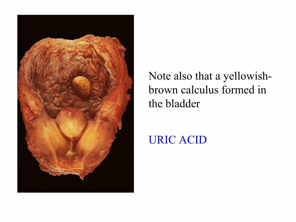
Note also that a yellowish-brown calculus formed in the bladder
URIC ACID

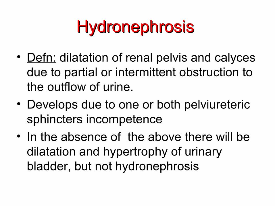
Hydronephrosis Hydronephrosis
• Defn: dilatation of renal pelvis and calyces due to partial or intermittent obstruction to the outflow of urine.
• Develops due to one or both pelviureteric sphincters incompetence
• In the absence of the above there will be dilatation and hypertrophy of urinary bladder, but not hydronephrosis

Hydronephrosis of the kidney, with marked dilation of the pelvis and calyces and thinning of the renal parenchyma

Case of hydronephrosis--a ureteral calculus
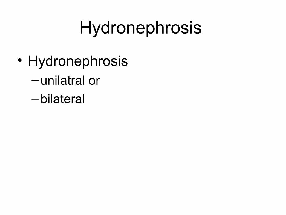
Hydronephrosis
• Hydronephrosis– unilatral or
– bilateral

Unilateral hydronephrosis
Ureteral obstruction at the level of pelviureteric junction
1. Intraluminal- calculi in ureter/renal pelvis
2. Intramural- cong PUJ obstruction– Atresia of ureter– Inflammatory stricture– Trauma
– Neoplasms of ureter or bladder 3. Extramural Obstruction of uppr part of ureter by inf renal artery/vein
Pressure on ureter from outside ex ca cx, prostate,rectum, caecum, retroperitoneal fibrosis

Bilateral hydronephrosis
• Congenital: Atresia of urethral meatus Cong posterior urethral valve• Acquired: Bladder tumor involving both ureteric
orifices Prostatic enlargement Ca prostate, prostatitis Bladder neck stenosis Inflammatory/traumatic urethral stricture & phimosis
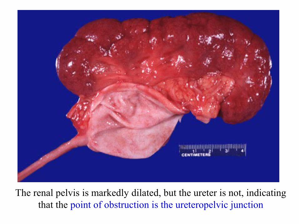
The renal pelvis is markedly dilated, but the ureter is not, indicating that the point of obstruction is the ureteropelvic junction

Pathologic changes
• Depends obstruction,
sudden / gradual
complete/incomplete
Intermittent
• Extrarenal / intrarenal
Extra renal hydronephrosis
• Dilatation of renal pelvis medially in the form of sac
• As the obstruction persists
-Progressive dilation of pelvis/ calyces- pressure atrophy of renal parenchyma
• Dilated – pelvicalyceal cystem extends deep in to renal cortex- thin rim of renal cortex streches over calyces- lobulation

Microscopy –hydronehrosis.
• Wall of hydronephrotic sac-
fibrous thickening –scarring
inflammatory cell infiltrates
• Progressive atrophy of tubules, glomeruli
• Stasis of urine- infection pyonephrosis.

E N D
Related Documents