Uro-radiology 2.

Uro-radiology
Oct 14, 2014
role of radiology in kidney disease
Welcome message from author
This document is posted to help you gain knowledge. Please leave a comment to let me know what you think about it! Share it to your friends and learn new things together.
Transcript

Uro-radiology 2.

Our menu.
• Usual introductory remarks.• Tools of the trade in uroradiology.• Trends in uroradiology.• Discuss select pathologies in
uroradiology.• The classics of uroradiology. • Epilogue.
INTRODUCTION
Imaging modalities in uroradiology have undergone a dramatic transformation in the recent past.
Earlier plain films were the only available study.
Subsequently for no less than 7 decades IVU was the pillar of uroradiology.
Today cross-sectional imaging studies have taken centre stage albeit without a definite flag bearer....
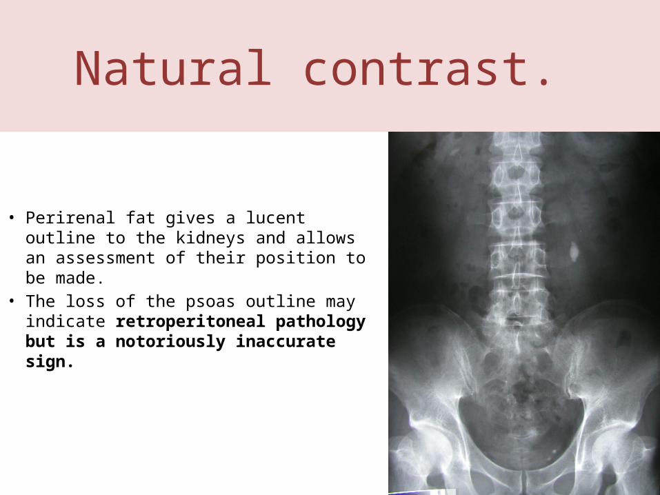
Natural contrast.
• Perirenal fat gives a lucent outline to the kidneys and allows an assessment of their position to be made.
• The loss of the psoas outline may indicate retroperitoneal pathology but is a notoriously inaccurate sign.

IVU
• The excretory urogram is a valuable examination of the urinary tract.
• It gives excellent anatomical images of the kidneys and to some extent an indication of their function.

Patient preparation.
• Bowel preparation is useful but no longer essential. Plumber unnecessary.
• Fluid restriction unless severe does not influence radiographic densities.
• At this severe level it becomes counter productive when it influences tubular function.
• Patients with diabetes and myeloma specifically require hydration.
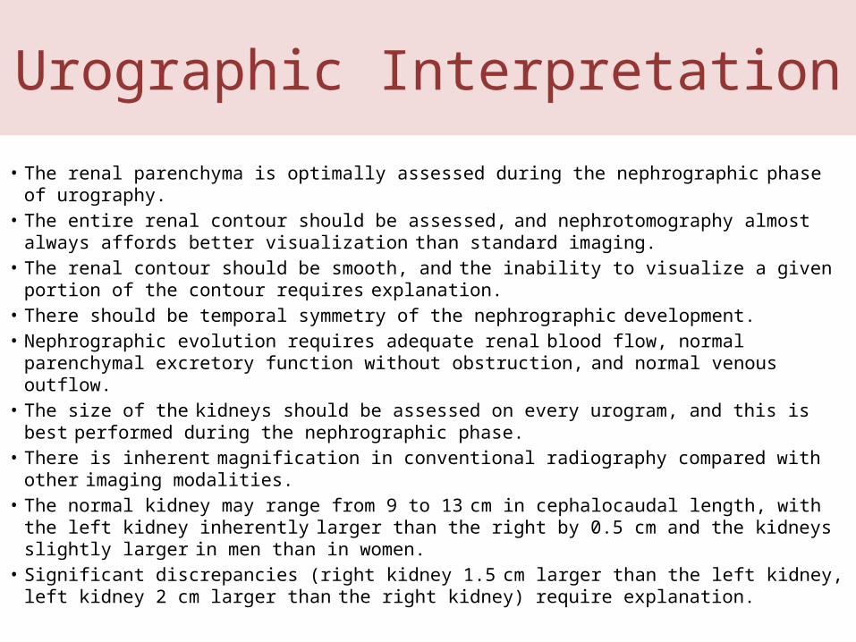
Urographic Interpretation• The renal parenchyma is optimally assessed during the nephrographic phase of
urography. • The entire renal contour should be assessed, and nephrotomography almost
always affords better visualization than standard imaging. • The renal contour should be smooth, and the inability to visualize a given
portion of the contour requires explanation. • There should be temporal symmetry of the nephrographic development. • Nephrographic evolution requires adequate renal blood flow, normal
parenchymal excretory function without obstruction, and normal venous outflow. • The size of the kidneys should be assessed on every urogram, and this is best
performed during the nephrographic phase. • There is inherent magnification in conventional radiography compared with
other imaging modalities. • The normal kidney may range from 9 to 13 cm in cephalocaudal length, with the
left kidney inherently larger than the right by 0.5 cm and the kidneys slightly larger in men than in women.
• Significant discrepancies (right kidney 1.5 cm larger than the left kidney, left kidney 2 cm larger than the right kidney) require explanation.

Normal nephrogram
• Final image of a tomographic sequence demonstrates symmetric nephrograms and pyelograms.
• Renal size is normal.
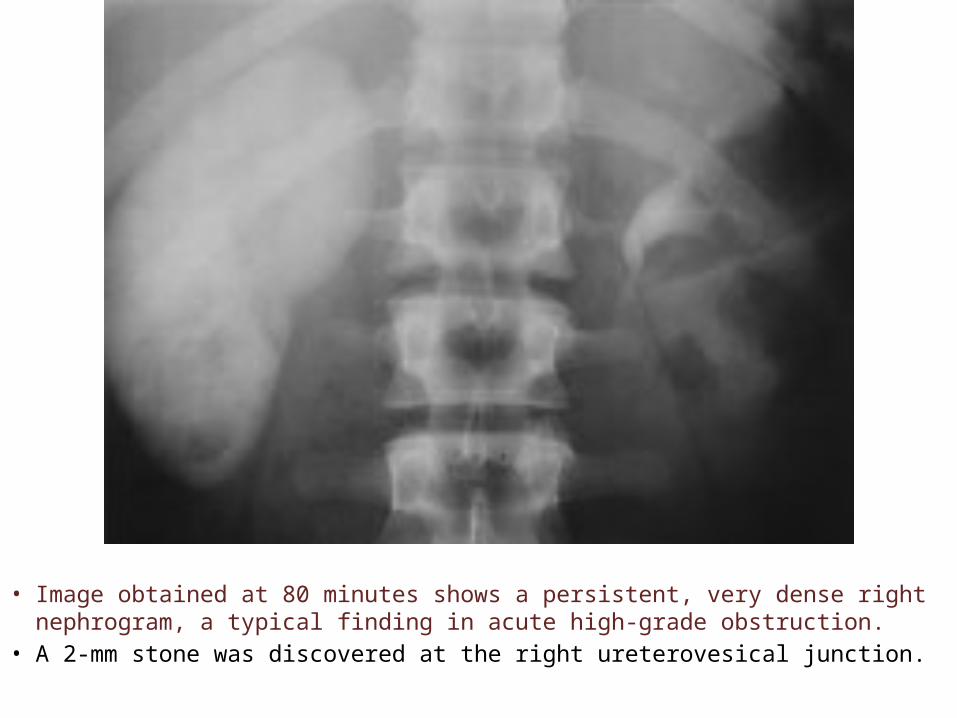
• Image obtained at 80 minutes shows a persistent, very dense right nephrogram, a typical finding in acute high-grade obstruction.
• A 2-mm stone was discovered at the right ureterovesical junction.

• Enlarged kidneys in a young patient with early, asymmetric findings of autosomal dominant polycystic kidney disease.
• Nephrotomogram shows enlarged kidneys, the left more so than the right.
• Note the multiple parenchymal defects ("Swiss cheese" nephrogram).
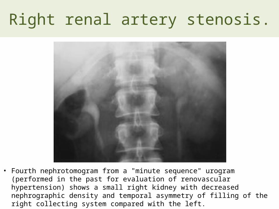
Right renal artery stenosis.
• Fourth nephrotomogram from a "minute sequence" urogram (performed in the past for evaluation of renovascular hypertension) shows a small right kidney with decreased nephrographic density and temporal asymmetry of filling of the right collecting system compared with the left.

Simple cyst.
• Nephrotomogram shows a nephrographic defect in the midportion of the left kidney (arrow) with increased parenchymal thickness and distortion of the underlying collecting system. US helped confirm a simple cyst.

Renal cell carcinoma.
• Nephrotomogram shows a mass in the midportion of the left kidney (arrows) producing increased parenchymal thickness and distorting the collecting system.
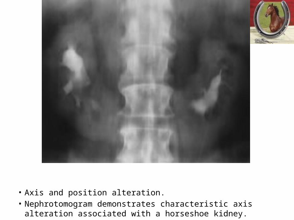
• Axis and position alteration. • Nephrotomogram demonstrates characteristic axis alteration
associated with a horseshoe kidney.

Epitaph for the Urogram1 E. Stephen Amis, Jr, MD 1
From the Department of Radiology, Albert Einstein College of Medicine and Montefiore Medical Center, 111 E 210th St, Bronx, NY 10467. Received July 29, 1999; accepted August 9.
Address reprint requests to the author (e-mail: [email protected]).

“The excretory urogram is terminally ill. It survives only because of clinicians and radiologists who do not fully appreciate that
more accurate delineation of the urinary tract can be gained with other techniquesIt is now time to move on to different techniques.”
Stephen Amis, Jr, MD

What has happened…
• The advent of cross-sectional imaging methods and their increasing sophistication over the quarter century of their existence has prompted uroradiologists to question the continued effectiveness of the urogram.
Hematuria…..
• The remaining major indication for excretory urography is hematuria.
• Patients with hematuria require evaluation of both the renal parenchyma and the urothelium.
• The traditional evaluation has been with cystoscopy and excretory urography (supplemented with retrograde ureteropyelography as needed).
The last IVU sermon….
“In the near future, excretory urography will be replaced by nonenhanced helical CT to evaluate for stones and by CT urography to evaluate for hematuria and other genitourinary conditions.
• CT urography should be viewed simply as an evolution of urography, which survived for 7 decades as one of the mainstays in imaging of the urinary tract.


Urethrography.

• Posterior urethral rupture extending through the urogenital diaphragm to involve the bulbous urethra following blunt trauma (type III urethral injury

• Gonococcal urethral stricture. Retrograde urethrogram reveals a segment of irregular, beaded narrowing in the distal bulbous urethra with opacification of the left Cowper duct (arrow).
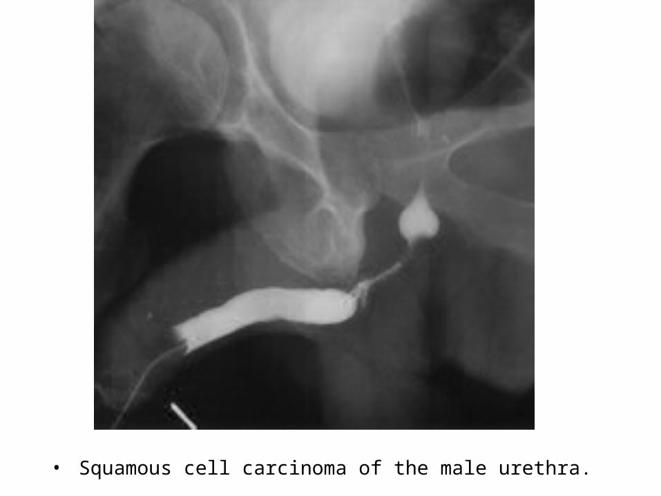
• Squamous cell carcinoma of the male urethra.
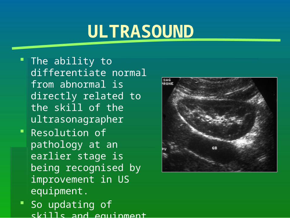
ULTRASOUND The ability to
differentiate normal from abnormal is directly related to the skill of the ultrasonagrapher
Resolution of pathology at an earlier stage is being recognised by improvement in US equipment.
So updating of skills and equipment is also needed
STRENGTHS of U/S
Very sensitive for upper tract dilatation.
Kidney size and the bladder well demonstrated.
Renal calculi and nephrocalcinosis seen.
Doppler evaluation of arterial and venous waveforms possible even with uncooperative child.
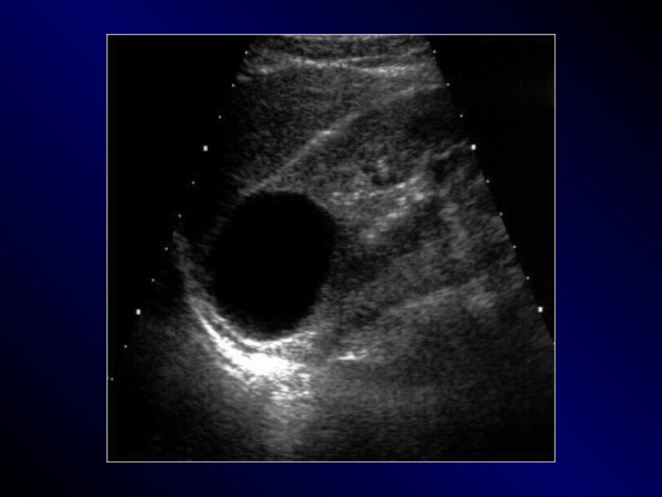
Cyst from solid differentiation is excellent.

WEAKNESSES of U/S Cannot detect focal renal scarring,
VUR or renal arterial abnormalities Assessment of post-micturition
volumes may be falsely high in presence of upper tract dilatation
Acurate comparison of small changes in renal pelvic dilatation on sequential US is difficult
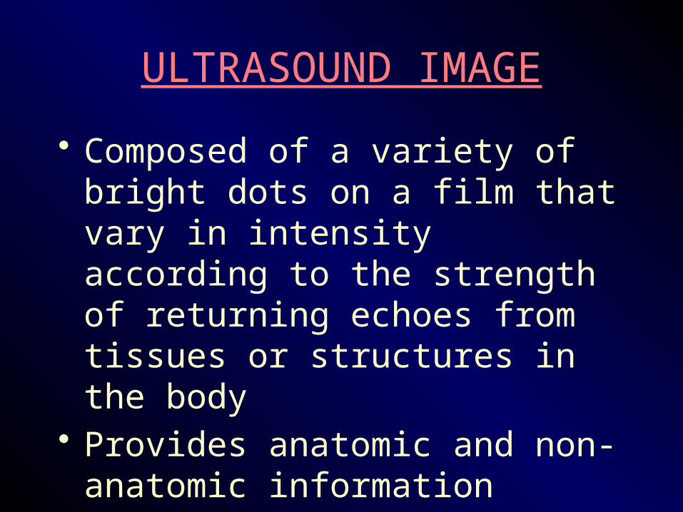
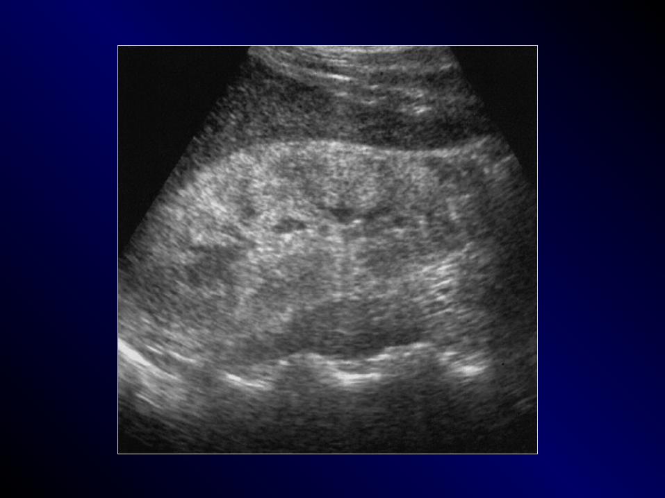
ULTRASOUND IMAGE
• Composed of a variety of bright dots on a film that vary in intensity according to the strength of returning echoes from tissues or structures in the body
• Provides anatomic and non-anatomic information

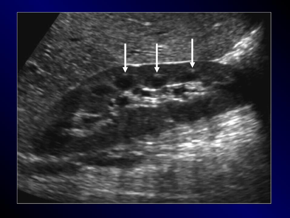
NORMAL KIDNEY
Capsule HyperechoicCortex HypoechoicMedulla Almost anechoicSinus HyperechoicPerirenal fat Hyperechoic

RENAL SIZES
• Vary with age, height, weight, sex
• Renal lengths 9-13 cm• RK & LK should be within 2 cm
in length• Decreases with age• Compensatory hypertrophy
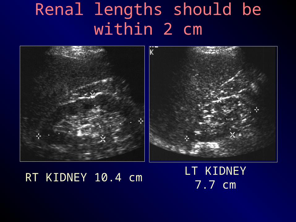
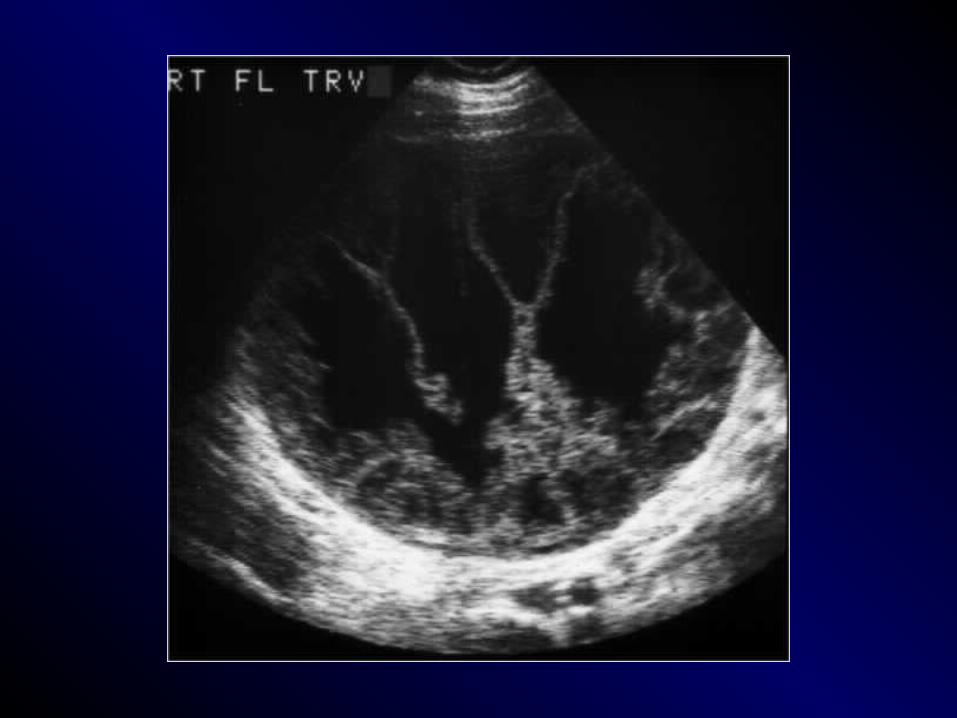
RT KIDNEY 10.4 cm LT KIDNEY 7.7 cm
Renal lengths should be within 2 cm

Multi-Detector Row CT of the Kidneys and Urinary Tract
• Multi–detector row helical computed tomography (CT) offers considerable advantages in evaluation of the urinary tract.
• It has the potential to become the single imaging modality used for comprehensive evaluation and treatment planning of most conditions affecting the kidneys and urinary tract, making conventional diagnostic techniques such as intravenous urography and angiography nearly obsolete.

• With the introduction of multidetector technology, CT urography, to date, has emerged as the initial heir apparent to intravenous
urography; many years of experience have now clearly demonstrated that CT is the test of choice for many urologic problems, including urolithiasis, renal masses, urinary tract infection, trauma, and
obstructive uropathy.
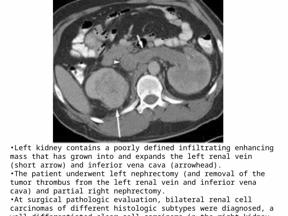
•Left kidney contains a poorly defined infiltrating enhancing mass that has grown into and expands the left renal vein (short arrow) and inferior vena cava (arrowhead). •The patient underwent left nephrectomy (and removal of the tumor thrombus from the left renal vein and inferior vena cava) and partial right nephrectomy. •At surgical pathologic evaluation, bilateral renal cell carcinomas of different histologic subtypes were diagnosed, a well-differentiated clear cell carcinoma in the right kidney and infiltrating carcinoma in the left kidney.

Phases of Contrast Enhancement in the Kidney and Collecting System
Phases of renal enhancement at multidetector CT. (a)Unenhanced axial CT at level of renal hilum. (b)Late arterial phase axial CT at level of renal
hilum. There is good opacification of the left renal artery (black arrow) and vein (white arrow).
(c) Nephrographic phase axial CT at level of renal hilum.
(d)Excretory phase axial CT at level of renal hilum.

• Right renal vein thrombosis in a 46-year-old woman with Escherichia coli urinary tract infection and pyelonephritis.

CT Angiography of the Renal Arteries.
• The main applications of CT angiography include evaluation of
potential renal donors, evaluation of suspected renal artery stenosis or aneurysms, and demonstration and location of crossing vessels prior to pyeloplasty for ureteropelvic junction obstruction.

• Emphysematous pyelonephritis in a 45-year-old woman with sepsis.
MR Urography: Techniques and Clinical Applications
• Magnetic resonance (MR) urography comprises an evolving group of techniques with the potential for allowing optimal noninvasive evaluation of many abnormalities of the urinary tract.
• MR urography is clinically useful in the evaluation of suspected urinary tract obstruction, hematuria, and congenital anomalies, as well as surgically altered anatomy, and can be particularly beneficial in pediatric or pregnant patients or when ionizing radiation is to be avoided.

• Prostate cancer metastatic to lymph nodes in a 53-year-old man.
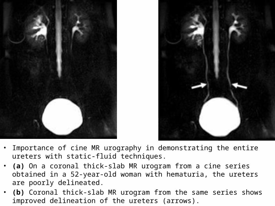
• Importance of cine MR urography in demonstrating the entire ureters with static-fluid techniques.
• (a) On a coronal thick-slab MR urogram from a cine series obtained in a 52-year-old woman with hematuria, the ureters are poorly delineated.
• (b) Coronal thick-slab MR urogram from the same series shows improved delineation of the ureters (arrows).
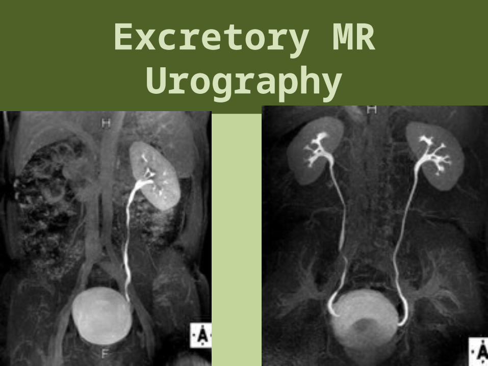
Excretory MR Urography

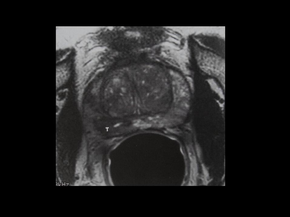
Prostate gland.

Transabdominal sonography

TRUS. TRX.
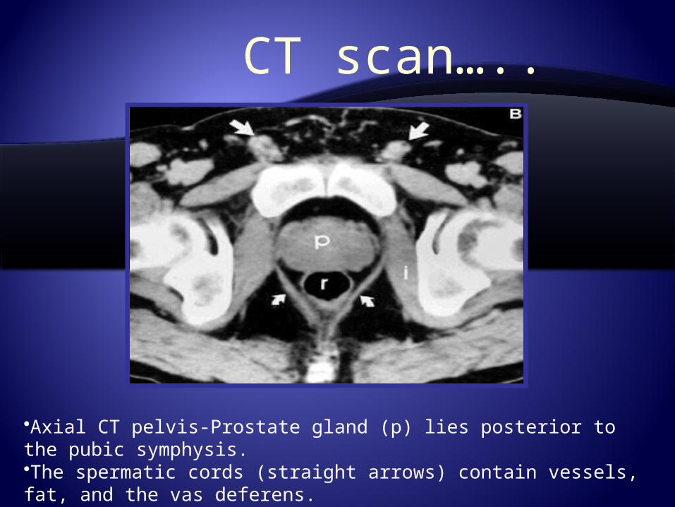
CT scan…..
•Axial CT pelvis-Prostate gland (p) lies posterior to the pubic symphysis. •The spermatic cords (straight arrows) contain vessels, fat, and the vas deferens. •Curved arrows, levator ani; r, rectum; i, obturator internus5.
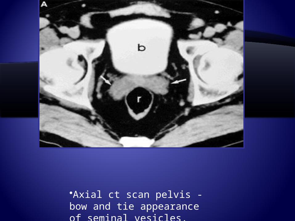
•Axial ct scan pelvis -bow and tie appearance of seminal vesicles.


NUCLEAR MEDICINE
“Unclear Medicine” ?The use of small amounts of
radioactive materials to diagnose and treat disease.
NUCLEAR MEDICINE SHOWS PHYSIOLOGYWhereas
RADIOLOGY SHOWS ANATOMY

GENERATOR SYSTEMThe basis of the generator system (the cow) is that the daughter is physically separable from the parent. This process is termed “milking”. The eluate contains sodium pertechnetate. Since the generator system is small and can be shipped easily, it makes the routine use of 99mTc possible in nuclear medicine
Molybdenum-99

BILATERAL TESTICULAR INFARCTION
Testicular torsion leads to testicular infarction. Testicular ischemia may also result from epididymitis as in this pt.Early scintigraphic flow images show markedly increased flow to scrotal regions bilaterally. Tissue-phase scintigram shows increased peri-testicular activity with bilateral central regions of diminished activity.
SCROTAL RNI

Uroradiology classics.

Radioglyphics…
• The language of radiology is rich with descriptions of imaging findings, often metaphorical, which have found common usage in the day-to-day practice of genitourinary radiology.
• These "classic signs" give us confidence in our diagnosis.
• Some of the signs have become so familiar to us that they are referred to as an "Aunt Minnie."

Staghorn
• A renal stone described as a staghorn implies a branched renal calculus that resembles the antlers of a stag.
• It is usually composed of struvite; but less commonly, it is formed from cystine or uric acid.

• (a) Staghorns. (b) On a scout image obtained before excretory urography, a calculus fills nearly the entirety of a bifid right renal collecting system, giving it a branched appearance that resembles the antlers of a stag.

• (a) A bear’s paws. (Photograph entitled "Bad Boys of the Arctic" reprinted with permission from Thomas D. Mangelsen, Inc.)
• (b) Contrast material-enhanced CT scan (same patient as in Fig 2) demonstrates a centrally obstructing stone with replacement of the renal parenchyma by low-attenuation collections in a "hydronephrotic" pattern. Note the lack of dilatation of the renal pelvis and infundibula.
• (c) CT scan obtained at a slightly lower level shows the fragments of a staghorn calculus within the parenchymal collections, which exhibit marginal enhancement. The pattern seen at CT resembles a bear’s paw.
Bear’s paws sign.
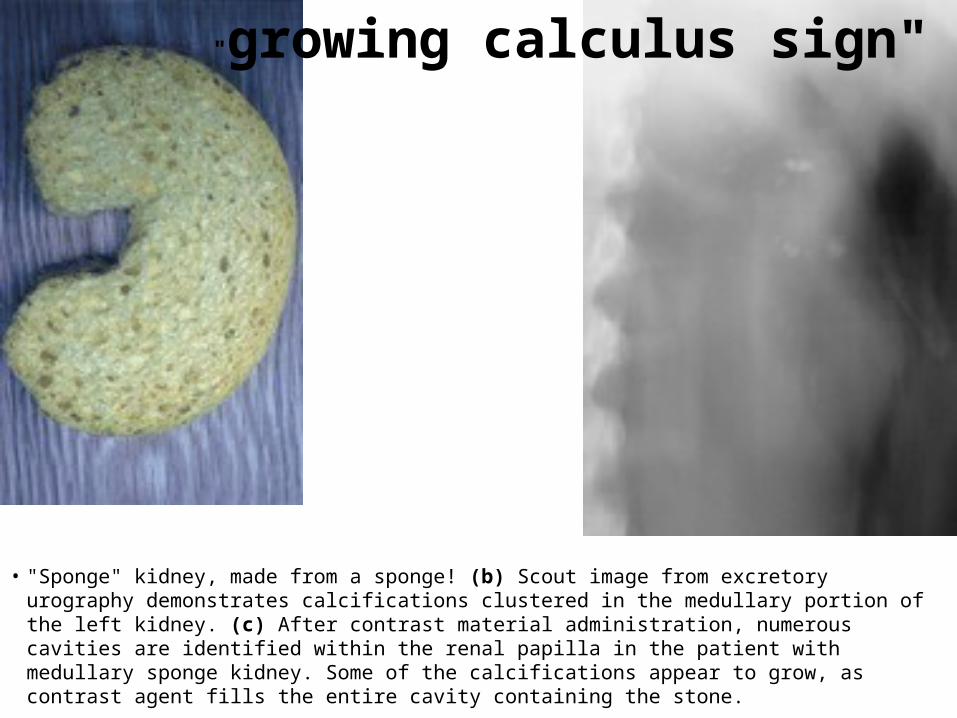
• "Sponge" kidney, made from a sponge! (b) Scout image from excretory urography demonstrates calcifications clustered in the medullary portion of the left kidney. (c) After contrast material administration, numerous cavities are identified within the renal papilla in the patient with medullary sponge kidney. Some of the calcifications appear to grow, as contrast agent fills the entire cavity containing the stone.
"growing calculus sign"
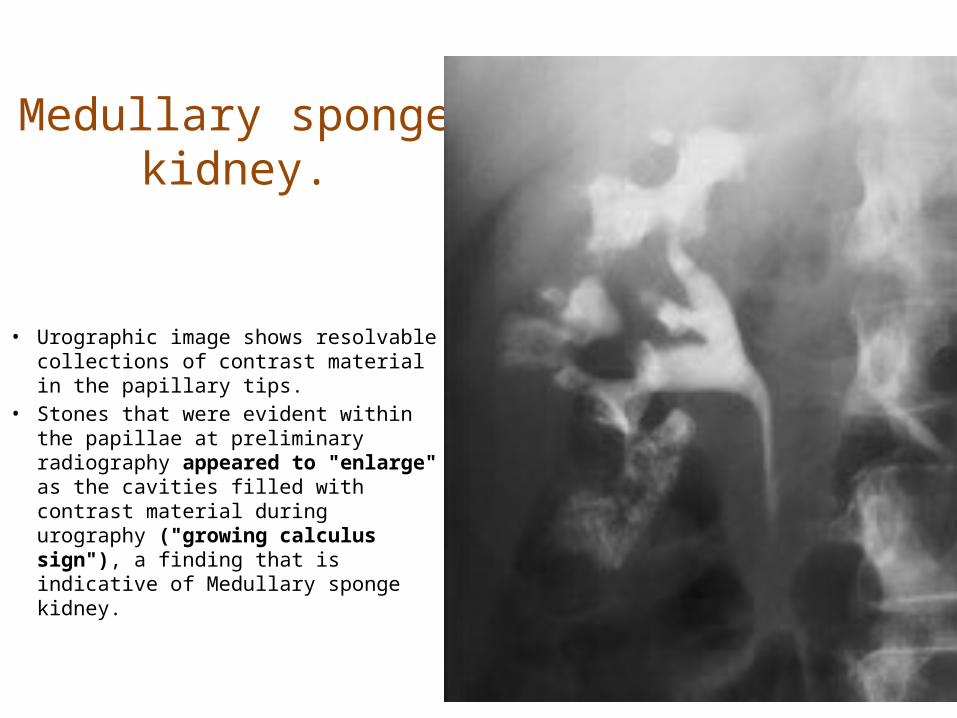
Medullary sponge kidney.
• Urographic image shows resolvable collections of contrast material in the papillary tips.
• Stones that were evident within the papillae at preliminary radiography appeared to "enlarge" as the cavities filled with contrast material during urography ("growing calculus sign"), a finding that is indicative of Medullary sponge kidney.

• Papillary necrosis caused by analgesic abuse. • Pyelographic image shows central cavities within
multiple papillae (arrows).
Egg in cup

Perirenal cobwebs.
• Perirenal cobwebs were initially attributed to collateral vessels seen in the perinephric space in patients with renal vein thrombosis.
• As our ability to image the perinephric space with CT improved, it became clear that a number of disease processes were manifest
by development of prominent perinephric structures.

Cobwebs. • (b) CT scan obtained after extracorporeal lithotripsy reveals a
subcapsular hematoma and exaggeration of Kunin septa on the left. Renofascial and renorenal septa are especially well identified.
• (c) On another CT scan obtained at the lower aspect of the left kidney, fasciofascial septa are nicely seen, and the cobweb appearance is particularly well developed.

• Comet. (Photograph entitled "Comet Hyakutake" reprinted with permission from Bill and Sally Fletcher.) (b) CT scan shows a calcification (the comet nucleus) (arrow) with a soft-tissue tail that represents a pelvic vein (arrowhead). Together, this appearance constitutes the comet sign. Note the stone at the left ureterovesical junction.
comet sign.

Spotted Nephrogram
• Irregular, patchy enhancement in the renal parenchyma, referred to as the spotted nephrogram, may occur as a result of small vessel occlusion, which can be seen with necrotizing vasculitis (periarteritis nodosa), scleroderma, and hypertensive nephrosclerosis
• Although first observed on angiograms, the abnormal perfusion pattern can be identified on CT and MR images
after contrast material administration.
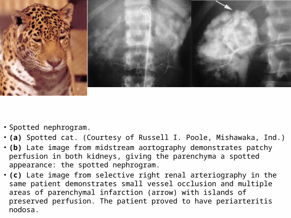
• Spotted nephrogram. • (a) Spotted cat. (Courtesy of Russell I. Poole, Mishawaka, Ind.) • (b) Late image from midstream aortography demonstrates patchy perfusion in both
kidneys, giving the parenchyma a spotted appearance: the spotted nephrogram. • (c) Late image from selective right renal arteriography in the same patient
demonstrates small vessel occlusion and multiple areas of parenchymal infarction (arrow) with islands of preserved perfusion. The patient proved to have periarteritis nodosa.
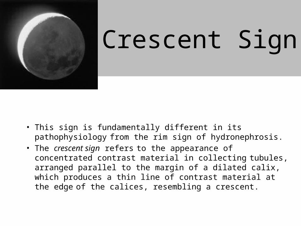
Crescent Sign
• This sign is fundamentally different in its pathophysiology from the rim sign of hydronephrosis.
• The crescent sign refers to the appearance of concentrated contrast material in collecting tubules, arranged parallel to the margin of a dilated calix,
which produces a thin line of contrast material at the edge of the calices, resembling a crescent.

• (a) Crescent. (Photograph entitled "Moon with Earthshine" reprinted with permission from Bill and Sally Fletcher.) (b) Crescent sign. CT image obtained during the corticomedullary phase of enhancement shows decreased thickness of the parenchyma surrounding the dilated collecting system in the left kidney. (c) Concentrated contrast material crescents surround the dilated collecting system elements (arrows) on this delayed image, which also shows a urine-contrast agent level in the dependent aspect.

Balloon on a String Sign.
• The balloon on a string sign may be seen with the rim sign of hydronephrosis, with the crescent sign, or as an isolated finding.
• This sign refers to the appearance of a high and somewhat eccentric exit point of the ureter from a dilated renal pelvis and is a typical finding of ureteropelvic junction obstruction.
• The possibility of an associated crossing vessel should always be evaluated before therapeutic intervention.
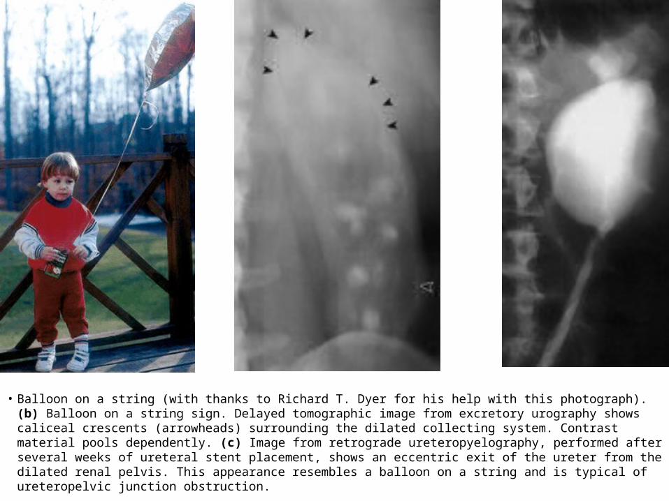
• Balloon on a string (with thanks to Richard T. Dyer for his help with this photograph). (b) Balloon on a string sign. Delayed tomographic image from excretory urography shows caliceal crescents (arrowheads) surrounding the dilated collecting system. Contrast material pools dependently. (c) Image from retrograde ureteropyelography, performed after several weeks of ureteral stent placement, shows an eccentric exit of the ureter from the dilated renal pelvis. This appearance resembles a balloon on a string and is typical of ureteropelvic junction obstruction.

Epilogue.
• Thank you for offering me 90 minutes of your precious time.
• This is but the foundation of uroradiology.
• We can irrigate this nascent discipline then tropicalize it.
Related Documents