Urinary Prostaglandins IDENTIFICATION AND ORIGIN J URGEN C. FROLICH, THOMAS W. WILSON, BRIAN J. SWEETMAN, MURRAY SMIGEL, ALAN S. NIES, KEITH CARR, J. THROCK WATSON, and JOHN A. OATES From the Departments of Medicine and Pharmacology, Vanderbilt University, Nashville, Tennessee 37232 A BSTRA CT Human urine was analyzed by mass spectrometry for the presence of prostaglandins. Pros- taglandin E2 and F2. were detected in urine from fe- males by selected ion monitoring of the prostaglandin E2-methylester-methoxime bis-acetate and the prosta- glandin F2a-methyl ester-Tris-trimethylsilylether de- rivative. Additional evidence for the presence of pros- taglandin F2. was obtained by isolating from female urine an amount of this prostaglandin sufficient to yield a complete mass spectrum. The methods utilized per- mitted quantitative analysis. The origin of urinary prostaglandin was determined by stimulating renal prostaglandin synthesis by arachi- donic acid or angiotensin infusion. Arachidonic acid, the precursor of prostaglandin E2, when infused into one renal artery of a dog led to a significant increase in the excretion rate of this prostaglandin. Similarly, infusion of angiotensin II amide led to a significantly increased ipsilateral excretion rate of prostaglandin E2 and F2. in spite of a simultaneous decrease in the crea- tinine clearance. In man, i.v. infusion of angiotensin also led to an increased urinary elimination of pros- taglandin E. These results show that urinary prostaglandins may originate from the kidney, indicating that renally syn- thesized prostaglandins diffuse or are excreted into the tubule. Thus, urinary prostaglandins are a reflection of renal prostaglandin synthesis and have potential as a tool to delineate renal prostaglandin physiology and pathology. INTRODUCTION When prostaglandins (PG) 1 are infused i.v., they can- not be recovered unchanged in the urine (1, 2), indi- Received for publication 31 July 1974 and in revised form 26 November 1974. 1 Abbreviationis used in this paper: CPBA, competitive cating little direct excretory clearance of the primary prostaglandins from the circulation. The kidney itself, however, is a prime site of prostaglandin synthesis (3- 5), and it was conceived that PG synthesized in the kidney might diffuse directly into the urine. Certainly some substances such as tryptamine (6) and cyclic AMP (7) appear in the urine totally or in part as a reflection of their intrarenal synthesis. Accordingly, an attempt was made to ascertain whether renal production of PG might result in their release directly into the tubular fluid. We have previ- ously obtained preliminary evidence for the presence of primary prostaglandins in human urine (8, 9), and others have obtained bioassay (10) and radioimmuno- assay (11) data suggesting the presence of PG in the urine of other species. This report provides defini- tive evidence regarding the existence and quantity of PG in human urine, and demonstrates that PG in the urine can originate from intrarenal synthesis. METHODS Analytical methods The identification of PGE2 and PGF2. was based on mass spectrometry. PGE2 and PGF2a were purified from 200-ml samples of urine, employing 2.5 mg of 3,3,4,4-tetradeutero PGE2 and 3,3,4,4-tetradeutero PGF2a as internal standards and carriers, together with tritiated PGE1 and tritiated PGF2a (Amersham/Searle Corp., Arlington Heights, Ill.; specific activity 60 and 15 Ci/mmol, respectively) as tracers during purification. Purification consisted of washing with an equal volume of benzene: butylchloride 1: 1 (vol/vol) at pH 6.5. The pH of the urine was then adjusted to 3.2 with formic acid and extraction was performed with an equal vol- ume of chloroform. The chloroform was removed by evap- oration and the residue dissolved in CHCI3: heptane: ethanol: acetic acid (100: 100: 30: 2) (vol/vol) and applied to an protein-binding assay; PG, prostaglandins; PGE2-ME-MO- bis-Ac, PGE2 methyl ester converted to methoxime bis-ace- tate; PGF2a-ME-Tris-TMS, PGF2. methyl ester converted to its Tris-trimethylsilylether; SIM, selected ion monitoring. The Journal of Clinical Investigation Volume 55 April 1975*763-770 76)3

Welcome message from author
This document is posted to help you gain knowledge. Please leave a comment to let me know what you think about it! Share it to your friends and learn new things together.
Transcript
Urinary Prostaglandins
IDENTIFICATION ANDORIGIN
J URGEN C. FROLICH, THOMASW. WILSON, BRIAN J. SWEETMAN,MURRAYSMIGEL, ALAN S. NIES, KEITH CARR, J. THROCKWATSON,andJOHNA. OATESFrom the Departments of Medicine and Pharmacology, Vanderbilt University,Nashville, Tennessee 37232
A BSTRACT Human urine was analyzed by massspectrometry for the presence of prostaglandins. Pros-taglandin E2 and F2. were detected in urine from fe-males by selected ion monitoring of the prostaglandinE2-methylester-methoxime bis-acetate and the prosta-glandin F2a-methyl ester-Tris-trimethylsilylether de-rivative. Additional evidence for the presence of pros-taglandin F2. was obtained by isolating from femaleurine an amount of this prostaglandin sufficient to yielda complete mass spectrum. The methods utilized per-mitted quantitative analysis.
The origin of urinary prostaglandin was determinedby stimulating renal prostaglandin synthesis by arachi-donic acid or angiotensin infusion. Arachidonic acid,the precursor of prostaglandin E2, when infused intoone renal artery of a dog led to a significant increasein the excretion rate of this prostaglandin. Similarly,infusion of angiotensin II amide led to a significantlyincreased ipsilateral excretion rate of prostaglandin E2and F2. in spite of a simultaneous decrease in the crea-tinine clearance. In man, i.v. infusion of angiotensinalso led to an increased urinary elimination of pros-taglandin E.
These results show that urinary prostaglandins mayoriginate from the kidney, indicating that renally syn-thesized prostaglandins diffuse or are excreted intothe tubule. Thus, urinary prostaglandins are a reflectionof renal prostaglandin synthesis and have potential asa tool to delineate renal prostaglandin physiology andpathology.
INTRODUCTIONWhen prostaglandins (PG) 1 are infused i.v., they can-not be recovered unchanged in the urine (1, 2), indi-
Received for publication 31 July 1974 and in revised form26 November 1974.
1 Abbreviationis used in this paper: CPBA, competitive
cating little direct excretory clearance of the primaryprostaglandins from the circulation. The kidney itself,however, is a prime site of prostaglandin synthesis (3-5), and it was conceived that PG synthesized in thekidney might diffuse directly into the urine. Certainlysome substances such as tryptamine (6) and cyclicAMP (7) appear in the urine totally or in part as areflection of their intrarenal synthesis.
Accordingly, an attempt was made to ascertainwhether renal production of PG might result in theirrelease directly into the tubular fluid. We have previ-ously obtained preliminary evidence for the presenceof primary prostaglandins in human urine (8, 9), andothers have obtained bioassay (10) and radioimmuno-assay (11) data suggesting the presence of PG inthe urine of other species. This report provides defini-tive evidence regarding the existence and quantity ofPG in human urine, and demonstrates that PG in theurine can originate from intrarenal synthesis.
METHODS
Analytical methodsThe identification of PGE2 and PGF2. was based on mass
spectrometry. PGE2 and PGF2a were purified from 200-mlsamples of urine, employing 2.5 mg of 3,3,4,4-tetradeuteroPGE2 and 3,3,4,4-tetradeutero PGF2a as internal standardsand carriers, together with tritiated PGE1 and tritiatedPGF2a (Amersham/Searle Corp., Arlington Heights, Ill.;specific activity 60 and 15 Ci/mmol, respectively) as tracersduring purification. Purification consisted of washing withan equal volume of benzene: butylchloride 1: 1 (vol/vol) atpH 6.5. The pH of the urine was then adjusted to 3.2 withformic acid and extraction was performed with an equal vol-ume of chloroform. The chloroform was removed by evap-oration and the residue dissolved in CHCI3: heptane: ethanol:acetic acid (100: 100: 30: 2) (vol/vol) and applied to an
protein-binding assay; PG, prostaglandins; PGE2-ME-MO-bis-Ac, PGE2 methyl ester converted to methoxime bis-ace-tate; PGF2a-ME-Tris-TMS, PGF2. methyl ester convertedto its Tris-trimethylsilylether; SIM, selected ion monitoring.
The Journal of Clinical Investigation Volume 55 April 1975*763-770 76)3

LH-20 column (13 X 1 cm) packed in the same solvent mix-ture (12). All the prostaglandins were eluted in one peak.Material from this peak was separated into PGE and PGFby reversed phase partition chromatography on 4.5 g of sup-port using solvent system C50 (13, 14). Because [3H]PGE1was used for location of PGE2 during chromatography, andbecause PGE1 and PGE2 separate on this column, the rela-tive retention volume of PGE2 was established by applyinga mixture of [3H]PGE1 and PGE2 on a separate reversedphase column. An aliquot of each fraction was analyzed forradioactivity and another aliquot (after conversion to PGBby treatment with 0.5 M methanolic KOH) for absorbanceat 278 nm. PGE2 had a retention volume of 65-100 ml andPGE1 of 95-125 ml. The fractions containing PGE2 andPGF2a as well as their deuterated internal standards werederivatized to the methyl esters with redistilled diazomethaneprepared from N-nitroso-N-methylurea (15). The PGF2amethyl ester was further converted to its Tris-trimethyl-silylether (PGF2a-ME-Tris-TMS) by treatment with N,O-bis-trimethylsilylacetamide in pyridine (1: 1, vol/vol) (9).The PGE2 methyl ester was converted to the methoximebis-acetate (PGE2-ME-MO-bis-Ac) by treatment withmethoxyamine hydrochloride and acetic acid anhydride (16).Final analysis was accomplished using a LKB 9,000 massspectrometer-PDP 12-computer system (LKB Instruments,Inc., Rockville, Md.) adjusted for selected ion monitoring(SIM) (17). For analysis of PGE2-ME-MO-bis-Ac by SIMat mr/e 419/423, a 0.5-m, 2-mm (ID) column of 1.2% Dexsilon Supelcoport 100/200 mesh (Supelco Inc., Bellefonte, Pa.)was used at 2200C with the helium flow rate at 20 ml/min.Source temperature was 2500 C, trap current 60 4uA, andionizing voltage 32.5 eV. The multiplier slit was adjustedto a setting slightly wider than for normal scan use. Sourcefocus was adjusted to optimize both sensitivity and resolu-tion at rn/e 419. The magnet was equilibrated at this settingfor about 12 h before the run. During the run stability andfocus were checked frequently by the use of appropriatestandard mixtures.
For the analysis of PGF2a as PGF2.-ME-Tris-TMS bySIM at m/e 423/427 and m/e 513/517, a 2-m, 2-mm (ID)column of 2% OV-7 on chromosorb G was used at 245'Cwith a helium flow rate of 20 ml/min. Otherwise, instru-mental conditions were similar to those used for PGE2-ME-MO-bis-Ac.
In addition to data obtained by selected ion monitoring, asufficient amount of PGF2a was isolated and purified to ob-tain a complete mass spectrum. For this, 2 jig tetradeuteroPGF2a were added to 1,500 ml of human urine from females.The urine was acidified to pH 3.4 and percolated throughan Amberlite XAD-2 column (Mallinkrodt Chemical Works,St. Louis, Mo.) (18). Subsequent purification consisted ofLH-20 chromatography on a column 5 cm in diameter and20 cm in height, reversed phase partition chromatographyon system C40 (14), methylation with diazomethane, chro-matography on reversed phase system F55 (14), and finalpurification on a 500-mg silicic acid column which waseluted with redistilled ethylacetate: benzene, 7: 3 (vol/vol).
To analyze aliquots of urine smaller than 50 ml, massspectrometry was impractical and therefore alternate methodswere found. For analysis of PGEa newly developed competi-tive protein-binding assay (CPBA) was employed (19).The PGE assay utilized a high-affinity binding site on rat-liver cell membranes that has selective affinity for PGE1and PGE2. The affinities of this binding site for PGF1a,PGF2a, PGB2, 15-keto-PGE2, and arachidonic acid were100-1,000 times less than those for PGE2. The sensitivityextends into the low picogram range.
The radioimmunoassay for PGF2a utilizes a commercialantibody (Calbiochem, San Diego, Calif.) specific for PGF2,.PGF2a standards in ethanol (25-400 pg) in triplicate andaliquots of the unknown were evaporated under reducedpressure in 12 X 75-mm polystyrene tubes. Amounts of anti-body sufficient to bind 50% of tritiated PGFa (60 Ci/mmol,Amersham/Searle Corp., Arlington Heights, Ill.) wereadded dissolved in 0.6 ml of buffer (0.01 M sodium phos-phate, 0.1% gelatin, 1 mMEDTA, pH 7.5). After incuba-tion for 4 h at 4VC, 0.5 ml of charcoal/dextran was addedto all but the tubes used for the counting of total radio-activity. The tubes were centrifuged for 20 min at 2,500 g,and the supernate was poured into liquid scintillation vialsand analyzed by liquid scintillation counting.
The counting results of both assays were evaluated bycomputerized Scatchard regression that incorporates a cor-rection for the [3H]PGE1 added to urine for estimation ofrecovery (20). The purity of the [3H]PGE1 used waschecked periodically using thin-layer radio chromatography(21). The material was purified by thin-layer chromatog-raphy if a contamination of >5% was detected. Both assaysshow linear Scatchard regressions between 25 and 250 pg.The SE of 10 replicate determinations of the same urine is+5%.
Biological studiesANIMAL STUDIES
Arachidonic acid infusion. Mongrel dogs (it = 6) wereanesthetized with sodium pentothal by i.v. drip. An endo-tracheal tube was inserted and artificial respiration insti-tuted. A renal artery was exposed by abdominal approachand the ipsilateral ureter catheterized. In a 90-min equilibra-tion period 0.225% NaCl was infused at a rate sufficient toproduce a urine flow of 0.3-1 ml/min from the investigatedkidney. Creatinine was infused at a rate of 7.04 mg/min.After a 30-min control period sodium arachidonate was in-fused at the rate of 10 ,ug/min in 0.2 ml of saline into therenal artery via a 25-gauge needle f or 30 min. Sodiumarachidonate was prepared freshly for each experimentunder conditions that avoided in vitro formation of PGE2from arachidonic acid (5,8,11,14-tetraenoic acid, Nu CheckPrep Inc., Elysian, Minn.) by opening the vials undernitrogen atmosphere, dissolving the material in 80% ethanolwith phenolphthalein indicator, and titration to pH 7 with0.1 N NaOH. The solution was evaporated under a gentlestream of nitrogen and dissolved in saline. Analysis of thismaterial by gas chromatography-mass spectrometry re-vealed that it contained less than 0.0259 PGE2 (22). Bloodwas drawn in the middle of the control and infusion periodfor estimation of glomerular filtration rate.
Angiotensin infusion. Similarly prepared dogs (n = 6)were infused with angiotensin (angiotensin II amide, Hyper-tensin, Ciba Pharmaceutical Co., Summit, N. J.) at a rateof 0.5, 5, and 50 ng/kg per min into a renal artery, and ipsi-lateral urine was collected at each dose level for 30 minafter a 30-min control period.
Another group of dogs (it = 5) received angiotensin (20ng/kg per min) into one renal artery during a 30-minperiod. In these animals urine was collected from bothureters.
HUMANSTUDIES
Studies in man were approved by the Vanderbilt HumanInvestigation Committee, Nashville, Tenn. Informed consentwas obtained from the volunteers.
764 Frolich, Wilson, Sweetman, Smigel, Nies, Carr, Watson, and Oates
Human urines were collected under refrigeration fromfemales only. Participants in the studies were instructed toabstain from sexual intercourse 2 days before and duringthe study. During acute studies a vaginal tampon was in-serted. Urines were collected only outside the menstrualperiod. Urine specimens were obtained by voiding and keptat -20'C until analyzed. 24-h urine specimens were collectedfrom female volunteers who had not been taking any drugsfor at least 14 days.
Angiotensin was given i.v. to six subjects in a total ofnine studies. The subjects fasted overnight. After voidingin the morning they received 500 ml of 0.45% saline in5%o dextrose orally, and subsequently 50 ml 5% dextroseevery 30 min for 2 h. 3 h later urine was collected, 500 mlof 0.45%c saline in 5% dextrose were given again, and anangiotensin infusion (Hypertensin, Ciba, Summit, N. J.)was started at the rate of 2.3 ng/kg per min. Blood pres-sure was taken every 2 min, and after 30 min the infusionrate was increased to 4.4 ng/kg per min in 1.1 ml of 5%odextrose. Urine was collected for 3 h after the beginningof the angiotensin infusion.
RESULTS
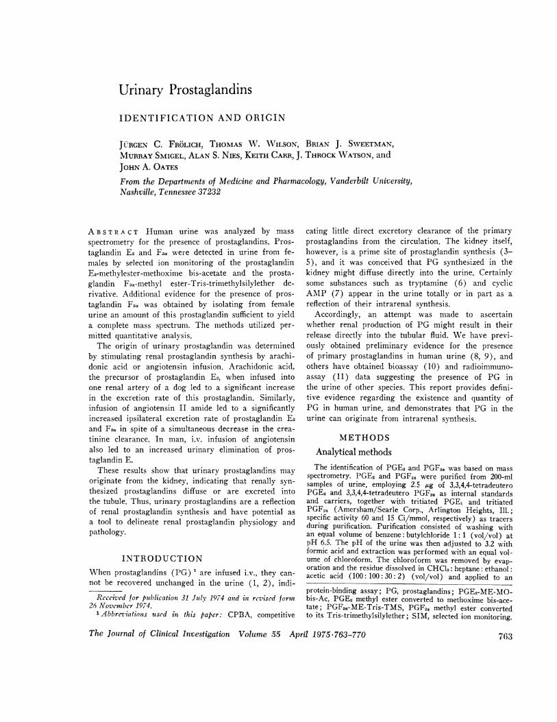
Identification and incasurernent of PGEe and PGFtain humnan urine. PGE2 and PGF2. were identified inurine of human females by mass spectrometry as thePGE2-ME-M\O-bis-Ac and PGF2.-Me-Tris-TMIS. Rep-resentative data from selective ion monitoring are shownin Fig. 1 and Fig. 2. These are photographs takenfrom the computer oscilloscope (17) showing ion pro-files obtained in the analysis of PGE2 (ions rtt/e 419
A419423
and 423, Fig. 1) and PGF2a (ions rn/e 423 and 427,Fig. 2). It was also possible to demonstrate the pres-ence of PGE2 and PGF2a by monitoring ions mre 328and rn/e 513, respectively. These findings confirmearlier SIM data obtained by converting urinary PGE2to PGB2 (analysis as PGB2-MAE-TMIS) and PGF2a to
PGF2a-Me-triacetate (8). Repeated analysis (n = 10)of the same urine sample revealed that a level of 590pg/ml of urine can be measured with a SD of 6.4%.Based on the amount of deuterated internal standardadded to each aliquot of urine, 24-h urine samplesfrom ambulatory females were found to have levels ofPGE2 of 374±77 pg/ml (n = 8, mean+SE) and werethus in the same range as the levels obtained by CBPA(319±53 pg/ml, n= 8). Levels of PGF2a measured bySIM were 386±78 pg/ml (n = 10).
In order to obtain a complete mass spectrum of PGF2afrom human urine, this prostaglandin was purifiedfrom a 1,500-ml sample of pooled female urine afteraddition of tetradeutero PGF2a. Fig. 3 shows that themass spectra of PGF2a-Me-Tris-TMS standard and thematerial extracted from urine are virtually identical.The spectrum of the tetradeutero PGF2a added to theurine and isolated together with the urinary PGF2.provided additional support for the identification.
Investigation of the renal origin of urinary prosta-glandins. Infusion of sodium arachidonate into the
B 419
~~~~~~~~~~~~~~~~
c 4423 D419
FIGURE 1 PGE2 from human urine. The vertical axis represents abundance, the horizontalaxis time. Ions monitored are nm/e 423 for the deuterated internal standard and rn/e 419 forthe protonated (biological) prostaglandin derivative. (A) Biological sample with internalstandard (ml/e 419 and 423). (B) Same as A, but magnified 32 times, nl/e 419 only. Arrowindicates the expected retention time of PGE-ME-MO-bis-Ac. Ion 1n/e 423 is deleted by thecomputer. (C) Internal standard alone (rl/e 423). (D) Protium blank created by internalstandard, magnified 32 times (in/e 419).
Identification and Origin of Urinary Prostaglandins
1% . .
V .,I'.
t
76.4

423427
A B423
* 'f. ~ ~~~~.
I
"I 4 K
FIGURE 2 PGF2a from human urine. The vertical axis represents abundance, the horizontalaxis time. Ions monitored are mle 423 for the internal standard and in/e 427 for the protonated(biological) prostaglandin derivative. (A) Biological sample with internal standard (}7/e 423and 427). (B) Same as A, but magnified 32 times (mle 423 only). Arrow indicates expectedretention time of PGF2,-Me-Tris-TMS. Blank correction as in Fig. 1.
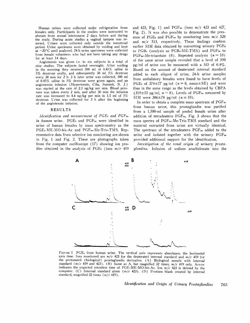
renal artery lead to a significantly increase in ipsi-lateral concentration and excretion rate of PGE (mea-sured by CPBA) (Fig. 4). It is known that arachidonicacid may be converted to prostaglandin-like materialin vitro (23). In order to exclude PG formation from
arachidonate in the urine or during the analytical pro-cedure as a possible explanation for our findings, tri-tiated arachidonate (100 Ci/mmol, New England Nu-clear, Boston, Mass.) was added to unlabeled arachi-donate and infused into the renal artery of a dog as
PGF20C- Me-Tris-TMS
Standard
191
73
L.
173
L
307/8333
L, LL I IALL ..AL. IL70 l00 150 200 250 300 350
m/e
I 43
1 423
14Q4
3971-1 41 I11
494
513
569 584I, i
400 450 560 550 600
PGF2oc - Me -Tris-TM
Urine Sample19
173
'I li .-I. 1. LL 6. -
IS
31 ~~~~~~~~~14 t
333 1404
307/8
LA1.LLLL L[ . .1 f LA../2 1337 1.397L 408
3
494
513
I-
427
198 5157 569 584Lt "1,, 11 E ~~~~~1.5?3i588
70 loo 150 200 250 300 350 400 450 500 550 600m/e
FIGURE 3 Mass spectrum of PGF2a from human urine. Top is mass spectrum of PGF2.-Me-Tris-TMS standard with prominent ions mile 584 (M+), 569, 513, 494, 423, and 404. Bottom ismass spectrum of deuterated internal standard with prominent ions mle 588 (M+), 573, 517,498, 427, and 408 together with mass spectrum of protonated (biological) prostaglandin withprominent ions rn/e 584 (M+), 569, 513, 494, 423, and 404.
766 Frblich, Wilson, Sweetman, Smigel, Nies, Carr, Watson, and Oates
z
80-z
3 60-w 40-
< 20--j
w
zo 104zo 84zm 6(
, 4(
< 24-Jw~
0-
LL I Anr
URINE FROMA RACHDONATEINFUSION
1,2001 dpm/FRACTION
600:
0 -
CONTROL INFUSION
FIGURE 4 Effect of infusion of sodium arachidonate on uri-nary concentration and excretion rate of PGE in the dog(it = 6, mean+SE). Blank bars: concentration in pg/mil,and cross-labeled bars: excretion rate in picogram perminute.
in the other experiments. Coincident with the be-ginning of the infusion a portion of the labeled andunlabeled arachidonic acid was added to the dogs urinecollected during the control period. Immediately at theend of the infusion both urine specimens were ex-
tracted by the methods described above, the materialapplied to 1-g silicic acid columns (6 X 100 mm) andeluted with a discontinuous gradient of ethylacetateand toluene. This column separates arachidonic acid,PGE, and PGF (24). The results of this chromatog-raphy are shown in Fig. 5 and indicate that only theurine obtained after intra-arterial infusion of arachi-donate contained significant amounts of PGEand PGF.Therefore, generation of PGE2 from arachidonate inthe urine or during the isolation procedure cannot ac-
count for the increased amounts of PGE2 appearing inurine during arachidonate administration.
The results of infusing increasing amounts of angio-tensin into the canine renal artery are shown in TableI. Both PGE (measured by CPBA) and PGF (mea-sured by radioimmunoassav) excretion rates increasedin a dose-dependent manner in response to angiotensin.This increase became significant (P < 0.05) for PGFat the 5 ng/kg per min and for PGE at the 50 ng/
1:9 3:7 4:6 6:4 9:1PGE PGF
ARACH DONATEADDEDTO URINEIN VITRO
r!Wl 6,400
1:9 3:7 4:6 6:4 9:1PGE PGF
FIGURE 5 Silicic acid column chromatography of extractfrom ureteral urine after administration of tritiated sodiumarachidonate into the ipsilateral renal artery or after itsaddition directly to the urine. Numbers on the abscissa indi-cate ratios of ethylacetate to toluene in the eluating fractions.Fraction volume is 35 ml.
kg per min infusion rate of angiotensin. These changesalso were seen when the excretion rate was expressedin terms of nanograms of prostaglandin per minute permilligram creatinine.
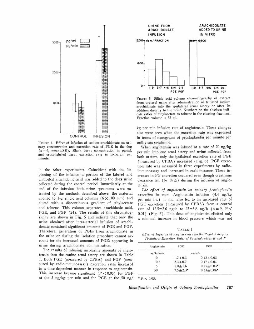
When angiotensin was infused at a rate of 20 ng/kgper min into one renal artery and urine collected fromboth ureters, only the ipsilateral excretion rate of PGE(measured by CPBA) increased (Fig. 6). PGF excre-tion rate was measured in three experiments by radio-immunoassay and increased in each instance. These in-creases in PG excretion occurred even though creatinineclearance fell (by 38%) during the infusion of angio-tensin.
The effect of angiotensin on urinary prostaglandinexcretion in manl. Angiotensin infusion (4.4 ng/kgper min i.v.) in man also led to an increased rate ofPGE excretion (measured by CPBA) from a controlrate of 12.5±2.6 ng/h to 27±3.8 ng/h (n = 9, P <0.01) (Fig. 7). This dose of angiotensin elicited onlya minimal increase in blood pressure which was not
TABLE IEffect of Infusion of A ngiotensin into the Renal Artery on
Ipsilateral Excretion Rates of Prostaglandins E and F
Angiotensin PGE PGF
ng/kg/nzin ng/mnin0 1.7±0.3 0.1240.030.5 2.340.7 0.17±0.045 5.0±+1.6 0.25±0.03*
50 7.5±2.3* 0.53+0.06*
* P < 0.05.
Identification and Origin of Urinary Prostaglandins
pg/ml Lpg/min
2000 -
767
-I
o UPGEV(ng/min)
3.
2-
I .
0 UPGEV(ng/min)
3-
2-
I t
INFUSED KIDNEY
IJ
* P< 0.05
CCR E3-40-30
cm -20
*20CONTROL ANGIO I
FIGURE 6 Effect of infusion of angiotensin into dog renalartery on ipsilateral and contralateral PGE excretion ratesand creatinine clearances.
statistically significant. Urine flow rate was not sig-nificantly altered by angiotensin.
DISCUSSIONOur preliminary report of PGE2 and PGF2a in humanurine was based on gas chromatographic analysis em-
ng PGE/3 h
80-
40-
+P <0.01
CONTROL INFUSION
FIGURE 7 Effect of angiotensin on urinary excretion rateof PGEin human females (n = 9, mean±4SD). Top is urineflow (ml/3 h) and bottom is PGEexcretion rate (ng/3 h).
ploying selected ion monitoring with the mass spec-trometer of the methyl ester trimethylsilylether ofPGB2, which was obtained by converting PGE2 to PGB2and the methyl ester Tris-acetate derivative of PGFsa(8). Although these data provided evidence for thepresence of PGE2and PGF2a in urine, these approacheswere not useful for quantification. The present com-munication provides a more complete identification ofurinary prostaglandins by measuring two additional de-rivatives, namely the methyl ester-methoxime-bis ace-tate of PGE2 and the methyl ester-Tris-TMS of PGF2a.Selected ion monitoring of both derivatives confirmedthe presence of PGE2 and PGF2a in urine and offereda precise method for quantification. In the case ofPGFsa a complete mass spectrum was obtained.
A renal origin for urinary PG was suggested by thefinding that the primary PG are not recovered un-changed in the urine after their i.v. administration(1, 2). This implied that the high capacity of the lung(14) and kidney (25) to metabolize PG as well as thebinding of PG to plasma protein (26) left little oppor-tunity for clearance of unmetabolized PG directly intothe urine. However, because we have found that in-fusion of large amounts of PGE2 directly into the renalartery can lead to an increase in urinary PGE2 (un-published observation), additional evidence was soughtin support of the release of PG synthesized in thekidney directly into the urine. One approach would beto determine the clearance of PG from the blood intothe urine and to compute from that clearance the sepa-rate contribution of the kidney and the blood to urinaryPG, analogpus to the method for assessing renal 3',5'-cyclic AMP-production (7). This would require, how-ever, precise quantification of prostaglandins present inarterial blood, and because of ready release of PG fromblood platelets (27) and that the most specific methodfor measurement (by mass spectrometry) (28) yieldsvalues only a fraction higher than the blanks, attemptsto assess prerenal contribution to urinary PG fromclearance information was thought to be impractical.Accordingly, the release of renal PG directly into theurine was evaluated by determining whether urinaryPG excretion could be increased by two maneuvers thatincrease renal prostaglandin synthesis by quite differentmechanisms.
An increase in renal PG synthesis by infusion of thePG precursor, arachidonic acid, was found to producean increase in urinary PG excretion. In separate ex-
periments, it was demonstrated that glomerular filtra-tion is little affected by arachidonic acid infusion, buturine flow rate and sodium excretion do increase onlyon the ipsilateral side, effects.blocked by inhibiting PGsynthesis (29).
768 Frfhch, Wilson, Sweetman, Smigel, Nies, Carr, Watson, and Oates
I
Confirmation of the release of renally synthesized PGdirectly into the urine was obtained by stimuling renalPG formation with angiotensin II (30). Infusion ofangiotensin II into the renal artery raised urinary PGexcretion rate even though creatinine clearance on thatside decreased. The increase in PG excretion only onthe ipsilateral side precludes a prerenal source forthis increase in urinary PG.
These experiments show that an increase in renalPG synthesis is reflected by an increase in urinary PGexcretion rates. That a similar response to angiotensinoccurs in man is suggested by the finding of an in-creased urinary excretion rate of PGE during angio-tensin infusion.
The finding of PG in urine adds a new dimension toassessment of their overall svnthesis. Production ratesbased on measurements of the major urinary metabo-lites does not adequately describe the turnover of PGsince a fraction of the primary PG is excreted directlywith the urine. The amount excreted with urine in manis not large and represents only a small fraction of thesecretion rate, as measured by the major metabolites(31). The direct excretion into the urine of renallysynthesized PG, however, raises the possibility thatsome early metabolites also may exit directly into theurine without entering into the pool from which themajor metabolite if formed.
While the quantitative aspects of urinary PG interms of total body synthesis appear to be of minorimportance, the close association with renal PG pro-duction suggests that urinary PG might serve as indi-cators of renal PG synthesis. Clearly, measurement ofPG in urine can be accomplished more readily thanin plasma where platelets generate PG rapidly (27, 32)and may be an important factor in causing the largediscrepancies in reported plasma levels (28, 33). Also,renal venous blood sampling is limited to the animalmodel or to the acute study in man. In contrast, uri-nary levels of PG are found to be much higher thanplasma levels, and repeated or prolonged sampling ofurine can easily be accomplished facilitating studies inman.
To ascertain more precisely the extent to which uri-nary PG may be employed as indicators of renal PGsynthesis, other parameters that influence their excre-tion need to be investigated. In particular the weakacidic properties of PG suggests that their excretioncould be influenced by urinary pH and flow rate.
Studies from this and other laboratories have dem-onstrated that PG synthesized intrarenally will affectblood flow to the cortex, chiefly that to the juxtamed-ullary nephrons (29, 34). As the synthesis of PG inthe kidney occurs almost entirely in the medulla (35),
the question arises as to how the PG are transportedfrom medulla to the point of blood flow regulation inthe cortex. The finding of appreciable quantities ofPG in urine provides the basis for a hypothesis thatthe medullo-cortical transport occurs via the tubularfluids to the site where the tubule is in direct appositionto glomerular arterioles.
ACKNOWLEDGMENTSThe prostaglandins used in these studies were kindly pro-vided by Drs. John E. Pike and Udo Axen, The UpjohnCompany, Kalamazoo, Mich. The authors want to thankMiss Annetta Cooper, Mr. Robert Rush, and Mr. JerryStrother for expert technical assistance. Ms. Evelyn Squiresprovided excellent secretarial help.
This wtork was supported by a Center Grant for ClinicalPharmacology and Drug Toxicology (National Institutesof Health GMS-14531) and a Clinical Research CenterGrant (U. S. Public Health Service 5-MO1-RR-00095).
REFERENCES
1. Granstr6m, E. 1967. On the metabolism of prostaglandinE1 in man. Prostaglandins and related factors. Prog.Biochem. Pharmacol. 3: 89-93.
2. Granstr6m, E., and B. Samuelsson. 1969. The struc-ture of a urinary metabolite of prostaglandin F2a inman. J. Amn. Chem. Soc. 91: 3398-3400.
3. Daniels, E. G., J. W. Hinman, B. E. Leach, and E. E.Muirhead. 1967. Identification of prostaglandin E2 asthe principal vasodepressor lipid of rabbit renal medulla.Natuire (Loud.). 215: 1298-1299.
4. Hamberg, M. 1969. Biosynthesis of prostaglandins inthe renal medulla of rabbit. FEBS (Fed. Eur. Biocheni.Soc.) Lett. 5: 127-130.
5. AnggArd, E., S. 0. Bohman, J. E. Griffin, III, C. Lars-son, and A. B. Maunsbach. 1972. Subcellular localizationof the prostaglandin system in the rabbit renal papilla.Acta Physiol. Scanid. 84: 231-246.
6. Oates, J. A. 1961. Measurement of urinary tryptamine,tyramine, and serotonin. Methods Med. Res. 9: 169-174.
7. Broadus, A. E., N. I. Kaminsky, J. G. Hardman, E. W.Sutherland, and G. W. Liddle. 1970. Kinetic parametersand renal clearances of plasma adenosine 3',5'-mono-phosphate and guanosine 3',5'-monophosphate in man J.Clint. Invest. 49: 2222-2236.
8. Frdlich, J. C., B. J. Sweetman, K. Carr, J. Splawin-ski, J. T. Watson, E. Angga'rd, and J. A. Oates. 1973.Occurrence of prostaglandins in human urine. In Adv.Biosc. 9: 321-330.
9. Sweetman, B. J., J. T. Watson, K. Carr, J. A. Oates,and J. C. Frolich. 1973. Quantitative vapor-phase analy-sis of prostaglandin F2e in female human urine. Pros-taglandinhs. 3: 385-387.
10. Magotteaux, G., C. Deby, and G. Barac. 1971. Urinaryelimination of smooth muscle-acting prostaglandins inthe dog. Arch. hIt. Physiol. Biochim. 79: 799-801.
11. Skarnes, R. C., and M. J. K. Harper. 1972. Relationshipbetween endotoxin-induced abortion and the synthesisof prostaglandin F. Prostaglandidns. 1: 191-203.
12. Jouvenaz, G. H., D. H. Nugteren, and D. A. van Dorp.1973. Gas chromatographic determination of nanogramamounts of prostaglandin E and F. Prostaglaudidbs. 3:175-187.
Identification and Origin of Urinary Prostaglandins 769
13. Norman, A. 1953. Separation of conjugated bile acidsby partition chromatography. Acta Chem. Scand. 7:1413-1419.
14. Angga'rd, E., and B. Samuelsson. 1964. Prostaglandinsand related factors. Metabolism of prostaglandin E1 inguinea pig lung. The structure of two metabolites. J.Biol. Chem. 239: 4097-4102.
15. Sweetman, B. J., J. C. Frblich, and J. T. Watson. 1973.Quantitative determination of prostaglandins A, B, andE in the sub-nanogram range. Prostaglandins. 3: 75-87.
16. Axen, U., K. Green, D. Hbrlin, and B. Samuelsson.1971. Mass spectrometric determination of picomoleamounts of prostaglandin E2 and F2, using syntheticdeuterium labeled carriers. Biochem. Biophys. Res. Com-mun. 45: 519-525.
17. Watson, J. T., D. R. Pelster, B. J. Sweetman, J. C.Fr6lich, and J. A. Oates. 1973. A display-oriented datasystem for multiple ion detection with gas chromatog-raphy-mass spectrometry in quantifying biomedically-important compounds. Anal. Chem. 45: 2071-2078.
18. Green, K. 1971. The metabolism of prostaglandin F2lin the rat. 1971. Biochim. Biophys. Acta. 231: 419-444.
19. Fr6lich, J. C., T. W. Wilson, M. Smigel, and J. A.Oates. 1974. A competitive protein binding assay specificfor prostaglandin E. Biochim. Biophys. Acta. 348: 241-248.
20. Smigel, M., J. C. Frolich, and S. Fleischer. 1974. Char-acterization of prostaglandin E receptors in membranesand their use in the assay of prostaglandin E. MethodsEnzymol. 32: 109-123.
21. Green, K., and B. Samuelsson. 1964. Prostaglandins andrelated factors. XIX. Thin layer chromatography ofprostaglandins. J. Lipid Res. 5: 117-120.
22. Splawinski, J. A., A. S. Nies, B. J. Sweetman, andJ. A. Oates. 1973. The effects of arachidonic acid, pros-taglandin E2, and prostaglandin F2a on the longitudinalstomach strip of the rat. J. Pharmacol. Exp. Ther. 187:501-510.
23. Nugteren, D. H., H. Vonkeman, and D. A. van Dorp.1967. Nonenzymic conversion of al-cis 8, 11, 14 eicosa-trienoic acid into prostaglandin E1. Rec. Trav. Chim. 86:1237-1245.
24. Bygdeman, M., and B. Samuelsson. 1966. Analyses ofprostaglandin in human semen. Clin. Chim. Acta. 13:465474.
25. Angga'rd, E., C. Larsson, and B. Samuelsson. 1971.The distribution of 15-hydroxy prostaglandin dehydroge-nase and prostaglandin-A'3-reductase in tissue of theswine. Acta. Physiol. Scanid. 81: 396-404.
26. Raz, A. 1972. Interactions of prostaglandins with bloodplasma proteins Biochemn. J. 130: 631-636.
27. Smith, J. B., and A. L. Willis. 1970. Formation andrelease of prostaglandins by platelets in response tothrombin. Br. J. Pharmacol. 40: 545P (Abstr.)
28. Green, K., E. Granstr6m, and B. Samuelsson. 1973.Methods for quantitative analysis of PGF2,, PGE2, 9a,1la-dihydroxy-15-ketoprost-5-enoic acid, and 9a,11a,15-trihydroxy-prost-5-enoic acid from body fluids usingdeuterated carriers and gas-chromatography-mass spec-trometry. Anal. Biochemn. 54: 434-453.
29. Nies, A. S., J. Tannenbaum, J. Splawinski, and J. A.Oates. 1973. Prostaglandin formed endogenously canaffect renal function. Clin. Res. 21: 701. (Abstr.)
30. McGiff, J. C., K. Crowshaw, N. A. Terragno, and A.J. Lonigro. 1971. Renal prostaglandins: their biosynthe-sis, release, effects and fate. In Renal Pharmacology.J. W. Fisher and E. J. Cafruny, editors. New York.Appleton-Century-Crofts, Inc. 211-240.
31. Hamberg, M., and B. Samuelsson. 1971. On the metab-olism of prostaglandin E1 and E2 in man. J. Biol. Chenw.246: 6713-6721.
32. Silver, M. J., J. B. Smith, C. Ingerman, and J. J.Kocsis. 1972. Human blood prostaglandins: formationduring clotting. Prostaglandins. 1: 429-436.
33. Jaffe, B. M., H. R. Behrman, and C. W. Parker. 1973.Radioimmunoassay measurement of prostaglandins E,A, and F in human plasma. J. Clin. Invest. 52: 398-405.
34. Lonigro, A. J., H. D. Itskovitz, K. Crowshaw, and J.McGiff. 1973. Dependency of renal blood flow on pros-taglandin synthesis in the dog. Circ. Res. 32: 712-717.
35. Crowshaw, K., and J. Z. Szlyk. 1970. Distribution ofprostaglandins in rabbit kidney. Biochcm. J. 116: 421-424.
770 Frolich, Wilson, Sweetman, Smigel, Nies, Carr, Watson, and Oates
Related Documents


