J. clin. Path. (1969), 22, 67-75 Urinary excretion of glycosaminoglycans in the various forms of gargoylism G. MANLEY AND U. WILLIAMS From the Nuffield Department of Clinical Biochemistry, the Radcliffe Infirmary, Oxford SYNOPSIS The urinary excretion of glycosaminoglycans in 28 cases of gargoylism, embracing the Hurler, Hunter, Sanfilippo, Morquio, and Scheie syndromes (McKusick, 1966), has been examined using the cetylpyridinium chloride (CPC) turbidity test, the uronic acid/creatinine ratio, and the electrophoretic pattern of urine concentrates, as routine procedures. Ion-exchange column chromatographic techniques were also employed for the fractionation of glycosaminoglycans and aminosugars. Molecular weights were investigated by gel filtration and ultracentrifugation. The CPC turbidity test was positive in every case. The uronic acid/creatinine ratio provided a sensitive index of increased glycosaminoglycan excretion. Cases of the Hurler syndrome showed the highest, and cases of the Morquio and Scheie syndromes the lowest, ratios. A correlation was observed between the uronic acid/creatinine ratio and the clinical severity of the disease. Cellulose acetate electrophoresis differentiated clearly between the two major forms of gargoylism, the Hurler and Sanfilippo syndromes, but differentiation between the Hurler, Hunter, and Scheie syndromes was more difficult on electrophoretic data alone. Results obtained with cases diagnosed as the Morquio syndrome were disappointing. The existence of formes frustes of the Sanfilippo syndrome among the mentally subnormal is predicted. Errors caused by bacterial contamination of urine samples are emphasized. The atypical behaviour of urinary glycosaminoglycans in analytical procedures is discussed. Molecular weight studies suggested heterogeneity. The nature of the basic defect in gargoylism is discussed. The condition 'gargoylism' has been known for about 60 years, the first detailed description being given by Hunter in 1917. Early histological studies showed vacuolated cells in many tissues, and in the belief that these were caused by fat dissolving out during processing, the term lipochondrodystrophy came into use (Washington, 1966). Then glycogen was thought to be involved in the disorder (de Lange, Gerlings, de Kleyn, and Lettinga, 1944) and only 11 years ago it was realized that glycosaminoglycans were primarily involved in the disease (Dorfman and Lorincz, 1957). Since then, studies of the urinary glycosaminoglycans in gargoylism, together with careful clinical and genetic documentation, has led to the recognition of six distinct entities within the group, which McKusick (1966) has called the Hurler, Hunter, Sanfilippo, Morquio, Scheie, and Maroteaux-Lamy syndromes. Though many of the techniques used in the isolation and characterization of glycosamino- glycans cannot easily be employed in hospital Received for publication 22 May 1968. laboratories, several simple tests have emerged for the detection of increased urinary glycosamino- glycan excretion (Berry and Spinanger, 1960; Denny and Dutton, 1962; Segni, Romano, and Tortorolo, 1964; Manley and Hawksworth, 1966). It has also been shown that simple cellulose acetate electrophoresis of urinary concentrates will differ- entiate between at least two types of gargoylism (Manley and Hawksworth, 1966). This paper presents a laboratory investigation of urinary glycosaminoglycans in 28 cases of gargoylism, in the light of current clinical classification. MATERIAL AND METHODS MATERIAL Twenty-eight cases of gargoylism form the basis of this study. A proforma listing relevant clinical features was completed by the clinician in charge of each case, and this data enabled each case to be assigned to one of the six mucopolysaccharidoses of McKusick. Urine was collected over a period of 24 hours, and preserved with merthiolate. When immediate analysis was impossible, the samples were stored at - 20°C. 67 on January 12, 2022 by guest. Protected by copyright. http://jcp.bmj.com/ J Clin Pathol: first published as 10.1136/jcp.22.1.67 on 1 January 1969. Downloaded from

Welcome message from author
This document is posted to help you gain knowledge. Please leave a comment to let me know what you think about it! Share it to your friends and learn new things together.
Transcript
J. clin. Path. (1969), 22, 67-75
Urinary excretion of glycosaminoglycans in thevarious forms of gargoylism
G. MANLEY AND U. WILLIAMS
From the Nuffield Department of Clinical Biochemistry, the Radcliffe Infirmary, Oxford
SYNOPSIS The urinary excretion of glycosaminoglycans in 28 cases of gargoylism, embracing theHurler, Hunter, Sanfilippo, Morquio, and Scheie syndromes (McKusick, 1966), has been examinedusing the cetylpyridinium chloride (CPC) turbidity test, the uronic acid/creatinine ratio, andthe electrophoretic pattern of urine concentrates, as routine procedures. Ion-exchange columnchromatographic techniques were also employed for the fractionation of glycosaminoglycans andaminosugars. Molecular weights were investigated by gel filtration and ultracentrifugation.The CPC turbidity test was positive in every case. The uronic acid/creatinine ratio provided a
sensitive index of increased glycosaminoglycan excretion. Cases of the Hurler syndrome showed thehighest, and cases of the Morquio and Scheie syndromes the lowest, ratios. A correlation was
observed between the uronic acid/creatinine ratio and the clinical severity of the disease. Celluloseacetate electrophoresis differentiated clearly between the two major forms of gargoylism, theHurler and Sanfilippo syndromes, but differentiation between the Hurler, Hunter, and Scheiesyndromes was more difficult on electrophoretic data alone. Results obtained with cases diagnosedas the Morquio syndrome were disappointing. The existence of formes frustes of the Sanfilipposyndrome among the mentally subnormal is predicted. Errors caused by bacterial contaminationof urine samples are emphasized. The atypical behaviour of urinary glycosaminoglycans inanalytical procedures is discussed. Molecular weight studies suggested heterogeneity. The natureof the basic defect in gargoylism is discussed.
The condition 'gargoylism' has been known forabout 60 years, the first detailed description beinggiven by Hunter in 1917. Early histological studiesshowed vacuolated cells in many tissues, and in thebelief that these were caused by fat dissolving outduring processing, the term lipochondrodystrophycame into use (Washington, 1966). Then glycogenwas thought to be involved in the disorder (de Lange,Gerlings, de Kleyn, and Lettinga, 1944) and only11 years ago it was realized that glycosaminoglycanswere primarily involved in the disease (Dorfman andLorincz, 1957). Since then, studies of the urinaryglycosaminoglycans in gargoylism, together withcareful clinical and genetic documentation, has ledto the recognition of six distinct entities within thegroup, which McKusick (1966) has called theHurler, Hunter, Sanfilippo, Morquio, Scheie, andMaroteaux-Lamy syndromes.Though many of the techniques used in the
isolation and characterization of glycosamino-glycans cannot easily be employed in hospitalReceived for publication 22 May 1968.
laboratories, several simple tests have emerged forthe detection of increased urinary glycosamino-glycan excretion (Berry and Spinanger, 1960;Denny and Dutton, 1962; Segni, Romano, andTortorolo, 1964; Manley and Hawksworth, 1966).It has also been shown that simple cellulose acetateelectrophoresis of urinary concentrates will differ-entiate between at least two types of gargoylism(Manley and Hawksworth, 1966). This paperpresents a laboratory investigation of urinaryglycosaminoglycans in 28 cases of gargoylism, in thelight of current clinical classification.
MATERIAL AND METHODS
MATERIAL Twenty-eight cases of gargoylism form thebasis of this study. A proforma listing relevant clinicalfeatures was completed by the clinician in charge of eachcase, and this data enabled each case to be assigned toone of the six mucopolysaccharidoses of McKusick.Urine was collected over a period of 24 hours, andpreserved with merthiolate. When immediate analysiswas impossible, the samples were stored at - 20°C.
67
on January 12, 2022 by guest. Protected by copyright.
http://jcp.bmj.com
/J C
lin Pathol: first published as 10.1136/jcp.22.1.67 on 1 January 1969. D
ownloaded from
G. Manley and U. Williams
ROUTINE METHODS The CPC turbidity, CPC-precipitableuronic acid level, and uronic acid/creatinine ratio weredetermined as described previously (Manley, Severn, andHawksworth, 1968).
CONCENTRATION OF URINE FOR ELECTROPHORESIS Thiswas carried out as described by Manley and Hawksworth(1966), with some modifications. Centrifuged urine(50 ml) was concentrated. A vacuum was applied to theWoulff's bottle by gas-ballast pump, and a regulatoryvalve, fitted with a vacuum gauge, was interposed betweenthe pump and the Woulff's bottle so that the suctionapplied could be set exactly at -710 mm Hg. The waterin the Woulff's bottle was mixed by magnetic stirring.Concentration was carried out for 24 hours. Theapplication of a controlled vacuum for a fixed time wasfound to favour reproducibility, for in this way theinevitable loss of glycosaminoglycans through thedialysis sac was standardized. The residue remainingin the sac after 24 hours was transferred quantitativelyto AutoAnalyzer pots, and dried over silica gel.
SEPARATION OF GLYCOSAMINOGLYCANS BY ELECTRO-PHORESIS The dried urine concentrates were dissolvedin 0 5 ml water, and samples equivalent to 5 to 20 l&guronic acid were applied to cellulose acetate strips asdescribed previously (Manley and Hawksworth, 1966).After electrophoresis in Michaelis's veronal acetatebuffer, pH 9-2, at 20 v/cm for 80 minutes in a constanttemperature room at 20°C, the strips were stained for30 minutes in a solution of 1 % alcian blue in 2% aceticacid. After staining, the strips were transferred to a slowlyrotating frame in a running-water bath, to wash for30 minutes. Quantitation of the alcian blue-positivefractions was carried out in a Joyce Chromoscan asdescribed previously.
DIGESTION WITH TESTICULAR HYALURONIDASE This wascarried out as described in the previous paper (Manleyet al, 1968), the glycosaminoglycans remaining in thedigests and saline controls being separated byelectrophoresis.
SEPARATION OF GLYCOSAMINOGLYCANS BY COLUMNCHROMATOGRAPHY Urine concentrates and urinaryglycosaminoglycans prepared by CPC precipitation,containing an equivalent of 600 ,ug of uronic acid, wereapplied to a 1 x 40 cm column of Dowex 1 Cl x 2-400,and eluted with an increasing NaCl gradient as describedin the previous paper. The NaCl molarity of the columneffluent was recorded automatically from the change inconductivity between two platinum-black electrodes setat either side of the column outlet. A small capillaryplaced between the electrodes conveyed the effluent to anAutoAnalyzer which performed continuous flow uronicacid analysis of the column effluent, employing anAutoAnalyzer adaptation of Bitter and Muir's (1962)modification of the carbazole method. Thus the NaCland uronic acid concentration of the column effluentwere recorded automatically.
HEXOSAMINE ASSAY Aliquots of urine concentrates,
equivalent to 40 ,ug uronic acid, were transferred toPyrex tubes, and dried in vacuo over silica gel. Hydro-chloric acid (0 5 ml), twice distilled and adjusted to 4 N,was added to each. The tubes were filled with nitrogenand sealed. Hydrolysis was carried out at 100°C foreight hours. HCI was removed in vacuo over KOH.Water (10 ml) was added and the hexosamine con-centration determined by Boas' (1953) modification of theElson-Morgan reaction. For the column separation ofglucosamine and galactosamine, an aliquot of the urineconcentrate equivalent to 800 pg of uronic acid washydrolysed in the same way.
COLUMN CHROMATOGRAPHIC SEPARATION OF GLUCOSAMINEAND GALACTOSAMINE Hydrolysates of urinary concen-trates equivalent to 800 pLg of uronic acid were appliedto a 1 x 40 cm. column of Dowex H+ x 8-400 andeluted with 0-3 M HCI at 6 ml/hr as described byGardell (1953). Effluent fractions (1-0 ml) were collectedin AutoAnalyzer pots by an LKB Radi Rac fractioncollector. The hexosamine content of each fraction wasdetermined by an AutoAnalyzer adaptation of theElson-Morgan reaction, using 0-3 M trisodium phosphateinstead of sodium carbonate for the acetylation-con-densation procedure.
GEL-FILTRATION COLUMN CHROMATOGRAPHY OF GLYCO-SAMINOGLYCANS Urinary glycosaminoglycans preparedby CPC precipitation, equivalent to 200 pg uronic acid,were dissolved in 0 5 M NaCl (1-0 ml) and applied to a1 x 100 cm column of Sephadex G 50 (fine) in 0-5M NaCI. The column was eluted with 0 5 M NaCl at6 ml/hr, and the effluent collected in fractions of1I0 ml in AutoAnalyzer pots. The uronic acid content ofeach fraction was determined by the AutoAnalyzeradaptation of the carbazole method of Bitter and Muir(1962). Standard vascular chondroitin-6-sulphate,Mw. 48,000, and tetrasaccharides produced by the actionof testicular hyaluronidase on chondroitin-6-sulphatewere prepared as described by Mullinger, Lloyd, andManley (1969).
ULTRACENTRIFUGAL SEDIMENTATION OF GLYCOSAMINO-GLYCANS Urinary glycosaminoglycans were preparedas their sodium salts by CPC and alcohol precipitation.They were dissolved in 0 15 M NaCl to give a uronicacid concentration of 2-0 mg/ml. Sedimentation velocityruns were carried out at 59,780 rev/min using Schlierenoptics in a Beckman model E analytical ultracentrifuge.
TERMINOLOGY Gargoylism is used as an umbrella termto include all the mucopolysaccharidoses. The suggestionsof Jeanloz (1960) are followed for the nomenclature ofmucopolysaccharides.
RESULTS
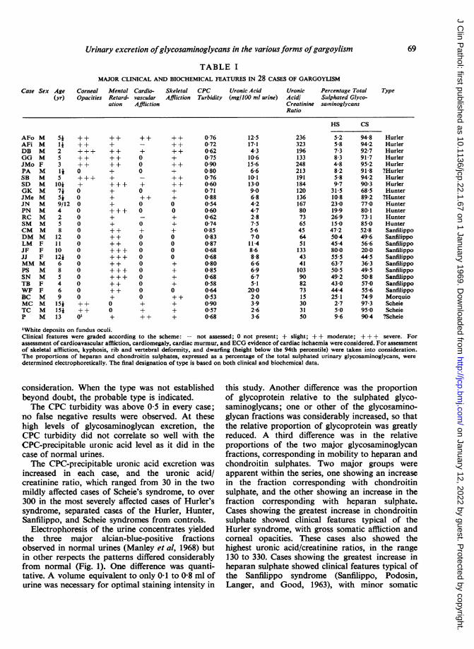
The age, sex, and relevant clinical features of thecases studied are shown in Table I, together with themajor chemical findings. For the typing of eachcase, clinical and chemical features were taken into
68
on January 12, 2022 by guest. Protected by copyright.
http://jcp.bmj.com
/J C
lin Pathol: first published as 10.1136/jcp.22.1.67 on 1 January 1969. D
ownloaded from
UronicAcidlCreatinineRatio
236323196133248213191184120136167807365456451133434110390827315303150
Percentage TotalSulphated Glyco-saininoglycans
69
Type
HS CS
5-2 94-8 Hurler5-8 94-2 Hurler7-3 92-7 Hurler8-3 91-7 Hurler4-8 95-2 Hurler8-2 91-8 ?Hurler5-8 94-2 Hurler9 7 90 3 Hurler315 68-5 Hunter10-8 89-2 ?Hunter23-0 77-0 Hunter19-9 80-1 Hunter26-9 73-1 Hunter15 0 85-0 Hunter47-2 52-8 Sanfilippo50 4 49-6 Sanfilippo45-4 56-6 Sanfilippo80-0 20-0 Sanfilippo55 5 44-5 Sanfilippo63-7 36-3 Sanfilippo50 5 49 5 Sanfilippo49-2 50-8 Sanfilippo43-0 57-0 Sanfilippo44-4 55-6 Sanfilippo25-1 74-9 Morquio2-7 97-3 Scheie5*0 95-0 Scheie9-6 904 ?Scheie
"White deposits on fundus oculi.Clinical features were graded according to the scheme: - not assessed; 0 not present; + slight; + + moderate; +++ severe. Forassessment of cardioavascular affliction, cardiomegaly, cardiac murmur, and ECG evidence of cardiac ischaemia were considered. For assessmentof skeletal affliction, kyphosis, rib and vertebral deformity, and dwarfing (height below the 94th percentile) were taken into consideration.The proportions of heparan and chondroitin sulphates, expressed as a percentage of the total sulphated urinary glycosaminoglycans, weredetermined electrophoretically. The final designation of type is based on both clinical and biochemical data.
consideration. When the type was not establishedbeyond doubt, the probable type is indicated.The CPC turbidity was above 0-5 in every case;
no false negative results were observed. At thesehigh levels of glycosaminoglycan excretion, theCPC turbidity did not correlate so well with theCPC-precipitable uronic acid level as it did in thecase of normal urines.The CPC-precipitable uronic acid excretion was
increased in each case, and the uronic acid/creatinine ratio, which ranged from 30 in the twomildly affected cases of Scheie's syndrome, to over300 in the most severely affected cases of Hurler'ssyndrome, separated cases of the Hurler, Hunter,Sanfilippo, and Scheie syndromes from controls.
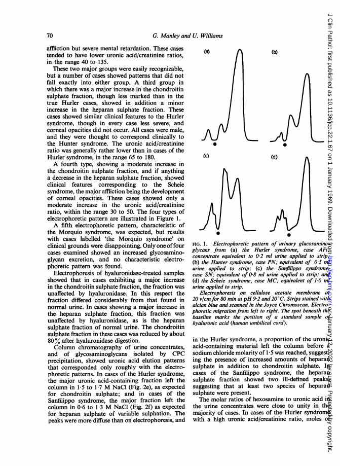
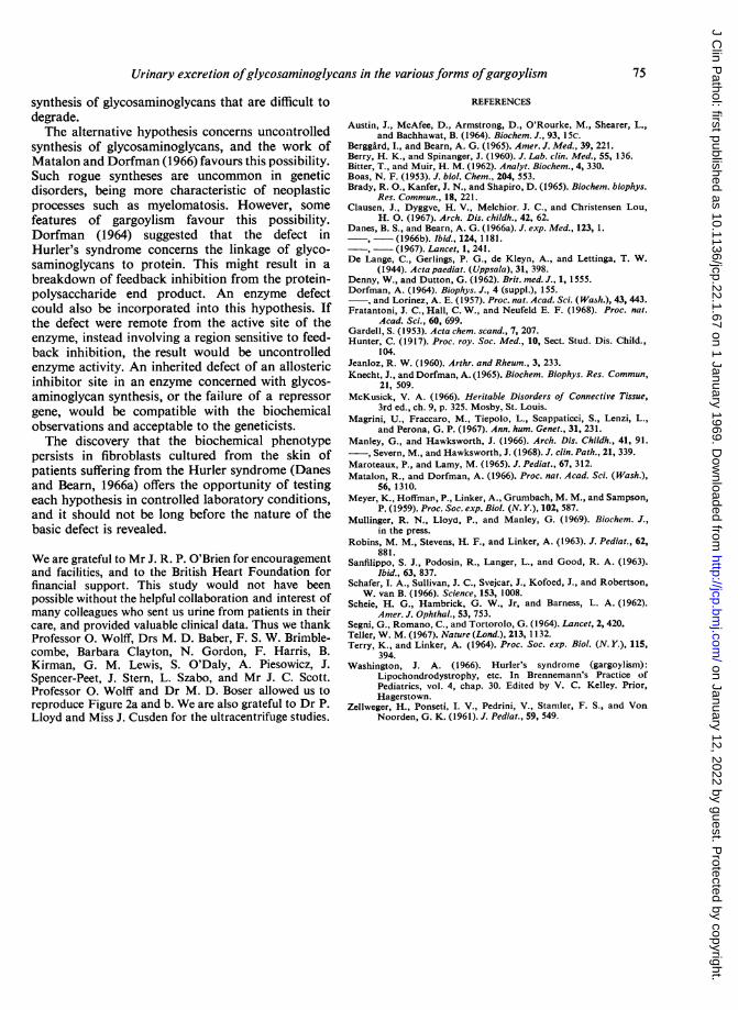
Electrophoresis of the urine concentrates yieldedthe three major alcian-blue-positive fractionsobserved in normal urines (Manley et al, 1968) butin other respects the patterns differed considerablyfrom normal (Fig. 1). One difference was quanti-tative. A volume equivalent to only 0-1 to 0-8 ml ofurine was necessary for optimal staining intensity in
this study. Another difference was the proportionof glycoprotein relative to the sulphated glyco-saminoglycans; one or other of the glycosamino-glycan fractions was considerably increased, so thatthe relative proportion of glycoprotein was greatlyreduced. A third difference was in the relativeproportions of the two major glycosaminoglycanfractions, corresponding in mobility to heparan andchondroitin sulphates. Two major groups wereapparent within the series, one showing an increasein the fraction corresponding with chondroitinsulphate, and the other showing an increase in thefraction corresponding with heparan sulphate.Cases showing the greatest increase in chondroitinsulphate showed clinical features typical of theHurler syndrome, with gross somatic affliction andcorneal opacities. These cases also showed thehighest uronic acid/creatinine ratios, in the range130 to 330. Cases showing the greatest increase inheparan sulphate showed clinical features typical ofthe Sanfilippo syndrome (Sanfilippo, Podosin,Langer, and Good, 1963), with minor somatic
Urinary excretion ofglycosaminoglycans in the variousforms oJ gargoylism
TABLE IMAJOR CLINICAL AND BIOCHEMICAL FEATURES IN 28 CASES OF GARGOYLISM
Case Sex Age Corneal Mental Cardio- Skeletal CPC Uronic Acid(yr) Opacities Retard- vascular Affliction Turbidity (mg/100 ml urine)
ation Affliction
AFo MAFi MDB MGG MJMo FPA MSB MSD MGK MJMe MJN MPN MRC MSM MCM MDM MLM FJF FJJ FMM MPS MSN MTB FWF FBC MMC MTC MP M
Si142S3145
7i5i9/124258
12111012468546915415i13
+++++++++0
+0
000000000000000
++
++01
++++++++++++++++++++++++
+++
+++
++
++
++
+000
+0++00
0+0000000000++++
++++++++++++++
++
00
++
+0000+++++
0-760-720-620750900-800-760-600-710-880-540-600-620740-850-830-870-680-680-800-850-680-580-640530900570-68
12-517-14-310-61566-610-1130906-84-24-72-87-55-67-0
11 48-68-86-66-96-75-1
20-02-03.92-63-6
on January 12, 2022 by guest. Protected by copyright.
http://jcp.bmj.com
/J C
lin Pathol: first published as 10.1136/jcp.22.1.67 on 1 January 1969. D
ownloaded from
G. Manley and U. Williams
affliction but severe mental retardation. These casestended to have lower uronic acid/creatinine ratios,in the range 40 to 135.These two major groups were easily recognizable,
but a number of cases showed patterns that did notfall exactly into either group. A third group inwhich there was a major increase in the chondroitinsulphate fraction, though less marked than in thetrue Hurler cases, showed in addition a minorincrease in the heparan sulphate fraction. Thesecases showed similar clinical features to the Hurlersyndrome, though in every case less severe, andcorneal opacities did not occur. All cases were male,and they were thought to correspond clinically tothe Hunter syndrome. The uronic acid/creatinineratio was generally rather lower than in cases of theHurler syndrome, in the range 65 to 180.A fourth type, showing a moderate increase in
the chondroitin sulphate fraction, and if anythinga decrease in the heparan sulphate fraction, showedclinical features corresponding to the Scheiesyndrome, the major affliction being the developmentof corneal opacities. These cases showed only amoderate increase in the uronic acid/creatinineratio, within the range 30 to 50. The four types ofelectrophoretic pattern are illustrated in Figure 1.A fifth electrophoretic pattern, characteristic of
the Morquio syndrome, was expected, but resultswith cases labelled 'the Morquio syndrome' onclinical grounds were disappointing. Only one offourcases examined showed an increased glycosamino-glycan excretion, and no characteristic electro-phoretic pattern was found.
Electrophoresis of hyaluronidase-treated samplesshowed that in cases exhibiting a major increasein the chondroitin sulphate fraction, the fraction wasunaffected by hyaluronidase. In this respect thefraction differed considerably from that found innormal urine. In cases showing a major increase inthe heparan sulphate fraction, this fraction wasunaffected by hyaluronidase, as is the heparansulphate fraction of normal urine. The chondroitinsulphate fraction in these cases was reduced by about80% after hyaluronidase digestion.Column chromatography of urine concentrates,
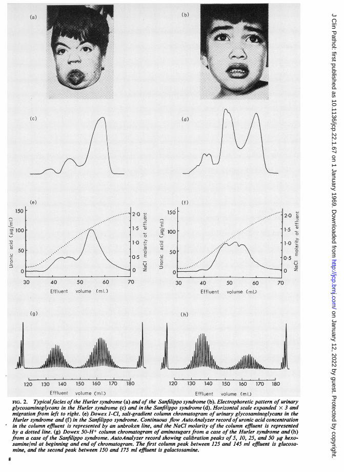
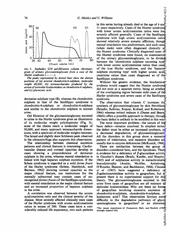
and of glycosaminoglycans isolated by CPCprecipitation, showed uronic acid elution patternsthat corresponded only roughly with the electro-phoretic patterns. In cases of the Hurler syndrome,the major uronic acid-containing fraction left thecolumn in 1-5 to 1-7 M NaCl (Fig. 2e), as expectedfor chondroitin sulphate; and in cases of theSanfilippo syndrome, the major fraction left thecolumn in 0-6 to 1-3 M NaCl (Fig. 2f) as expectedfor heparan sulphate of variable sulphation. Thepeaks were more diffuse than on electrophoresis, and
(a) (b)
.
(c) (d)
FIG. 1. Electrophoretic pattern of urinary glucosamino-glycans from (a) the Hurler syndrome, case AFi;concentrate equivalent to 0-2 ml urine applied to strip;(b) the Hunter syndrome, case PN; equivalent of 0 5 mlurine applied to strip; (c) the Sanfilippo syndrome,case SN; equivalent of 0-8 ml urine applied to strip; and(d) the Scheie syndrome, case MC; equivalent of 1J0 mlurine applied to strip.
Electrophoresis on cellulose acetate membrane at20 vlcm for 80 min at pH9-2 and 20°C. Strips stained withalcian blue and scanned in the Joyce Chromoscan. Electro-phoretic migration from left to right. The spot beneath thebaseline marks the position of a standard sample ofhyaluronic acid (human umbilical cord).
in the Hurler syndrome, a proportion of the uronicacid-containing material left the column before asodium chloride molarity of 1-5 was reached, suggest-ing the presence of increased amounts of heparansulphate in addition to chondroitin sulphate. Incases of the Sanfilippo syndrome, the heparansulphate fraction showed two ill-defined peaks,suggesting that at least two species of heparansulphate were present.The molar ratios of hexosamine to uronic acid in
the urine concentrates were close to unity in themajority of cases. In cases of the Hurler syndromewith a high uronic acid/creatinine ratio, moles of
70
on January 12, 2022 by guest. Protected by copyright.
http://jcp.bmj.com
/J C
lin Pathol: first published as 10.1136/jcp.22.1.67 on 1 January 1969. D
ownloaded from
(a)
(c) (d)
(e) (f)
c
a)
0
E
Cuz
CD
0:
30 40 50 60 70
EfflLent volume (ml.)
(g)
120 130 140 150 160 170 180
Effluent volume (ml.)
20 ,MI
1 5 -0
1 0faso
I 0E
0*5CX
I0Z30 40 50 60 70
Effluent volume (ml.)
(h)
1,I I I .
120 130 140 150 160 170 180
Effluent volume (ml.)FIG. 2. Typicalfacies ofthe Hurler syndrome (a) and of the Sanfilippo syndrome (b). Electrophoretic pattern of urinaryglycosaminoglycans in the Hurler syndrome (c) and in the Sanfilippo syndrome (d). Horizontal scale expanded x 3 andmigration from left to right. (e) Dowex 1-Cl, salt-gradient column chromatogram of urinary glycosaminoglycans in theHurler syndrome and (f) in the Sanfilippo syndrome. Continuous flow AutoAnalyzer record ofuronic acid concentrationin the column effluent is represented by an unbroken line, and the NaCl molarity of the column effluent is representedby a dotted line. (g) Dowex 50-H+ column chromatogram of aminosugars from a case of the Hurler syndrome and (h)from a case of the Sanfilippo syndrome. AutoAnalyzer record showing calibration peaks of 5, 10, 25, and SO ,ug hexo-samine/ml at beginning and end of chromatogram. The first column peak between 125 and 145 ml effluent is glucosa-mine, and the second peak between 150 and 175 ml effluent is galactosamine.
(b)
Arli
W"NW. ll:il.-...
,W
on January 12, 2022 by guest. Protected by copyright.
http://jcp.bmj.com
/J C
lin Pathol: first published as 10.1136/jcp.22.1.67 on 1 January 1969. D
ownloaded from
G. Manley and U. Williams
uronic acid outnumbered moles of hexosamine byapproximately two to one. In the case of Morquio'ssyndrome, more hexosamine than uronic acid waspresent (ratio 192: 1).Column chromatographic separation of the
aminosugars derived from the urine concentratesby hydrolysis showed two major fractions in eachcase, corresponding to glucosamine hydrochlorideand galactosamine hydrochloride. Cases of theHurler syndrome showed a major increase in thegalactosamine fraction, but also some increase in theglucosamine fraction (Fig. 2g). The glucosamine :gal-actosamine ratio was 1:1-8 after correcting for thelower colour yield of galactosamine in the Elson-Morgan reaction. Cases of the Sanfilippo syndromeshowed a major increase in the glucosamine fraction(Fig. 2h), giving a glucosamine :galactosamineratio of 1:0-6.
Molecular weight examinations were carried outon the CPC-precipitated glycosaminoglycans fromcases of the Hurler and Sanfilippo syndromes, butbecause of the difficulties involved in the physicalexamination of these highly charged polyanions,only approximate, comparative measurements weremade. 'Molecular sieving' chromatography onSephadex G 50 showed that none of the moleculeswere totally excluded from the gel, and althoughthe major peak emerged close to the void volume,uronic acid-containing molecules continued to beeluted until the total volume was approached.Arterial chondroitin-6-sulphate of molecular weight48,000 was totally excluded, being eluted with thevoid volume; tetrasaccharides derived from thetesticular hyaluronidase-digestion of arterial chon-droitin-6-sulphate were eluted close to the totalvolume, and a standard sample of glucuronic acid(British Drug Houses Ltd) was eluted with the totalvolume.
Ultracentrifugal sedimentation of the urinaryglycosaminoglycans showed broad and slowly-sedimenting Schlieren peaks in both the Hurler andSanfilippo syndromes. The peaks were slightly skew,due to extension of the trailing edge. At the con-centration employed, sedimentation rates were 1-0for the heparan sulphate excreted in the Sanfilipposyndrome and 1-1 for the chondroitin sulphateexcreted in the Hurler syndrome.
DISCUSSION
It is clear that the urinary glycosaminoglycans ingargoylism do not present a stiaightforward prob-lem and sometimes exhibit atypical behaviour inanalytical procedures. However, the excretion ofglycosaminoglycans in most cases of gargoylism isso different from normal that the problem of lab-oratory diagnosis is straightforward.
The CPC turbidity test is a simple and reliablescreening procedure, clearly differentiating gargoylesfrom the great majority of non-gargoyles, and falsenegative results have not been encountered. Theprevious paper (Manley et a!, 1968) emphasized thedanger of the normally high glycosaminoglycanexcretion in infancy, and the presence of excessiveamounts of glycoprotein at all ages, producing anincrease in the CPC turbidity, but these factorscannot lead to a false diagnosis of gargoylism if theuronic acid/creatinine ratio and electrophoreticpattern are determined in all cases with a CPCturbidity above 0 5.The CPC turbidity is an unreliable guide to the
total glycosaminoglycan excretion when this isexcessively high. One reason for this is that if theurine glycosaminoglycan concentration is excessive,not all of it is precipitated by the amount of CPCnormally added.A serious source of error is the presence of bacteria
in the urine sample. Many bacteria can elaborateenzymes that degrade glycosaminoglycans, andunless the concentration of merthiolate in the urineis at least 1:10,000 (w/v) bacterial contaminationcan completely alter the glycosaminoglycans present.The degradative effect of endogenous testicularhyaluronidase in the urine sample must also beborne in mind when dealing with adult males.The CPC-precipitable uronic acid/creatinine ratio
is the most reliable method of detecting excessiveglycosaminoglycan excretion that is suitable forroutine use, and clearly differentiates cases ofgargoylism (with the exception of the Morquiosyndrome) from other disorders. The excessivelyhigh concentration of glycosaminoglycans presentin the urine of some casesofthe Hurlersyndrome canbe a source of slight error in this test, and aftersedimenting the CPC-glycosaminoglycan complexin the centrifuge, it is advisable to add more CPCto the clear supernatant to ensure that all theglycosaminoglycans have been precipitated.The electrophoretic pattern of glycosaminoglycans
differentiates clearly between the two major formsof gargoylism: the Hurler and the Sanfilippo syn-dromes. These two distinct patterns were thought,in an earlier publication (Manley and Hawksworth,1966), to differentiate between the autosomalHurler syndrome and the sex-linked Hunter syn-drome, mainly on the basis of evidence presented byTerry and Linker (1964), but it is now clear that theHurler and Hunter syndromes show a similarurinary glycosaminoglycan pattern on electro-phoresis. This observation does not detract from thework of Terry and Linker, for it is clear that the twotechniques do not give identical results.The differentiation between the Hurler and
72
on January 12, 2022 by guest. Protected by copyright.
http://jcp.bmj.com
/J C
lin Pathol: first published as 10.1136/jcp.22.1.67 on 1 January 1969. D
ownloaded from
Urinary excretion ofglycosaminoglycans in the various forms oJ gargoylism
Hunter syndromes is best made at the clinical level,where evidence of X-linkage in the pedigree andabsence of corneal opacities favours the Huntersyndrome. In the laboratory, the presence of anexcessive excretion of testicular hyaluronidase-resistant chondroitin sulphate with a minor increasein heparan sulphate and a moderately elevateduronic acid/creatinine ratio in the range 65 to 180is confirmatory. However, the tissue culture workof Danes and Bearn (1966a) offers a clear differentia-tion between the Hurler and Hunter syndromes atthe laboratory level and exciting possibilities tothose interested in the basic biochemical lesion.The electrophoretic pattern in the Sanfilippo
syndrome is unmistakable, a striking increase in theheparan sulphate fraction being accompanied bylittle or no increase in the chondroitin sulphatefraction. There is one serious source of error, againrelated to bacterial contamination, that must beborne in mind. Bacterial action in the urine of a caseof the Hurler syndrome can reduce the electro-phoretic mobility of the chondroitin sulphatepresent to that of heparan sulphate. This effectcould result in a case of the Hurler syndrome beingmisdiagnosed as a case of the Sanfilippo syndrome,and the importance of adding sufficient preservativeto the urine cannot be overemphasized.The Scheie syndrome presents a clear electro-
phoretic pattern, with a sharp increase in thetesticular hyaluronidase-resistant chondroitin sul-phate fraction, and no increase, or even a decrease,in the heparan sulphate fraction. There is only amodest increase in the uronic acid/creatinine ratio.The disappointing results obtained with thesetechniques from cases diagnosed clinically as theMorquio syndrome are puzzling. The clinical group'Morquio's syndrome' may be heterogeneous,including some conditions that do not have adisorder of glycosaminoglycans as the basic defect.The heterogeneity of this clinical group has also beensuggested by skin fibroblast cultures (Danes andBearn, 1967; Magrini, Fraccaro, Tiepolo, Scap-paticci, Lenzi, and Perona, 1967). Techniques basedon uronic acid assay will not detect an increasedkeratan sulphate excretion. Reliance on the totalhexose or hexosamine/uronic acid ratio of urinaryconcentrates, or CPC precipitates, for the diagnosisof keratansulphaturia may be misleading, forincreased glycoprotein excretion will also increasethis ratio. The presence of excessive amounts ofkeratan sulphate in the urine of patients sufferingfrom the 'Morquio-Ullrich syndrome' seems estab-lished (Zellweger, Ponseti, Pedrini, Stamler, andVon Noorden, 1961; Robins, Stevens, and Linker,1963; Maroteaux and Lamy, 1965) and accepted aspart of the Morquio syndrome by McKusick (1966),
but it seems that further studies are necessary beforea simple method for the diagnosis of the Morquiosyndrome can be introduced to hospital laboratories.The discrepancy between column fractionation
and electrophoretic separation of urinary glyco-saminoglycans in the Hurler syndrome is interesting.Although electrophoresis shows that more than 90%of the urinary glycosaminoglycan has an electro-phoretic migration identical with that of chondroitinsulphate, a proportion of the uronic acid-containingmaterial is eluted from a column of Dowex 1-Clat a sodium chloride concentration of between0-7 and 1-3 M, suggesting the presence of increasedamounts of heparan sulphate. The measurement ofglucosamine and galactosamine in acid hydrolysatesof urine concentrates shows that the concentrationof glucosamine is increased in addition to gal-actosamine, again suggesting that heparan sulphateis increased in addition to chondroitin sulphate.An increased urinary excretion of heparan and
chondroitin sulphates in cases of the Hurlersyndrome is well supported (Meyer, Hoffman,Linker, Grumbach, and Sampson, 1959; Berggardand Beam, 1965; Teller, 1967) but the electrophoretictechnique used in this study shows that in fresh,uncontaminated urine, about 90% of the glyco-saminoglycans excreted have a mobility correspond-ing with chondroitin sulphate. One explanation ofthis discrepancy is that an oversulphated heparansulphate is excreted in cases of the Hurler syndrome,and this accompanies chondroitin sulphate oncellulose acetate electrophoresis, where net molecularcharge is the overwhelming criterion for fraction-ation. Another possibility is that hybrid moleculesare produced in the Hurler syndrome, withglucosamine being built into the chondroitinsulphate chain. A third possibility is that glyco-saminoglycan fragments of low molecular weightmay behave atypically on ion-exchange columns,being eluted at a lower salt concentration thanexpected. Cellulose acetate electrophoresis has theadvantage of being relatively free from molecularweight effects.The two heparan sulphate peaks in column
chromatograms from cases of the Sanfilippo syn-drome supports the finding of two electrophoreticfractions of heparan sulphate (Manley and Hawks-worth, 1966). Knecht and Dorfman (1965) foundtwo major fractions of heparan sulphate in Hurlertissues, differing in the degree of N sulphation. Thedata reported here are consistent with two speciesof heparan sulphate in urine from cases of theSanfilippo syndrome.
Testicular hyaluronidase digestion shows that thechondroitin sulphate excreted in urine from cases ofthe Hurler, Hunter, and Scheie syndromes is of the
73
on January 12, 2022 by guest. Protected by copyright.
http://jcp.bmj.com
/J C
lin Pathol: first published as 10.1136/jcp.22.1.67 on 1 January 1969. D
ownloaded from
G. Manley and U. Williams
E
340
20
0
25 30 40 50 60 70 80Effluent volume (ml.)
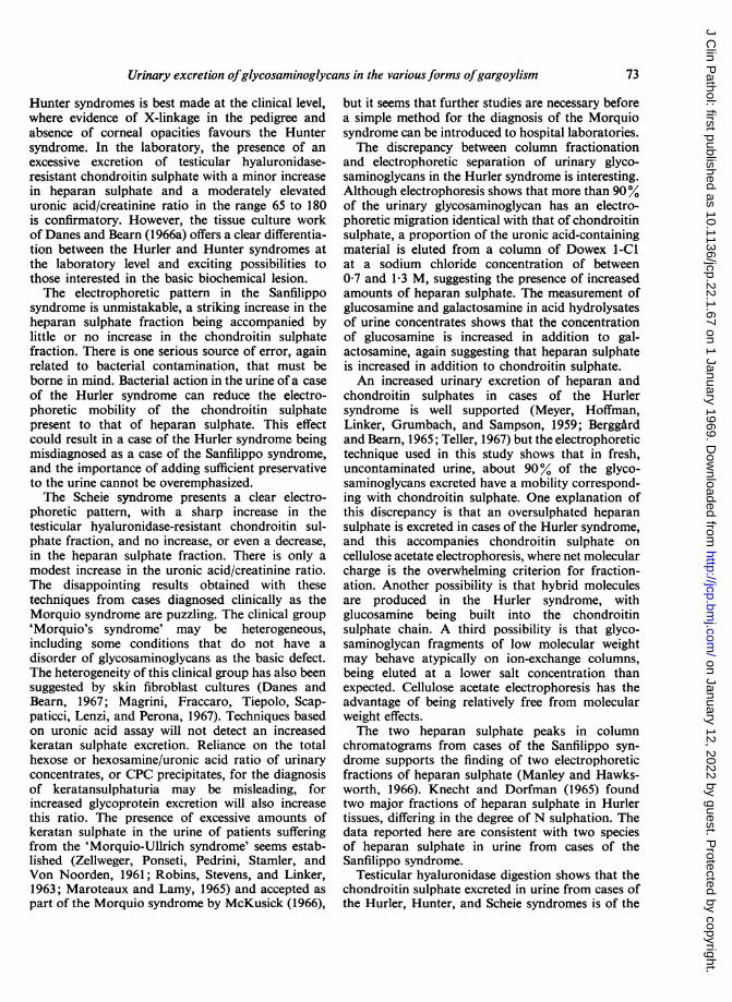
FIG. 3. Sephadex G50 gel-filtration column chromato-gram of urinary glycosaminoglycans from a case of theHurler syndrome ( ).
The peaks represented by dotted lines show the elutionpositions of (a) arterial chondroitin-6-sulphate, molecularweight 48,000; (b) tetrasaccharides produced by theaction oftesticular hyaluronidase on chondroitin-6-sulphate,and (c) glucuronic acid.
dermatan sulphate type (B), whereas the chondroitinsulphate in that of the Sanfilippo syndrome ischondroitin-4-sulphate or chondroitin-6-sulphateand similar to the chondroitin sulphate in normalurine.
Gel filtration of the glycosaminoglycans excretedin urine in the Hurler syndrome gives an illustrationof its molecular weight polydispersity (Fig. 3);none of the chains reach a molecular weight of50,000, and many approach tetrasaccharide dimen-sions, with a spectrum of molecular weights between.The broad and slightly skew Schlieren peak observedin the ultracentrifuge also supports this observation.The relationship between chemical excretion
patterns and clinical features is interesting. Cardio-vascular disease and corneal opacities develop incases showing a preponderance of dermatansulphate in the urine, whereas mental retardation islinked with high heparan sulphate excretion. If theScheie syndrome is regarded as a mild forme frusteof the Hurler syndrome (Scheie, Hambrick, andBarness, 1962) showing corneal opacities as themajor clinical feature, our institutions for thementally subnormal may contain cases of un-recognizedformesfrustes of the Sanfilippo syndrome,with mental retardation as the major clinical featureand an increased proportion of heparan sulphatein the urine.A correlation was observed between the uronic
acid/creatinine ratio and the clinical severity of thedisease. Most severely affected clinically were casesof the Hurler syndrome with uronic acid/creatinineratios in excess of 200. These cases have a con-
siderably reduced life expectancy, two such patients
in this series having already died at the age of 6 and11 years respectively. Cases of the Hunter syndromewith lower uronic acid/creatinine ratios were lessseverely affected generally. Cases of the Sanfilipposyndrome with high uronic acid/creatinine ratiosshowed relatively severe somatic affliction, thoughmental retardation was predominant, and such cases(when male) were often diagnosed clinically asthe Hunter syndrome. Clinically diagnosed cases ofthe Hunter syndrome were found, on examinationof the urinary glycosaminoglycans, to be dividedbetween the 'chondroitin sulphate excreting type'with lower uronic acid/creatinine ratios than casesof the true Hurler syndrome, and the 'heparansulphate excreting type' with higher uronic acid/creatinine ratios than cases diagnosed as of theSanfilippo syndrome.Without the genetic evidence, the biochemical
evidence would suggest that the Hunter syndromedid not exist as a separate entity, being an artefactof the overlapping region between mild cases of theHurler syndrome and severe cases of the Sanfilipposyndrome.The observation that vitamin C increases the
synthesis of glycosaminoglycans by skin fibroblasts(Schafer, Sullivan, Svejcar, Kofoed, and Robertson,1966) whereas retinol reduces it (Danes and Beam,1966b) offers a possible approach to therapy, thoughthe basic defect is unlikely to be modified in this way.The most important problem-the nature of the
basic defect-remains unsolved. In simplest terms,the defect must be either an increased synthesis, ora decreased degradation, of glycosaminoglycans.All the disorders in this group show a recessivepattern of inheritance, and recessive disorders areusually due to enzyme deficiencies (McKusick, 1966).There are similarities between the group of
disorders considered here, and the lipoidoses. Thereis evidence for a deficiency of ,B-glucosidase activityin Gaucher's disease (Brady, Kanfer, and Shapiro,1965) and of sulphatase activity in metachromaticleucodystrophy (Austin, McAfee, Armstrong,O'Rourke, Shearer, and Bachhawat, 1964). Clausenet al (1967) have suggested a deficiency offl-galactosaminidase activity in gargoylism, but atpresent there is no experimental support for thistheory. The glycosaminoglycans excreted in theurine from cases of gargoylism are all resistant totesticular hyaluronidase. Why are there no formsof gargoylism involving excessive excretion ofchondroitin-6-sulphate, chondroitin-4-sulphate, orhyaluronic acid? This observation favours somedifficulty in the degradative pathways of glyco-saminoglycans in gargoylisml or an abnormal'The recent experiences of Fratantoni, Hall, and Neufeld (1968)strongly support this.
74
on January 12, 2022 by guest. Protected by copyright.
http://jcp.bmj.com
/J C
lin Pathol: first published as 10.1136/jcp.22.1.67 on 1 January 1969. D
ownloaded from
Urinary excretion ofglycosaminoglycans in the various forms ofgargoylism
synthesis of glycosaminoglycans that are difficult todegrade.The alternative hypothesis concerns uncointrolled
synthesis of glycosaminoglycans, and the work ofMatalon and Dorfman (1966) favours this possibility.Such rogue syntheses are uncommon in geneticdisorders, being more characteristic of neoplasticprocesses such as myelomatosis. However, somefeatures of gargoylism favour this possibility.Dorfman (1964) suggested that the defect inHurler's syndrome concerns the linkage of glyco-saminoglycans to protein. This might result in abreakdown of feedback inhibition from the protein-polysaccharide end product. An enzyme defectcould also be incorporated into this hypothesis. Ifthe defect were remote from the active site of theenzyme, instead involving a region sensitive to feed-back inhibition, the result would be uncontrolledenzyme activity. An inherited defect of an allostericinhibitor site in an enzyme concerned with glycos-aminoglycan synthesis, or the failure of a repressorgene, would be compatible with the biochemicalobservations and acceptable to the geneticists.The discovery that the biochemical phenotype
persists in fibroblasts cultured from the skin ofpatients suffering from the Hurler syndrome (Danesand Bearn, 1966a) offers the opportunity of testingeach hypothesis in controlled laboratory conditions,and it should not be long before the nature of thebasic defect is revealed.
We are grateful to Mr J. R. P. O'Brien for encouragementand facilities, and to the British Heart Foundation forfinancial support. This study would not have beenpossible without the helpful collaboration and interest ofmany colleagues who sent us urine from patients in theircare, and provided valuable clinical data. Thus we thankProfessor 0. Wolff, Drs M. D. Baber, F. S. W. Brimble-combe, Barbara Clayton, N. Gordon, F. Harris, B.Kirman, G. M. Lewis, S. O'Daly, A. Piesowicz, J.Spencer-Peet, J. Stem, L. Szabo, and Mr J. C. Scott.Professor 0. Wolff and Dr M. D. Boser allowed us toreproduce Figure 2a and b. We are also grateful to Dr P.Lloyd and Miss J. Cusden for the ultracentrifuge studies.
REFERENCES
Austin, J., McAfee, D., Armstrong, D., O'Rourke, M., Shearer, L.,and Bachhawat, B. (1964). Biochem. J., 93, 15c.
Berggard, I., and Bearn, A. G. (1965). Amner. J. Med., 39, 221.Berry, H. K., and Spinanger, J. (1960). J. Lab. clin. Med., 55, 136.Bitter, T., and Muir, H. M. (1962). Analyt. Biochem., 4, 330.Boas, N. F. (1953). J. biol. Chem., 204, 553.Brady, R. O., Kanfer, J. N., and Shapiro, D. (1965). Biochem. biophys.
Res. Commun., 18, 221.Clausen, J., Dyggve, H. V., Melchior. J. C., and Christensen Lou,
H. 0. (1967). Arch. Dis. childh., 42, 62.Danes, B. S., and Bearn, A. G. (1966a). J. exp. Med., 123, 1.
(1966b). Ibid., 124, 1181.(1967). Lancet, 1, 241.
De Lange, C., Gerlings, P. G., de Kleyn, A., and Lettinga, T. W.(1944). Acta paediat. (Uppsala), 31, 398.
Denny, W., and Dutton, G. (1962). Brit. med. J., 1, 1555.Dorfman, A. (1964). Biophys. J., 4 (suppl.), 155.
, and Lorinez, A. E. (1957). Proc. nat. Acad. Sci. (Wash.), 43, 443.Fratantoni, J. C., Hall, C. W., and Neufeld E. F. (1968). Proc. nat.
Acad. Sci., 60, 699.Gardell, S. (1953). Acta chem. scand., 7, 207.Hunter, C. (1917). Proc. roy. Soc. Med., 10, Sect. Stud. Dis. Child.,
104.Jeanloz, R. W. (1960). Arthr. and Rheum., 3, 233.Knecht, J., and Dorfman, A. (1965). Biochem. Biophys. Res. Commun,
21, 509.McKusick, V. A. (1966). Heritable Disorders of Connective Tissue,
3rd ed., ch. 9, p. 325. Mosby, St. Louis.Magrini, U., Fraccaro, M., Tiepolo, L., Scappaticci, S., Lenzi, L.,
and Perona, G. P. (1967). Ann. hum. Genet., 31, 231.Manley, G., and Hawksworth, J. (1966). Arch. Dis. Childh., 41, 91.-, Severn, M., and Hawksworth, J. (1968). J. clin. Path., 21, 339.Maroteaux, P., and Lamy, M. (1965). J. Pediat., 67, 312.Matalon, R., and Dorfman, A. (1966). Proc. nat. Acad. Sci. (Wash.),
56, 1310.Meyer, K., Hoffman, P., Linker, A., Grumbach, M. M., and Sampson,
P. (1959). Proc. Soc. exp. Biol. (N. Y.), 102, 587.Mullinger, R. N., Lloyd, P., and Manley, G. (1969). Biochem. J.,
in the press.Robins, M. M., Stevens, H. F., and Linker, A. (1963). J. Pediat., 62,
881.Sanfilippo, S. J., Podosin, R., Langer, L., and Good, R. A. (1963).
Ibid., 63, 837.Schafer, I. A., Sullivan, J. C., Svejcar, J., Kofoed, J., and Robertson,
W. van B. (1966). Science, 153, 1008.Scheie, H. G., Hambrick, G. W., Jr, and Barness, L. A. (1962).
Amer. J. Ophthal., 53, 753.Segni, G., Romano, C., and Tortorolo, G. (1964). Lancet, 2, 420.Teller, W. M. (1967). Nature (Lond.), 213, 1132.Terry, K., and Linker, A. (1964). Proc. Soc. exp. Biol. (N. Y.), 115,
394.Washington, J. A. (1966). Hurler's syndrome (gargoylism):
Lipochondrodystrophy, etc. In Brennemann's Practice ofPediatrics, vol. 4, chap. 30. Edited by V. C. Kelley. Prior,Hagerstown.
Zellweger, H., Ponseti, I. V., Pedrini, V., Stamler, F. S., and VonNoorden, G. K. (1961). J. Pediat., 59, 549.
75
on January 12, 2022 by guest. Protected by copyright.
http://jcp.bmj.com
/J C
lin Pathol: first published as 10.1136/jcp.22.1.67 on 1 January 1969. D
ownloaded from
Related Documents