Article Urinary Biomarkers and Progression of AKI in Patients with Cirrhosis Justin M. Belcher,* †‡ Guadalupe Garcia-Tsao, ‡§| Arun J. Sanyal, ¶ Heather Thiessen-Philbrook,** Aldo J. Peixoto, †| Mark A. Perazella, †| Naheed Ansari, †† Joseph Lim, § Steven G. Coca,* †‡ and Chirag R. Parikh* †‡ for the TRIBE-AKI Consortium Abstract Background and objectives AKI is a common and severe complication in patients with cirrhosis. AKI progression was previously shown to correlate with in-hospital mortality. Therefore, accurately predicting which patients are at highest risk for AKI progression may allow more rapid and targeted treatment. Urinary biomarkers of structural kidney injury associate with AKI progression and mortality in multiple settings of AKI but their prognostic performance in patients with liver cirrhosis is not well known. Design, setting, participants, & measurements A multicenter, prospective cohort study was conducted at four tertiary care United States medical centers between 2009 and 2011. The study comprised patients with cirrhosis and AKI defined by the AKI Network criteria evaluating structural (neutrophil gelatinase–associated lipocalin, IL-18, kidney injury molecule-1 [KIM-1], liver-type fatty acid–binding protein [L-FABP], and albuminuria) and functional (fractional excretion of sodium [FENa]) urinary biomarkers as predictors of AKI progression and in- hospital mortality. Results Of 188 patients in the study, 44 (23%) experienced AKI progression alone and 39 (21%) suffered both progression and death during their hospitalization. Neutrophil gelatinase–associated lipocalin, IL-18, KIM-1, L-FABP, and albuminuria were significantly higher in patients with AKI progression and death. These biomarkers were independently associated with this outcome after adjusting for key clinical variables including model of end stage liver disease score, IL-18 (relative risk [RR], 4.09; 95% confidence interval [95% CI], 1.56 to 10.70), KIM-1 (RR, 3.13; 95% CI, 1.20 to 8.17), L-FABP (RR, 3.43; 95% CI, 1.54 to 7.64), and albuminuria (RR, 2.07; 95% CI, 1.05– 4.10) per log change. No biomarkers were independently associated with progression without mortality. FENa demonstrated no association with worsening of AKI. When added to a robust clinical model, only IL-18 inde- pendently improved risk stratification on a net reclassification index. Conclusions Multiple structural biomarkers of kidney injury, but not FENa, are independently associated with progression of AKI and mortality in patients with cirrhosis. Injury marker levels were similar between those without progression and those with progression alone. Clin J Am Soc Nephrol 9: 1857–1867, 2014. doi: 10.2215/CJN.09430913 Introduction AKI is common in patients with cirrhosis, complicat- ing 20% of hospitalizations (1–3). The risk of death increases with peak severity of AKI (4–8) and pro- gression of AKI to a higher stage defined by the AKI Network (AKIN) criteria. AKI progression is as- sociated with mortality independent of the model of end stage liver disease (MELD) score (4). Intervening to prevent progression therefore may reduce mortal- ity. Despite the overall grim prognosis for patients with cirrhosis and AKI, disease-specific treatments carrying the potential to improve outcomes, if cor- rectly and judiciously applied, are available for pa- tients with hepatorenal syndrome (HRS) (9–12). In addition, patients with progressive acute tubular ne- crosis (ATN) may be managed with dialysis, and those with severe, irreversible disease benefit from combined liver-kidney transplantation (13). For rea- sons of safety and equity, such aggressive therapies should ideally be offered only to those patients at greatest risk for progressive renal dysfunction and death. However, in practice, where clinically distinguish- ing the cause of AKI is frequently challenging, patients often receive a “kitchen sink” approach of multiple ag- gressive therapies irrespective of whether they are at high risk for AKI progression or death. If a patient is unlikely to progress or die, aggressive management can likely be held while time is taken to clarify the cause of AKI. However, if a patient is at high risk for adverse outcomes, early and aggressive action should be taken. Unfortunately, predicting which patients will suffer progressive AKI, and identifying those progressors who will proceed to death, is clinically challenging. In patients with cirrhosis, an enlarged volume of fluid *Program of Applied Translational Research, Sections of † Nephrology and § Digestive Diseases, Yale University School of Medicine, New Haven, Connecticut; ‡ Clinical Epidemiology Research Center, Veterans Affairs Medical Center, West Haven, Connecticut; | Veterans Affairs- Connecticut Healthcare System, West Haven, Connecticut; ¶ Division of Gastroenterology, Hepatology, and Nutrition, Department of Internal Medicine, Virginia Commonwealth University School of Medicine, Richmond, Virginia; **Division of Nephrology, Department of Medicine, University of Western Ontario, London, Ontario, Canada; and †† Division of Nephrology, Department of Internal Medicine, Jacobi Medical Center, South Bronx, New York Correspondence: Dr. Chirag R. Parikh, Section of Nephrology, Yale University and Veterans Affairs Medical Center, Temple Medical Center, 60 Temple Street, Suite 6C, New Haven, CT 06510. Email: chirag.parikh@ yale.edu www.cjasn.org Vol 9 November, 2014 Copyright © 2014 by the American Society of Nephrology 1857

Welcome message from author
This document is posted to help you gain knowledge. Please leave a comment to let me know what you think about it! Share it to your friends and learn new things together.
Transcript
Article
Urinary Biomarkers and Progression of AKI in Patientswith Cirrhosis
Justin M. Belcher,*†‡ Guadalupe Garcia-Tsao,‡§| Arun J. Sanyal,¶ Heather Thiessen-Philbrook,** Aldo J. Peixoto,†|
Mark A. Perazella,†| Naheed Ansari,†† Joseph Lim,§ Steven G. Coca,*†‡ and Chirag R. Parikh*†‡ for the TRIBE-AKIConsortium
AbstractBackground and objectivesAKI is a common and severe complication in patients with cirrhosis. AKI progressionwas previously shown to correlatewith in-hospital mortality. Therefore, accurately predictingwhich patients areat highest risk for AKI progression may allow more rapid and targeted treatment. Urinary biomarkers ofstructural kidney injury associate with AKI progression and mortality in multiple settings of AKI but theirprognostic performance in patients with liver cirrhosis is not well known.
Design, setting, participants, & measurements A multicenter, prospective cohort study was conducted at fourtertiary care United States medical centers between 2009 and 2011. The study comprised patients with cirrhosisand AKI defined by the AKI Network criteria evaluating structural (neutrophil gelatinase–associated lipocalin,IL-18, kidney injury molecule-1 [KIM-1], liver-type fatty acid–binding protein [L-FABP], and albuminuria) andfunctional (fractional excretion of sodium [FENa]) urinary biomarkers as predictors of AKI progression and in-hospital mortality.
Results Of 188 patients in the study, 44 (23%) experienced AKI progression alone and 39 (21%) suffered bothprogression and death during their hospitalization. Neutrophil gelatinase–associated lipocalin, IL-18, KIM-1,L-FABP, and albuminuria were significantly higher in patients with AKI progression and death. These biomarkerswere independently associatedwith this outcome after adjusting for key clinical variables includingmodel of endstage liver disease score, IL-18 (relative risk [RR], 4.09; 95% confidence interval [95% CI], 1.56 to 10.70), KIM-1(RR, 3.13; 95% CI, 1.20 to 8.17), L-FABP (RR, 3.43; 95% CI, 1.54 to 7.64), and albuminuria (RR, 2.07; 95% CI, 1.05–4.10) per log change. No biomarkers were independently associated with progression without mortality. FENademonstrated no association with worsening of AKI. When added to a robust clinical model, only IL-18 inde-pendently improved risk stratification on a net reclassification index.
Conclusions Multiple structural biomarkers of kidney injury, but not FENa, are independently associated withprogression of AKI and mortality in patients with cirrhosis. Injury marker levels were similar between thosewithout progression and those with progression alone.
Clin J Am Soc Nephrol 9: 1857–1867, 2014. doi: 10.2215/CJN.09430913
IntroductionAKI is common in patients with cirrhosis, complicat-ing 20% of hospitalizations (1–3). The risk of deathincreases with peak severity of AKI (4–8) and pro-gression of AKI to a higher stage defined by theAKI Network (AKIN) criteria. AKI progression is as-sociated with mortality independent of the model ofend stage liver disease (MELD) score (4). Interveningto prevent progression therefore may reduce mortal-ity. Despite the overall grim prognosis for patientswith cirrhosis and AKI, disease-specific treatmentscarrying the potential to improve outcomes, if cor-rectly and judiciously applied, are available for pa-tients with hepatorenal syndrome (HRS) (9–12). Inaddition, patients with progressive acute tubular ne-crosis (ATN) may be managed with dialysis, andthose with severe, irreversible disease benefit from
combined liver-kidney transplantation (13). For rea-sons of safety and equity, such aggressive therapiesshould ideally be offered only to those patients atgreatest risk for progressive renal dysfunction anddeath. However, in practice, where clinically distinguish-ing the cause of AKI is frequently challenging, patientsoften receive a “kitchen sink” approach of multiple ag-gressive therapies irrespective of whether they are athigh risk for AKI progression or death. If a patient isunlikely to progress or die, aggressive management canlikely be held while time is taken to clarify the cause ofAKI. However, if a patient is at high risk for adverseoutcomes, early and aggressive action should be taken.Unfortunately, predicting which patients will suffer
progressive AKI, and identifying those progressorswho will proceed to death, is clinically challenging. Inpatients with cirrhosis, an enlarged volume of fluid
*Program of AppliedTranslationalResearch, Sections of†Nephrology and§Digestive Diseases,Yale University Schoolof Medicine, NewHaven, Connecticut;‡ClinicalEpidemiologyResearch Center,Veterans AffairsMedical Center, WestHaven, Connecticut;|Veterans Affairs-ConnecticutHealthcare System,West Haven,Connecticut;¶Division ofGastroenterology,Hepatology, andNutrition, Departmentof Internal Medicine,VirginiaCommonwealthUniversity School ofMedicine, Richmond,Virginia; **Division ofNephrology,Department ofMedicine, Universityof Western Ontario,London, Ontario,Canada; and††Division ofNephrology,Department ofInternal Medicine,Jacobi Medical Center,South Bronx, NewYork
Correspondence:Dr. Chirag R. Parikh,Section ofNephrology, YaleUniversity andVeterans AffairsMedical Center,Temple MedicalCenter, 60 TempleStreet, Suite 6C, NewHaven, CT 06510.Email: [email protected]
www.cjasn.org Vol 9 November, 2014 Copyright © 2014 by the American Society of Nephrology 1857
distribution, low protein intake, and decreased creatinineproduction secondary to muscle atrophy and liver dys-function significantly dissociate creatinine levels fromreflecting the true presence and severity of kidney dys-function (14). Correspondingly, creatinine fluctuationsearly in the course of AKI are difficult to interpret, takingseveral days to resolve into a definitive trend demonstrat-ing progression. As a result of this delay, potentially ben-eficial treatments may be deferred. An accurate, objective,and reproducible means of anticipating AKI progressionor death at the time of AKI diagnosis is urgently needed toallocate treatments, stratify patients for inclusion in trials,and prioritize liver and kidney transplantations.Research into structural AKI has been revolutionized by
investigation of multiple urinary biomarkers of kidneytubular injury that independently predict AKI progressionin multiple clinical settings (15–17). Among the mostpromising are neutrophil gelatinase–associated lipocalin(NGAL), IL-18, kidney injury molecule-1 (KIM-1), andliver-type fatty acid–binding protein (L-FABP). AlthoughNGAL has been studied for early detection of AKI afterliver transplantation (18,19) and for differential diagnosisof AKI in cirrhosis (20,21), few studies have evaluatedthese biomarkers for prognosis in patients with cirrhosisand AKI (20,21). With a unique mix of functional (HRS)and structural disease (ATN and GN), the association incirrhosis between tubular injury biomarker levels and out-comes is unclear. In this setting, it is possible that biomark-ers of tubular function, such as fractional excretion ofsodium (FENa), and traditional markers of both glomeru-lar and tubular injury, such as urine albumin, may alsoprovide additional prognostic accuracy. Indeed, albumin-uria is predictive of impending AKI in patients with cir-rhosis (22). We conducted a multicenter prospective studyevaluating urinary biomarkers of kidney injury and tubu-lar function for prediction of AKI progression and progres-sion with mortality in patients with cirrhosis.
Materials and MethodsStudy DesignThe details of the cohort and study design were pre-
viously described (4). This prospective, multicenter obser-vational cohort study was conducted over 29 monthsbetween 2009 and 2011, at four tertiary care academic cen-ters in the United States. Eligible patients were admittedwith AKI (see independent variables below for definitions)or developed it during the course of hospitalization. Inclu-sion criteria included a known diagnosis of cirrhosis (seeindependent variables below for definitions), age$18 years,and availability of a documented serum creatinine within 1year before AKI. Major exclusion criteria included priorkidney or liver transplantation and advanced CKD orRRT at the time of enrollment. All consecutive eligible pa-tients were enrolled within 5 days of meeting AKI criteria.Informed consent was obtained from all patients or theirproxy decision-makers. The study was approved by the in-stitutional review board at each participating institution.
Sample Collection and Biomarker MeasurementA fresh 10-ml urine sample was collected daily for 3 days.
Samples were immediately refrigerated and centrifuged at
50003g for 10 minutes at 24°C. Aliquots of 1 ml of super-natant were stored within 6 hours of collection at280°C. Noadditives or protease inhibitors were utilized. All biomarkerswere measured from frozen aliquots that did not undergoany additional freeze-thaw cycles. Laboratory measurementswere performed by personnel blinded to patient informa-tion. ELISA methods, coefficients of variation, and detectionranges were as previously described for measurement ofNGAL (23), IL-18 (24), KIM-1 (25), and L-FABP (25). Urinecreatinine was measured by the modified Jaffe reaction.
VariablesIndependent Variables. Independent variables included
cirrhosis, AKI, and baseline serum creatinine. Eligible pa-tients carried a documented diagnosis of cirrhosis based onliver biopsy, when available, or on a combination of clinical,biochemical, imaging, and endoscopic findings.AKI was defined as a rise in creatinine of 0.3 mg/dl or
50% from baseline as recommended by a working groupcomposed of members of the International Ascites Club(IAC) and the Acute Dialysis Quality Initiative who basedthis cutoff on stage 1 of the AKIN criteria (26). Becausedocumentation of urine output was incomplete, this aspectof the criteria was not utilized.Baseline serum creatinine was defined as the most recent
stable measurement before admission because the use ofoutpatient values results in less misclassification of AKIincidence, severity, and prognosis compared with utilizinghospital admission, hospital nadir, or imputed values (27).The median interval between creatinine utilized for base-line and hospital admission in this study was 26 days (in-terquartile range [IQR], 9–73).Other Variables. Baseline GFR was estimated via the
Modification of Diet in Renal Disease 4 equation (28).CKD was defined as a GFR,60 ml/min per 1.73 m2. MELDand Child–Pugh scores were calculated on the day of firstsample collection. HRS was diagnosed via the 2007 IACcriteria (29).Outcomes. Our primary outcomes consisted of progres-
sion to a higher AKIN stage and progression to a higherstage with subsequent death, which were compared sepa-rately with patients who did not progress. If patients whopresented with stage 3 AKI but not requiring RRT sub-sequently required dialysis, this was considered progression.Patients who died without progression were excluded fromthe primary analysis because death may have been a com-peting risk for progression for these patients. Biomarker valuesfor these excluded patients did not differ from those withprogression and death (Supplemental Table 1).
Statistical AnalysesCategorical variables were expressed as proportions and
compared using chi-squared and Fisher’s exact tests, asappropriate. Normally or near-normally distributed vari-ables were reported as means with SDs and were comparedby the t test. Non-normally distributed continuous vari-ables were reported as medians with IQRs and were com-pared by the Wilcoxon rank-sum test. Normality wasassessed using the Kolmogorov–Smirnov test. NGAL val-ues were bounded at an upper limit of 1000 ng/ml with nolower bound. KIM-1 was bounded at an upper limit of 60ng/ml and a lower limit of 0.056 ng/ml. L-FABP was
1858 Clinical Journal of the American Society of Nephrology

Tab
le1.
Baselinean
ddem
ograp
hic
charac
teristics
Cha
racteristic
Total
(N=18
8)NoProg
ression
(n=95
)Prog
ressionAlone
(n=44
)Prog
ressionwithDeath
(n=39
)Death
Alone
(n=10
)PValue
Age
(yr)
5569.3
55.769.6
54.768.9
54.769
50.867
0.10
Men
133(71)
66(69)
32(73)
29(74)
6(60)
0.83
BMI(kg/
m2 )
30.1
(25.5–
35.1)
30(24.5–
35.3)
29.6
(26–
33.9)
30.9
(27–
34.8)
26.7
(25.7–
32.7)
0.40
Race/ethnicity
White
134(71)
70(74)
37(84)
21(54)
6(60)
0.01
Black
27(14)
14(15)
3(7)
9(23)
1(10)
0.26
Hispa
nic
23(12)
10(11)
3(7)
8(21)
2(20)
0.06
CKD
a65
(35)
31(33)
21(48)
12(31)
1(10)
0.48
eGFR
(ml/min
per1.73
m2 )
70(55–
98)
70(57–
93)
64(43–
88)
81(54–
100)
101(78–
115)
0.14
Proteinu
riab
22(12)
14(15)
3(7)
5(13)
0(0)
0.36
BaselineSC
r(m
g/dl)
1(0.8–1.3)
1(0.8–1.3)
1.1(0.8–1.5)
1(0.8–1.3)
0.7(0.6–1)
0.09
Creatinineat
meeting
AKIN
criteria
(mg/
dl)
2.2(1.6–2.9)
2.1(1.6–2.7)
2.4(1.6–3.3)
2.1(1.5–2.8)
2.3(1.6–4.1)
0.50
Peak
creatinine
2.7(1.9–4.2)
2.2(1.7–3.1)
3.4(2.4–4.8)
4(3.1–5.5)
2.5(2.2–5.1)
,0.00
1Diabe
tes
49(26)
29(31)
10(23)
9(23)
1(10)
0.56
Activecanc
er21
(11)
10(11)
5(11)
4(10)
2(20)
0.98
Cau
seof
cirrhosis
Alcoh
ol55
(29)
27(28)
12(27)
10(26)
6(60)
0.17
Alcoh
olan
dHCV
52(28)
26(27)
13(30)
12(31)
1(10)
0.57
HCV
32(17)
14(15)
10(23)
6(15)
2(20)
0.87
NASH
17(9)
10(11)
5(11)
2(5)
0(0)
0.62
Cryptog
enic
12(6)
7(7)
3(7)
2(5)
0(0)
0.96
Autoimmun
e11
(6)
6(6)
0(0)
4(10)
1(10)
0.26
Other
10(5)
5(5)
1(2)
4(10)
0(0)
0.38
Previou
sco
mplication
sAscites
142(76)
71(75)
35(81)
30(77)
6(60)
0.91
Hep
aticen
cepha
lopa
thy
126(67)
61(64)
31(70)
28(72)
6(60)
0.77
Varicealb
leed
44(23)
23(24)
12(27)
7(18)
2(20)
0.31
SBP
30(16)
13(14)
7(16)
9(23)
1(10)
0.53
Dataarepresen
tedas
themean6
SD,m
edian(interqu
artilerang
e),o
rn(%
).Pva
lues
werecalculated
usingtheun
iversalF
testacross
four
grou
ps.B
MI,bo
dymassindex
;SCr,serum
creatinine
;AKIN
,AKINetwork;
HCV,h
epatitisCvirus;NASH
,non
alcoho
licsteatohe
patitis;S
BP,spo
ntan
eous
bacterialp
eriton
itis.
a CKD
isdefi
nedas
aneG
FR,60
ml/min
per1.73
m2by
theMod
ification
ofDietinRen
alDisease
equa
tion
.bMicroalbu
minuria
isdefi
nedas
$30
mg/
dlo
ndipsticktestingor
quan
titative
measu
remen
tbe
fore
admission
.
Clin J Am Soc Nephrol 9: 1857–1867, November, 2014 Biomarkers and Progression of AKI in Cirrhosis, Belcher et al. 1859

Tab
le2.
Hospital
even
tsan
dco
mplica
tions
Eve
ntsan
dCom
plications
Total
(n=18
8)NoProg
ression
(n=95
)Prog
ressionAlone
(n=44
)Prog
ressionwithDeath
(n=39
)Death
Alone
(n=10
)PValue
Rea
sonforad
mission
Hep
aticen
cepha
lopa
thy
50(27)
30(32)
8(18)
10(26)
2(20)
0.33
Refractoryascites/
edem
a23
(12)
11(12)
7(16)
5(13)
0(0)
0.53
AKI
22(12)
12(13)
7(16)
1(3)
2(20)
0.23
Gastrointestina
lbleed
ing
14(7)
8(8)
2(5)
3(8)
1(10)
0.97
Abd
ominal
pain
14(7)
5(5)
5(11)
2(5)
2(20)
0.67
Jaund
ice
10(5)
5(5)
1(2)
3(8)
1(10)
0.58
Transplant
work-up
6(3)
5(5)
0(0)
0(0)
1(10)
0.43
SBP
6(3)
2(2)
1(2)
2(5)
1(10)
0.21
Other
39(21)
17(18)
12(27)
13(33)
0(0)
0.45
Child–Pugh
classa
,0.00
1b
A4(2)
4(4)
0(0)
0(0)
0(0)
B60
(32)
42(45)
14(32)
4(10)
0(0)
C12
3(66)
48(51)
30(68)
35(90)
10(100
)Hos
pital
complication
sUTI
53(28)
14(15)
18(41)
16(41)
5(50)
,0.00
1SB
P37
(20)
12(13)
11(25)
12(31)
2(20)
0.00
2Pn
eumon
ia35
(19)
11(12)
4(9)
15(38)
5(50)
,0.00
1Bacteremia
33(18)
11(12)
8(18)
11(28)
3(30)
0.06
ICU
admission
90(48)
24(25)
20(45)
38(97)
8(80)
,0.00
1Mecha
nicalv
entilation
62(33)
9(9)
13(30)
34(87)
6(60)
,0.00
1Vasop
ressor
therap
y49
(26)
6(6)
8(18)
29(74)
6(60)
,0.00
1Dialysis
44(23)
0(0)
16(36)
27(69)
1(10)
,0.00
1Child–Pu
ghscore
10.5
(9–12
)10
(8–11
)10
(9–12
)12
(11–
13)
12.5
(11–
14)
,0.00
1MELD
scorea
26.369.5
22.168.4
26.566.9
34.369.3
34.366.1
,0.00
1Serum
sodium
aten
rollm
ent(mmol/L)
133(130
–13
8)13
4(130
–13
7)13
2(128
–13
7)13
3(130
–13
8)13
4(124
–14
3)0.31
Hyp
onatremia
aten
rollm
entc
62(33)
29(31)
17(39)
13(33)
3(30)
0.30
WBC(n=17
0)(3
1000
/ml)
7.4(5–10
.9)
6.1(4.4–8.5)
7.2(5.2–10
.1)
12.4
(7.3–10
.3)
13.9
(10.4–
14)
,0.00
1Hbg
(n=17
0)(g/dl)
8.7(7.9–10
.1)
8.9(8.2–10
.1)
8.8(7.3–10
.3)
8.1(7.5–9.1)
8.3(8–11
.1)
0.02
AST
(n=15
5)(U
/L)
64(39–
113)
56(39–
89)
50(32–
88)
118(49–
276)
116(69–
272)
,0.00
1ALT(n=15
5)(U
/L)
33(18–
51)
29(20–
46)
23(14–
47)
39(24–
106)
44(30–
79)
0.00
1Midod
rine
80(45)
32(34)
25(57)
23(59)
6(60)
,0.00
1Octreotide
81(46)
32(34)
25(57)
24(62)
7(70)
,0.00
1Len
gthof
stay
d12
(6–19
)9(6–14
)16
(10–
35)
15(8–31
)17
(5–26
),0.00
1
Dataarepresen
tedas
themean6
SD,m
edian(interqu
artilerang
e),orn
(%).Pva
lues
werecalculated
usingtheun
iversalF
testacross
four
grou
ps.U
TI,urinarytractinfection
;ICU,inten
sive
care
unit;M
ELD,m
odel
foren
dstag
eliv
erdisease;W
BC,w
hite
bloo
dcellcoun
t;Hgb
,hem
oglobin;
AST
,asp
artate
aminotransferase;A
LT,a
lanine
aminotransferase.
a Child–Pu
ghclassan
dMELD
scoreareat
thetimeof
enrollm
ent.
bJonc
kheere–Terpstra
tren
dtest.
c Serum
sodium
,13
0mEq/
L.
dDay
sfrom
admission
untild
isch
arge
ordeath.
1860 Clinical Journal of the American Society of Nephrology
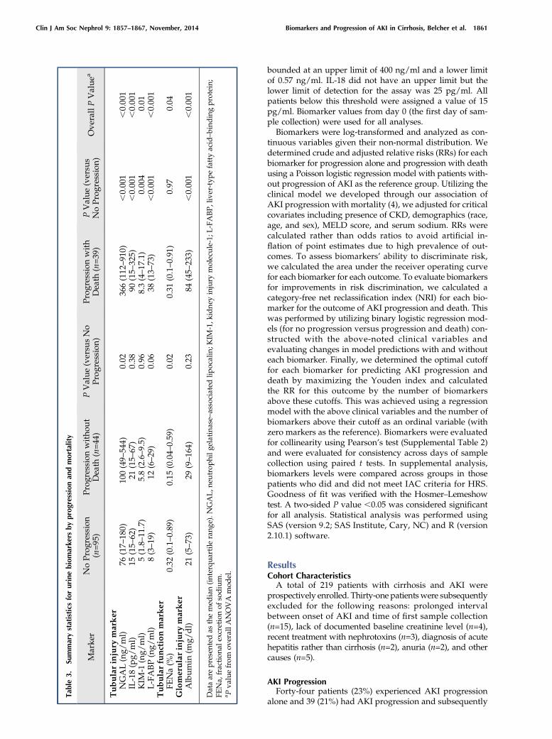
bounded at an upper limit of 400 ng/ml and a lower limitof 0.57 ng/ml. IL-18 did not have an upper limit but thelower limit of detection for the assay was 25 pg/ml. Allpatients below this threshold were assigned a value of 15pg/ml. Biomarker values from day 0 (the first day of sam-ple collection) were used for all analyses.Biomarkers were log-transformed and analyzed as con-
tinuous variables given their non-normal distribution. Wedetermined crude and adjusted relative risks (RRs) for eachbiomarker for progression alone and progression with deathusing a Poisson logistic regression model with patients with-out progression of AKI as the reference group. Utilizing theclinical model we developed through our association ofAKI progression with mortality (4), we adjusted for criticalcovariates including presence of CKD, demographics (race,age, and sex), MELD score, and serum sodium. RRs werecalculated rather than odds ratios to avoid artificial in-flation of point estimates due to high prevalence of out-comes. To assess biomarkers’ ability to discriminate risk,we calculated the area under the receiver operating curvefor each biomarker for each outcome. To evaluate biomarkersfor improvements in risk discrimination, we calculated acategory-free net reclassification index (NRI) for each bio-marker for the outcome of AKI progression and death. Thiswas performed by utilizing binary logistic regression mod-els (for no progression versus progression and death) con-structed with the above-noted clinical variables andevaluating changes in model predictions with and withouteach biomarker. Finally, we determined the optimal cutofffor each biomarker for predicting AKI progression anddeath by maximizing the Youden index and calculatedthe RR for this outcome by the number of biomarkersabove these cutoffs. This was achieved using a regressionmodel with the above clinical variables and the number ofbiomarkers above their cutoff as an ordinal variable (withzero markers as the reference). Biomarkers were evaluatedfor collinearity using Pearson’s test (Supplemental Table 2)and were evaluated for consistency across days of samplecollection using paired t tests. In supplemental analysis,biomarkers levels were compared across groups in thosepatients who did and did not meet IAC criteria for HRS.Goodness of fit was verified with the Hosmer–Lemeshowtest. A two-sided P value ,0.05 was considered significantfor all analysis. Statistical analysis was performed usingSAS (version 9.2; SAS Institute, Cary, NC) and R (version2.10.1) software.
ResultsCohort CharacteristicsA total of 219 patients with cirrhosis and AKI were
prospectively enrolled. Thirty-one patientswere subsequentlyexcluded for the following reasons: prolonged intervalbetween onset of AKI and time of first sample collection(n=15), lack of documented baseline creatinine level (n=4),recent treatment with nephrotoxins (n=3), diagnosis of acutehepatitis rather than cirrhosis (n=2), anuria (n=2), and othercauses (n=5).
AKI ProgressionForty-four patients (23%) experienced AKI progression
alone and 39 (21%) had AKI progression and subsequently
Tab
le3.
Summarystatistics
forurinebiomarke
rsbyprogressionan
dmortality
Marke
rNoProg
ression
(n=95
)Prog
ressionwitho
ut
Death
(n=44
)PValue
(versu
sNo
Prog
ression)
Prog
ressionwith
Death
(n=39
)PValue
(versu
sNoProg
ression)
Ove
rallPValue
a
Tubularinjury
marker
NGAL(ng/
ml)
76(17–
180)
100(49–
544)
0.02
366(112
–91
0),0.00
1,0.00
1IL-18(pg/
ml)
15(15–
62)
21(15–
67)
0.38
90(15–
325)
,0.00
1,0.00
1KIM
-1(ng/
ml)
5(1.8–11
.7)
5.8(2.6–9.5)
0.96
8.3(4–17
.1)
0.00
40.01
L-FABP(ng/
ml)
8(3–19
)12
(6–29
)0.06
38(13–
73)
,0.00
1,0.00
1Tubularfu
nctionmarker
FENa(%
)0.32
(0.1–0.89
)0.15
(0.04–
0.59
)0.02
0.31
(0.1–0.91
)0.97
0.04
Glomerularinjury
marker
Album
in(m
g/dl)
21(5–73
)29
(9–16
4)0.23
84(45–
233)
,0.00
1,0.00
1
Dataarepresen
tedas
themed
ian(interqu
artile
rang
e).N
GAL,n
eutrop
hilg
elatinase–associated
lipocalin;K
IM-1,k
idne
yinjury
molecule-1;
L-FABP,liver-typ
efattyacid–bind
ingprotein;
FENa,
fraction
alexcretionof
sodium.
a Pva
luefrom
overallA
NOVA
mod
el.
Clin J Am Soc Nephrol 9: 1857–1867, November, 2014 Biomarkers and Progression of AKI in Cirrhosis, Belcher et al. 1861

died during their hospitalization. Ten patients (5%) diedwithout progression and were excluded from the primaryanalysis. Baseline demographic, clinical, and laboratorydata of the entire cohort and of those patients with andwithout AKI progression are shown in Tables 1 and 2.Neither baseline GFR nor the presence of CKD varied be-tween the three groups. The D creatinine between baselineand admission did not differ between the three groups (0.8mg/dl versus 0.8 versus 0.5; P=0.28). Baseline proteinuriawas only present in 22 patients (12%) and was similaracross groups. The majority of patients had decompensatedcirrhosis as evidenced by the history of ascites (76%), hepaticencephalopathy (67%), variceal bleeding (23%), and spon-taneous bacterial peritonitis (16%). Reasons for admission
were similar across groups but rates of urinary tract infec-tions and pneumonia differed during the course of hospi-talization. The median Child–Pugh score was 10.5 and themedian MELD score was 26.3. Both Child–Pugh (12 versus10 versus 10; P,0.001) and MELD scores (34.3 versus 26.5versus 22.1; P,0.001) were higher in those patients who pro-gressed and died than in those with progression alone orthose without progression.Three urine samples were collected in 134 participants
(71%), two samples in 42 participants (22%), and only onesample was collected in 12 participants (6%). The first sam-ple was collected at a median 2 days (IQR, 1–3) after firstmeeting AKIN criteria. Median values for biomarkers areshown in Table 3. Sensitivity analysis using raw biomarker
Figure 1. | Biomarker levels for patients with no progression, progression alone, and progression with death. Biomarker values are presentedfor patients who did not have progression of AKI, those who had progression alone and those with progression and death. Data are presented asbox plots with the horizontal black line representing the median and the shaded region representing the interquartile range. Blue bars depictpatientswithout progression (n=95), green bars are patientswith progression alone (n=44), and red bars are patientswith progression and death(n=35). *Groups in which the biomarker level is statistically higher than in patients without progression; #Groups in which the biomarker levelis significantly higher than patients with progression alone. KIM-1, kidney injury molecule-1; L-FABP, liver-type fatty acid–binding protein;NGAL, neutrophil gelatinase–associated lipocalin.
1862 Clinical Journal of the American Society of Nephrology
values and those corrected for urine creatinine showedminimal variation (data not shown). To facilitate cross-studycomparison of results, NGAL, IL-18, KIM-1, and L-FABP aretherefore presented as raw values. Log-transformed bio-markers demonstrated moderate correlations between eachother (Supplemental Table 2).Median values for all biomarkers varied across the three
groups. Tubular injury markers were highest in patientswith progression and death. Although all tubular injurymarkers distinguished patients with progression and deathfrom no progression, only NGAL distinguished progres-sion alone from no progression. Microalbuminuria washigher in patients with progression and death than thosewith progression alone or no progression (84 mg/dl [IQR,45–233] versus 29 [IQR, 9–164] versus 21 [IQR, 5–73]), re-spectively, and this distinction persisted when correctingfor urinary creatinine. FENa was significantly lower in pa-tients with progression alone but did not differ betweenthose without progression and those with progression anddeath. In subgroup analysis, similar trends were seen inpatients who did not meet IAC criteria for HRS but not inthose who did (Supplemental Table 3). The medians and
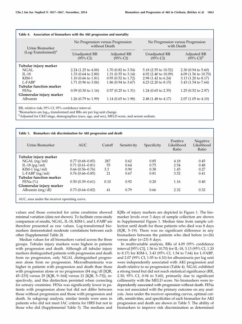
IQRs of injury markers are depicted in Figure 1. The bio-marker levels over 3 days of sample collection are shownin Supplemental Figure 1. Median time from sample col-lection until death for those patients who died was 8 days(IQR, 5–19). There was no significant difference in anybiomarkers between the patients who died before (n=26)versus after (n=23) 8 days.In multivariable analysis, RRs of 4.09 (95% confidence
interval [95% CI], 1.56 to 10.70) for IL-18, 3.13 (95% CI, 1.20to 8.17) for KIM-1, 3.43 (95% CI, 1.54 to 7.64) for L-FABP,and 2.07 (95% CI, 1.05 to 4.10) for albuminuria per log unitwere independently associated with AKI progression anddeath relative to no progression (Table 4). NGAL exhibiteda strong trend but did not reach statistical significance (RR,2.30; 95% CI, 0.94 to 5.60), primarily due to significantcollinearity with the MELD score. No biomarkers were in-dependently associated with progression without death. FENawas not associated with the primary outcome on any anal-ysis. Area under the receiver operating curves, optimal cut-offs, sensitivities, and specificities of each biomarker for AKIprogression and death are shown in Table 5. The ability ofbiomarkers to improve risk discrimination as determined
Table 4. Association of biomarkers with the AKI progression and mortality
Urine Biomarker(Log-Transformed)a
No Progression versus Progressionwithout Death
No Progression versus Progressionwith Death
Unadjusted RR(95% CI)
Adjusted RR(95% CI)
Unadjusted RR(95% CI)
Adjusted RR(95% CI)b
Tubular injury markerNGAL 2.24 (1.25 to 4.00) 1.70 (0.82 to 3.54) 5.18 (2.55 to 10.52) 2.30 (0.94 to 5.60)IL-18 1.33 (0.64 to 2.80) 1.31 (0.55 to 3.14) 4.92 (2.40 to 10.09) 4.09 (1.56 to 10.70)KIM-1 1.10 (0.66 to 1.81) 0.95 (0.52 to 1.72) 2.98 (1.42 to 6.24) 3.13 (1.20 to 8.17)L-FABP 1.71 (0.96 to 3.06) 1.86 (0.94 to 3.67) 4.23 (2.20 to 8.15) 3.43 (1.54 to 7.64)
Tubular function markerFENa 0.59 (0.30 to 1.16) 0.57 (0.25 to 1.31) 1.24 (0.65 to 2.35) 1.25 (0.52 to 2.97)
Glomerular injury markerAlbumin 1.26 (0.79 to 1.99) 1.14 (0.65 to 1.98) 2.48 (1.48 to 4.17) 2.07 (1.05 to 4.10)
RR, relative risk; 95% CI, 95% confidence interval.aBiomarkers are log10 transformed and RRs are per log-unit change.bAdjusted for CKD stage, demographics (race, age, and sex), MELD score, and serum sodium.
Table 5. Biomarkers risk discrimination for AKI progression and death
Urine Biomarker AUC Cutoff Sensitivity SpecificityPositive
LikelihoodRatio
NegativeLikelihood
Ratio
Tubular injury markerNGAL (ng/ml) 0.77 (0.68–0.85) 287 0.62 0.85 4.18 0.45IL-18 (pg/ml) 0.71 (0.61–0.81) 55 0.64 0.75 2.54 0.48KIM-1 (ng/ml) 0.66 (0.56–0.76) 3.3 0.90 0.38 1.45 0.27L-FABP (ng/ml) 0.76 (0.66–0.85) 21 0.67 0.81 3.52 0.41
Tubular function markerFENa (%) 0.50 (0.39–0.61) 0.10 0.92 0.20 1.16 0.40
Glomerular injury markerAlbumin (mg/dl) 0.73 (0.64–0.82) 41 0.79 0.66 2.32 0.32
AUC, area under the receiver operating curve.
Clin J Am Soc Nephrol 9: 1857–1867, November, 2014 Biomarkers and Progression of AKI in Cirrhosis, Belcher et al. 1863
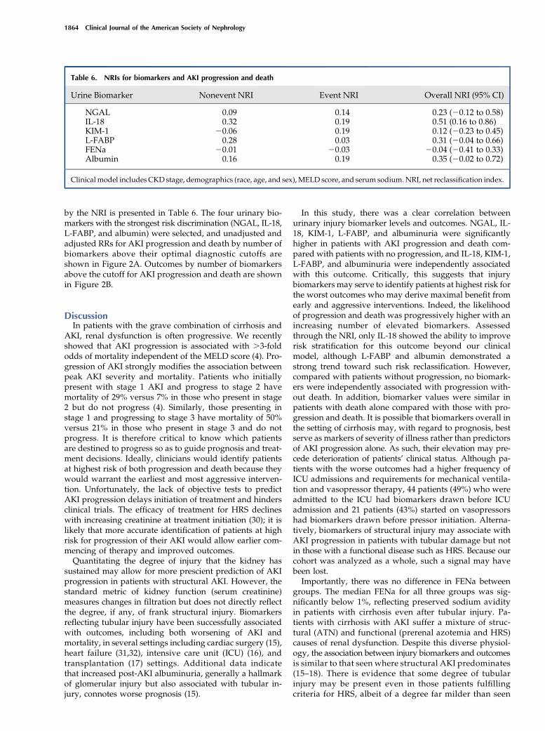
by the NRI is presented in Table 6. The four urinary bio-markers with the strongest risk discrimination (NGAL, IL-18,L-FABP, and albumin) were selected, and unadjusted andadjusted RRs for AKI progression and death by number ofbiomarkers above their optimal diagnostic cutoffs areshown in Figure 2A. Outcomes by number of biomarkersabove the cutoff for AKI progression and death are shownin Figure 2B.
DiscussionIn patients with the grave combination of cirrhosis and
AKI, renal dysfunction is often progressive. We recentlyshowed that AKI progression is associated with .3-foldodds of mortality independent of the MELD score (4). Pro-gression of AKI strongly modifies the association betweenpeak AKI severity and mortality. Patients who initiallypresent with stage 1 AKI and progress to stage 2 havemortality of 29% versus 7% in those who present in stage2 but do not progress (4). Similarly, those presenting instage 1 and progressing to stage 3 have mortality of 50%versus 21% in those who present in stage 3 and do notprogress. It is therefore critical to know which patientsare destined to progress so as to guide prognosis and treat-ment decisions. Ideally, clinicians would identify patientsat highest risk of both progression and death because theywould warrant the earliest and most aggressive interven-tion. Unfortunately, the lack of objective tests to predictAKI progression delays initiation of treatment and hindersclinical trials. The efficacy of treatment for HRS declineswith increasing creatinine at treatment initiation (30); it islikely that more accurate identification of patients at highrisk for progression of their AKI would allow earlier com-mencing of therapy and improved outcomes.Quantitating the degree of injury that the kidney has
sustained may allow for more prescient prediction of AKIprogression in patients with structural AKI. However, thestandard metric of kidney function (serum creatinine)measures changes in filtration but does not directly reflectthe degree, if any, of frank structural injury. Biomarkersreflecting tubular injury have been successfully associatedwith outcomes, including both worsening of AKI andmortality, in several settings including cardiac surgery (15),heart failure (31,32), intensive care unit (ICU) (16), andtransplantation (17) settings. Additional data indicatethat increased post-AKI albuminuria, generally a hallmarkof glomerular injury but also associated with tubular in-jury, connotes worse prognosis (15).
In this study, there was a clear correlation betweenurinary injury biomarker levels and outcomes. NGAL, IL-18, KIM-1, L-FABP, and albuminuria were significantlyhigher in patients with AKI progression and death com-pared with patients with no progression, and IL-18, KIM-1,L-FABP, and albuminuria were independently associatedwith this outcome. Critically, this suggests that injurybiomarkers may serve to identify patients at highest risk forthe worst outcomes who may derive maximal benefit fromearly and aggressive interventions. Indeed, the likelihoodof progression and death was progressively higher with anincreasing number of elevated biomarkers. Assessedthrough the NRI, only IL-18 showed the ability to improverisk stratification for this outcome beyond our clinicalmodel, although L-FABP and albumin demonstrated astrong trend toward such risk reclassification. However,compared with patients without progression, no biomark-ers were independently associated with progression with-out death. In addition, biomarker values were similar inpatients with death alone compared with those with pro-gression and death. It is possible that biomarkers overall inthe setting of cirrhosis may, with regard to prognosis, bestserve as markers of severity of illness rather than predictorsof AKI progression alone. As such, their elevation may pre-cede deterioration of patients’ clinical status. Although pa-tients with the worse outcomes had a higher frequency ofICU admissions and requirements for mechanical ventila-tion and vasopressor therapy, 44 patients (49%) who wereadmitted to the ICU had biomarkers drawn before ICUadmission and 21 patients (43%) started on vasopressorshad biomarkers drawn before pressor initiation. Alterna-tively, biomarkers of structural injury may associate withAKI progression in patients with tubular damage but notin those with a functional disease such as HRS. Because ourcohort was analyzed as a whole, such a signal may havebeen lost.Importantly, there was no difference in FENa between
groups. The median FENa for all three groups was sig-nificantly below 1%, reflecting preserved sodium avidityin patients with cirrhosis even after tubular injury. Pa-tients with cirrhosis with AKI suffer a mixture of struc-tural (ATN) and functional (prerenal azotemia and HRS)causes of renal dysfunction. Despite this diverse physiol-ogy, the association between injury biomarkers and outcomesis similar to that seen where structural AKI predominates(15–18). There is evidence that some degree of tubularinjury may be present even in those patients fulfillingcriteria for HRS, albeit of a degree far milder than seen
Table 6. NRIs for biomarkers and AKI progression and death
Urine Biomarker Nonevent NRI Event NRI Overall NRI (95% CI)
NGAL 0.09 0.14 0.23 (20.12 to 0.58)IL-18 0.32 0.19 0.51 (0.16 to 0.86)KIM-1 20.06 0.19 0.12 (20.23 to 0.45)L-FABP 0.28 0.03 0.31 (20.04 to 0.66)FENa 20.01 20.03 20.04 (20.41 to 0.33)Albumin 0.16 0.19 0.35 (20.02 to 0.72)
Clinical model includes CKD stage, demographics (race, age, and sex),MELD score, and serum sodium.NRI, net reclassification index.
1864 Clinical Journal of the American Society of Nephrology

Figure 2. | Association between the biomarker panel and AKI progression and death, as well as biomarker elevation and outcomes. (A)Association between the number of biomarkers above their optimal cutoff for AKI progression and death and the unadjusted and adjustedrelative risk for this outcome. All values are relative to having nomarkers over their cutoffs (n=65). Markers used in the panel includeNGAL, IL-18, L-FABP, and albumin. The adjusted model is adjusted for CKD stage, demographics (race, age, and sex), MELD score, and serum sodium.The 95% confidence intervals for adjusted relative risks are as follows: one marker, 1.21 to 10.67; two markers, 1.25 to 11.40; three markers,1.62 to 13.72; and four markers, 2.32 to 20.46. Biomarker cutoffs are as follows: NGAL, 287 ng/ml; IL-18, 55 pg/ml; L-FABP, 21 ng/ml; andalbumin, 41 mg/dl. (B) Association between biomarker elevation and outcomes. The percentage of patients without AKI progression or death,progression alone and progression and death by the number of biomarkers of structural injury above their optimal cutoff for prediction ofprogression and death. Biomarkers included in the panel include NGAL, IL-18, L-FABP, and albumin. MELD, model of end stage liver disease.
Clin J Am Soc Nephrol 9: 1857–1867, November, 2014 Biomarkers and Progression of AKI in Cirrhosis, Belcher et al. 1865
with ATN (21). It is striking then that, along with thenegative findings regarding FENa, our results suggestthe primacy of structural injury in determining out-comes when generally applied to a cohort composed ofpatients with cirrhosis and both “functional” and “structural”AKI.This study has several important strengths. Unlike many
studies of AKI and cirrhosis, it is not restricted to ICUpatients, improving the generalizability of the findings. Thesize of this cohort is one of the largest in the literature forthis difficult-to-study population. The evaluation of mul-tiple biomarkers is critical in cirrhosis in which AKI isphysiologically distinct from other settings such as surgery,sepsis, or the ICU. Finally, the prospective design allowedfor robust and complete data collection on multiple criticalcovariates.Our study is not without limitations. The cause of AKI
was not considered and thus patients likely suffered from amix of prerenal azotemia, ATN, and HRS. However,accurate adjudication of the cause of AKI must frequentlybe done retrospectively and thus would not be available toclinicians at the time of biomarker measurement. Poten-tially divergent associations by the cause of AKI betweenbiomarkers and outcomes would, if anything, be expectedto bias our results toward the null. The use of outpatientvalues for baseline creatinine results in the least misclas-sification of AKI incidence, severity, and prognosis, but thisapproach does mean that the exact timeframe for a rise increatinine is unknown (27). Kidney injury biomarkers inhospitalized patients with cirrhosis without AKI are onlyminimally above normal ranges (J.M. Belcher, G. Garcia-Tsao, A.J. Sanyal, H. Thiessen-Philbrook, A.J. Peixoto,M.A. Perazella, N. Ansari, J. Lim, S.G. Coca, and C.R. Parikh,unpublished data) and the significantly elevated valueseven in those patients who do not experience progressionor death therefore suggests that AKI is indeed ongoing athospital admission.In conclusion, this study confirms that multiple structural
biomarkers of kidney injury, but not FENa, are indepen-dently associated with progression of AKI and mortalityin patients with cirrhosis. Elevated injury markers wereseen in patients who ultimately progressed and died, butlevels were similar between those without progressionand those with progression alone. Further research in alarger cohort is required to validate this finding and todetermine whether biomarkers may identify patients withcirrhosis most likely to benefit from disease-specific AKItreatments.
AcknowledgmentsThe biomarker assays for KIM-1 and L-FABP were donated by
Seikisui Diagnostics. Neither the granting agency nor Seikisui Di-agnostics participated in the protocol development, analysis, orinterpretation of results.
The research reported in this articlewas fundedbygrants from theNational Institutes of Health (NIH) National Institute of Diabetesand Digestive and Kidney Diseases (1R21-DK078714 to C.R.P., sup-ported by NIH P-30DK034989) and the George M. O’Brien KidneyCenter at Yale (P30-DK079310). J.M.B. was supported by an insti-tutional fellowship training grant from the NIH. C.R.P. is the co-inventor on the IL-18 patent (no commercial value) licensed to theUniversity of Colorado.
Drs. Isabel Butrymowicz and Harjit Bhogal are collaborators ofthe Translational Research Investigating Biomarker Endpoints inAKI Consortium.
DisclosuresNone.
References1. Arabi Y, Ahmed QA, Haddad S, Aljumah A, Al-Shimemeri A:
Outcome predictors of cirrhosis patients admitted to the in-tensive care unit. Eur J Gastroenterol Hepatol 16: 333–339, 2004
2. Cardenas A,Gines P, Uriz J, Bessa X, Salmeron JM,Mas A,OrtegaR, Calahorra B, De Las Heras D, Bosch J, Arroyo V, Rodes J: Renalfailure after upper gastrointestinal bleeding in cirrhosis: In-cidence, clinical course, predictive factors, and short-termprognosis. Hepatology 34: 671–676, 2001
3. Tandon P, Garcia-Tsao G: Renal dysfunction is the most impor-tant independent predictor of mortality in cirrhotic patients withspontaneous bacterial peritonitis. Clin Gastroenterol Hepatol 9:260–265, 2011
4. Belcher JM, Garcia-Tsao G, Sanyal AJ, Bhogal H, Lim JK, AnsariN, Coca SG, Parikh CR; TRIBE-AKI Consortium: Association ofAKI with mortality and complications in hospitalized patientswith cirrhosis. Hepatology 57: 753–762, 2013
5. Jenq CC, Tsai MH, Tian YC, Lin CY, Yang C, Liu NJ, Lien JM, ChenYC, Fang JT, Chen PC, Yang CW: RIFLE classification can predictshort-term prognosis in critically ill cirrhotic patients. IntensiveCare Med 33: 1921–1930, 2007
6. Cholongitas E, Calvaruso V, Senzolo M, Patch D, Shaw S,O’Beirne J, Burroughs AK: RIFLE classification as predictivefactor of mortality in patients with cirrhosis admitted to intensivecare unit. J Gastroenterol Hepatol 24: 1639–1647, 2009
7. de Carvalho JR, Villela-Nogueira CA, Luiz RR, Guzzo PL, da SilvaRosa JM, Rocha E, Moraes Coelho HS, de Mello Perez R: Acutekidney injury network criteria as a predictor of hospital mortalityin cirrhotic patients with ascites. J Clin Gastroenterol 46: e21–e26, 2012
8. du Cheyron D, Bouchet B, Parienti JJ, Ramakers M, CharbonneauP: The attributable mortality of acute renal failure in critically illpatients with liver cirrhosis. Intensive Care Med 31: 1693–1699,2005
9. Dobre M, Demirjian S, Sehgal AR, Navaneethan SD: Terlipressinin hepatorenal syndrome: A systematic review andmeta-analysis.Int Urol Nephrol 43: 175–184, 2011
10. Appenrodt B, Zielinski J, Brensing KA, Heller J, Sauerbruch T,Schepke M: Degree of hepatic dysfunction and improvement ofrenal function predict survival in patients with HRS type I: Aretrospective analysis. Eur J Gastroenterol Hepatol 21: 1428–1432, 2009
11. Colle I, Durand F, Pessione F, Rassiat E, Bernuau J, Barriere E,LebrecD, VallaDC,Moreau R: Clinical course, predictive factorsand prognosis in patients with cirrhosis and type 1 hepatorenalsyndrome treated with Terlipressin: A retrospective analysis.J Gastroenterol Hepatol 17: 882–888, 2002
12. Northup PG, Argo CK, Bakhru MR, Schmitt TM, Berg CL, RosnerMH: Pretransplant predictors of recovery of renal function afterliver transplantation. Liver Transpl 16: 440–446, 2010
13. Nadim MK, Sung RS, Davis CL, Andreoni KA, Biggins SW,Danovitch GM, Feng S, Friedewald JJ, Hong JC, Kellum JA, KimWR, Lake JR, Melton LB, Pomfret EA, Saab S, Genyk YS: Simul-taneous liver-kidney transplantation summit: Current state andfuture directions. Am J Transplant 12: 2901–2908, 2012
14. Francoz C, Prie D, Abdelrazek W, Moreau R, Mandot A, BelghitiJ, Valla D, Durand F: Inaccuracies of creatinine and creatinine-based equations in candidates for liver transplantation with lowcreatinine: Impact on themodel for end-stage liver disease score.Liver Transpl 16: 1169–1177, 2010
15. Koyner JL, Garg AX, Coca SG, Sint K, Thiessen-Philbrook H, PatelUD, Shlipak MG, Parikh CR; TRIBE-AKI Consortium: Biomarkerspredict progression of acute kidney injury after cardiac surgery.J Am Soc Nephrol 23: 905–914, 2012
16. Siew ED, Ikizler TA, Gebretsadik T, Shintani A, Wickersham N,Bossert F, Peterson JF, Parikh CR, May AK, Ware LB: Elevated
1866 Clinical Journal of the American Society of Nephrology

urinary IL-18 levels at the time of ICU admission predict adverseclinical outcomes. Clin J Am Soc Nephrol 5: 1497–1505, 2010
17. Hall IE, Yarlagadda SG, Coca SG,Wang Z, DoshiM,Devarajan P,Han WK, Marcus RJ, Parikh CR: IL-18 and urinary NGAL predictdialysis and graft recovery after kidney transplantation. J Am SocNephrol 21: 189–197, 2010
18. Wagener G, Minhaz M, Mattis FA, Kim M, Emond JC, Lee HT:Urinary neutrophil gelatinase-associated lipocalin as a marker ofacute kidney injury after orthotopic liver transplantation.NephrolDial Transplant 26: 1717–1723, 2011
19. Portal AJ, McPhail MJ, Bruce M, Coltart I, Slack A, Sherwood R,Heaton ND, Shawcross D, Wendon JA, Heneghan MA: Neutro-phil gelatinase—associated lipocalin predicts acute kidney in-jury in patients undergoing liver transplantation. Liver Transpl16: 1257–1266, 2010
20. Fagundes C, Pepin MN, Guevara M, Barreto R, Casals G, Sola E,Pereira G, Rodrıguez E, Garcia E, Prado V, Poch E, Jimenez W,Fernandez J,ArroyoV,GinesP:Urinaryneutrophilgelatinase-associatedlipocalin as biomarker in the differential diagnosis of impairment ofkidney function in cirrhosis. J Hepatol 57: 267–273, 2012
21. Verna EC, Brown RS, Farrand E, Pichardo EM, Forster CS, Sola-Del Valle DA, Adkins SH, Sise ME, Oliver JA, Radhakrishnan J,Barasch JM, Nickolas TL: Urinary neutrophil gelatinase-associatedlipocalin predicts mortality and identifies acute kidney injury incirrhosis. Dig Dis Sci 57: 2362–2370, 2012
22. Slack AJ, McPhail MJ, Ostermann M, Bruce M, Sherwood R,Musto R, Dew T, Auzinger G, Bernal W, O’Grady J, HeneghanMA, Moore K, Wendon JA: Predicting the development of acutekidney injury in liver cirrhosis—an analysis of glomerular filtra-tion rate, proteinuria and kidney injury biomarkers. AlimentPharmacol Ther 37: 989–997, 2013
23. Mishra J, Dent C, Tarabishi R, Mitsnefes MM,Ma Q, Kelly C, RuffSM, Zahedi K, Shao M, Bean J, Mori K, Barasch J, Devarajan P:Neutrophil gelatinase-associated lipocalin (NGAL) as a bio-marker for acute renal injury after cardiac surgery. Lancet 365:1231–1238, 2005
24. Shibata M, Hirota M, Nozawa F, Okabe A, Kurimoto M, OgawaM: Increased concentrations of plasma IL-18 in patients withhepatic dysfunction after hepatectomy. Cytokine 12: 1526–1530, 2000
25. Parikh CR, Thiessen-Philbrook H, Garg AX, Kadiyala D, ShlipakMG, Koyner JL, Edelstein CL, Devarajan P, Patel UD, ZappitelliM, Krawczeski CD, Passik CS, Coca SG; TRIBE-AKI Consortium:Performance of kidney injury molecule-1 and liver fatty acid-
binding protein and combined biomarkers of AKI after cardiacsurgery. Clin J Am Soc Nephrol 8: 1079–1088, 2013
26. Wong F, Nadim MK, Kellum JA, Salerno F, Bellomo R, Gerbes A,Angeli P, Moreau R, Davenport A, Jalan R, Ronco C, Genyk Y,Arroyo V: Working Party proposal for a revised classificationsystem of renal dysfunction in patients with cirrhosis. Gut 60:702–709, 2011
27. Siew ED, Matheny ME, Ikizler TA, Lewis JB, Miller RA, WaitmanLR, Go AS, Parikh CR, Peterson JF: Commonly used surrogates forbaseline renal function affect the classification and prognosis ofacute kidney injury. Kidney Int 77: 536–542, 2010
28. Levey AS, Bosch JP, Lewis JB, Greene T, Rogers N, Roth D;Modification of Diet in Renal Disease Study Group: A more ac-curate method to estimate glomerular filtration rate from serumcreatinine: A new prediction equation. Ann Intern Med 130:461–470, 1999
29. Salerno F, Gerbes A, Gines P, Wong F, Arroyo V: Diagnosis,prevention and treatment of hepatorenal syndrome in cirrhosis.Gut 56: 1310–1318, 2007
30. Boyer TD, Sanyal AJ, Garcia-Tsao G, Blei A, Carl D, Bexon AS,Teuber P; Terlipressin Study Group: Predictors of response toterlipressin plus albumin in hepatorenal syndrome (HRS) type 1:Relationship of serumcreatinine to hemodynamics. J Hepatol 55:315–321, 2011
31. Aghel A, Shrestha K, Mullens W, Borowski A, Tang WH: Serumneutrophil gelatinase-associated lipocalin (NGAL) in predictingworsening renal function in acute decompensated heart failure. JCard Fail 16: 49–54, 2010
32. Haase M, Devarajan P, Haase-Fielitz A, Bellomo R, Cruz DN,Wagener G, Krawczeski CD, Koyner JL, Murray P, Zappitelli M,Goldstein SL, Makris K, Ronco C, Martensson J, Martling CR,Venge P, Siew E,Ware LB, Ikizler TA,Mertens PR: The outcome ofneutrophil gelatinase-associated lipocalin-positive subclinicalacute kidney injury: Amulticenter pooled analysis of prospectivestudies. J Am Coll Cardiol 57: 1752–1761, 2011
Received: September 11, 2013 Accepted: July 28, 2014
Published online ahead of print. Publication date available at www.cjasn.org.
This article contains supplemental material online at http://cjasn.asnjournals.org/lookup/suppl/doi:10.2215/CJN.09430913/-/DCSupplemental.
Clin J Am Soc Nephrol 9: 1857–1867, November, 2014 Biomarkers and Progression of AKI in Cirrhosis, Belcher et al. 1867
Related Documents