Urinalysis Tidbits

Urinalysis Tidbits
Dec 31, 2015
Urinalysis Tidbits. Stains. Sternheimer-Malbin Crystal Violet and Safranin O Lipid Stains Oil Red O and Sudan III Stain Trigs and Neutral Fat but Not Cholesterol Cholesterol polarizes Cholesterol Drops Maltese Cross Cholesterol Crystals Birefringence. Microscopic Sediment Prep. - PowerPoint PPT Presentation
Welcome message from author
This document is posted to help you gain knowledge. Please leave a comment to let me know what you think about it! Share it to your friends and learn new things together.
Transcript

Urinalysis Tidbits

Stains
• Sternheimer-Malbin
Crystal Violet and Safranin O
• Lipid Stains• Oil Red O and Sudan III • Stain Trigs and Neutral Fat but Not Cholesterol
Cholesterol polarizes• Cholesterol Drops Maltese Cross • Cholesterol Crystals Birefringence
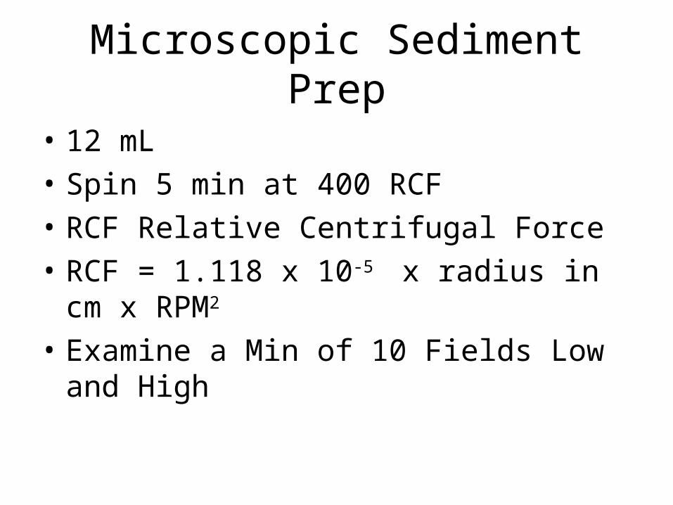
Microscopic Sediment Prep
• 12 mL
• Spin 5 min at 400 RCF
• RCF Relative Centrifugal Force
• RCF = 1.118 x 10⁻⁵ x radius in cm x RPM2
• Examine a Min of 10 Fields Low and High

Microscopy
• Bright field
• Polarized
• Phase Contrast
• Interference

Red Blood Cells
• Pale red
• Ghost Cells Dilute or Alkaline Lyse
• Hypertonic Conditions Crenate
• 2% Acetic Acid Lyse
• Yeast and Calcium Oxalate do not disappear with 2 % acidic acid

WBC
• Granular and often visible nucleus
• Hypertonic will shrink
• Hypotonic “Glitter” cells associated with Infection and Irritation & Pyelonephritis
• Glitter cells show Brownian movement

Epithelial Cells
• Squamous Contamination
• Transitional Bladder
• Renal Tubular Renal Tubules

Squamous Cells
• Large, flat irregular shape• Small nucleus• Urethra and contamination• Not considered Clinically Sig

Clue Cell
• Vaginal Infection w Gardnerella vaginalis• Squamous cell covered w coccobacillus• Covers most of cell and extends beyond edges• Vaginal wet prep but may appear in urine

Renal Tubular Cell
• Slightly larger than WBC• Eccentric nucleus• Increased numbers Tubular damage• Pyelonephritis, Acute Tubular necrosis

Hemosiderin
• Not birefringent unlike urates or phosphates
• Prussian blue dye stains iron of Hb blue

Transitional Cells
• Smaller than Squamous• Several Forms Caudate, spherical and polyhedral• Central nucleus• Renal Pelvis, calyses, ureters and bladder• Not Clinical Sig catheterization

Starch granules
• Source Powdered Gloves
• Maltese cross Polarized light
• Look like a pouch

Crab
• Pediculosis pubis

Pinworm
• Enterobius vermicularis
• Ova one side flat
• Adult male

Schistosoma hematobium
• World wide problem Egypt, Iraq, Syria, Iran Africa
• US lacks the snail intermediate host• Trematode Terminal spine

Triple Phosphate
• Tri Phosphate going into solution

RTE

Mite
• Adult mite• Scabies• Smaller than crab

UA Web Sites
• Comprehensive Review
• http://www.aafp.org/afp/2005/0315/p1153.html
Related Documents