Upstate Medical University

Welcome message from author
This document is posted to help you gain knowledge. Please leave a comment to let me know what you think about it! Share it to your friends and learn new things together.
Transcript

Upstate Medical University

DNV requires physicians who use or operate fluoroscopic x-ray systems to be properly credentialed. This training shall include radiation safety, management of fluoroscopic radiation and operation of fluoroscope used by the physician. This is in addition to any specific training required to perform the clinical procedure.

The hospital requires documentation of appropriate training before granting fluoroscopic privileges.
In order to become credentialed, a physician must do the following: View this powerpoint presentation Complete the self-assessment quiz Obtain certificate from the radiation safety office. Present
this certificate to the department contact person. It is up to the each department to establish other additional
training and education requirements needed to obtain fluoroscopy privileges.

X-ray Production Units of Radiation Exposure and Absorbed
Dose Mobile C-arms Factors Influencing Fluoroscopy Exposure Rate Sources of Radiation Exposure Radiation Protection Personnel Monitoring Basic Radiation Biology Example of a Skin Injury from Fluoroscopy Radiation and Pregnancy
X-rays are produced in an x-ray tube when electrons are accelerated through a high voltage (50,000 – 150,000 volts or 50 - 150 kVp) and allowed to hit a target composed of high atomic number materials such as tungsten.

Electrons are released from an electrically heated filament and are accelerated to the target by the high voltage (kV). This flow of electrons from the filament to the target is known as the tube current (mA). Fluoroscopy is usually performed using an accelerating voltage of 70 to 120 kV.
The amount of x-rays produced is determined by the tube current (mA) and the high voltage (kV). X-ray production is directly proportional to the tube current therefore doubling the tube current (mA) doubles the # of x-rays produced at a particular kVp.
However, x-ray production increases more rapidly with kVp than mA therefore increasing the kV by 15% is equivalent to doubling the mA. NOTE: Higher kV values also provides a more penetrating x-ray beam
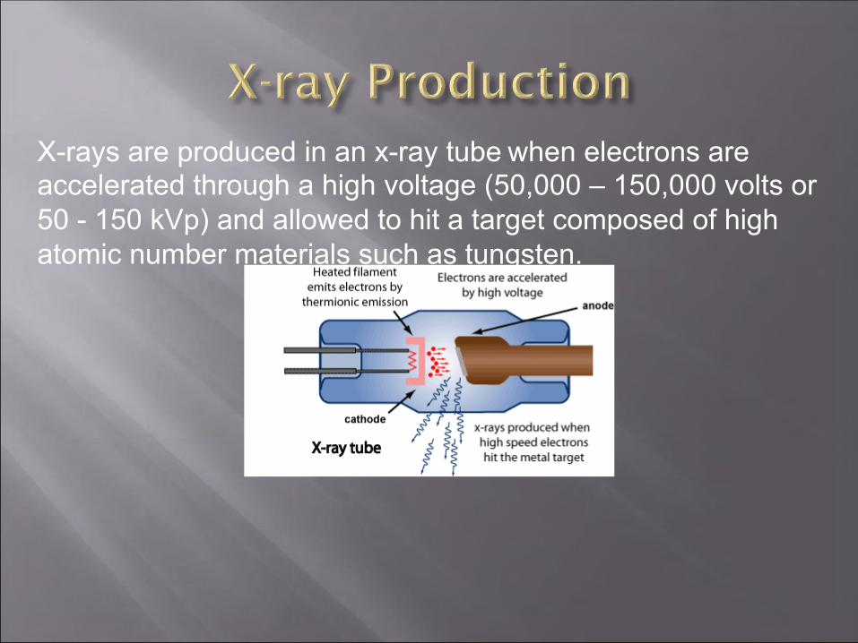
" Exposure – The quantity of x-rays or
gamma radiation required to produce an amount of ionization (electric charge) in air
– Units: Roentgen (R) – Usually expressed in terms
of exposure rate: i.e., R/hour or fluoroscopy output measured as R/minute.
" Absorbed Dose – The amount of ionizing
radiation energy absorbed per unit mass of tissue.
– Units: rad (radiation absorbed dose)
1 rad = 0.01 Joules/kg 1 rad = 0.01 Gray
– For x-rays used in fluoroscopy an exposure of 1R results in an absorbed dose of approximately 1 rad.

Effective Dose Equivalent (EDE) The comparative risk of potential health effects when
only a portion of the body is irradiated is smaller than when the whole body is exposed.
The EDE is a calculation of the risk to an individual posed by a partial body irradiation, such as exposure of the patient during a fluoroscopic examination
The EDE can also used to estimate the equivalent whole body exposure for fluoroscopy staff wearing protective aprons for comparison to annual personnel dose limits for radiation exposure

Moved around ORs to visualize anatomy during surgery
Metallic c-arm contains x-ray tube at one end and image receptor at the other
An associated cart contains the display

Modern fluoroscopy units produce images with an image intensifier (II) which brightens the image level sufficiently so that the image may be displayed on a TV screen. Fluoroscopy units are usually operated in an automatic brightness control (ABC) mode.
These units will automatically adjust the brightness by increasing the kV to increase x-ray penetration and increasing the mA to increase intensity.
Note: Exposure to a thick patient will be greater than to a thin patient and also abdominal fluoro will require a greater exposure than a chest fluoro due to increased thickness and tissue density in the abdomen.

Recommendation # 1: The image intensifier input should be positioned as close to the patient as practicable. This results in a lower patient dose and sharper image.

Recommendation # 2: Use the exposure pedal as sparingly as possible.
Radiation exposure during fluoroscopy is directly proportional to the length of time the unit is activated by the foot pedal. Depression of the foot pedal determines the length of exposure. The fluoroscopy time is an important determinant of patient and staff radiation dose. Fluoroscopy units are equipped with a timer and an alarm which sounds at the end of every 5 minutes of fluoroscopy time. The alarm serves as a reminder of the elapsed time.

Recommendation # 3: Use “last-image” hold and pulsed fluoro whenever possible.
All modern fluoro units are equipped with “last-image” hold, which stores the last fluoro image and allows viewing without having to expose the patient again. Many fluoro units also offer a “pulsed mode”, in which the x-ray beam is pulsed rapidly on and off and results in a lower radiation dose without significantly degrading the appearance of the image on the display.


Recommendation # 4: Use the smallest field of view practicable. Effective dose depends on x-ray field size. Keeping the x-ray field as small as possible (by using collimators) will decrease dose to BOTH the patient and staff in the fluoroscopy suite. Restricting the field size will also produce better image quality. The contrast between various tissue types will be greater for the smallest field of view possible.

Recommendation # 5: High dose or detail modes should be used only sparingly. Many fluoro units will have various dose modes, such as low dose, medium dose, high dose or boost mode.
It is important to recognize that fluoroscopic image quality can be adversely affected by too low of a dose; the image can be noisy with poor contrast. More tissue contrast is produced by the “high dose” mode which will improve the image quality at the expense of increased patient dose.

Recommendation # 6: Magnification should be used only when necessary.
Fluoroscopy units are capable of using different magnification modes. Image resolution is improved with magnification but patient radiation dose is increased. Patient dose is minimized by using the lowest magnification possible.
Under Normal mode, there is little magnification with the whole beam used to generate a bright image. Under Mag 1 mode, a smaller beam area is projected to the same II output. The resulting object size is larger, but the image is dimmer due to the less beam input. The ABC system senses the brightness loss and either boosts machine X-ray output, increases tube voltage, or a combination of both.


6 inch mag FOV increases dose by a factor of 4 over non-mag image
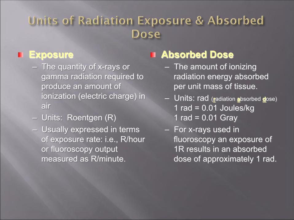
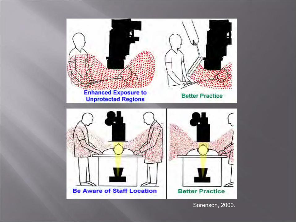
Recommendation # 7: For C-arm type fluoroscopy units the patient should be positioned as far from the x-ray tube as practicable to minimize patient entrance dose. To reduce personnel exposure the x-ray tube should be positioned beneath the patient.

In the case of portable C-Arm systems, eliminating the air gap between the I-I and the patient ensures that the table top is as far away as possible from the X-ray tube, minimizing radiation exposure to the patient’s skin.

In C-arm fluoroscopy the distance between the x-ray tube and image intensifier is fixed. The patient can be positioned in close proximity to the x-ray tube which increases the entrance skin dose drastically.
It is preferable to locate the C-arm x-ray tube underneath the patient. Since the radiation transmitted through the patient is typically only 5 – 10 % of the entrance dose, inadvertent exposure to the operator hand on the exit side of the patient will result in a smaller dose compared to the dose to the hand on the entrance side of the patient. Also the amount of scatter radiation the operator is exposed to on the beam exit side of the patient is significantly less than on the beam entrance side.

Note: The benefit is exaggerated - some operator dose occurs on the image intensifier (I-I) side.
Sorenson, 2000.
X-ray tube
X-ray tube

Care should be taken whenever the image view angle is changed during the procedure (e.g, changing from an ANT to a steep LAO). The I-I is often moved away from the patient while changing X-ray tube position. Large air gaps can result if the table or I-I height remains unadjusted.
Sorenson, 2000.
DIRECT EXPOSURE Entrance Skin Exposure (ESE) rates (where the x-rays enter the
patient) are limited to a maximum of 10 R/minute (NOTE: At ESE rates of 10 R/min, 30 minutes of fluoroscopy can deliver 300 R in skin dose.)
ESE rates for typical fluoroscopy procedures are usually less than 2 R/min.
On some machines an operator can deliberately choose a setting that will increase the output. The use of higher radiation rates or "boost" modes are useful in situations requiring high video image resolution. An ESE up to 20 R/min is permitted for a short time duration. Special operator reminders, such as audible alarms, are activated during "boost" modes.

SCATTER EXPOSURE TO PERSONNEL Almost all of the radiation
exposure received by the operator or other personnel in the fluoroscopy suite is due to scatter radiation from the patient.
The operator will be exposed to a dose rate of approximately one one-thousandth (1/1000) of the ESE rate at a distance of 1 meter from the center of the fluoroscopy field.
Sorenson, 2000.

Factors which increase the dose from scatter radiation: Size of the patient and angle of view: Larger patients will
cause the automatic brightness control (ABC) to adjust the kV and mA to higher values causing greater amounts of scatter radiation. Lateral views produce higher amounts of scatter than an A/P view.
Size of the x-ray field, a larger x-ray field will result in an increase in scatter radiation,
The length of time the fluoroscopy unit is on. Complex interventional cases will require greater procedure time, increasing dose to both the patient and operator

Other sources of exposure to the operator may be associated with the following: A very small percentage of exposure to the operator
may be due to leakage radiation through the x-ray tube housing.
C-arm operators should be aware that the shielding built in to “fixed” fluoroscopy systems is not available for protection against backscatter. This may be of greater concern if the C-arm is rotated out of the normal vertical plane.

The three most productive means of reducing radiation dose is: Time: Minimize time spent in the radiation field.
Use of “last-image-hold” and pulse fluoro features are technical advantages in reducing the total time x-rays are produced
Distance: Radiation dose rates increase or decrease according to the inverse square law Ex: Double your distance from the source and decrease your exposure by a factor of 4
Shielding: Use of lead garments, lead gloves, thyroid shields, leaded eyeglasses, lead drapes and clear leaded glass barriers between the patient and operator

Sorenson, 2000.

Even when radiation protection techniques and engineering controls are in place to reduce personnel exposure, individual dose monitoring is required.
Badges are assigned to an individual and must not be shared.
A badge designed to measure the whole body (torso including head) should be worn at the collar – OUTSIDE the lead apron.

Quarterly Investigational Levels (mrems)
Level I Level II Body Badge (DDE) 125 375 Collar Badge 125 375
Eye (LDE) 375 1125
Ring/Wrist (SDE) or Extremity 500 1,500
DDE = Deep Dose Equivalent
LDE = Lens Dose Equivalent
SDE = Shallow Dose Equivalent

Level I: Each incident will be noted on the personnel badge report by the Radiation Safety Office. A notification letter is sent to the employee.
Level II: The Radiation Safety Office will investigate each such incident. A report will be generated and the results of each investigation will be presented to the hospital radiation safety committee.
Physicians performing fluoroscopy that receive Level II collar badge readings will get a notification letter that includes their effective dose.

The effective dose equivalent (EDE) may be calculated in the following manner: A two-badge system (waist and collar badges) is used to calculate
an individual’s EDE by taking into account the protective factor of the lead apron. In this situation, one badge is worn OUTSIDE the lead apron (collar) and a second badge is worn UNDERNEATH the lead apron (waist). The EDE is calculated as follows: EDE = [1.5 x (waist)] + [0.04 x (collar)]
A one-badge system (collar badge ONLY) is where one badge is worn on the OUTSIDE of the lead apron. The EDE is calculated as follows: EDE = [0.3 x (collar)]

Dosimeters must be promptly turned in and exchanged each month to give accurate assessments.
Badge reports are reviewed by the Radiation Safety Office. Notification letters are sent to individuals who exceed monthly “Level I” exposure limits
Copies of badge reports are kept in the radiation safety office
If your badge is misplaced call the radiation safety office (464-6510) for a replacement.
Turn in badges that are found later, no matter how old.

RADIATION WORKERS ANNUAL LIMIT Whole Body 5,000 mrem
Lens of the Eye 15,000 mrem Skin, extremities 50,000 mrem
Embryo/Fetus 500 mrem gestation 50 mrem/month
For hospital radiation workers, annual doses rarely exceed 10 % of these values.

X-rays from fluoroscopy interact with biological materials by transferring their energy to an electron which subsequently interacts with the target molecule to produce an ion or a free radical.
Indirect action is the creation of free radicals from interactions with water molecules. Free radicals may then chemically interact with biologically sensitive molecules (DNA, RNA, proteins) causing damage
Direct action is the interaction of ionizing radiation with biologically sensitive molecules such as DNA causing direct destruction or mutation
Since water molecules are much more numerous than biologically sensitive molecules, indirect action is the most common form of biological damage.

Cells can sustain a variable amount of radiation and still repair themselves from sub-lethal damage.
Continuous high intensity radiation will produce greater damage than an equivalent fractionated (multiple smaller) dose since fractionation allows for cell repair.

A given organ’s response to radiation depends on: Total dose Dose rate Fractionated scheme Volume of irradiated tissue Inherent tissue radiation sensitivity
A large total dose, high dose rate and small fractionated schedule (which is all possible in fluoroscopy) will cause a greater degree of damage.
Major concern in fluoroscopy is the possibility of acute, direct or deterministic, radiation damage which manifests as a skin injury. The severity of skin injury is dose-dependent; more dose means more severe symptoms

Note that the time to expression of symptoms is long enough that the patient may no longer be in the hospital when symptoms appear. The physician performing the fluoroscopy cannot discern the damage by observing the patient immediately following the procedure.
FDA Specification of Radiation-Induced Skin Injuries
Threshold Dose Typical Fluoro-On Time in Minutes
Skin Effect rem Gy Normal mode @ 10 R/min
High Dose mode @ 20 R/min
Time to Onset
Early Transient Erythema 200 2 20 minutes 10 minutes Hours
Temporary Epilation 300 3 30 minutes 15 minutes 20 days
Basal Cell Erythema 600 6 60 minutes 30 minutes 10 days
Permanent Epilation 700 7 70 minutes 35 minutes 20 days
Dry Desquamation 1000 10 100 minutes 50 minutes 30 days

These threshold doses to cause an observable effect cannot be considered exact due to many variables such as individual biological response, age, characteristics of the individual exposed and the area exposed.
A patient may exceed the threshold dose without showing symptoms. This may be due to: The x-ray beam may not have been concentrated on a
single area of the skin for the entire time and because 10 R/min or 20 R/min are maximum outputs for very thick patients.

This case, patient A is that of a 40-year-old male who underwent coronary angiography, coronary angioplasty and a second angiography procedure due to complications, followed by a coronary artery by-pass graft, all on March 29, 1990. The area of injury six to eight weeks following the procedures. The injury was described as "turning red about one month after the procedure and peeling a week later."

In mid-May 1990, it had the appearance of a second-degree burn. The condition in late summer 1990, exact date unknown, with the appearance of a healed burn, except for a small ulcerated area present near the center.
Example of a Skin Injury from Fluoroscopy (cont.)
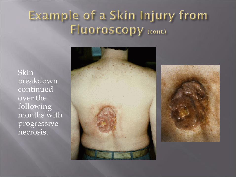
Skin breakdown continued over the following months with progressive necrosis.

The injury eventually required a skin graft. The magnitude of the skin dose received by this patient is not known. However, from the nature of the injury, it is probable that the dose exceeded 20 Gy.
Wagner LK, Eifel PJ, Geise RA. Potential Biological Effects Following High X-ray Dose Interventional Procedures. JVIR 1994; 5:71-8

Threshold dose below which no effect is observed Severity increases with dose Examples: skin erythema, dermatitis,
desquamation cataracts

Incidence increases with dose No dose threshold assumed Basis for ALARA principle of radiation protection Example: cancer

The decision to perform a radiological procedure on a patient who may be pregnant is a medical decision and shall be made by a physician in consultation with a radiologist and the patient.
Shielding shall be used to shield the abdomen from radiation provided it does not interfere with the procedure.
Every attempt must be made to minimize direct exposure to the fetus according to the principles described in this presentation.
Medical emergency radiological procedures however take precedence over pregnancy status.

PAUSE to properly plan and prepare for study Activate dose saving features of equipment No exposures unless necessary Depress last image hold and last image grab instead PULSE at lowest possible rate

PAUSE: Clinical indication, appropriateness of study, questions to be answered, unusual anatomy or prior surgery, and type of study to be performed should be clarified as much as possible. Explain procedure, risks and required immobilization to patient &/or parents. PULSE COLLIMATE / NO MAGNIFICATION: Bring the patient as close as possible to the image intensifier. Preset the collimators to the likely field of view and position the unit over the anatomic location of interest prior to beginning fluoroscopy STEP LIGHTLY: Step lightly on the fluoroscopy pedal. Use intermittent visualization only as needed. Most images obtained during the study can be screen saves without any additional radiation. If more detail is needed some images can be camera spots. FLUOROSCOPY TIME: Check fluoroscopic time used, document time/dose information as per the policy of hospital/ department
Any institution that uses radiation for diagnostic and/or therapeutic purposes must name a radiation expert as their Radiation Safety Officer (RSO). This individual is responsible for the day-to-day safe use of radiation at the institution.
The RSO at University Hospital is Tom LaVoy 464-5088 [email protected].
Related Documents