REVIEW Update on laboratory diagnosis of amoebiasis Syazwan Saidin 1,2 & Nurulhasanah Othman 1 & Rahmah Noordin 1 Received: 12 July 2018 /Accepted: 7 September 2018 /Published online: 25 September 2018 # Springer-Verlag GmbH Germany, part of Springer Nature 2018 Abstract Amoebiasis, an enteric protozoan disease caused by Entamoeba histolytica, is a public health problem in many developing countries, causing up to 100,000 fatal cases annually. Detection of the pathogenic E. histolytica and its differentiation from the non-pathogenic Entamoeba spp. play a crucial role in the clinical management of patients. Laboratory diagnosis of intestinal amoebiasis in developing countries still relies on labour-intensive and insensitive methods involving staining of stool sample and microscopy. Newer and more sensitive methods include a variety of antigen detection ELISAs and rapid tests; however, their diagnostic sensitivity and specificity seem to vary between studies, and some tests do not distinguish among the Entamoeba species. Molecular detection techniques are highly sensitive and specific and isothermal amplification approaches may be developed into field-applicable tests; however, cost is still a barrier for their use as a routine laboratory test method in most endemic areas. Laboratory diagnosis of extraintestinal amoebiasis faces challenges of lack of definitive detection of current infection and commercially available point-of-care tests. For both types of amoebiasis, there is still a need for highly sensitive and specific tests that are rapid and cost-effective for use in developing countries where the disease is prevalent. In recent years, new molecules of diagnostic value are being discovered and new tests developed. The advances in ‘omics’ technologies are enabling discoveries of new biomarkers that may help distinguish between different infection stages. Keywords Amoebiasis . Entamoeba histolytica . Entamoeba dispar . Laboratory . Diagnosis Introduction Amoebiasis is still a big challenge to public health in many regions, especially in the ‘bottom billion’ countries where poverty and low income is prevalent, and complex challenges are hindering their economic development. Areas with high rates of amoebic infection include parts of India, Bangladesh, tropical African countries, Brazil and Mexico, China, and South-east Asia [1, 2]. It is esti- mated to affect 50 million people worldwide and causes up to 100,000 deaths annually [1, 3]. Approximately 90% of infected individuals are asymptomatic carriers; the oth- er 10% show clinical symptoms such as colitis, dysentery and extraintestinal amoebiasis [3]. The most common clinical manifestation of extraintestinal infection is amoe- bic liver abscess (ALA) and a delay in diagnosis and treatment may cause fatality [4]. Despite the prevalence of amoebiasis, there is still no vaccine to prevent this disease [5]. Human infection is usually found in areas with poor sanitary conditions, inadequate water treatment and low socio-economic status. The only reservoir is hu- man, and infection occurs via food, water or hands con- taminated with cyst-containing fecal material. Human to human transmission has been reported through oral- genital and oral-anal contact, especially among homosex- uals [6] and those with poor personal hygiene [7]. Diagnosis of intestinal amoebiasis relies on clinical symptoms and laboratory test results. Continuous im- provement of health programmes, as well as monitoring and mapping the prevalence of amoebiasis is needed and this requires good diagnostic tools. This review describes the laboratory diagnosis of amoebiasis. Other than the conventional methods, a substantial amount of work has been carried out to develop new and improved serological and molecular diagnostic tests for both clinical and re- search purposes. * Rahmah Noordin [email protected] 1 Institute for Research in Molecular Medicine, Universiti Sains Malaysia, 11800 Penang, Malaysia 2 Department of Biology, Faculty of Science and Mathematics, Sultan Idris Education University, 35900 Tanjung Malim, Perak, Malaysia European Journal of Clinical Microbiology & Infectious Diseases (2019) 38:15–38 https://doi.org/10.1007/s10096-018-3379-3

Welcome message from author
This document is posted to help you gain knowledge. Please leave a comment to let me know what you think about it! Share it to your friends and learn new things together.
Transcript
REVIEW
Update on laboratory diagnosis of amoebiasis
Syazwan Saidin1,2& Nurulhasanah Othman1
& Rahmah Noordin1
Received: 12 July 2018 /Accepted: 7 September 2018 /Published online: 25 September 2018# Springer-Verlag GmbH Germany, part of Springer Nature 2018
AbstractAmoebiasis, an enteric protozoan disease caused by Entamoeba histolytica, is a public health problem in many developingcountries, causing up to 100,000 fatal cases annually. Detection of the pathogenic E. histolytica and its differentiation from thenon-pathogenic Entamoeba spp. play a crucial role in the clinical management of patients. Laboratory diagnosis of intestinalamoebiasis in developing countries still relies on labour-intensive and insensitive methods involving staining of stool sample andmicroscopy. Newer and more sensitive methods include a variety of antigen detection ELISAs and rapid tests; however, theirdiagnostic sensitivity and specificity seem to vary between studies, and some tests do not distinguish among the Entamoebaspecies. Molecular detection techniques are highly sensitive and specific and isothermal amplification approaches may bedeveloped into field-applicable tests; however, cost is still a barrier for their use as a routine laboratory test method in mostendemic areas. Laboratory diagnosis of extraintestinal amoebiasis faces challenges of lack of definitive detection of currentinfection and commercially available point-of-care tests. For both types of amoebiasis, there is still a need for highly sensitive andspecific tests that are rapid and cost-effective for use in developing countries where the disease is prevalent. In recent years, newmolecules of diagnostic value are being discovered and new tests developed. The advances in ‘omics’ technologies are enablingdiscoveries of new biomarkers that may help distinguish between different infection stages.
Keywords Amoebiasis .Entamoeba histolytica . Entamoeba dispar . Laboratory . Diagnosis
Introduction
Amoebiasis is still a big challenge to public health inmany regions, especially in the ‘bottom billion’ countrieswhere poverty and low income is prevalent, and complexchallenges are hindering their economic development.Areas with high rates of amoebic infection include partsof India, Bangladesh, tropical African countries, Braziland Mexico, China, and South-east Asia [1, 2]. It is esti-mated to affect 50 million people worldwide and causesup to 100,000 deaths annually [1, 3]. Approximately 90%of infected individuals are asymptomatic carriers; the oth-er 10% show clinical symptoms such as colitis, dysenteryand extraintestinal amoebiasis [3]. The most common
clinical manifestation of extraintestinal infection is amoe-bic liver abscess (ALA) and a delay in diagnosis andtreatment may cause fatality [4]. Despite the prevalenceof amoebiasis, there is still no vaccine to prevent thisdisease [5]. Human infection is usually found in areaswith poor sanitary conditions, inadequate water treatmentand low socio-economic status. The only reservoir is hu-man, and infection occurs via food, water or hands con-taminated with cyst-containing fecal material. Human tohuman transmission has been reported through oral-genital and oral-anal contact, especially among homosex-uals [6] and those with poor personal hygiene [7].
Diagnosis of intestinal amoebiasis relies on clinicalsymptoms and laboratory test results. Continuous im-provement of health programmes, as well as monitoringand mapping the prevalence of amoebiasis is needed andthis requires good diagnostic tools. This review describesthe laboratory diagnosis of amoebiasis. Other than theconventional methods, a substantial amount of work hasbeen carried out to develop new and improved serologicaland molecular diagnostic tests for both clinical and re-search purposes.
* Rahmah [email protected]
1 Institute for Research in Molecular Medicine, Universiti SainsMalaysia, 11800 Penang, Malaysia
2 Department of Biology, Faculty of Science and Mathematics, SultanIdris Education University, 35900 Tanjung Malim, Perak, Malaysia
European Journal of Clinical Microbiology & Infectious Diseases (2019) 38:15–38https://doi.org/10.1007/s10096-018-3379-3
Laboratory diagnosis of amoebiasis
Laboratory diagnostic methods for amoebiasis are based onparasitological, immunological and molecular techniques.The microscopic observation of the parasite in stool, bodyfluid or tissue sample is considered as the ‘gold standard’ indiagnosis. Patients in endemic areas with clinical signs andsymptoms that include gastrointestinal discomfort and wateryor bloody diarrhoea should be suspected of intestinal amoebi-asis. The laboratory diagnosis of intestinal disease can bemade by microscopy, culture, isoenzyme analysis, antigen de-tection test, molecular-based test and point-of-care (POC) test.
The laboratory diagnosis of extraintestinal amoebiasis isdifferent from intestinal amoebiasis in two ways. First, mostpatients with extraintestinal amoebiasis, especially ALA, donot have concurrent amoebic colitis. Thus, analysis of stoolsample is generally not performed for suspected ALA cases,unless intestinal symptoms are present as well. Second, ma-jority of patients with intestinal amoebiasis have been exposedto Entamoeba histolytica, and developed IgG antibodies tothis parasite which may persist for some time. Thus, definitivediagnosis using the available IgG antibody detection assays isa challenge because of the difficulty in differentiating past andcurrent infections [8, 9].
Intestinal amoebiasis
Microscopic examination
The visual demonstration ofE. histolytica cysts and/or tropho-zoites in stool or colonic mucosa of patients can be performedby microscopic examination. This technique is still frequentlypracticed in many parasitology diagnostic laboratories, partic-ularly in developing countries [3]. The direct examination of asaline wet mount of fresh sample (with or without iodine astemporary stain) under a microscope is not a sensitive method[10]. To identify motile trophozoites (which may contain redblood cells), the stool samples need to be examined within 1 hof collection. Therefore, if the examination cannot be per-formed immediately, the stool sample should be preserved inpolyvinyl alcohol (PVA), Schaudinn’s fixative or sodiumacetate-acetic acid-formalin (SAF) [11]. The possibility of ob-serving trophozoites is higher in loose stools, which containmucous, pus and trace amounts of occult blood, whereas cystscan be observed in both formed and loose stools [12].Permanent stain of the stool smear should be examined toenable the morphology, size and number of the nuclei to beclearly observed. Stains such as methylene blue, Giemsa,Wright’s and iodine-trichrome can be used for the staining,however for routine use, the modified iron haematoxylin andWheatley’s trichrome stains are recommended [3]. Eventhough microscopic examination allows visualization of theparasite and hence provides a definitive diagnosis, it has
several limitations. Before the morphological similar non-pathogenic strain E. dispar was discovered, misdiagnosisand over-treatment were common. The morphologies ofE. histolytica, E. dispar and E. moshkovskii under the micro-scope are indistinguishable, although the presence of ingestedred blood cells most likely indicates infection withE. histolytica. Moreover, although these three species can bedifferentiated morphologically from the other common amoe-bas (E. coli, E. hartmanni, E. polecki and I. butschlii), it is stilla challenge for an inexperienced technician. Thus, the diag-nostic sensitivity and specificity of microscopic examinationto detect E. histolytica in stool is considered low [13–17].
Biochemical methods: culture and isoenzyme analysis
Previously, stool culture followed by isoenzyme analysis wascommonly used as a gold standard method to differentiatebetween E. histolytica and E. dispar [18]. Other than faecalsample, rectal biopsy or liver abscess aspirate can also be usedto culture E. histolytica. Pus aspirate from liver of an ALApatient is normally sterile thus it is necessary to add a bacteri-um or a trypanosomatid before introducing the amoebae into axenic culture [18–20].
From the cultured amoeba, isoenzyme analysis is per-formed using zymodeme enzymes as markers [21]. Azymodeme is a cluster of amoeba strains that has the sameelectrophoretic pattern and mobility for a few enzymes.Examples of the enzymes are hexokinase, decarboxylatingmalate dehydrogenase, glucose phosphate isomerase andphosphoglucomutase isoenzyme [22]. There are 24 differentestablished zymodemes in which 21 are from human isolates(nine E. histolytica and 12 E. dispar) and another threezymodemes from experimentally cultured amoeba strains.Since E. histolytica and E. dispar have genetically differenthexokinase enzymes, it is reliable in discriminating betweenthe two species. Three zymodeme bands for E. histolytica (II,XIV, and XIX) as compared to only one band for E. dispar (I)can be used to differentiate the two Entamoeba species [23].
However, isoenzyme analysis requires the use of culturedamoeba trophozoites which is tedious and time consuming[12, 24–26]. Four to 10 days are needed to grow the tropho-zoites to a significant amount prior to performing starch-gelelectrophoresis, and the culture may not be always successful[16]. In reference laboratories, the success rate of establishingE. histolytica culture was reported to be between 50 and 70%[18]. The isoenzyme analysis of E. histolytica culture fromclinical samples often gives false-negative result. There werealso many samples that were positive by microscopy but wereculture-negative [27]. In addition, a major problem that mayarise during E. histolytica culture is the overgrowth of bacte-ria, other protozoan or fungi [18]. Therefore, due to its lowsensitivity, culture in combination with isoenzyme analysis, isnot routinely used in diagnosis [28]. This technique is more
16 Eur J Clin Microbiol Infect Dis (2019) 38:15–38
suitable for research rather than as a primary diagnostic tool.Molecular diagnosis has now replaced isoenzyme analysis asthe preferred method to identify Entamoeba species.
Antigen detection ELISA
The disadvantages of the traditional parasitological techniqueshave led to the current use of coproantigen ELISAs for labo-ratory diagnosis of intestinal amoebiasis. ELISAs are usefulfor clinical and epidemiological studies, especially where mo-lecular assays are not practical or available [25, 26]. The im-munoassay is relatively simple and rapid, and can be per-formed in most laboratories. TechLab E. histolytica IIELISA (TechLab, Blacksburg, VA, USA) and EntamoebaCELISA PATH kit (Cellabs, Brookvale, Australia) use mono-clonal antibodies against E. histolytica Gal/GalNAc lectin.Other commercial ELISA kits include Optimum S kit(Merlin Diagnostika, Bernheim-Hersel, Germany) andProSpecT ELISA (Remel Inc., Lenexa, KS, USA). The for-mer detects serine-rich antigen of E. histolyticawhile the latterdetects a specific antigen (EHSA) from E. histolytica/E.dispar [15]. Comparison of diagnostic sensitivity and speci-ficity of these kits obtained from different studies are present-ed in Table 1. The most commonly used antigen detection testis the E. histolytica TechLab kit. It is the first generation kit inELISA format produced in 1993 to specifically detectE. histolytica Gal/GalNAc lectin in stool samples [26, 27].This lectin protein is highly immunogenic and conserved,and can be used to specifically detect E. histolytica due tothe antigenic differences between the lectins of E. histolyticaand E. dispar. According to Haque et al. [27], this test showedan excellent correlation with nested PCR when tested withstool samples from people with diarrhoea. Moreover, this testwas reported to have higher sensitivity (80 to 94%) and spec-ificity (94 to 100%), as compared to both microscopy andculture [13, 25]. However, in a study by Gonin and Trudel[55], E. histolytica TechLab kit was found to show re-duced diagnostic sensitivity and specificity compared tomicroscopy and PCR in discriminating betweenE. histolytica and E. dispar.
Due to some limitations observed in the first generationTechLab ELISA kit, a second version of the kit calledTechLab E. histolytica II was produced. In a study performedin Bangladesh, it was reported to display higher sensitivity(86% to 95%) and specificity (93% to 100%) when comparedto microscopy and culture [13, 28]. It also demonstrated goodlevels of sensitivity (71 to 79%) and specificity (96 to 100%)when compared to real-time PCR for the diagnosis ofE. histolytica [30, 31, 55]. In addition, Haque et al. [56] alsoused this kit on serum and liver abscess samples of patients.The result showed that, prior to treatment with metronidazole,96% (22/23) and 100% (3/3) of the ALA patients can bedetected to have the lectin antigen in their serum and liver
abscess samples, respectively. However, after several days ofthe treatment, the diagnostic sensitivity decreased to 33% (32/98) and 41% (11/27) for serum and liver abscess, respectively.This is likely due to the reduction of lectin in the samples post-therapy. In contrast, another study which was performed in thevillage of Borbòn, Ecuador, reported that TechLab IIE. histolytica showed low diagnostic sensitivity (14.3%) fordetection of E. histolytica antigen when compared to cultureand zymodeme analysis [29]. Visser et al. [31] also found thatthe kit lacked sensitivity for a reliable diagnosis ofE. histolytica infection among carriers of the parasite in anon-endemic area. In other studies conducted in North Indiaand Baghdad, Iraq, the sensitivity and specificity of this testwas reported to be 20–60% and 86.7–93.4%, respectively,when compared with microscopy-positive E. histolytica,E. dispar, or E. moshkovskii [32, 33].
ProSpecT ELISA is amicroplate immunoassay assay thatdetects bothE. histolytica andE. dispar antigens. In compar-ison with conventional microscopy, the sensitivity and spec-ificity of this test was reported to be 78% and 99%, respec-tively [35]. In another studybyGatti et al. [29], this assaywasfound to be 54.5%sensitive and 94%specific for detection ofE. histolytica/E. dispar as compared to culture andzymodeme analysis. In Australia, Stark and colleagues [57]evaluated the use of the CELISA PATH and TechLabE. histolytica II kits to detect E. histolytica using PCR asthe reference standard. Both kits use monoclonal antibodyagainst the Gal/GalNAc lectin of E. histolytica. The formershowed 28% sensitivity and 100% specificity, while the lat-ter showed very low sensitivity and specificity. The TechLabE. histolytica II kit required 10,000 trophozoites/well forpositive result, hence less sensitive as compared toEntamoeba CELISA PATH kit, which required approxi-mately 1 000 trophozoites/well. It was suggested that thedifferent amounts of antibody used to coat the wells of theplates might contribute to the differences in performancebetween the two ELISAs [58]. A recent study on 288 stoolsamples of children in a community village in Budhni,Peshawar, using Entamoeba CELISA PATH showed diag-nostic sensitivity and specificity of 27% and 98.4% respec-tively, compared tomicroscopy-positiveEntamoeba species[34].Meanwhile, Pillai et al. [36] evaluated the usefulness ofOptimum S kit to detect E. histolytica in 72 stool samplespositive for E. histolytica/E. dispar complex; it showed only4.2% sensitivity as compared to the combined results of twoother coproantigens ELISAs.
Besides the commercially available kits, several laboratorybased-assays have been developed using monoclonal andpolyclonal antibodies against various E. histolytica antigenssuch as lipophosphoglycan, lectin-rich surface antigen andpyruvate phosphate dikinase [PPDK] [54, 59, 60]. In addition,a 170-kDa amoebic adherence lectin was reportedly detectedin saliva of amoebiasis patients [61].
Eur J Clin Microbiol Infect Dis (2019) 38:15–38 17
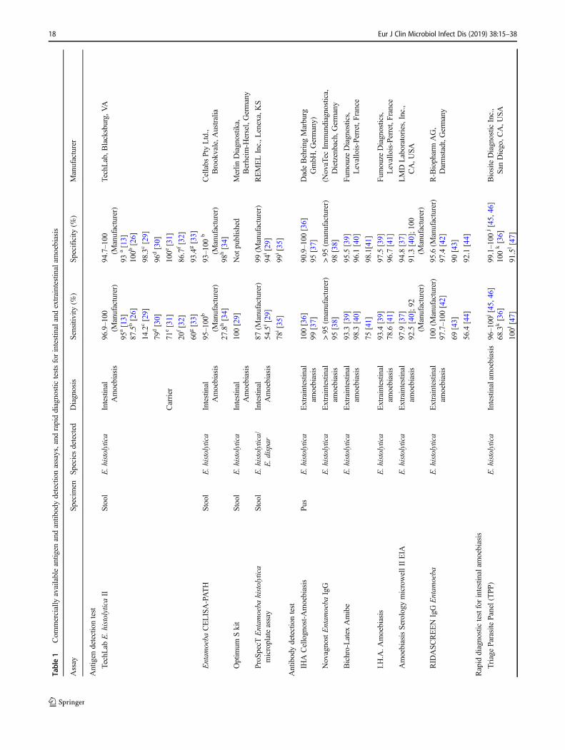
Table1
Com
mercially
availableantig
enandantib
odydetectionassays,and
rapiddiagnostictestsforintestinalandextraintestin
alam
oebiasis
Assay
Specimen
Speciesdetected
Diagnosis
Sensitiv
ity(%
)Specificity
(%)
Manufacturer
Antigen
detectiontest
TechLab
E.histolyticaII
Stool
E.histolytica
Intestinal
Amoebiasis
96.9–100
(Manufacturer)
94.7–100
(Manufacturer)
TechLab,B
lacksburg,VA
95a[13]
93a[13]
87.5b[26]
100b
[26]
14.2c[29]
98.3c[29]
79d[30]
96d[30]
Carrier
71e[31]
100e
[31]
20f[32]
86.7f[32]
60g[33]
93.4g[33]
Entam
oeba
CELISA-PATH
Stool
E.histolytica
Intestinal
Amoebiasis
95–100
b
(Manufacturer)
93–100
b
(Manufacturer)
Cellabs
Pty
Ltd.,
Brookvale,A
ustralia
27.8h[34]
98h[34]
Optim
umSkit
Stool
E.histolytica
Intestinal
Amoebiasis
100[29]
Not
published
Merlin
Diagnostik
a,Berheim
-Hersel,Germany
ProSp
ecTEntam
oeba
histolytica
microplateassay
Stool
E.histolytica/
E.dispar
Intestinal
Amoebiasis
87(M
anufacturer)
99(M
anufacturer)
REMELInc.,L
enexa,KS
54.5i[29]
94i[29]
78i[35]
99i[35]
Antibodydetectiontest
IHACellognost-Amoebiasis
Pus
E.histolytica
Extraintestinal
amoebiasis
100[36]
90.9–100
[36]
DadeBehring
Marburg
GmbH
,Germany)
99[37]
95[37]
NovagnostEntam
oeba
IgG
E.histolytica
Extraintestinal
amoebiasis
>95
(manufacturer)
>95
(manufacturer)
(NovaTec
Immundiagnostica,
Dietzenbach,G
ermany
95[38]
98[38]
Bichro-Latex
Amibe
E.histolytica
Extraintestinal
amoebiasis
93.3[39]
95.5[39]
Fumouze
Diagnostics,
Levallois-Perret,France
98.3[40]
96.1[40]
75[41]
98.1[41]
I.H.A.A
moebiasis
E.histolytica
Extraintestinal
amoebiasis
93.4[39]
97.5[39]
Fumouze
Diagnostics,
Levallois-Perret,France
78.6[41]
96.7[41]
AmoebiasisSerology
microwellIIEIA
E.histolytica
Extraintestinal
amoebiasis
97.9[37]
94.8[37]
LMDLaboratories,Inc.,
CA,U
SA92.5[40];9
2(M
anufacturer)
91.3[40];1
00(M
anufacturer)
RID
ASC
REENIgGEntam
oeba
E.histolytica
Extraintestinal
amoebiasis
100(M
anufacturer)
95.6(M
anufacturer)
R-Biopharm
AG,
Darmstadt,Germany
97.7–100
[42]
97.4[42]
69[43]
90[43]
56.4[44]
92.1[44]
Rapid
diagnostictestforintestinalam
oebiasis
TriageParasite
Panel(TPP)
E.histolytica
Intestinalam
oebiasis
96–100
j[45,46]
99.1–100
j[45,46]
BiositeDiagnostic
Inc.,
SanDiego,C
A,U
SA68.3k[36]
100
k[36]
100l[47]
91.5l[47]
18 Eur J Clin Microbiol Infect Dis (2019) 38:15–38
Tab
le1
(contin
ued)
Assay
Specimen
Speciesdetected
Diagnosis
Sensitiv
ity(%
)Specificity
(%)
Manufacturer
RID
A®QUICK
Cryptosporidium
/Giardia/Entam
oeba
Com
biE.histolytica/E.
dispar
Intestinalam
oebiasis
100[48]
80–88[48]
R-BioPharm
,Darmstadt,Germany
62[49]
96[49]
ImmunoC
ardSTA
T!®
CGE
E.histolytica
Intestinalam
oebiasis
88m[50]
100
m[50]
MeridianBioscienceInc.,
Luckenw
alde,G
ermany
RID
AQuick
Entam
oeba
test
28.6p[51]
86p[51]
R-Biopharm
AG,D
armstadt,Germany
E.histolyticaQuikChek
E.histolytica
Intestinalam
oebiasis
100n
[52]
100[52]
TechLab,B
lacksburg,VA,U
SA97
o[53]
100
n[53]
Prototypeof
lateralflowdipsticktest
Intestinalam
oebiasis
65.4q[54]
92q[54]
aCom
paredto
cultu
reandmicroscopy
bCom
paredto
isoenzym
eanalysis
cSensitivity
andspecificity
comparedto
cultu
reandmicroscopy
dCom
paredto
real-tim
ePCR
eCom
paredto
real-tim
ePCR
fCom
paredto
microscopy
gCom
paredto
microscopy
hCom
paredto
microscopy
iCom
paredto
microscopy(w
etmountsandconcentration)
jCom
paredto
microscopy
kCom
paredto
ProS
pecT
Entam
oeba
histolyticamicroplateassay
lCom
paredto
microscopy
mCom
paredto
real-tim
ePC
RnCom
paredto
ELISAantig
enoCom
paredto
ProS
pecT
Entam
oeba
histolyticamicroplateassay
pCom
paredto
TechlabE.histolyticaII
qCom
paredto
real-tim
ePCR
Eur J Clin Microbiol Infect Dis (2019) 38:15–38 19
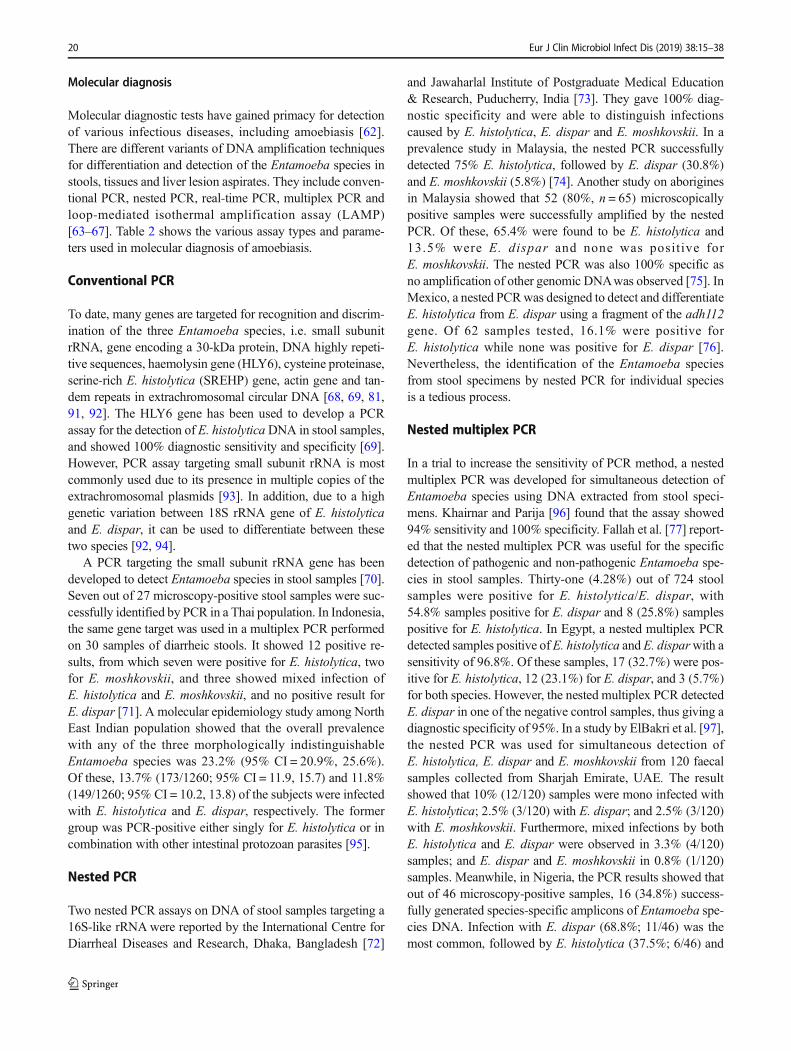
Molecular diagnosis
Molecular diagnostic tests have gained primacy for detectionof various infectious diseases, including amoebiasis [62].There are different variants of DNA amplification techniquesfor differentiation and detection of the Entamoeba species instools, tissues and liver lesion aspirates. They include conven-tional PCR, nested PCR, real-time PCR, multiplex PCR andloop-mediated isothermal amplification assay (LAMP)[63–67]. Table 2 shows the various assay types and parame-ters used in molecular diagnosis of amoebiasis.
Conventional PCR
To date, many genes are targeted for recognition and discrim-ination of the three Entamoeba species, i.e. small subunitrRNA, gene encoding a 30-kDa protein, DNA highly repeti-tive sequences, haemolysin gene (HLY6), cysteine proteinase,serine-rich E. histolytica (SREHP) gene, actin gene and tan-dem repeats in extrachromosomal circular DNA [68, 69, 81,91, 92]. The HLY6 gene has been used to develop a PCRassay for the detection of E. histolyticaDNA in stool samples,and showed 100% diagnostic sensitivity and specificity [69].However, PCR assay targeting small subunit rRNA is mostcommonly used due to its presence in multiple copies of theextrachromosomal plasmids [93]. In addition, due to a highgenetic variation between 18S rRNA gene of E. histolyticaand E. dispar, it can be used to differentiate between thesetwo species [92, 94].
A PCR targeting the small subunit rRNA gene has beendeveloped to detect Entamoeba species in stool samples [70].Seven out of 27 microscopy-positive stool samples were suc-cessfully identified by PCR in a Thai population. In Indonesia,the same gene target was used in a multiplex PCR performedon 30 samples of diarrheic stools. It showed 12 positive re-sults, from which seven were positive for E. histolytica, twofor E. moshkovskii, and three showed mixed infection ofE. histolytica and E. moshkovskii, and no positive result forE. dispar [71]. A molecular epidemiology study among NorthEast Indian population showed that the overall prevalencewith any of the three morphologically indistinguishableEntamoeba species was 23.2% (95% CI = 20.9%, 25.6%).Of these, 13.7% (173/1260; 95% CI = 11.9, 15.7) and 11.8%(149/1260; 95% CI = 10.2, 13.8) of the subjects were infectedwith E. histolytica and E. dispar, respectively. The formergroup was PCR-positive either singly for E. histolytica or incombination with other intestinal protozoan parasites [95].
Nested PCR
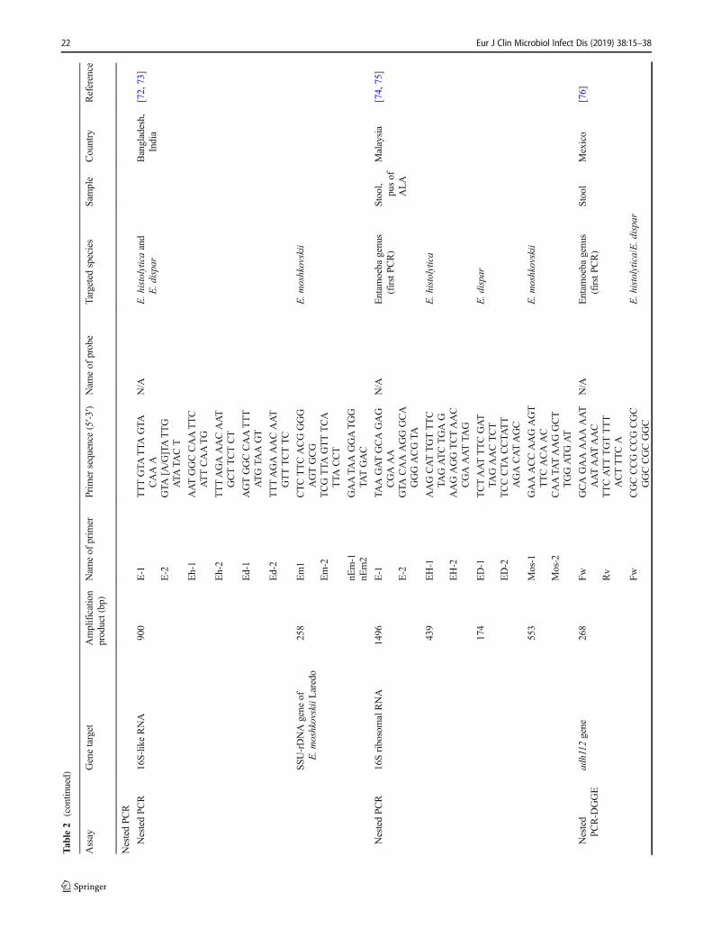
Two nested PCR assays on DNA of stool samples targeting a16S-like rRNAwere reported by the International Centre forDiarrheal Diseases and Research, Dhaka, Bangladesh [72]
and Jawaharlal Institute of Postgraduate Medical Education& Research, Puducherry, India [73]. They gave 100% diag-nostic specificity and were able to distinguish infectionscaused by E. histolytica, E. dispar and E. moshkovskii. In aprevalence study in Malaysia, the nested PCR successfullydetected 75% E. histolytica, followed by E. dispar (30.8%)and E. moshkovskii (5.8%) [74]. Another study on aboriginesin Malaysia showed that 52 (80%, n = 65) microscopicallypositive samples were successfully amplified by the nestedPCR. Of these, 65.4% were found to be E. histolytica and13.5% were E. dispar and none was positive forE. moshkovskii. The nested PCR was also 100% specific asno amplification of other genomic DNAwas observed [75]. InMexico, a nested PCRwas designed to detect and differentiateE. histolytica from E. dispar using a fragment of the adh112gene. Of 62 samples tested, 16.1% were positive forE. histolytica while none was positive for E. dispar [76].Nevertheless, the identification of the Entamoeba speciesfrom stool specimens by nested PCR for individual speciesis a tedious process.
Nested multiplex PCR
In a trial to increase the sensitivity of PCR method, a nestedmultiplex PCR was developed for simultaneous detection ofEntamoeba species using DNA extracted from stool speci-mens. Khairnar and Parija [96] found that the assay showed94% sensitivity and 100% specificity. Fallah et al. [77] report-ed that the nested multiplex PCR was useful for the specificdetection of pathogenic and non-pathogenic Entamoeba spe-cies in stool samples. Thirty-one (4.28%) out of 724 stoolsamples were positive for E. histolytica/E. dispar, with54.8% samples positive for E. dispar and 8 (25.8%) samplespositive for E. histolytica. In Egypt, a nested multiplex PCRdetected samples positive ofE. histolytica and E. disparwith asensitivity of 96.8%. Of these samples, 17 (32.7%) were pos-itive for E. histolytica, 12 (23.1%) for E. dispar, and 3 (5.7%)for both species. However, the nested multiplex PCR detectedE. dispar in one of the negative control samples, thus giving adiagnostic specificity of 95%. In a study by ElBakri et al. [97],the nested PCR was used for simultaneous detection ofE. histolytica, E. dispar and E. moshkovskii from 120 faecalsamples collected from Sharjah Emirate, UAE. The resultshowed that 10% (12/120) samples were mono infected withE. histolytica; 2.5% (3/120) with E. dispar; and 2.5% (3/120)with E. moshkovskii. Furthermore, mixed infections by bothE. histolytica and E. dispar were observed in 3.3% (4/120)samples; and E. dispar and E. moshkovskii in 0.8% (1/120)samples. Meanwhile, in Nigeria, the PCR results showed thatout of 46 microscopy-positive samples, 16 (34.8%) success-fully generated species-specific amplicons of Entamoeba spe-cies DNA. Infection with E. dispar (68.8%; 11/46) was themost common, followed by E. histolytica (37.5%; 6/46) and
20 Eur J Clin Microbiol Infect Dis (2019) 38:15–38
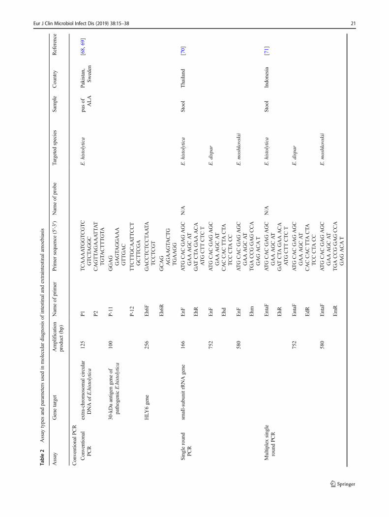
Table2
Assay
typesandparametersused
inmolecular
diagnosisof
intestinalandextraintestin
alam
oebiasis
Assay
Genetarget
Amplification
product(bp)
Nam
eof
prim
erPrim
ersequence
(5′-3
′)Nam
eof
probe
Targeted
species
Sam
ple
Country
Reference
ConventionalP
CR
Conventional
PCR
extra-chromosom
alcircular
DNAof
E.histolytica
125
P1
TCAAAATGGTCGTC
GTCTA
GGC
E.histolytica
pusof
ALA
Pakistan,
Sweden
[68,69]
P2
CAGTTA
GAAATTA
TTGTA
CTTTGTA
30-kDaantig
engene
ofpathogenicE.histolytica
100
P-11
GGAG
GAGTA
GGAAA
GTTGAC
P-12
TTCTTGCAATTCCT
GCTTCGA
HLY
6gene
256
Eh6F
GACCTCTCCTA
ATA
TCCTCGT
Eh6R
GCAG
AGAAGTA
CTG
TGAAGG
Singleround
PCR
small-subunitrRNAgene
166
EnF
ATGCACGAGAGC
GAAAGCAT
N/A
E.histolytica
Stool
Thailand
[70]
EhR
GATCTA
GAAACA
ATGCTTCTCT
752
EnF
ATGCACGAGAGC
GAAAGCAT
E.dispar
Ehd
CACCACTTA
CTA
TCCCTA
CC
580
EnF
ATGCACGAGAGC
GAAAGCAT
E.m
oshkovskii
Ehm
TGACCGGAGCCA
GAGACAT
Multip
lexsingle
roundPC
REntaF
ATGCACGAGAGC
GAAAGCAT
N/A
E.histolytica
Stool
Indonesia
[71]
EhR
GATCTA
GAAACA
ATGCTTCTCT
752
EntaF
ATGCACGAGAGC
GAAAGCAT
E.dispar
EdR
CACCACTTA
CTA
TCCCTA
CC
580
EntaF
ATGCACGAGAGC
GAAAGCAT
E.m
oshkovskii
EmR
TGACCGGAGCCA
GAGACAT
Eur J Clin Microbiol Infect Dis (2019) 38:15–38 21
Tab
le2
(contin
ued)
Assay
Genetarget
Amplification
product(bp)
Nam
eof
prim
erPrim
ersequence
(5′-3
′)Nam
eof
probe
Targeted
species
Sam
ple
Country
Reference
NestedPC
R
NestedPCR
16S-likeRNA
900
E-1
TTTGTA
TTA
GTA
CAAA
N/A
E.histolyticaand
E.dispar
Bangladesh,
India
[72,73]
E-2
GTA
[A/G]TATTG
ATA
TACT
Eh-1
AATGGCCAATTC
ATTCAATG
Eh-2
TTTAGAAACAAT
GCTTCTCT
Ed-1
AGTGGCCAATTT
ATGTA
AGT
Ed-2
TTTAGAAACAAT
GTTTCTTC
SSU-rDNAgene
ofE.m
oshkovskiiLaredo
258
Em1
CTCTTCACGGGG
AGTGCG
E.m
oshkovskii
Em-2
TCGTTA
GTTTCA
TTA
CCT
nEm-1
GAATA
AGGATGG
TATGAC
nEm2
NestedPCR
16SribosomalRNA
1496
E-1
TAAGATGCAGAG
CGAAA
N/A
Entam
oeba
genus
(firstPC
R)
Stool,
pusof
ALA
Malaysia
[74,75]
E-2
GTA
CAAAGGGCA
GGGACGTA
439
EH-1
AAGCATTGTTTC
TAGATCTGAG
E.histolytica
EH-2
AAGAGGTCTAAC
CGAAATTA
G
174
ED-1
TCTAATTTCGAT
TAGAACTCT
E.dispar
ED-2
TCCCTA
CCTA
TT
AGACATAGC
553
Mos-1
GAAACCAAGAGT
TTCACAAC
E.m
oshkovskii
Mos-2
CAATA
TAAGGCT
TGGATGAT
Nested
PCR-D
GGE
adh112
gene
268
FwGCAGAAAAAAAT
AATAATAAC
N/A
Entam
oeba
genus
(firstPC
R)
Stool
Mexico
[76]
Rv
TTCATTTGTTTT
ACTTTCA
Fw
CGCCCGCCGCGC
GGCCGCGGC
E.histolytica/E.dispar
22 Eur J Clin Microbiol Infect Dis (2019) 38:15–38
Tab
le2
(contin
ued)
Assay
Genetarget
Amplification
product(bp)
Nam
eof
prim
erPrim
ersequence
(5′-3
′)Nam
eof
probe
Targeted
species
Sam
ple
Country
Reference
CGGCCGGGGGCA
CGCGGCGGC
AGAAAAAAATA
ATA
ATA
A
Rv
TTCATTTGTTTT
ACTTTCA
Nestedmultip
lex
PCR
16S-
likerRNAgene
E-1
TAAGATGCACGAGA
GCGAAA
N/A
Entam
oeba
genus
(firstPC
R)
Stool
India,Iran,
Nigeria
[77–79]
E-2
GTA
CAAAGGGCAGG
GACGTA
439
EH-1
AAGCATTGTTTCTA
GATCTGAG
E.histolytica
(secondPC
R)
EH-2
AAGA
GGTCTA
ACCG
AAATTA
G
174
ED-1
TCTA
ATTTCGATTA
GAACTCT
E.dispar
(secondPC
R)
ED-2
TCCCTA
CCTA
TTA
GACATA
GC
553
Mos-1
GAAA
CCAAGAGTTT
CACAAC
E.m
oshkovskii
(secondPC
R)
Mos-2
CAATA
TAAGGCTTG
GATGAT
Real-tim
ePCR
Real-tim
ePCR
(Light
Cycler)
18SrRNA
307
Eh-S2
6CGTA
CAAAATGGC
CAATTCATTCAA
LC-Red
640-TCG
AACCCCAAT
TCCTCGTTA
TCC
Eh/Ed-25-fluores-
cein
(fluorescein-G
CC-
ATCTGTAAA
GCTCCCTCT
CCGAX
E.histolytica
Stool,
pusof
ALA
USA
[66,80]
Ed-27C
GTA
CAAAGTGGC
CAATTTATGTA
AGCA
E.dispar
Eh/Ed-AS2
5GAATTGATTTTA
CTCAACTCT
AGAG
E.histolyticaand
E.dispar
Real-tim
ePCR
(Taq
Man)
18SrRNA
172
Ehd-239F
ATTGTCGTGGC
ATCCTA
ACTCA
VIC-5′-T
CAT
TGAATGAATT
GGCCAT
TT-3′-nonfluores-
cent
quencher
E.histolytica
Stool,
pusof
ALA
Netherlands,
Egypt
[81–83]
Ehd-88R
GCGGACGGCTCATT
AT A
ACA
Eur J Clin Microbiol Infect Dis (2019) 38:15–38 23
Tab
le2
(contin
ued)
Assay
Genetarget
Amplification
product(bp)
Nam
eof
prim
erPrim
ersequence
(5′-3
′)Nam
eof
probe
Targeted
species
Sam
ple
Country
Reference
Real-tim
ePCR
(Taqman)
smallsubunitrRNAgene
134
Eh-f
AACAGTA
ATA
GTTT
CTTTGGTTA
GTA
AAA
YYT,
5′-ATTAGT
ACAAACTGG
CCAATTCAT
TCA-3′(Eclipse)
E.histolytica
pusof
ALA
Bangladesh
[63]
Real-tim
ePCR
(Molecular
Beacon)
18srRNA
134
Ehf
AACAGTA
ATA
GTTT
CTTTGGTTA
GTA
AAA
Texas
Red-G
CGAGC--
ATTA
GTA
CAAAATG
GCCAAT
TCATTC
A-G
CTCGC-dR
Elle
E.histolytica
Stool,
pusof
ALA
Bangladesh
[30]
Ehr
CTTA
GAATGTCATT
TCTCAATTCAT
Realtim
ePCR
16srRNA
134
Ehf
AACAGTA
ATA
GTTT
CTTTGGTTA
GTA
AAA
SYBRGreen
Superm
ixE.histolytica
Stool
Mexico
[85]
Ehr
CTTA
GAATGTCATT
TCTCAATTCAT
Multip
lexReal
timePC
Rsm
all-subunitrRNAgene
sequences
222
EhsmF
EhdmR
CGAAAGCATTTCAC
TCAACTG
TCCCCCTGAAGTCC
ATA
AACTC
Ehdm-FL:5
′-ACTA
TAAACGATGT
CAAC
CAAGGATT
GGAT
GAAA-FITC-3′
E.histolytica
Stool
Thailand
[86]
Ehd-640:5
′-TCAG
ATGTA
CAAAG
ATA
GAGAAGCAT
TGTT
TCTA
-phosphat-
e-3
E.dispar
Em-705:5
′-AAGA
AATTCGCGGA
TGAA
GAA
ACATTGTT
T-phosphate-3
E.m
oshkovskii
Multip
robe
real-tim
ePC
RE.histolyticaSSU
rRNA
110
EntaTaq-L
GGACACATTTCAAT
TGTCCTA
YAK-5’TGTA
GTTA
TCTA
AT
TTCGGT
TAGACC-3′
E. histolytica
Stool
Taiwan
[58]
EntaTaq-R
CATCACAGACCTGT
TATTGCTG
111
EntaTaq-L
GGACACATTTCAAT
TGTCCTA
FAM-5’TGTTA
GTT
ATCTA
ATTTCGA
TTA
GAACTC-3’
E.dispar
EntaTaq-R
CATCACAGACCTGT
TATTGCTG
24 Eur J Clin Microbiol Infect Dis (2019) 38:15–38
Tab
le2
(contin
ued)
Assay
Genetarget
Amplification
product(bp)
Nam
eof
prim
erPrim
ersequence
(5′-3
′)Nam
eof
probe
Targeted
species
Sam
ple
Country
Reference
Loop-mediatedisothemalam
plificationassay(LAMP)
LAMP
small-subunitribosom
alDNAin
theE.histolytica
genome
447
Outer
prim
erEHd1-F3
Ehd1-F3
-AAAGATA
ATA
CTTGAGACGA
TCC
E.histolytica
Stool
Taiwan,
India
[17,87]
Outer
prim
erEHd1-B3
Ehd1-B3-TCGTTA
TC
CGTTA
TAATCTT
GG
Innerprim
erEh1-FIP
Eh1-FIP
GCATCCTA
ACTCACTTA
GAA
TGTCAAGTA
CAA
AATGGCCAATTC
ATTC
Innerprim
erEhd1-BIP
Ehd1-BIP-CACGA-
CAATTGTA
GAACACACAGTTC
CTCGATA
CTA
CC
AACTGAT
LAMP
E.histolyica
hemolysin
gene,
HLY
6
Outer prim
erEh-2F
3Eh-2F
3-GCACTA
TACTTGAACGGATT
G
E.histolytica
Stool
Philip
pines
[88]
Outer
prim
erEh-2B
3Eh-2B
3-GTTTGACA
AGATGTTGAGTG
A
Innerprim
erEh-2F
IPEh-2F
IP-TCGCCCTA
TACTCAAATA
TG
ACAAGACTTTGG
TGGAAGATTCAC
G
Innerprim
erEh-2F
IPEh-2F
IP-ATCTA
GTA
GCTGGTTCCACC
TGAACACCTA
AT
CATTA
TCTTTA
CCAATC
Aditio
nalp
rimer
Eh-2F
2Eh-2F
2-A
CTTTGGT
GGAAGATTCACG
Aditio
nalp
rimer
Eh-2B
2Eh-2B
2-CACCTA
AT
CATTA
TCTTTA
CCAATC
Stem
LAMP
18Ssm
allsubunitribosomal
RNA
207
F3
AAATA
CAAGGATA
GCTTTGTG
E.histolytica
Stool
Kenya
[89]
B3
Eur J Clin Microbiol Infect Dis (2019) 38:15–38 25
Tab
le2
(contin
ued)
Assay
Genetarget
Amplification
product(bp)
Nam
eof
prim
erPrim
ersequence
(5′-3
′)Nam
eof
probe
Targeted
species
Sam
ple
Country
Reference
AAGCTCCCTCTCCG
ATGTC
FIP
CTCAATTCATTGAA
TGAATTGGCATG
ATA
AAGATA
ATA
CTTGAGAC
BIP
CAATGAGAATTTCT
GATCTA
TCCGTT
ATCCGTTA
TAAT
CTTGG
LF
TTTGTA
CTA
ATA
CA
AACTGGATC
LB
CAGTTGGTA
GTA
TC
GAGGAC
SF
CGACAATTGTA
GAA
CACACAG
SBATCCTA
ACTCACTT
AGAATGTC
LAMP-NAL-
FIA
serine-rich
E.histolytica
protein(
SREHP
)gene
Eh-FIP-SER
GCTTCGTTCTTTA
AAAATA
CACCGTC
ATTCTTGATTTG
GATCAAGAAGT
E.histolytica
pusof
ALA
Malaysia
[90]
Eh-BIP-SER-FITC
AGTA GCTCAGCAAA
ACCAGAATCACT
TGCTTTTTCATC
TTCATCA
Eh-F3-SER
TGCATTCACTA
GTG
CAACT
Eh-B3-SE
RGCTTGATTCTGAGT
TATCACTTG
Eh-LB-SER-Biotin
AAGTTCAAATGA
AGATA
ATGAA
Entam
oeba
spp.(LSU-rRNAgene
Eh-FIP-H
LYTA
CGCCATTTCGTT
TCCTTA
CTCGAT
TTCTTA
ACTGAT
ACTCGACCG
E.histolytica
Eh-BIP-H
LY-FITC
AGATTGAAACTGTC
CTTA
GTGCAGCA
GTTCTA
AGATGT
TTTTTTCCTC
26 Eur J Clin Microbiol Infect Dis (2019) 38:15–38
Tab
le2
(contin
ued)
Assay
Genetarget
Amplification
product(bp)
Nam
eof
prim
erPrim
ersequence
(5′-3
′)Nam
eof
probe
Targeted
species
Sam
ple
Country
Reference
Eh-F3-HLY
CCTGAAAATGGATG
GCATTA
Eh-B3-HLY
CCCTA
ATCCAAGTA
ATGTTGTT
Enta-LB-H
LY-Tex
CTTGGTGGTA
GTA
GCAAATA
CTA
AG
Eur J Clin Microbiol Infect Dis (2019) 38:15–38 27
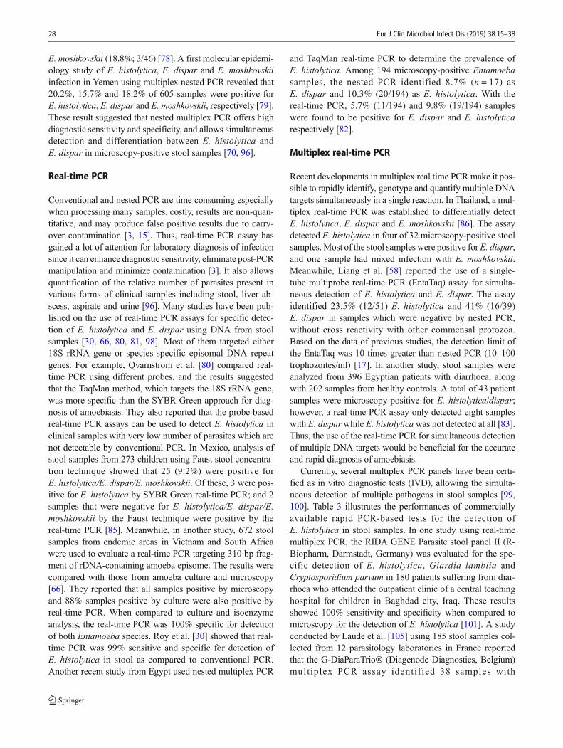
E. moshkovskii (18.8%; 3/46) [78]. A first molecular epidemi-ology study of E. histolytica, E. dispar and E. moshkovskiiinfection in Yemen using multiplex nested PCR revealed that20.2%, 15.7% and 18.2% of 605 samples were positive forE. histolytica, E. dispar and E. moshkovskii, respectively [79].These result suggested that nested multiplex PCR offers highdiagnostic sensitivity and specificity, and allows simultaneousdetection and differentiation between E. histolytica andE. dispar in microscopy-positive stool samples [70, 96].
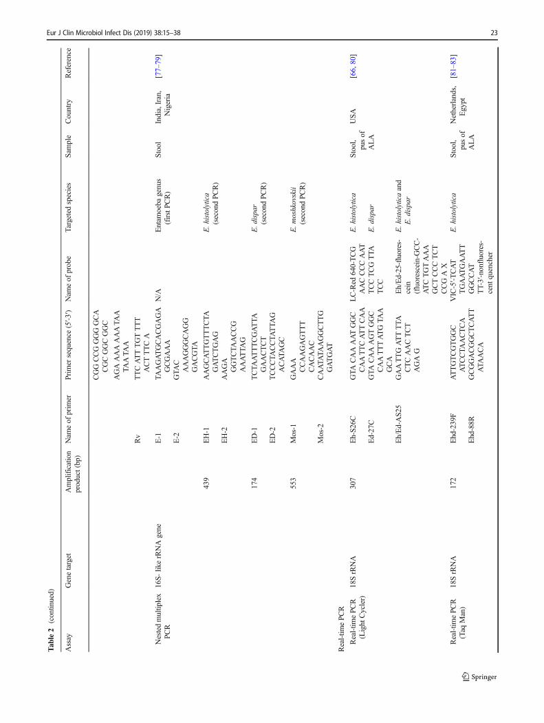
Real-time PCR
Conventional and nested PCR are time consuming especiallywhen processing many samples, costly, results are non-quan-titative, and may produce false positive results due to carry-over contamination [3, 15]. Thus, real-time PCR assay hasgained a lot of attention for laboratory diagnosis of infectionsince it can enhance diagnostic sensitivity, eliminate post-PCRmanipulation and minimize contamination [3]. It also allowsquantification of the relative number of parasites present invarious forms of clinical samples including stool, liver ab-scess, aspirate and urine [96]. Many studies have been pub-lished on the use of real-time PCR assays for specific detec-tion of E. histolytica and E. dispar using DNA from stoolsamples [30, 66, 80, 81, 98]. Most of them targeted either18S rRNA gene or species-specific episomal DNA repeatgenes. For example, Qvarnstrom et al. [80] compared real-time PCR using different probes, and the results suggestedthat the TaqMan method, which targets the 18S rRNA gene,was more specific than the SYBR Green approach for diag-nosis of amoebiasis. They also reported that the probe-basedreal-time PCR assays can be used to detect E. histolytica inclinical samples with very low number of parasites which arenot detectable by conventional PCR. In Mexico, analysis ofstool samples from 273 children using Faust stool concentra-tion technique showed that 25 (9.2%) were positive forE. histolytica/E. dispar/E. moshkovskii. Of these, 3 were pos-itive for E. histolytica by SYBR Green real-time PCR; and 2samples that were negative for E. histolytica/E. dispar/E.moshkovskii by the Faust technique were positive by thereal-time PCR [85]. Meanwhile, in another study, 672 stoolsamples from endemic areas in Vietnam and South Africawere used to evaluate a real-time PCR targeting 310 bp frag-ment of rDNA-containing amoeba episome. The results werecompared with those from amoeba culture and microscopy[66]. They reported that all samples positive by microscopyand 88% samples positive by culture were also positive byreal-time PCR. When compared to culture and isoenzymeanalysis, the real-time PCR was 100% specific for detectionof both Entamoeba species. Roy et al. [30] showed that real-time PCR was 99% sensitive and specific for detection ofE. histolytica in stool as compared to conventional PCR.Another recent study from Egypt used nested multiplex PCR
and TaqMan real-time PCR to determine the prevalence ofE. histolytica. Among 194 microscopy-positive Entamoebasamples, the nested PCR identified 8.7% (n = 17) asE. dispar and 10.3% (20/194) as E. histolytica. With thereal-time PCR, 5.7% (11/194) and 9.8% (19/194) sampleswere found to be positive for E. dispar and E. histolyticarespectively [82].
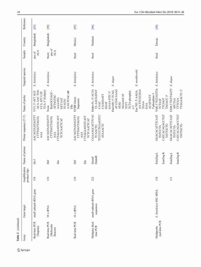
Multiplex real-time PCR
Recent developments in multiplex real time PCR make it pos-sible to rapidly identify, genotype and quantify multiple DNAtargets simultaneously in a single reaction. In Thailand, a mul-tiplex real-time PCR was established to differentially detectE. histolytica, E. dispar and E. moshkovskii [86]. The assaydetected E. histolytica in four of 32 microscopy-positive stoolsamples.Most of the stool samples were positive for E. dispar,and one sample had mixed infection with E. moshkovskii.Meanwhile, Liang et al. [58] reported the use of a single-tube multiprobe real-time PCR (EntaTaq) assay for simulta-neous detection of E. histolytica and E. dispar. The assayidentified 23.5% (12/51) E. histolytica and 41% (16/39)E. dispar in samples which were negative by nested PCR,without cross reactivity with other commensal protozoa.Based on the data of previous studies, the detection limit ofthe EntaTaq was 10 times greater than nested PCR (10–100trophozoites/ml) [17]. In another study, stool samples wereanalyzed from 396 Egyptian patients with diarrhoea, alongwith 202 samples from healthy controls. A total of 43 patientsamples were microscopy-positive for E. histolytica/dispar;however, a real-time PCR assay only detected eight sampleswith E. disparwhile E. histolyticawas not detected at all [83].Thus, the use of the real-time PCR for simultaneous detectionof multiple DNA targets would be beneficial for the accurateand rapid diagnosis of amoebiasis.
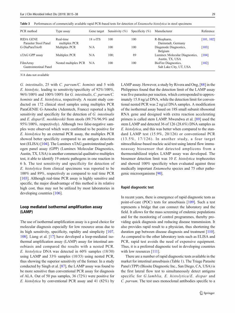
Currently, several multiplex PCR panels have been certi-fied as in vitro diagnostic tests (IVD), allowing the simulta-neous detection of multiple pathogens in stool samples [99,100]. Table 3 illustrates the performances of commerciallyavailable rapid PCR-based tests for the detection ofE. histolytica in stool samples. In one study using real-timemultiplex PCR, the RIDA GENE Parasite stool panel II (R-Biopharm, Darmstadt, Germany) was evaluated for the spe-cific detection of E. histolytica, Giardia lamblia andCryptosporidium parvum in 180 patients suffering from diar-rhoea who attended the outpatient clinic of a central teachinghospital for children in Baghdad city, Iraq. These resultsshowed 100% sensitivity and specificity when compared tomicroscopy for the detection of E. histolytica [101]. A studyconducted by Laude et al. [105] using 185 stool samples col-lected from 12 parasitology laboratories in France reportedthat the G-DiaParaTrio® (Diagenode Diagnostics, Belgium)multiplex PCR assay identif ied 38 samples with
28 Eur J Clin Microbiol Infect Dis (2019) 38:15–38
G. intestinalis, 25 with C. parvum/C. hominis and 5 withE. histolytic, leading to sensitivity/specificity of 92%/100%,96%/100% and 100%/100% for G. intestinalis, C. parvum/C.hominis and E. histolytica, respectively. A recent study con-ducted on 172 clinical stool samples using multiplex PCRParaGENIE G-Amoeba (Ademtech, France) reported a highsensitivity and specificity for the detection of G. intestinalisand E. dispar/E. moshkovskii from stools (89.7%/96.9% and95%/100%, respectively). Although two false-negative sam-ples were observed which were confirmed to be positive forE. histolytica by an external PCR assay, the multiplex PCRshowed better specificity compared to an antigen detectiontest (ELISA) [104]. The Luminex xTAG gastrointestinal path-ogen panel assay (GPP) (Luminex Molecular Diagnostics,Austin, TX, USA) is another commercial qualitative multiplextest, it able to identify 19 enteric pathogens in one reaction in6 h. The test sensitivity and specificity for detection ofE. histolytica from clinical specimens was reported to be100% and 89%, respectively as compared to real time PCR[103]. Although real-time PCR assay is highly sensitive andspecific, the major disadvantage of this method is its relativehigh cost, thus may not be utilized by most laboratories indeveloping countries [106].
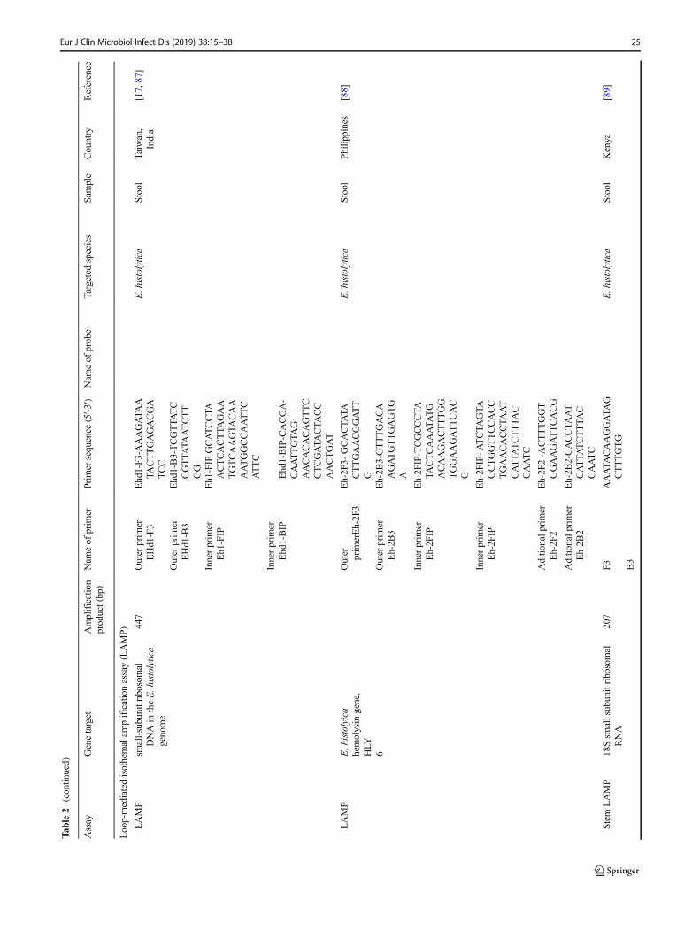
Loop mediated isothermal amplification assay(LAMP)
The use of isothermal amplification assay is a good choice formolecular diagnosis especially for low resource areas due toits high sensitivity, specificity, rapidity and simplicity [107,108]. Liang et al. [17] have developed a loop-mediated iso-thermal amplification assay (LAMP) assay for intestinal am-oebiasis and compared the results with a nested PCR.E. histolytica DNA was detected in 60% samples (18/30)using LAMP and 33% samples (10/33) using nested PCR,thus showing the superior sensitivity of the former. In a studyconducted by Singh et al. [87], the LAMP assay was found tobe more sensitive than conventional PCR assay for diagnosisof ALA. Out of 50 pus samples, 36 (72%) were positive forE. histolytica by conventional PCR assay and 41 (82%) by
LAMP assay. However, a study by Rivera and Ong, [88] in thePhilippines found that the detection limit of the LAMP assaywas five parasites per reaction, which corresponded to approx-imately 15.8 ng/μl DNA, while the detection limit for conven-tional nested PCR was 2 ng/μl DNA samples. A modificationof the isothermal assay based on 18S small subunit ribosomalRNA gene and designed with extra reaction acceleratingprimers is called stem LAMP. Mwendwa et al. [89] used thestem LAMP and detected 36 of 126 (28.6%) DNA samples asE. histolytica, and this was better when compared to the stan-dard LAMP test (15.9%, 20/126) or conventional PCR(13.5%, 17/126). In another study, a four targetnitrocellulose-based nucleic acid test using lateral flow immu-noassay biosensor that detected amplicons from athermostabilized triplex LAMP assay was developed. Thebiosensor detection limit was 10 E. histolytica trophozoitesand showed 100% specificity when evaluated against threemedically important Entamoeba species and 75 other patho-genic microorganisms [90].
Rapid diagnostic test
In recent years, there is emergence of rapid diagnostic tests aspoint-of-care (POC) tests for amoebiasis [109]. Such a testrepresents a bridge that can connect the laboratory and thefield. It allows for the mass screening of endemic populationsand for the monitoring of control programmes, thereby pro-viding quick diagnosis and reducing disease transmission. Italso provides rapid result to a physician, thus shortening theduration gap between disease diagnosis and treatment [110].As compared to the other laboratory tests such as ELISA andPCR, rapid test avoids the need of expensive equipment.Thus, it is a preferred diagnostic tool in developing countrieswith low resources [111].
There are a number of rapid diagnostic tests available in themarket for intestinal amoebiasis (Table 1). The Triage ParasitePanel (TPP) (Biosite Diagnostic Inc., San Diego, CA, USA) isthe first lateral flow test to simultaneously detect antigensspecific for G.lamblia, E. histolytica/E. dispar andC. parvum. The test uses monoclonal antibodies specific to a
Table 3 Performances of commercially available rapid PCR-based tests for detection of Entamoeba histolytica in stool specimens
PCR method Type assay Gene target Sensitivity (%) Specificity (%) Manufacturer Reference
RIDA GENEParasiter Stool Panel
Real-timemultiplex PCR
18 s-ITS 100 100 R-Biopharm,Darmstadt, Germany
[101, 102]
G-DiaParaTrio® Multiplex PCR N/A 100 100 Diagenode Diagnostics,Belgium
[103]
xTAG GPP assay Multiplex PCR N/A 100; 89 Luminex Molecular Diagnostics,Austin, TX, USA
[104]
FilmArrayGastrointestinal Panel
Nested multiplex PCR N/A 100 100 BioFire Diagnostics,Salt Lake City, UT, USA
[102]
N/A data not available
Eur J Clin Microbiol Infect Dis (2019) 38:15–38 29
29-kDa surface antigen of E. histolytica/E. dispar, G. lambliaalpha-1-giardin, and C. parvum protein disulfide isomerase.Two studies showed that the TPP kit had high diagnostic sen-sitivity (96% to 100%) and specificity (99.1% to 100%) fordetection of E. histolytica/E. dispar, when compared to mi-croscopy [45–47]. In contrast, Garcia et al. [45] and Pillai andKain [36] found that the sensitivity of the kit was low (68.3%),albeit with high specificity (100%) when compared withProSpecT test. This is corroborated by the work by Leivaet al. [112], who found that the sensitivity of TPP kit waslowwhen compared to PCR assay. In addition the kit is unableto differentiate among E. histolytica, E. dispar andE. moshkovskii. Furthermore, either fresh or fresh-frozennon-preserved stools should be used with the TPP kit whichmay be impractical in some situations [3].
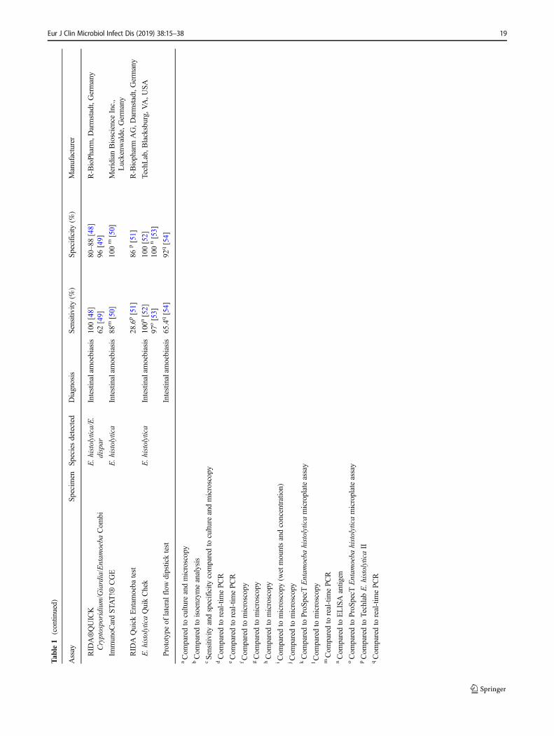
A retrospective study was performed by Van den Bosscheand his colleagues [48] to evaluate the lateral flowRIDA®QUICK Cryptosporidium/Giardia/EntamoebaCombi (R-BioPharm, Darmstadt, Germany), using stool sam-ples collected from patients at an outpatient clinic of theInstitute of Tropical Medicine (ITM), Antwerp, Belgium orthe Central Laboratory of Clinical Biology (CLKB). The kitdemonstrated 100% sensitivity, while the specificity rangedfrom 80% to 88% for detection of E. histolytica. This resultdiffered from Goñi et al. [49], which showed that the kit ex-hibited 62% sensitivity and 96% specificity for E. histolytica.The lower specificity in the study by Van den Bossche et al.[48] can be explained by the high number of E. dispar sam-ples, which substantially influences specificity since this kit isunable to differentiate between E. histolytica and E. dispar[15]. Another version of the one-step immunochromatographictest for the qualitative detection of C. parvum, G. lamblia andE. histolytica antigens in human stool samples has been intro-duced to the market by Meridian Bioscience Inc.(Luckenwalde, Germany) and is known as ImmunoCardSTAT!® CGE. This new ImmunoCard rapid antigen detectiontest exhibited 88% sensitivity and 92% specificity as comparedto real-time PCR in detection of E. histolytica, but it showedcross-reactivity with E. dispar [50]. A study was conducted ondiarrheic/dysenteric stool samples from clinically suspected in-dividuals from Beni-Suef, Egypt, using RIDA®QUICKEntamoeba Test (R-Biopharm AG, Darmstadt, Germany), animmunochromatographic (ICT) rapid assay for the qualitativedetermination of E. histolytica / dispar, and TechlabE. histolytica II ELISA test was used as the reference. Of 7specimens that were positive by the ELISA, only 2 specimenswere positive by the ICT, thus a sensitivity of 28.6%, and thespecificity was reported as 86.1% [51].
The third generation of a rapid test known as E. histolyticaQuik Chek (TechLab, Blacksburg, VA, USA) was recentlyintroduced to the market. The antibody used in this kit isspecific against the E. histolytica adherence lectin [113]. Thetest is a modified version of the TechLab E. histolytica II
ELISA, but uses a flow-through format. An evaluation of thispoint-of-care rapid test in a cohort of children in Bangladeshshowed 100% sensitivity and specificity when compared to anELISA antigen detection assay [52]. When compared toProSpecT microplate assay, this kit exhibited 97% sensitivityand 100% specificity [53]. These findings indicated that thenew Quik Chek assay was robust and can specifically detectE. histolytica trophozoites in unfixed, frozen clinical stoolsamples. However, it requires an additional incubation of bothconjugate and chromogen which increases the processingtime. Furthermore, it requires cold-chain transportation.
Our group previously reported the development of a lateralflow dipstick test which detected E. histolytica PPDK in stoolsamples [54]. When compared to real-time PCR, the diagnos-tic sensitivity of the dipstick was 65.4% (n = 17/26), while thespecificity when tested with stool samples containing otherintestinal pathogens was 92% (23/25). Although not highlysensitive, it was superior to the performance of TechlabE. histolytica II ELISA which detected only 19.2% (5/26) ofthe same set of PCR-positive samples. Thus, the lateral flowdipstick test showed good potential to be further developedinto a stool rapid test for intestinal amoebiasis.
Extraintestinal amoebiasis
Clinical manifestations of ALA are highly variable, thus mak-ing the diagnosis difficult [56, 114]. Ultrasound, computedtomography and magnetic resonance are very useful tech-niques and have excellent sensitivity for detection of liverabscess arising from any cause [115]. However, these detec-tion methods cannot distinguish amoebic abscesses from pyo-genic abscesses or necrotic tumours. Most patients with ALAdo not have co-existing amoebic colitis. Only less than 10% ofamoeba have been identified in stool samples from ALA pa-tients using microscopy and antigen detection tests [116].Consequently, stool microscopy or antigen detection testsare not helpful for diagnosis of ALA [27, 56, 61].
Microscopic examination
ALA can be confirmed by microscopic examination of liverpus to look for amoebic trophozoites [117]. However, aspira-tion of liver pus in an ALA patient is not necessary for estab-lishing the diagnosis. When aspirated, they contain acellulardebris that forms a brown, thick fluid (‘anchovy paste’).Although the presence of amoebic trophozoites in aspiratedpus is confirmatory for ALA, it is not a sensitive diagnosticmethod. Trophozoites are seen in a minority of aspirates (<20%) and typically only seen when the wall of the cyst issampled [118, 119]. Hence, it is not surprising that many au-thors reported their total absence or very low incidence onexamination of liver pus. In general, trophozoites have beenseen in aspirated pus in 11–25% cases [120]. Parija and
30 Eur J Clin Microbiol Infect Dis (2019) 38:15–38
Khairnar [121] found trophozoites of E. histolytica in pus in7.2% cases, while Haque et al. [56] showed that 11% of aspi-rated liver pus samples were positive for the organism.
Microscopic examination of the pus may show dead anddeformed hepatocytes, red blood cells and some polymorphs.A good staining method is necessary to visualize the morpho-logical changes in the liver tissue and also differentiates theamoebas against the surrounding cells. The common stainingtechniques are haematoxylin and eosin (H&E), periodic-acidSchiff (PAS) and immunostaining. With H&E and PAS stains,amoebic trophozoites are difficult to differentiate from mac-rophages because of similarities in size and morphology[122]. Examination of fixed and stained biopsy samples usingH&E stain is challenging due to lack of differentiation be-tween the stained trophozoites and surrounding tissues[123].With PAS stain, the amoebae are intensely PAS positivedue to the presence of glycogen in cytoplasm; however, livercells also contain glycogen [124]. Moreover, these two stain-ing methods require high technical expertise to identify andinterpret the results. In comparison, immunohistochemistry(IHC) is presumed to be more specific as it is the consequenceof specific reactions between amoebic antigens and antibodiesagainst them. Although IHC is still rarely reported for use ininvestigation of invasive amoebiasis [122], it is potentiallyvery useful as a confirmatory test for ALA when liver pusaspirate biopsy or autopsy is available.
Antibody detection test
Petri and Singh [60] reported that anti-amoebic antibodieswere detectable inmore than 90% of patients with ALAwithin7 to 10 days after symptoms began [117]. Therefore, serologyis used for diagnosis of ALA, in combination with observa-tions from clinical manifestations and result of radiologicalimaging. Several assays are commonly used to detect theanti-amoebic antibodies, such as ELISA, indirecthaemagglutination (IHA), indirect immunofluorescence assay(IFA), latex agglutination, immunoelectrophoresis,counterimmunoelectrophoresis (CIE), amoebic gel diffusiontest, immunodiffusion and complement fixation test [125].Of these, ELISA is the most popular technique and has beenused to investigate the epidemiology of symptomatic amoebi-asis due to its reliability and ease of performance [113, 126].
A study was performed to compare the performance ofseveral antibody detection assays on patients in Kuwaitsuspected of ALA using IHA (Collegnost Amoebiasis,Germany), Amoebiasis Serology Microwell II EIA (LMDLAboratories, CA) and ImmunoTab test (Institute Virion,Germany). The true positive cases were defined as patientspositive by CT scan or ultrasonography with an IHA titer ≥1:256. The sensitivity and specificity of the IHA, ImmunoTaband Microwell II EIAwas found to be 99% and 99.8%, 98%and 95.5% and 97.9% and 94.8%, respectively [37]. In other
studies, the sensitivity of the Microwell II EIA for the detec-tion of specific E. histolytica antibodies in ALA patients wasreported to be 97.6–99% [125, 127].
Hung et al. [128] reported that IHA showed high specificity(99.1%) when tested with samples from patients with gastro-intestinal symptoms, including HIV-infected patients.However, it showed lower sensitivity when compared toELISA due to false-negative results [129]. In a village inWest Kalimantan, Borneo, the seroprevalence of anti-amoebic antibodies among the population with IHA titersequal or greater than 1:128 was found to be 7% [130].Meanwhile, the seroprevalence among blood donors fromKelantan, Malaysia, was reported to be 16% [131]. A studyconducted by Dhanalakshmi et al. [43] using IHA, based on arecombinant calcium binding domain-containing protein, onserum of suspected amoebiasis patients showed 62% sensitiv-ity and 96% specificity. In comparison, an antigen detectionELISA on the same set of samples showed 69% sensitivityand 90% specificity.
Garcia et al. [132] reported that the IFA was a reliable,reproducible and a rapid tool to differentiate ALA from othernon-amoebic etiologies. In addition, it helps to differentiatecurrent and past (treated) infected patients. The sensitivity ofIFA was reported to be 93.6% (higher than ELISA) and thespecificity was 96.7% [133]. However, this technique is diffi-cult to perform routinely since it requires skill to culture theparasite and prepare the antigen [134].
The common commercially available antibody assays fordetection of E. histolytica antibodies in human serum are in-cluded in Table 1. Most of these commercial tests are coatedwith crude soluble trophozoite antigen for the detection ofanti-amoebic antibodies. Several studies have demonstratedhigh sensitivity of crude soluble and excretory-secretory anti-gens in detecting amoebic antibodies in ALA patients [38–40,42, 135, 136, 137]. However, the drawbacks using crude an-tigens are the high cost and tediousness of maintainingE. histolytica culture, and other problems associated with themass production of the antigen. In addition, the crude amoebicantigen gave false positive results, thus decreasing the testspecificity [138]. In endemic settings, it tends to produce highbackground readings; thus, the antibody cut-off titre may beneeded to be adjusted from that recommended by the manu-facturer. In general, the use of the current commercial anti-body detection tests is only reliable in developed countrieswhere amoebiasis is not endemic. However, if the vast major-ity of patients in these countries are immigrants from devel-oping countries, the usefulness of this test in developed coun-tries may also be doubtful [60]. In endemic areas, individualsare constantly exposed to E. histolytica, and therefore theavailable IgG antibody detection tests are unable to definitive-ly distinguish between past and current infections [27, 139].The test may remain positive in patients after a few years ofinfection, and thus is a major problem that must be addressed.
Eur J Clin Microbiol Infect Dis (2019) 38:15–38 31
To overcome this limitation, a standardized serological testbased on a well-defined antigen is required. A heavy subunitof E. histolytica lectin (152 kDa) and PPDK (110 kDa) forserodiagnosis of ALA have been shown to possess diagnosticsensitivities of above 80% and no cross-reactivity in Westernblot analysis [137]. The recombinant form of the PPDK hasbeen produced and Western blot using a panel of serumsamples probed with horseradish conjugated anti-humanIgG4 showed a high diagnostic sensitivity (93.3%) andspecificity (100%), when compared to blots using IgG andIgG1 as secondary antibodies [140].
Other recombinant proteins that has been used as anti-gens in amoebiasis serology included serine rich protein,170 kDa subunit galactose-specific adhesin, cysteine pro-teinase, putative alcohol dehydrogenase and phosphogluco-mutase [141–143, 144, 145].
Molecular diagnosis
Table 2 includes information on the assays that have been usedin detecting E. histolytica DNA in liver abscess/pus samples.Detection of ALA by conventional PCR showed varied diag-nostic sensitivity, ranging from 33% to 100% [56, 68, 121,125, 146]. When PCR using specific primers for E. histolytica18S rDNAwas used to detect the parasite in liver pus samples,only 33% sensitivity was recorded; whereas 100% sensitivitywas obtained when a second primer pair specific for a geneencoding a 30-kDa antigen was used [125, 147]. Zaman et al.[68] was successful in obtaining 100% diagnostic sensitivityby using two pairs of published primers namely P1-P2 andP11-P12 which targeted the extrachromosomal circular DNAof E. histolytica and the 30-kDa antigen gene, respectively. Inanother study, PCR was performed which targetedE. histolytica HLY6 gene using pus samples from ALA pa-tients. The hemo-PCR detected 89% of the samples, as com-pared to 77% and 28% detection when PCR assays were per-formed using primers based on the 30-kDa antigen gene and18S rDNA, respectively [69]. In Canada, a total of 25 ALAsamples were assessed by microscopy, serology, and nestedPCR. The results showed that microscopy was negative forE. histolytica in all samples, and nested PCR as well as serol-ogy were positive in 11 samples [148].
In another study, Parija and Khairnar [121] reported that anested multiplex PCR detected E. histolytica DNA in 100%(n = 37) liver abscess pus samples from ALA patients collect-ed prior to metronidazole treatment, as compared to only70.6% (53/75) after therapy. The decline in diagnostic sensi-tivity can be attributed to the parasite DNA clearance from theabscess subsequent to the parasite destruction post-treatment.However, pus aspiration carries the risk of bacterial infectionor spillage of abscess content; thus, it is performed only ifthere is a danger of rupture of the abscess or if the size ofabscess is greater than10 cm in diameter. Abscess should also
be drained if it does not respond to medical therapy within 3 to5 days [149, 150]. In addition, it was reported that the PCRdetected E. histolytica DNA in 21 of 53 (39.6%) urine speci-mens of ALA patients. Among them, four of 23 (17.4%) urinespecimens were collected prior to metronidazole treatment,and 17 of 30 (56.7%) were collected after treatment [121]. Astudy conducted in Malaysia showed that real-time PCR suc-cessfully detected E. histolytica DNA in 76.7% of 30 liverabscess samples, whereas IHA detected the presence of anti-E. histolytica antibodies in 46.7%(14/30) of the correspondingserum samples [56]. Meanwhile, a study conducted inBangladesh by Haque et al. [63], using real-time PCR on 98ALA patients showed that the assay was able to detectE. histolytica DNA in 49%, 77% and 69% of blood, urineand saliva samples, respectively. Ahmad et al. [84] showedthat E. histolytica detection by real-time PCR had higher di-agnostic sensitivity than antigen detection method. In a studyby Roy et al. [30], a real-time PCR assay on liver pus samplesutilizing a molecular-beacon probe was found to be compara-ble to the TaqMan-based method by Blessmann et al. [66].
Weitzel et al. [102] reported the first use of Rida®GeneParasitic Stool Panel (R-Biopharm, Darmstadt, Germany)and FilmArray Gastrointestinal Panel (BioFire Diagnostics,Salt Lake City, UT, USA) on samples from aspirated pus ofpatients with cystic focal liver lesions. Both commercial kitsconfirmed the diagnosis of ALA within a short time, withFilmArray system giving a faster result than the Rida®Genetest (1 h vs. 3 h) and required less hands-on time (5 min vs.45 min).
Rapid diagnostic test
To date, there is no commercial rapid diagnostic test for thediagnosis of ALA. However, a proof-of-concept of a lateralflow dipstick test for rapid detection of ALA has been devel-oped. The test is based on detection of anti-PPDK IgG4 anti-body in infected patients. Initial evaluation of the rapid testshowed 87% diagnostic sensitivity and 100% specificity[140]. Recently, an immunochromatographic test using fluo-rescent silica nanoparticles coated with C-terminal region ofthe intermediate subunit of E. histolytica Gal/GalNAc lectinprotein has been reported. The kit showed 100% sensitivityand 97.6% specificity when tested with sera from healthycontrols and patients with other infectious diseases [151].Both of the above tests seemed to have good potential forrapid diagnosis of ALA and merit further multicentre valida-tion using a much larger sample size.
Future perspectives
Quick and correct diagnosis of amoebic infection is importantto avoid morbidity, death and disease transmission. Sincemost endemic areas are underdeveloped and lack resources
32 Eur J Clin Microbiol Infect Dis (2019) 38:15–38
(financial, facility, skilled manpower), tests which fulfilled theWorld Health Organization ASSURED criteria would be wel-comed, i.e. affordable, highly sensitive and specific, user-friendly, robust and rapid, equipment-free and deliverable tothose who need them [111]. Thus far, the tests developed foramoebiasis fulfilled some but not all of the above criteria.Thus, scientists working on laboratory diagnosis of amoe-biasis should try to develop tests that fully comply withthe above criteria.
Other than identifying infected individuals, it would bevery useful to identify biomarkers that can predict individualswho are susceptible to acquire symptomatic infection, distin-guish between the different stages of an infection, and monitorwhether treatment leads to cure. The combination of well-characterized patient samples and advances in ‘omics’ tech-nologies may make this possible. Genomics technologies maybe able to assess host susceptibility based on the variations insingle nucleotide polymorphism (SNP) of a certain loci [152].Meanwhile, proteomics and metabolomics may help to distin-guish the different stages of E. histolytica infection. Massiveproteomic data on amoeba, especially E. histolytica, havebeen generated, whereby its proteome and sub-proteomeshave been explored [153–158]. Hence, the biological process-es of the E. histolytica proteins could be deduced. This willhelp us to better understand the role of certain E. histolyticaproteins in the pathogenesis even though not all proteins arewell characterized. Using quantitative proteomic, someE. histolytica proteins of a virulent variant have been con-firmed to be differentially abundant when compared to itsnon-virulent variant or other non-pathogenic Entamoeba spe-cies [159, 160]. In addition, a comparison betweenE. histolytica life stages has been made using this approach[152]. Proteins that showed increased in abundance duringcertain life stages can be explored as potential biologicalmarkers for E. histolytica diagnosis, as well for drug and vac-cine development.
Conclusion
Laboratory diagnosis of both intestinal and extraintestinal am-oebiasis has improved with more sensitive and specific tests.For intestinal infection, molecular diagnostic is the way for-ward, with PCR-based assays for well-equipped laboratoriesand LAMP for low-resource settings. For extraintestinal in-fection, improvement in serodiagnosis is needed to accuratelydetect active infection since the exclusion of past infection is achallenge for the current IgG-based assays. Similarly, the sen-sitivity of rapid POC diagnostic tests for ALA can also befurther improved and be made commercially available to en-able its wider application. Further research areas that can beexplored include discovery of biomarkers that can predict
individuals prone to symptomatic infection, distinguish be-tween the different stages of infection and monitor treatmentsuccess.
Funding information We would like to acknowledge the funding assis-tance from the Malaysian Ministry of Education through the HigherInstitution Centre of Excellence Program (HICoE) [Grant no. 311/CIPPM/4401005].
References
1. Stanley SL (2003) Amoebiasis. Lancet 361:1025–10342. Collier P (2007) The bottom billion: why the poorest countries are
failing and what can be done about it. Oxford University Press,United Kingdom
3. Fotedar R, Stark D, Beebe N, Marriott D, Ellis J, Harkness J(2007a) Laboratory diagnostic techniques for Entamoeba species.Clin Microbiol Rev 20:511–532
4. Zlobl TL (2001) Amebiasis. Prim Care Update Ob Gyns 8:65–685. Stanley SL (2006) Vaccines for amoebiasis: barriers and opportu-
nities. Parasitology 133:S81–S866. Hung CC, Ji DD, Sun HY, Lee YT, Hsu SY, Chang SY, Wu CH,
Chan YH, Hsiao CF, Liu WC, Colebunders R (2008) Increasedrisk for Entamoeba histolytica infection and invasive amebiasis inHIV seropositive men who have sex with men in Taiwan. PLoSNegl Trop Dis 2:e175
7. Rivero LR, Fernández FAN, Robertson LJ (2008) Cuban parasi-tology in review: a revolutionary triumph. Trends Parasitol 24:440–448
8. Gathiram V, Jackson TF (1987) A longitudinal study of asymp-tomatic carriers of pathogenic zymodemes of Entamoebahistolytica. S Afr Med J 72:669–672
9. Caballero-Salcedo A, Viveros-Rogel M, Salvatierra B, Tapia-Conyer R, Sepulveda-Amor J, Gutierrez G, Ortiz-Ortiz L (1994)Seroepidemiology of amebiasis in Mexico. Am J Trop Med Hyg50:412–419
10. Hughes MA, Petri WA (2000) Amebic liver abscess. Infect DisClin N Am 14:565–582
11. Garcia LS, Shimizu RY (1998) Evaluation of intestinal protozoanmorphology in human fecal specimens preserved in EcoFix: com-parison of Wheatley’s trichrome stain and EcoStain. ClinMicrobiol 36:1974–1976
12. Gonzalez-Ruiz A, Haque R, Aguirre A, Castanon G, Hall A, GuhlF, Ruiz-Palacios G, Miles MA, Warhurst DC (1994) Value ofmicroscopy in the diagnosis of dysentery associated with invasiveEntamoeba histolytica. J Clin Pathol 47:236–239
13. Haque R, Neville LM, Hahn P, Petri WA (1995) Rapid diagnosis ofEntamoeba infection by using Entamoeba and Entamoebahistolytica stool antigen detection kits. J Clin Microbiol 33:2558–2561
14. World Health Organization (WHO) (1997) UNESCO report of aconsultation of experts on amoebiasis.WklyEpidemiol Rec 72:97–99
15. Tanyuksel M, Petri WA (2003) Laboratory diagnosis of amebiasis.Clin Microbiol Rev 16:713–729
16. Haque R, Petri WA (2006) Diagnosis of amebiasis in Bangladesh.Arch Med Res 37:272–275
17. Liang SY, Chan YH, Hsia KT, Lee JL, Kuo MC, Hwa KY, ChanCW, Chiang TY, Chen JS, Wu FT, Ji DD (2009) Development ofloop-mediated isothermal amplification assay for detection ofEntamoeba histolytica. J Clin Microbiol 47:1892–1895
18. Clark CG, Diamond LS (2002) Methods for cultivation ofluminal parasitic protists of clinical importance. ClinMicrobiol Rev 15:329–341
Eur J Clin Microbiol Infect Dis (2019) 38:15–38 33
19. Freedman L, Maddison SE, Elsdon-Dew B (1958) Monoxeniccultures of Entamoeba histolytica derived from human liver ab-scesses. Afr J Med Med Sci 23:9–12
20. Wang LT, Jen G, Cross JH (1973) Establishment of Entamoebahistolytica from liver abscess in monoxenic cultures with hemo-flagellates. Am J Trop Med Hyg 22:30–32
21. Sargeaunt PG, Jackson TF, Wiffen S, Bhojnani R, Williams JE,Felmingham D, Goldmeir D, Allason-Jones E, Mindel A, PhillipsE (1987) The reliability of Entamoeba histolytica zymodemes inclinical laboratory diagnosis. Arch Invst Med 18:69–75
22. Razmjou E, Haghighi A, Rezaian M, Kobayashi S, Nozaki T(2006) Genetic diversity of glucose phosphate isomerase fromEntamoeba histolytica. Parasitol Int 55:307–311
23. Jackson TF, Suparsad S (1996) Zymodeme stability ofEntamoebahistolytica and E. dispar. Arc Med Res 28:304–305
24. Strachan WD, Spice WM, Chiodini PL, Moody AH, Ackers JP(1988) Immunological differentiation of pathogenic and non-pathogenic isolates of Entamoeba histolytica. Lancet 331:561–563
25. HaqueR, Kress K,Wood S, Jackson TF, Lyerly D,Wilkins T, PetriWA (1993) Diagnosis of pathogenic Entamoeba histolytica infec-tion using a stool ELISA based on monoclonal antibodies to thegalactose-specific adhesin. J Infect Dis 167:247–249
26. Haque R, Faruque ASG, Hahn P, Lyerly DM, Petri WA (1997)Entamoeba histolytica andEntamoeba dispar infection in childrenin Bangladesh. J Infec Dis 175:734–736
27. Haque R, Ali IKM, Akther S, Petri WA (1998a) Comparison ofPCR, isoenzyme analysis, and antigen detection for diagnosis ofEntamoeba histolytica infection. J Clin Microbiol 36:449–452
28. Haque R, Ali IKM, Clark CG, Petri WA (1998b) A case report ofEntamoebamoshkovskii infection in a Bangladeshi child. ParasitolInt 47:201–202
29. Gatti S, Swierczynski G, Robinson F, Anselmi M, Corrales J,Moreira J, Montalvo G, Bruno A, Maserati R, Bisoffi Z, ScagliaM (2002) Amebic infections due to the Entamoeba histolytica-Entamoeba dispar complex: a study of the incidence in a remoterural area of Ecuador. Am J Trop Med Hyg 67:123–127
30. Roy S, Kabir M, Mondal D, Ali IKM, Petri WA, Haque R (2005)Real-time-PCR assay for diagnosis of Entamoeba histolytica in-fection. J Clin Microbiol 43:2168–2172
31. Visser LG, Verweij JJ, Van Esbroeck M, Edeling WM, Clerinx J,Polderman AM (2006) Diagnostic methods for differentiation ofEntamoeba histolytica and Entamoeba dispar in carriers: perfor-mance and clinical implications in a non-endemic setting. Int JMed Microbiol 296:397–403
32. Mohanty S, Sharma N, Deb M (2014) Microscopy versus enzymelinked immunosorbent assay test for detection of Entamoebahistolytica infection in stool samples. Trop Parasitol 4:136
33. Al-Basheer NM, Majeed IA, Jawad HM (2014) Evaluation ofantigen-based enzyme immuno-assay in reference to direct mi-croscopy for the diagnosis of Entamoeba histolytica in stool sam-ples. Al-Taqani 27:E54–E60
34. Akhtar T, Khan AG, Ahmed I, Nazli R, Haider J (2016)Prevalence of amoebiasis in a model research community andits confirmation using stool antigen elisa for Entamoebahistolytica. Pak J Pharm Sci 29:1587–1590
35. Ong SJ, Cheng MY, Liu KH, Horng CB (1996) Use of theProSpecT® microplate enzyme immunoassay for the detectionof pathogenic and non-pathogenic Entamoeba histolytica in faecalspecimens. Trans R Soc Trop Med Hyg 90:248–249
36. Pillai DR, Kain KC (1999) Immunochromatographic strip-baseddetection of Entamoeba histolytica-E. dispar and Giardia lambliacoproantigen. J Clin Microbiol 37:3017–3019
37. Hira PR, Iqbal J, Al-Ali FAIZA, Philip R, Grover S, D'Almeida E,Al-Eneizi AA (2001) Invasive amebiasis: challenges in diagnosis
in a nonendemic country (Kuwait). Am J Trop Med Hyg 65:341–345
38. van Hal SJ, Stark DJ, Fotedar R, Marriott D (2007) Amoebiasis:current status in Australia. Med J Aust 186:412
39. van Doorn HR, Hofwegen H, Koelewijn R, Gilis H, Peek R,Wetsteyn JC, van Genderen PJ, Vervoort T, van Gool T (2005)Use of rapid dipstick and latex agglutination tests and enzyme-linked immunosorbent assay for serodiagnosis of amebic liverabscess, amebic colitis, and Entamoeba histolytica cyst passage.J Clin Microbiol 43:4801–4806
40. Shenai BR, Komalam BL, Arvind AS, Krishnaswamy PR, RaoPV (1996) Recombinant antigen-based avidin-biotin microtiterenzyme-linked immunosorbent assay for serodiagnosis of inva-sive amebiasis. J Clin Microbiol 34:828–833
41. Uslu H, Aktas O, Uyanik MH (2016) Comparison of variousmethods in the diagnosis of Entamoeba histolytica in stool andserum specimens. Eur J Med 48:124
42. Knappik M, Börner U, Jelinek T (2005) Sensitivity and specificityof a new commercial enzyme-linked immunoassay kit for detect-ing Entamoeba histolytica IgG antibodies in serum samples. Eur JClin Microbiol Infect Dis 24:701–703
43. Dhanalakshmi S, Meenachi C, Parija SC (2016) Indirecthaemagglutination test in comparison with ELISA for detectionof antibodies against invasive amoebiasis. J Clin Diagn Res 10:DC05–DC08
44. Dhanalakshmi S, Parija SC (2016) Seroprevalence ofEntamoeba histolytica from a tertiary care hospital, SouthIndia. Trop Parasitol 6:78
45. Garcia LS, Shimizu RY, Bernard CN (2000) Detection of Giardialamblia, Entamoeba histolytica/Entamoeba dispar, andCryptosporidium parvum antigens in human fecal specimensusing the triage parasite panel enzyme immunoassay. J ClinMicrobiol 38:3337–3340
46. Sharp SE, Suarez CA, Duran Y, Poppiti RJ (2001) Evaluation ofthe triage micro parasite panel for detection of Giardia lamblia,Entamoeba histolytica/Entamoeba dispar, and Cryptosporidiumparvum in patient stool specimens. J Clin Microbiol 39:332–334
47. Gaafar MR (2011) Evaluation of enzyme immunoassay tech-niques for diagnosis of the most common intestinal protozoa infecal samples. Int J Infect Dis 15(8):e541–e544
48. Van den Bossche D, Cnops L, Verschueren J, Van Esbroeck M(2015) Comparison of four rapid diagnostic tests, ELISA, micros-copy and PCR for the detection of Giardia lamblia ,Cryptosporidium spp. and Entamoeba histolytica in feces. JMicrobiol Methods 110:78–84
49. Goñi P, Martín B, Villacampa M, Garcia A, Seral C, Castillo FJ,Clavel A (2012) Evaluation of an immunochromatographic dipstrip test for simultaneous detection of Cryptosporidium spp,Giardia duodenalis, and Entamoeba histolytica antigens in humanfaecal samples. Eur J Clin Microbiol Infect Dis 31:2077–2082
50. Formenti F, Perandin F, Bonafini S, Degani M, Bisoffi Z (2015)Evaluation of the new ImmunoCard STAT!® CGE test for thediagnosis of Amebiasis. Bull Soc Pathol Exot 108:171–174
51. Ibrahim SS, El-Matarawy OM, Ghieth MA, Sarea EYA, El-BadryAA (2015) Copro prevalence and estimated risk of Entamoebahistolytica in diarrheic patients at Beni-Suef, Egypt. World JMicrobiol Biotechnol 31:385–390
52. Korpe PS, Stott BR, Nazib F, Kabir M, Haque R, Herbein JF,Petri WA (2012) Evaluation of a rapid point-of-care fecalantigen detection test for Entamoeba histolytica. Am J TropMed Hyg 86:980–981
53. Verkerke HP, Hanbury B, Siddique A, Samie A, HaqueR,HerbeinJ, Petri WA (2015) Multisite clinical evaluation of a rapid test forEntamoeba histolytica in stool. J Clin Microbiol 53:493–497
54. Saidin S, Yunus MH, Othman N, Lim YAL, Mohamed Z, ZakariaNZ, Noordin R (2017) Development and initial evaluation of a
34 Eur J Clin Microbiol Infect Dis (2019) 38:15–38
lateral flow dipstick test for antigen detection of Entamoebahistolytica in stool sample. Pathog Glob Health 111:128–136
55. Gonin P, Trudel L (2003) Detection and differentiation ofEntamoeba histolytica and Entamoeba dispar isolates in clinicalsamples by PCR and enzyme-linked immunosorbent assay. J ClinMicrobiol 41:237–241
56. Haque R, Mollah NU, Ali IKM, Alam K, Eubanks A, Lyerly D,Petri WA (2000) Diagnosis of amebic liver abscess and intestinalinfection with the TechLab Entamoeba histolytica II antigen de-tection and antibody tests. J Clin Microbiol 38:3235–3239
57. Stark D, van Hal SJ, Matthews G, Harkness J, Marriott D (2008)Invasive amebiasis in men who have sex with men, Australia.Emerg Infect Dis 14:1141
58. Liang SY, Hsia KT, Chan YH, Fan CK, Jiang DDS, Landt O, JiDD (2010) Evaluation of a new single-tube multiprobe real-timePCR for diagnosis of Entamoeba histolytica and Entamoebadispar. J Parasitol 96:793–797
59. Mirelman D, Nuchamowitz Y, Stolarsky T (1997)Comparison of use of enzyme-linked immunosorbent assay-based kits and PCR amplification of rRNA genes for simul-taneous detection of Entamoeba histolytica and E. dispar. JClin Microbiol 35:2405–2407
60. Petri WA, Singh U (1999) Diagnosis and management of amebi-asis. Clin Infect Dis 29:1117–1125
61. Abd-Alla MD, Jackson TF, Reddy S, Ravdin JI (2000) Diagnosisof invasive amebiasis by enzyme-linked immunosorbent assay ofsaliva to detect amebic lectin antigen and anti-lectin immunoglob-ulin G antibodies. J Clin Microbiol 38:2344–2347
62. Gomes TDS, Garcia MC, Souza Cunha FD, Werneck de MacedoH, Peralta JM Peralta RHS (2014) Differential diagnosis ofEntamoeba spp. in clinical stool samples using SYBR Greenreal-time polymerase chain reaction. Sci World J. https://doi.org/10.1155/2014/645084
63. Haque R, Kabir M, Noor Z, Rahman SM, Mondal D, Alam F,Rahman I, Al Mahmood A, Ahmed N, Petri WA (2010)Diagnosis of amebic liver abscess and amebic colitis by detectionof Entamoeba histolytica DNA in blood, urine, and saliva by areal-time PCR assay. J Clin Microbiol 48:2798–2801
64. Khairnar K, Parija SC, Palaniappan R (2007) Diagnosis of intes-tinal amoebiasis by using nested polymerase chain reaction-restriction fragment length polymorphism assay. J Gastroenterol42:631–640
65. Calderaro A, Gorrini C, Bommezzadri S, Piccolo G, Dettori G,Chezzi C (2006) Entamoeba histolytica and Entamoeba dispar:comparison of two PCR assays for diagnosis in a non-endemicsetting. Trans R Soc Trop Med Hyg 100:450–457
66. Blessmann J, Buss H, Nu PA, Dinh BT, Ngo QT, Le Van A,Alla MD, Jackson TF, Ravdin JI, Tannich E (2002) Real-timePCR for detection and differentiation of Entamoebahistolytica and Entamoeba dispar in fecal samples. J ClinMicrobiol 40:4413–4417
67. Shah PH,MacFarlane RC, Bhattacharya D,Matese JC, Demeter J,Stroup SE Singh U (2005) Comparative genomic hybridizationsof Entamoeba strains reveal unique genetic fingerprints that cor-relate with virulence. Eukaryot Cell 4:504–515
68. Zaman S, Khoo J, Ng SW, Ahmed R, Khan MA, Hussain R,Zaman V (2000) Direct amplification of Entamoeba histolyticaDNA from amoebic liver abscess pus using polymerase chainreaction. J Parasitol Res 86:724–728
69. Zindrou S, Orozco E, Linder E, Téllez A, Björkman A (2001)Specific detection of Entamoeba histolytica DNA by hemolysingene targeted PCR. Acta Trop 78:117–125
70. Hamzah Z, Petmitr S, Mungthin M, Leelayoova S,Chavalitshewinkoon-Petmitr P (2006) Differential detection ofEntamoeba histolytica, Entamoeba dispar, and Entamoeba
moshkovskii by a single-round PCR assay. J Clin Microbiol 44:3196–3200
71. Sri-Hidajati BS, Basuki S, Pusarawati S, Kusmartisnawati K,Rossyanti L, Sulistyowati SW, Kartikasari DP, Arwati H,Tantula I, Darma A, Handajani R (2018) Comparison of multiplexsingle round PCR and microscopy in diagnosis of amoebiasis. AfrJ Infect Dis 12:120–126
72. Ali IK, Hossain MB, Roy S, Ayeh-Kumi PF, Petri WA Jr, HaqueR, Clark CG (2003) Entamoeba moshkovskii infections in chil-dren, Bangladesh. Emerg Infect Dis 9:580–584
73. Parija SC, Khairnar K (2005) Entamoeba moshkovskii andEntamoeba dispar-associated infections in Pondicherry, India. JHealth Popul Nutr 23:292–295
74. Ngui R, Angal L, Fakhrurrazi SA, Lian YLA, Ling LY, Ibrahim J,Mahmud R (2012) Differentiating Entamoeba histolytica,Entamoeba dispar and Entamoeba moshkovskii using nested po-lymerase chain reaction (PCR) in rural communities in Malaysia.Parasit Vectors 5:187
75. Lau YL, Anthony C, Fakhrurrazi SA, Ibrahim J, Ithoi I, MahmudR (2013) Real-time PCR assay in differentiating Entamoebahistolytica, Entamoeba dispar, and Entamoeba moshkovskii infec-tions in Orang Asli settlements in Malaysia. Parasit Vectors 6:250
76. López-López P, Martínez-López MC, Boldo-León XM,Hernández-Díaz Y, González-Castro TB, Tovilla-Zárate CA,Luna-Arias JP (2017) Detection and differentiation ofEntamoeba histolytica and Entamoeba dispar in clinical samplesthrough PCR-denaturing gradient gel electrophoresis. Braz J MedBiol Res. https://doi.org/10.1590/1414-431x20175997
77. Fallah E, Shahbazi A, Yazdanjoii M, Rahimi-Esboei B (2014)Differential detection of Entamoeba histolytica from Entamoebadispar by parasitological and nested multiplex polymerase chainreaction methods. J Anal Res Clin Med 2:27–32
78. Dawah IS, Inabo HI, Abdullahi IO, Machido AD (2016)Differentiation of Entamoeba histolytica, Entamoeba dispar andEntamoeba moshkovskii from diarrhoeic stools using polymerasechain reaction in Kaduna, Nigeria. Int J Biomed Sci 5:61–66
79. Al-Areeqi MA, Sady H, Al-Mekhlafi HM, Anuar TS, Al-AdhroeyAH, Atroosh WM et al (2017) First molecular epidemiology ofEntamoeba histolytica, E. dispar and E. moshkovskii infections inYemen: different species-specific associated risk factors. TropicalMed Int Health 22:493–504
80. Qvarnstrom Y, James C, Xayavong M, Holloway BP, VisvesvaraGS, Sriram R, da Silva AJ (2005) Comparison of real-time PCRprotocols for differential laboratory diagnosis of amebiasis. J ClinMicrobiol 43:5491–5497
81. Verweij JJ, Blangé RA, Templeton K, Schinkel J, Brienen EA, vanRooyen MA, van Lieshout L, Polderman AM (2004)Simultaneous detection of Entamoeba histolytica, Giardialamblia, and Cryptosporidium parvum in fecal samples by usingmultiplex real-time PCR. J Clin Microbiol 42:1220–1223
82. Roshdy MH, El-Kader NMA, Ali-Tammam M, Fuentes I,Mohamed MM, El-Sheikh NA, Rubio JM (2017) Molecular di-agnosis of Entamoeba spp. versus microscopy in the Great Cairo.Acta Parasitol 62:188–191
83. Nazeer JT, Khalifa KES, von Thien H, El-Sibaei MM, Abdel-Hamid MY, Tawfik RAS, Tannich E (2013) Use of multiplexreal-time PCR for detection of common diarrhea causing protozo-an parasites in Egypt. Parasitol Res 112:595–601
84. Ahmad N, Khan M, Hoque MI, Haque R, Mondol D(2007) Detection of Entamoeba histolytica DNA from liver ab-scess aspirate using polymerase chain reaction (PCR): a diagnos-tic tool for amoebic liver abscess. Bangladesh Med Res CouncBull 33:13-20
85. Aguayo-Patrón S, Castillo-Fimbres R, Quihui-Cota L, de la BarcaAMC (2017) Use of real-time polymerase chain reaction to
Eur J Clin Microbiol Infect Dis (2019) 38:15–38 35
identify Entamoeba histolytica in schoolchildren from northwestMexico. J Infect Dev Ctries 11:800–805
86. Hamzah Z, Petmitr S, Mungthin M, Leelayoova S,Chavalitshewinkoon-Petmitr P (2010) Development of multiplexreal-time polymerase chain reaction for detection of Entamoebahistolytica, Entamoeba dispar, and Entamoeba moshkovskii inclinical specimens. Am J Trop Med Hyg 83:909–913
87. Singh P, Mirdha BR, Ahuja V, Singh S (2013) Loop-mediatedisothermal amplification (LAMP) assay for rapid detection ofEntamoeba histolytica in amoebic liver abscess. World JMicrobiol Biotechnol 29:27–32
88. Rivera WL, Ong VA (2013) Development of loop-mediated iso-thermal amplification for rapid detection of Entamoebahistolytica. Asian Pac J Trop Med 6:457–461
89. Mwendwa F, Mbae CK, Kinyua J, Mulinge E, Mburugu GN, NjiruZK (2017) Stem loop-mediated isothermal amplification test: com-parative analysis with classical LAMP and PCR in detection ofEntamoeba histolytica in Kenya. BMC Res Notes 10:142
90. Foo PC, Chan YY, MohamedM,WongWK, Najian AN, Lim BH(2017) Development of a thermostabilised triplex LAMP assaywith dry-reagent four target lateral flow dipstick for detection ofEntamoeba histolytica and non-pathogenic Entamoeba spp. AnalChim Acta 966:71–80
91. Freitas MAR, Vianna EN, Martins AS, Silva EF, Pesquero JL,Gomes MA (2004) A single step duplex PCR to distinguishEntamoeba histolytica from Entamoeba dispar. Parasitology128:625–628
92. NúñezYO, FernándezMA, Torres-NúñezD, Silva JA,Montano I,Maestre JL, Fonte L (2001) Multiplex polymerase chain reactionamplification and differentiation of Entamoeba histolytica andEntamoeba dispar DNA from stool samples. Am J Trop MedHyg 64:93–297
93. Bhattacharya S, Som I, Bhattacharya A (1998) The ribosomalDNA plasmids of Entamoeba. Parasitol Today 14:181–185
94. Cruz-Reyes JA, Spice WM, Rehman T, Gisborne E, Ackers JP(1992) Ribosomal DNA sequences in the differentiation of path-ogenic and non-pathogenic isolates of Entamoeba histolytica.Parasitology 104:239–246
95. Nath J, Ghosh SK, Singha B, Paul J (2015) Molecular epidemiol-ogy of amoebiasis: a cross-sectional study among North EastIndian population. PLoS Neg Trop Dis 9:e0004225
96. Khairnar K, Parija SC (2007) A novel nested multiplex polymer-ase chain reaction (PCR) assay for differential detection ofEntamoeba histolytica,E. moshkovskii andE. disparDNA in stoolsamples. BMC Microbiol 7:1
97. ElBakri A, Samie A, Ezzedine S, Odeh RA (2013) Differentialdetection of Entamoeba histolytica, Entamoeba dispar andEntamoeba moshkovskii in fecal samples by nested PCR in theUnited Arab Emirates (UAE). Acta Parasitol 58:185–190
98. Verweij JJ, Laeijendecker D, Brienen EA, van Lieshout L,Polderman AM (2003) Detection and identification ofEntamoeba species in stool samples by a reverse line hybridiza-tion assay. J Clin Microbiol 41:5041–5045
99. Binnicker MJ (2015) Multiplex molecular panels for diagnosis ofgastrointestinal infection: performance, result interpretation, andcost-effectiveness. J Clin Microbiol 53:3723–3728
100. Zboromyrska Y, Vila J (2016) Advanced PCR-based moleculardiagnosis of gastrointestinal infections: challenges and opportuni-ties. Expert Rev Mol Diagn 16:631–640
101. Shakir JM (2015) Evaluation of multiplex real-time PCR for detec-tion of three diarrhea causing intestinal protozoa. Med Sci 5:783–786
102. Weitzel T, Cabrera J, Rosas R, Noriega LM, Schiappacasse G,Vollrath V, Porte L (2017) Enteric multiplex PCR panels: a newdiagnostic tool for amoebic liver abscess? New Microbes NewInfect 18:50–53
103. Navidad JF, Griswold DJ, Gradus MS, Bhattacharyya S (2013)Evaluation of Luminex xTAG® gastrointestinal pathogen analytespecific reagents for high-throughput, simultaneous detection ofbacteria, viruses, and parasites of clinical and public health impor-tance. J Clin Microbiol 51:3018–3024
104. Morio F, Valot S, Laude A, Desoubeaux G, ArgyN, Nourrisson C,Pomares C, Machouart M, Le Govic Y, Dalle F, Botterel F (2018)Evaluation of a new multiplex PCR assay (ParaGENIE G-Amoeba real-time PCR kit) targeting Giardia intestinalis,Entamoeba histolytica and Entamoeba dispar/Entamoebamoshkovskii from stool specimens: evidence for the limited per-formances of microscopy-based approach for amoeba speciesidentification. Clin Microbiol Infect. https://doi.org/10.1016/j.cmi.2018.02.007
105. Laude A, Valot S, Desoubeaux G, ArgyN, Nourrisson C, PomaresC, Machouart M, Le Govic Y, Dalle F, Botterel F, Bourgeois N(2016) Is real-time PCR-based diagnosis similar in performance toroutine parasitological examination for the identification ofGiardia intestinalis, Cryptosporidium parvum/Cryptosporidiumhominis and Entamoeba histolytica from stool samples?Evaluation of a new commercial multiplex PCR assay and litera-ture review. Clin Microbiol Infect. https://doi.org/10.1016/j.cmi.2015.10.019
106. Parija SC, Mandal J, Ponnambath DK (2014) Laboratory methodsof identification of Entamoeba histolytica and its differentiationfrom look-alike Entamoeba spp. Trop Parasitol 4:90
107. Notomi T, Okayama H, Masubuchi H, Yonekawa T, Watanabe K,Amino N, Hase T (2000) Loop-mediated isothermal amplificationof DNA. Nucleic Acids Res 28:e63–e63
108. Fernández-Soto P, Arahuetes JG, Hernández AS, Abán JL,Santiago BV, Muro A (2014) A loop-mediated isothermal ampli-fication (LAMP) assay for early detection of Schistosomamansoni in stool samples: a diagnostic approach in a murine mod-el. PLoS Negl Trop Dis 8:e3126
109. Banoo S, Bell D, Bossuyt P, Herring A, Mabey D, Poole F, SmithPG, Sriram N, Wongsrichanalai C, Linke R, O'brien R (2008)Evaluation of diagnostic tests for infectious diseases: general prin-ciples. Nat Rev Microbiol 8:S16–S28
110. Chin CD, Chin SY, Laksanasopin T, Sia SK (2013) Low-costmicron- devices for point-of-care testing. In: Issadore D,Westervelt RM (eds) Point-of-care diagnostics on a Chip.Springer, Berlin Heidelberg, pp 3–21
111. Peeling RW, Mabey D (2010) Point-of-care tests for diagnos-ing infections in the developing world. Clin Microbiol Infect16:1062–1069
112. Leiva B, Lebbad M, Winiecka-Krusnell J, Altamirano I, Tellez A,Linder E (2006) Overdiagnosis of Entamoeba histolytica andEntamoeba dispar in Nicaragua: a microscopic, triage parasitepanel and PCR study. Arch Med Res 37:529–534
113. Leo M, Haque R, Kabir M, Roy S, Lahlou RM, Mondal D,Tannich E, Petri WA (2006) Evaluation of Entamoeba histolyticaantigen and antibody point-of-care tests for the rapid diagnosis ofamebiasis. J Clin Microbiol 44:4569–4571
114. Mukhopadhyay M, Saha AK, Sarkar A, Mukherjee S (2010)Amoebic liver abscess: presentation and complications. Indian JSurg 72:37–41
115. Kasper DL, Braunwald E, Fauci AS, Hauser SL, Longo DL,Jameson JL (2005) Harrison's principles of internal medicine.McGraw-Hill companies Inc, USA
116. Katzenstein D, Rickerson V, Braude A (1982) New concepts ofamebic liver abscess derived from hepatic imaging, serodiagnosis,and hepatic enzymes in 67 consecutive cases in San Diego.Medicine 61:237–246
117. Mathur S, Gehlot RS, Mohta A, Bhargava N (2002) Clinical pro-file of amoebic liver abscess. J Indian Acad Clin Med 3:367–373
36 Eur J Clin Microbiol Infect Dis (2019) 38:15–38
118. Salles JM, Moraes LA, Salles MC (2003) Hepatic amebiasis. BrazJ Infect Dis 7:96–110
119. Othman N, Mohamed Z, Verweij JJ, Huat LB, Olivos-García A,Yeng C, Noordin R (2010) Application of real-time polymerasechain reaction in detection of Entamoeba histolytica in pus aspi-rates of liver abscess patients. Foodborne Pathog Dis 7:637–641
120. Sifri CD, Madoff LC (2010) Infectious disease of the liver andbiliary system. In: Mandell GL, Benett JE, Dolin R (eds)Principles and practice of infectious disease. ChurchillLivingstone Elsevier, Philadelphia, pp 1035–1044
121. Parija SC, Khairnar K (2007) Detection of excretory Entamoebahistolytica DNA in the urine, and detection of E. histolytica DNAand lectin antigen in the liver abscess pus for the diagnosis ofamoebic liver abscess. BMC Microbiol 7:41
122. Bancroft JD, Gamble M (2002) Theory and practice of histologi-cal techniaques, 5th edn. Churchill Livingstone, China
123. Rigothier MC, Khun H, Tavares P, Cardona A, Huerre M, GuillenN (2002) Fate of Entamoeba histolytica during establishment ofamoebic liver abscess analyzed by quantitative radioimaging andhistology. Infect Immun 70:3208–3215
124. Ning TZ, KinWW,Mustafa S, AhmedA,Noordin R, Cheong TG,Huat LB (2012) Detection ofEntamoeba histolytica in experimen-tally induced amoebic liver abscess: comparison of three stainingmethods. Asian Pac J Trop Biomed 2:61–65
125. Zengzhu G, Bracha R, Nuchamowitz Y, Cheng W, Mirelman D(1999) Analysis by enzyme-linked immunosorbent assay andPCR of human liver abscess aspirates from patients in China forEntamoeba histolytica. J Clin Microbiol 37:3034–3036
126. Karki BM, Parija SC (1999) Co-agglutination test for the detectionof circulating antigen in amebic liver abscess. Am J Trop MedHyg 60:498–501
127. Ravdin JI, Jackson TF, Petri WA Jr, Murphy CF, Ungar BL,Gathiram V, Skilogiannis J, Simjee AE (1990) Association ofserum antibodies to adherence lectin with invasive amebiasisand asymptomatic infection with pathogenic Entamoebahistolytica. J Infect Dis 162:768–772
128. Hung CC, Chen PJ, Hsieh SM, Wong JM, Fang CT, Chang SC,Chen MY (1999) Invasive amoebiasis: an emerging parasitic dis-ease in patients infected with HIV in an area endemic for amoebicinfection. Aids 13:2421–2428
129. del Carmen Sánchez-Guillén M, Velázquez-Rojas M, Salgado-Rosas H, Torres-Rasgado E, Pérez-Fuentes R, Martı́nez-Munguı́a J, Talamás-Rohana P (2000) Seroprevalence of anti-Entamoeba histolytica antibodies by IHA and ELISA assays inblood donors from Puebla, Mexico. Arch Med Res 31:S53–S54
130. Cross JH, Clarke MD, Cole WC, Lien JC, Partono F, Joesoef A,Oemijati S (1976) Parasitic infections in humans in WestKalimantan (Borneo), Indonesia. Trop Geogr Med 28:121–130
131. Mohamed Z, Bachok NA, Hasan H (2009) Analysis of indirecthemagglutination assay results among patients with amoebic liverabscess. Int Med J 16:195–199
132. Garcia LS, Bruckner DA, Brewer TC, Shimizu RY (1982)Comparison of indirect fluorescent-antibody amoebic serologywith counterimmunoelectrophoresis and indirect hemagglutina-tion amoebic serologies. J Clin Microbiol 15:603-605
133. Shamsuzzaman SM, Haque R, Ruhul Hasin SK, Hashiguchi Y(2000) Evaluation of indirect fluorescent antibody test andenzyme-linked immunosorbent assay for diagnosis of hepatic am-ebiasis in Bangladesh. J Parasitol 86:611-615
134. PattersonM, Schoppe LE (1982) Presentation of amoebiasis. MedClin North Am 66:689–705
135. Yang J, Kennedy MT (1979) Evaluation of enzyme-linked immu-nosorbent assay for the serodiagnosis of amebiasis. J ClinMicrobiol 10:778-785
136. Robert R, Mahaza C, Bernard C, Buffard C, Senet JM (1990)Evaluation of a new bicolored latex agglutination test for
immunological diagnosis of hepatic amoebiasis. J ClinMicrobiol 28:1422–1424
137. Wong WK, Tan ZN, Othman N, Lim BH, Mohamed Z, GarciaAO, Noordin R (2011) Analysis of Entamoeba histolytica-excretory-secretory antigen and identification of a new potentialdiagnostic marker. Clin Vaccine Immunol 18:1913–1917
138. Lotter H, Mannweiler E, Schreiber M, Tannich E (1992) Sensitiveand specific serodiagnosis of invasive amebiasis by using a recom-binant surface protein of pathogenic Entamoeba histolytica. J ClinMicrobiol 30:3163–3167
139. Weinke T, Scherer W, Neuber U, Trautmann M (1989) Clinicalfeatures and management of amebic liver abscess. Experiencefrom 29 patients. Klin Wochenschr 67:415–420
140. Saidin S, Yunus MH, Zakaria ND, Razak KA, Huat LB, OthmanN, Noordin R (2014) Production of recombinant Entamoebahistolytica pyruvate phosphate dikinase and its application in alateral flow dipstick test for amoebic liver abscess. BMC InfectDis 14:182
141. Myung K, Burch D, Jackson TF, Stanley SL Jr (1991)S e r o d i a g n o s i s o f i n v a s i v e am e b i a s i s u s i n g arecombinant Entamoeba histolytica antigen-based ELISA. ArchMed Res 23:285–288
142. Tachibana H, Cheng XJ, Masuda G, Horiki N, Takeuchi T (2004)Evaluation of recombinant fragments of Entamoebahistolytica Gal/GalNAc lectin intermediate subunit forserodiagnosis of amebiasis. J Clin Microbiol 42:1069–1074
143. Stanley SL (1997) Progress towards development of a vaccine foramebiasis. Clin Microbiol Rev 10:637–649
144. Kimura A, Hara Y, Kimoto T, Okuno Y, Minekawa Y,Nakabayashi T (1996) Cloning and expression of a putative alco-hol dehydrogenase gene of Entamoeba histolytica and its appli-cation to immunological examination. Clin Diagn Lab Immunol3:270–274
145. Ning TZ, Kin WW, Noordin R, Cun ST, Chong FP, Mohamed Z,Olivos-Garcia A, Huat LB (2013) Evaluation of Entamoebahistolytica recombinant phosphoglucomutase protein forserodiagnosis of amoebic liver abscess. BMC Infect Dis 13:144
146. Khan U, Mirdha BR, Samantaray JC, Sharma MP (2006)Detection of Entamoeba histolytica using polymerase chain reac-tion in pus samples from amebic liver abscess. Indian JGastroenterol 25:55
147. Tachibana H, Kobayashi S, Okuzawa E, Masuda G (1992)Detection of pathogenic Entamoeba histolytica DNA in liver ab-scess fluid by polymerase chain reaction. Int J Parasitol 22:1193–1196
148. MorshedM, Cherian SS, Lo T, LeeMK,WongQ, HoangL (2017)Superiority of PCR against microscopy for diagnosing Entamoebahistolytica in liver abscess samples. Can J Infect Dis MedMicrobiol 2:1–3
149. Pritt BS, Clark CG (2008) Amebiasis. Mayo Clin Proc 83:1154–1160
150. Dhawan VK (2008) Current diagnosis and treatment of amebiasis.US Infect Dis 4:59–61
151. Tachibana H, Kakino A, Kazama M, Feng M, Asai S, UmezawaK, Nozaki T, Makiuchi T, Kamada T, Watanabe H, Horiki N(2018) Development of a sensitive immunochromatographic kitusing fluorescent silica nanoparticles for rapid serodiagnosis ofamebiasis. Parasitology 9:1–6
152. Shastry BS (2002) SNP alleles in human disease and evolution. JHum Genet 47:561
153. Luna-Nácar M, Navarrete-Perea J, Moguel B, Bobes RJ, LacletteJP, Carrero JC (2016) Proteomic study of Entamoeba histolyticatrophozoites, cysts, and cyst-like structures. PLoS One 11:e0156018
154. Ujang JA, Kwan SH, Ismail MN, Lim BH, Noordin R, Othman N(2016) Proteome analysis of excretory-secretory proteins
Eur J Clin Microbiol Infect Dis (2019) 38:15–38 37
of Entamoeba histolytica HM1: IMSS via LC–ESI–MS/MS andLC–MALDI–TOF/TOF. Clin Proteomics 13:33
155. Perdomo D, Aït-Ammar N, Syan S, Sachse M, Jhingan GD,Guillén N (2015) Cellular and proteomics analysis of theendomembrane system from the unicellular Entamoebahistolytica. J Proteome 112:125–140
156. Biller L, Matthiesen J, Kuehne V, Lotter H, Handal G, Nozaki T,Saito-Nakano Y, Schuemann M, Roeder T, Tannich E, Krause E(2013) The cell surface proteome of Entamoeba histolytica. MolCell Proteomics. https://doi.org/10.1074/mcp.M113.031393
157. Okada M, Huston CD, Oue M, Mann BJ, Petri WA Jr, Kita K,Nozaki T (2006) Kinetics and strain variation of phagosome pro-teins of Entamoeba histolytica by proteomic analysis. MolBiochem Parasitol 145:171–183
158. Leitsch D, Radauer C, Paschinger K, Wilson IB, Breiteneder H,Scheiner O, DuchêneM (2005) Entamoeba histolytica: analysis ofthe trophozoite proteome by two-dimensional polyacrylamide gelelectrophoresis. Exp Parasitol 110:191–195
159. Davis PH, Chen M, Zhang X, Clark CG, Townsend RR, StanleySL Jr (2009) Proteomic compar ison of Entamoebah i s t o l y t i c a a nd En t amoeba d i s p a r a nd t h e r o l eof E. histolytica alcohol dehydrogenase 3 in virulence. PLoSNegl Trop Dis 3:e415
160. Davis PH, ZhangX, Guo J, Townsend RR, Samuel L. Stanley,(2006) Comparative proteomic analysis of two Entamoebahistolytica strains with different virulence phenotypes identifiesperoxiredoxin as an important component of amoebic virulence.Mol Microbiol 61:1523-1532
38 Eur J Clin Microbiol Infect Dis (2019) 38:15–38
Related Documents