Up-Regulation of Imp3 Confers In Vivo Tumorigenicity on Murine Osteosarcoma Cells Arisa Ueki 1,2 , Takatsune Shimizu 1,3,7 *, Kenta Masuda 1,2 , Sayaka I. Yamaguchi 1,4 , Tomoki Ishikawa 1,5 , Eiji Sugihara 1,7 , Nobuyuki Onishi 1 , Shinji Kuninaka 1 , Keita Miyoshi 6 , Akihiro Muto 3 , Yoshiaki Toyama 4 , Kouji Banno 2 , Daisuke Aoki 2 , Hideyuki Saya 1,7 1 Division of Gene Regulation, Institute for Advanced Medical Research, School of Medicine, Keio University, Tokyo, Japan, 2 Department of Obstetrics and Gynecology, School of Medicine, Keio University, Tokyo, Japan, 3 Department of Pathophysiology, School of Pharmacy and Pharmaceutical Sciences, Hoshi University, Tokyo, Japan, 4 Department of Orthopedic Surgery, School of Medicine, Keio University, Tokyo, Japan, 5 Kasai R&D Center, Daiichi Sankyo Co. Ltd., Tokyo, Japan, 6 Department of Molecular Biology, School of Medicine, Keio University, Tokyo, Japan, 7 Japan Science and Technology Agency, Core Research for Evolutional Science and Technology (CREST), Tokyo, Japan Abstract Osteosarcoma is a high-grade malignant bone tumor that manifests ingravescent clinical behavior. The intrinsic events that confer malignant properties on osteosarcoma cells have remained unclear, however. We previously established two lines of mouse osteosarcoma cells: AX cells, which are able to form tumors in syngeneic mice, and AXT cells, which were derived from such tumors and acquired an increased tumorigenic capacity during tumor development. We have now identified Igf2 mRNA-binding protein3 (Imp3) as a key molecule responsible for this increased tumorigenicity of AXT cells in vivo. Imp3 is consistently up-regulated in tumors formed by AX cells, and its expression in these cells was found to confer malignant properties such as anchorage-independent growth, loss of contact inhibition, and escape from anoikis in vitro. The expression level of Imp3 also appeared directly related to tumorigenic ability in vivo which is the critical determination for tumor-initiating cells. The effect of Imp3 on tumorigenicity of osteosarcoma cells did not appear to be mediated through Igf2-dependent mechanism. Our results implicate Imp3 as a key regulator of stem-like tumorigenic characteristics in osteosarcoma cells and as a potential therapeutic target for this malignancy. Citation: Ueki A, Shimizu T, Masuda K, Yamaguchi SI, Ishikawa T, et al. (2012) Up-Regulation of Imp3 Confers In Vivo Tumorigenicity on Murine Osteosarcoma Cells. PLoS ONE 7(11): e50621. doi:10.1371/journal.pone.0050621 Editor: Hirofumi Arakawa, National Cancer Center Research Institute, Japan Received August 28, 2012; Accepted October 22, 2012; Published November 30, 2012 Copyright: ß 2012 Ueki et al. This is an open-access article distributed under the terms of the Creative Commons Attribution License, which permits unrestricted use, distribution, and reproduction in any medium, provided the original author and source are credited. Funding: This work was supported by grants from the Ministry of Education, Culture, Sports, Science, and Technology of Japan and CREST, JST, Japan (TS #2401320) http://kaken.nii.ac.jp/d/r/40407101.en.html, (HS #22130007) http://www.cancer-stem-cell.com/, CREST: http://www.jst.go.jp/kisoken/crest/en/ research_area/ongoing/area03-1.html. The funders had no role in study design, data collection and analysis, decision to publish, or preparation of the manuscript. Competing Interests: Tomoki Ishikawa is employed by a commercial company Kasai R&D Center, Daiichi Sankyo Co. Ltd., Tokyo, Japan. He is a researcher at Biological Research Laboratories IV at Kasai R&D Center, Daiichi Sankyo Co. Ltd and joins to this study as a Research Student, Graduate School of Medicine, Keio University. This does not alter the authors’ adherence to all the PLOS ONE policies on sharing data and materials. * E-mail: [email protected] Introduction Malignant tumors are derived from transformed normal cells. As the disease course progresses, tumor cells acquire various malignant biological properties such as deregulated cell pro- liferation, anchorage-independent growth, increased invasiveness, as well as the potential to induce neovascularization and to undergo metastasis, the combination of all of which eventually becomes life threatening [1,2]. The cell-intrinsic molecular events that underlie the conversion of tumor cells from initial relatively benign state to high-grade malignant state remain largely un- known, however, as does whether master regulators of such malignant properties exist. We previously established a line of mouse osteosarcoma cells, designated AX, through overexpression of c-MYC in bone marrow stromal cells derived from Ink4a and Arf knockout mice. Subcutaneous injection of AX cells into syngeneic mice resulted in the formation of lethal osteosarcoma tumors that underwent metastasis, mimicking the pathology of human osteosarcoma [3]. We further established tumor-initiating cells, designated AXT, from such AX cell-derived subcutaneous tumors. Injection of AXT cells resulted in the generation of tumors that were identical histologically to those formed by AX cells but with a greatly shortened disease course, suggesting that tumorigenic capability of AXT cells increased during initial tumor formation in vivo. Further investigation revealed that AXT cells showed enhanced anchorage-independent growth and anoikis resistance compared with AX cells. Anchorage-independent growth and anoikis resistance, which reflect the ability of cells undergoing continuous proliferation and avoiding death after loss of contact with the extracellular matrix, have been found to correlate with transformation, tumorigenic activity, tumor progression, and metastasis [1,4]. Molecules that confer these properties on cancer cells have remained to be definitively identified, however. We have now compared the gene expression profiles of AX and AXT cells and have identified the gene for Imp3 as being highly overexpressed in AXT cells. We further found that Imp3 plays a key role in the anchorage- independent growth and anoikis resistance in vitro as well as in their tumorigenicity in vivo. Our findings thus indicate that Imp3 is a potential target for therapeutic control of the aggressiveness of osteosarcoma. PLOS ONE | www.plosone.org 1 November 2012 | Volume 7 | Issue 11 | e50621

Welcome message from author
This document is posted to help you gain knowledge. Please leave a comment to let me know what you think about it! Share it to your friends and learn new things together.
Transcript
Up-Regulation of Imp3 Confers In Vivo Tumorigenicityon Murine Osteosarcoma CellsArisa Ueki1,2, Takatsune Shimizu1,3,7*, Kenta Masuda1,2, Sayaka I. Yamaguchi1,4, Tomoki Ishikawa1,5,
Eiji Sugihara1,7, Nobuyuki Onishi1, Shinji Kuninaka1, Keita Miyoshi6, Akihiro Muto3, Yoshiaki Toyama4,
Kouji Banno2, Daisuke Aoki2, Hideyuki Saya1,7
1Division of Gene Regulation, Institute for Advanced Medical Research, School of Medicine, Keio University, Tokyo, Japan, 2Department of Obstetrics and Gynecology,
School of Medicine, Keio University, Tokyo, Japan, 3Department of Pathophysiology, School of Pharmacy and Pharmaceutical Sciences, Hoshi University, Tokyo, Japan,
4Department of Orthopedic Surgery, School of Medicine, Keio University, Tokyo, Japan, 5 Kasai R&D Center, Daiichi Sankyo Co. Ltd., Tokyo, Japan, 6Department of
Molecular Biology, School of Medicine, Keio University, Tokyo, Japan, 7 Japan Science and Technology Agency, Core Research for Evolutional Science and Technology
(CREST), Tokyo, Japan
Abstract
Osteosarcoma is a high-grade malignant bone tumor that manifests ingravescent clinical behavior. The intrinsic events thatconfer malignant properties on osteosarcoma cells have remained unclear, however. We previously established two lines ofmouse osteosarcoma cells: AX cells, which are able to form tumors in syngeneic mice, and AXT cells, which were derivedfrom such tumors and acquired an increased tumorigenic capacity during tumor development. We have now identified Igf2mRNA-binding protein3 (Imp3) as a key molecule responsible for this increased tumorigenicity of AXT cells in vivo. Imp3 isconsistently up-regulated in tumors formed by AX cells, and its expression in these cells was found to confer malignantproperties such as anchorage-independent growth, loss of contact inhibition, and escape from anoikis in vitro. Theexpression level of Imp3 also appeared directly related to tumorigenic ability in vivo which is the critical determination fortumor-initiating cells. The effect of Imp3 on tumorigenicity of osteosarcoma cells did not appear to be mediated throughIgf2-dependent mechanism. Our results implicate Imp3 as a key regulator of stem-like tumorigenic characteristics inosteosarcoma cells and as a potential therapeutic target for this malignancy.
Citation: Ueki A, Shimizu T, Masuda K, Yamaguchi SI, Ishikawa T, et al. (2012) Up-Regulation of Imp3 Confers In Vivo Tumorigenicity on Murine OsteosarcomaCells. PLoS ONE 7(11): e50621. doi:10.1371/journal.pone.0050621
Editor: Hirofumi Arakawa, National Cancer Center Research Institute, Japan
Received August 28, 2012; Accepted October 22, 2012; Published November 30, 2012
Copyright: � 2012 Ueki et al. This is an open-access article distributed under the terms of the Creative Commons Attribution License, which permits unrestricteduse, distribution, and reproduction in any medium, provided the original author and source are credited.
Funding: This work was supported by grants from the Ministry of Education, Culture, Sports, Science, and Technology of Japan and CREST, JST, Japan (TS#2401320) http://kaken.nii.ac.jp/d/r/40407101.en.html, (HS #22130007) http://www.cancer-stem-cell.com/, CREST: http://www.jst.go.jp/kisoken/crest/en/research_area/ongoing/area03-1.html. The funders had no role in study design, data collection and analysis, decision to publish, or preparation of the manuscript.
Competing Interests: Tomoki Ishikawa is employed by a commercial company Kasai R&D Center, Daiichi Sankyo Co. Ltd., Tokyo, Japan. He is a researcher atBiological Research Laboratories IV at Kasai R&D Center, Daiichi Sankyo Co. Ltd and joins to this study as a Research Student, Graduate School of Medicine, KeioUniversity. This does not alter the authors’ adherence to all the PLOS ONE policies on sharing data and materials.
* E-mail: [email protected]
Introduction
Malignant tumors are derived from transformed normal cells.
As the disease course progresses, tumor cells acquire various
malignant biological properties such as deregulated cell pro-
liferation, anchorage-independent growth, increased invasiveness,
as well as the potential to induce neovascularization and to
undergo metastasis, the combination of all of which eventually
becomes life threatening [1,2]. The cell-intrinsic molecular events
that underlie the conversion of tumor cells from initial relatively
benign state to high-grade malignant state remain largely un-
known, however, as does whether master regulators of such
malignant properties exist.
We previously established a line of mouse osteosarcoma cells,
designated AX, through overexpression of c-MYC in bone
marrow stromal cells derived from Ink4a and Arf knockout mice.
Subcutaneous injection of AX cells into syngeneic mice resulted in
the formation of lethal osteosarcoma tumors that underwent
metastasis, mimicking the pathology of human osteosarcoma [3].
We further established tumor-initiating cells, designated AXT,
from such AX cell-derived subcutaneous tumors. Injection of AXT
cells resulted in the generation of tumors that were identical
histologically to those formed by AX cells but with a greatly
shortened disease course, suggesting that tumorigenic capability of
AXT cells increased during initial tumor formation in vivo.
Further investigation revealed that AXT cells showed enhanced
anchorage-independent growth and anoikis resistance compared
with AX cells.
Anchorage-independent growth and anoikis resistance, which
reflect the ability of cells undergoing continuous proliferation and
avoiding death after loss of contact with the extracellular matrix,
have been found to correlate with transformation, tumorigenic
activity, tumor progression, and metastasis [1,4]. Molecules that
confer these properties on cancer cells have remained to be
definitively identified, however. We have now compared the gene
expression profiles of AX and AXT cells and have identified the
gene for Imp3 as being highly overexpressed in AXT cells. We
further found that Imp3 plays a key role in the anchorage-
independent growth and anoikis resistance in vitro as well as in
their tumorigenicity in vivo. Our findings thus indicate that Imp3
is a potential target for therapeutic control of the aggressiveness of
osteosarcoma.
PLOS ONE | www.plosone.org 1 November 2012 | Volume 7 | Issue 11 | e50621
Materials and Methods
Cell CultureMouse osteosarcoma AX and AXT cells were established as
previously described [3] and were cultured in DMEM High
Glucose (Invitrogen, Carlsbad, CA) supplemented with 10%
FBS and antibiotic-antimycotic (100 U/ml, Invitrogen). In the
experiments of inhibition of DNA methyltransferase and/or
histone deacetylase, AX cells were treated with 5-AZA-29-
DEOXYCYTIDINE (5AzaD) (Sigma-Aldrich, St. Louis, MO),
TRICHOSTATIN A (TSA) (SIGMA), Valproic acid (VPA)
(SIGMA) or SAHA (SIGMA) at the indicated concentration for
one day. Cells were collected and subjected to RT and real-
time PCR analysis.
RT and Real-time PCR AnalysisTotal RNA was extracted from cells or tumors with the use of
RNeasy Mini Spin columns (Qiagen, Hilden, Germany) and was
subjected to RT with a Prime Script RT-PCR kit (Takara, Shiga,
Japan). Real-time PCR analysis was performed with SYBR Premix
Ex TaqII and Thermal Cycler Dice (Takara). The sequences of
primers are shown in Table S1. Data were normalized by the
corresponding amount of Gapdh mRNA and are means 6 SD for
three independent experiments.
ImmunostainingImmunohistochemical analysis was performed according to
standard methods. Deparaffinized sections were stained with
rabbit polyclonal antibodies to GFP-FL (Santa Cruz Biotechnol-
ogy, Santa Cruz, CA) or IMP3 (MBL, Aichi, Japan). Immune
complexes were detected with Histofine (Nichirei Bioscience,
Tokyo, Japan) and Simple Stain kit (Nichirei Bioscience). For
immunofluorescence analysis, cells were fixed with acetone and
stained with primary antibodies and Alexa546-conjugated sec-
ondary antibodies (Invitrogen). Nuclei were stained with TOTO3
(Invitrogen). Samples were observed with LSM510 confocal
microscope (Zeiss, Gottingen, Germany) and analyzed with LSM
image browser (Zeiss).
Human Osteosarcoma Tissue ArrayAn array of human osteosarcoma specimens was obtained from
Folio Biosciences (Powell, OH) and was subjected to immunohis-
tochemical staining for IMP3 as described above.
Gene Expression ProfilingGene expression profiling was performed with a 3D-DNA chip
(Toray, Tokyo, Japan) as previously described [3].
Knockdown of Imp3 and Igf2AXT cells were infected with the pRePS retroviral vector
(kindly provided by T. Hara) as previously described [5] and were
then subjected to selection in the presence of puromycin (3 mg/ml). The sequences of the sense oligonucleotides for Imp3 and Igf2
shRNAs are shown in Table S1.
Plasmid and Retroviral Gene TransferMouse Imp3 cDNA was isolated from a cDNA library of AXT
cells and cloned into the PMXs-IP retroviral plasmid (kindly
provided by T. Kitamura). Retroviral gene transfer was performed
as previously described [6]. Infected AX cells were subjected to
selection in the presence of puromycin (3 mg/ml).
Flow CytometryCells were stained with FITC-conjugated annexin V and
propidium iodide (PI) with use of apoptosis detection kit (BD
Biosciences, Franklin Lakes, NJ) and were analyzed (10,000 cells
per sample) with FACS Calibur (BD Biosciences).
Cell Proliferation AssayCells (1000 per well) were transferred to 96-well tissue culture
plates (BD Biosciences) or 96-well ultra low-adherence plates
(Corning, NY, USA) and were cultured in DMEM supplemented
with 10% FBS and in the absence or presence of mouse Igf2 (R&D
Systems, Mineapolis, MN) as indicated. Cell proliferation was
assayed in triplicate with the use of a Cell Titer Glo assay kit
(Promega, Madison, WI). Quantitative data are expressed relative
to the value for time 0 and are means 6 SD for three independent
experiments.
Immunoblot AnalysisCells were lysed with Laemmli sample buffer (BioRad, Hercules,
CA) and subjected to immunoblot analysis according to standard
procedures. Primary antibodies included those to IMP3 (MBL), a-Tubulin (SIGMA), rpS6 (Cell Signaling Technology, Beverly,
MA), and Ago2 (Wako, Osaka, Japan).
Tumor Xenograft ModelSingle-cell suspensions were prepared in 100 ml of PBS and
were injected subcutaneously (16106 cells; bilaterally) or in-
traperitoneally (36106 cells) into 8-week-old syngeneic female
C57BL/6 mice. The weight of subcutaneous tumors was measured
after 28 days unless indicated otherwise.
Polysome AnalysisAXT cells were cultured in 10-cm dishes, washed with ice-cold
PBS, and lysed in a solution containing 20 mM Hepes–KOH
(pH7.4), 150 mM NaCl, 2.5 mM MgCl2, 0.1% NP-40, 1 mM
DTT and protease inhibitors (Roche, Mannheim, Germany). The
lysate was subjected to centrifugation at 15,0006g for 10 min at
4uC, and the resulting supernatant was applied to 5–30% (w/v)
sucrose density gradient prepared in the cell lysis buffer. The
gradient was then centrifuged at 40,000 r.p.m for 90 min at 4uC,after which the absorbance profile at 254 nm was recorded with
PGF fractionator (BIOCOMP, Fredericton, NB, Canada) and
gradient fractions were subjected to immunoblot analysis.
Statistical AnalysisQuantitative data are presented as means 6 SD and were
compared with Student’s t test. Kaplan-Meier survival curves were
compared with the log-rank test. The relation between tumor
weight and Igf2 expression was evaluated by calculation of the
correlation coefficient (CC).
Ethics StatementAnimal care and procedures were performed in accordance
with the guidelines of Keio University. The ethics committee of
Keio University specifically approved this study.
Results
Up-regulation of Imp3 Expression in AX Cells DuringTumor Formation in vivoConsistent with our previous observations [3], the weight of
tumors formed after subcutaneous cell injection in C57BL/6
syngeneic mice was significantly greater for AXT cells than for AX
Imp3 Activates Osteosarcoma Tumorigenesis In Vivo
PLOS ONE | www.plosone.org 2 November 2012 | Volume 7 | Issue 11 | e50621
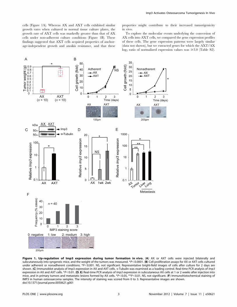
cells (Figure 1A). Whereas AX and AXT cells exhibited similar
growth rates when cultured in normal tissue culture plates, the
growth rate of AXT cells was markedly greater than that of AX
cells under non-adherent culture conditions (Figure 1B). These
findings suggested that AXT cells acquired properties of anchor-
age-independent growth and anoikis resistance, and that these
properties might contribute to their increased tumorigenicity
in vivo.
To explore the molecular events underlying the conversion of
AX cells into AXT cells, we compared the gene expression profiles
of these cells. The gene expression patterns were largely similar
(data not shown), but we extracted genes for which the AXT/AX
log2 ratio of normalized expression values was $3.0 (Table S2).
Figure 1. Up-regulation of Imp3 expression during tumor formation in vivo. (A) AX or AXT cells were injected bilaterally andsubcutaneously into syngeneic mice, and the weight of the tumors was measured. *P,0.0001. (B) Cell proliferation assays for AX or AXT cells culturedunder adherent or nonadherent conditions. *P,0.001. NS, not significant. Representative bright-field images of cells after culture for 2 days areshown. (C) Immunoblot analysis of Imp3 expression in AX and AXT cells. a-Tubulin was examined as a loading control. Real-time PCR analysis of Imp3expression in AX and AXT cells. *P,0.01. (D, E) Real-time PCR analysis of Imp3 expression in subcutaneous AX cells at 1 or 2 weeks after injection intomice, and in primary tumors and metastatic lesions formed by AX cells. *P,0.05, **P,0.01. NS, not significant. (F) Immunohistochemical staining ofIMP3 in human osteosarcoma samples. The intensity of staining was scored from 0 to 3. Representative images are shown.doi:10.1371/journal.pone.0050621.g001
Imp3 Activates Osteosarcoma Tumorigenesis In Vivo
PLOS ONE | www.plosone.org 3 November 2012 | Volume 7 | Issue 11 | e50621
From among the 38 identified genes, we selected candidate
molecules for further evaluation according to the following
criteria: The expression level (1) is low in normal tissue; (2) is
associated with poor prognosis in various human malignancies; (3)
increases during tumor development from AX cells; and (4) is
directly related to tumorigenic activity in vivo as revealed by
forced expression of the encoded protein in AX cells and its
depletion in AXT cells. We found that Imp3 meets all these
criteria, as shown below.
Imp3 is expressed predominantly during embryogenesis and in
various tumors [7–14], with its expression being limited to the
placenta and testis in normal adult mice [9]. Imp3 is thus
considered an oncofetal protein and is highly expressed in various
human malignancies [8,9,15,16]. The abundance of Imp3 was
markedly higher in AXT cells than in AX cells and Imp3
expression in AXT cells was .10 times that in AX cells
(Figure 1C). Of note, the amount of Imp3 mRNA in AX cells
after inoculation into syngeneic mice increased in a time-de-
pendent manner (Figure 1D), and it was significantly higher in
both primary and metastatic lesions than in parental AX cells
(Figure 1E). These results thus suggested that Imp3 expression in
AX cells is maintained at low level in vitro but is up-regulated
during tumor formation in vivo in association with the conversion
of AX cells into highly tumorigenic AXT cells.
IMP3 Expression in Human OsteosarcomaGiven that Imp3 expression appeared to be associated with an
aggressive phenotype of mouse osteosarcoma, we examined the
expression in human osteosarcoma. Immunohistochemical anal-
ysis of a tissue array containing 40 human osteosarcoma samples
showed that IMP3 was expressed in 36 (90%) of the specimens
(Figure 1F). Scoring of staining intensity from 0 to 3 revealed a high
expression level (score of 2 or 3) in 27 of the 40 samples (67.5%),
suggesting that deregulation of IMP3 expression occurs frequently
in human osteosarcoma.
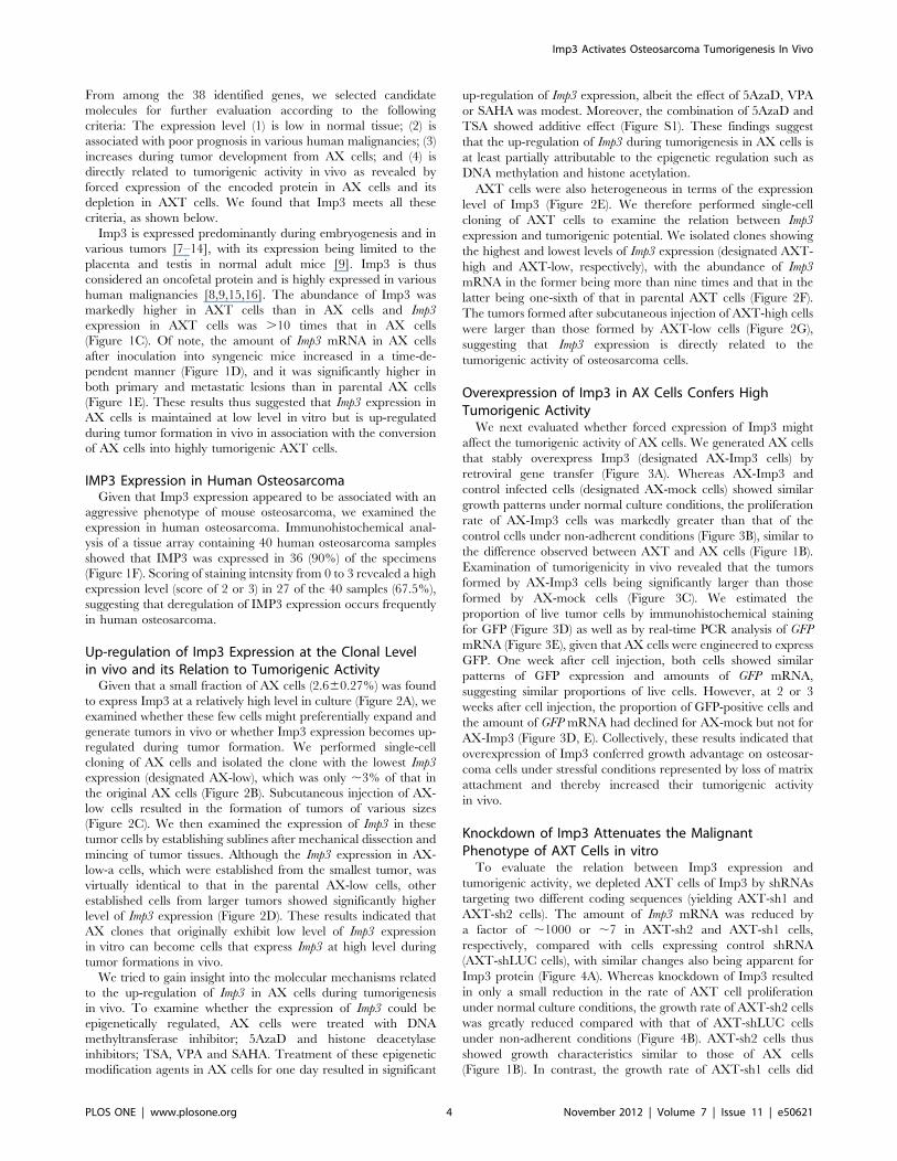
Up-regulation of Imp3 Expression at the Clonal Levelin vivo and its Relation to Tumorigenic ActivityGiven that a small fraction of AX cells (2.660.27%) was found
to express Imp3 at a relatively high level in culture (Figure 2A), we
examined whether these few cells might preferentially expand and
generate tumors in vivo or whether Imp3 expression becomes up-
regulated during tumor formation. We performed single-cell
cloning of AX cells and isolated the clone with the lowest Imp3
expression (designated AX-low), which was only ,3% of that in
the original AX cells (Figure 2B). Subcutaneous injection of AX-
low cells resulted in the formation of tumors of various sizes
(Figure 2C). We then examined the expression of Imp3 in these
tumor cells by establishing sublines after mechanical dissection and
mincing of tumor tissues. Although the Imp3 expression in AX-
low-a cells, which were established from the smallest tumor, was
virtually identical to that in the parental AX-low cells, other
established cells from larger tumors showed significantly higher
level of Imp3 expression (Figure 2D). These results indicated that
AX clones that originally exhibit low level of Imp3 expression
in vitro can become cells that express Imp3 at high level during
tumor formations in vivo.
We tried to gain insight into the molecular mechanisms related
to the up-regulation of Imp3 in AX cells during tumorigenesis
in vivo. To examine whether the expression of Imp3 could be
epigenetically regulated, AX cells were treated with DNA
methyltransferase inhibitor; 5AzaD and histone deacetylase
inhibitors; TSA, VPA and SAHA. Treatment of these epigenetic
modification agents in AX cells for one day resulted in significant
up-regulation of Imp3 expression, albeit the effect of 5AzaD, VPA
or SAHA was modest. Moreover, the combination of 5AzaD and
TSA showed additive effect (Figure S1). These findings suggest
that the up-regulation of Imp3 during tumorigenesis in AX cells is
at least partially attributable to the epigenetic regulation such as
DNA methylation and histone acetylation.
AXT cells were also heterogeneous in terms of the expression
level of Imp3 (Figure 2E). We therefore performed single-cell
cloning of AXT cells to examine the relation between Imp3
expression and tumorigenic potential. We isolated clones showing
the highest and lowest levels of Imp3 expression (designated AXT-
high and AXT-low, respectively), with the abundance of Imp3
mRNA in the former being more than nine times and that in the
latter being one-sixth of that in parental AXT cells (Figure 2F).
The tumors formed after subcutaneous injection of AXT-high cells
were larger than those formed by AXT-low cells (Figure 2G),
suggesting that Imp3 expression is directly related to the
tumorigenic activity of osteosarcoma cells.
Overexpression of Imp3 in AX Cells Confers HighTumorigenic ActivityWe next evaluated whether forced expression of Imp3 might
affect the tumorigenic activity of AX cells. We generated AX cells
that stably overexpress Imp3 (designated AX-Imp3 cells) by
retroviral gene transfer (Figure 3A). Whereas AX-Imp3 and
control infected cells (designated AX-mock cells) showed similar
growth patterns under normal culture conditions, the proliferation
rate of AX-Imp3 cells was markedly greater than that of the
control cells under non-adherent conditions (Figure 3B), similar to
the difference observed between AXT and AX cells (Figure 1B).
Examination of tumorigenicity in vivo revealed that the tumors
formed by AX-Imp3 cells being significantly larger than those
formed by AX-mock cells (Figure 3C). We estimated the
proportion of live tumor cells by immunohistochemical staining
for GFP (Figure 3D) as well as by real-time PCR analysis of GFP
mRNA (Figure 3E), given that AX cells were engineered to express
GFP. One week after cell injection, both cells showed similar
patterns of GFP expression and amounts of GFP mRNA,
suggesting similar proportions of live cells. However, at 2 or 3
weeks after cell injection, the proportion of GFP-positive cells and
the amount of GFP mRNA had declined for AX-mock but not for
AX-Imp3 (Figure 3D, E). Collectively, these results indicated that
overexpression of Imp3 conferred growth advantage on osteosar-
coma cells under stressful conditions represented by loss of matrix
attachment and thereby increased their tumorigenic activity
in vivo.
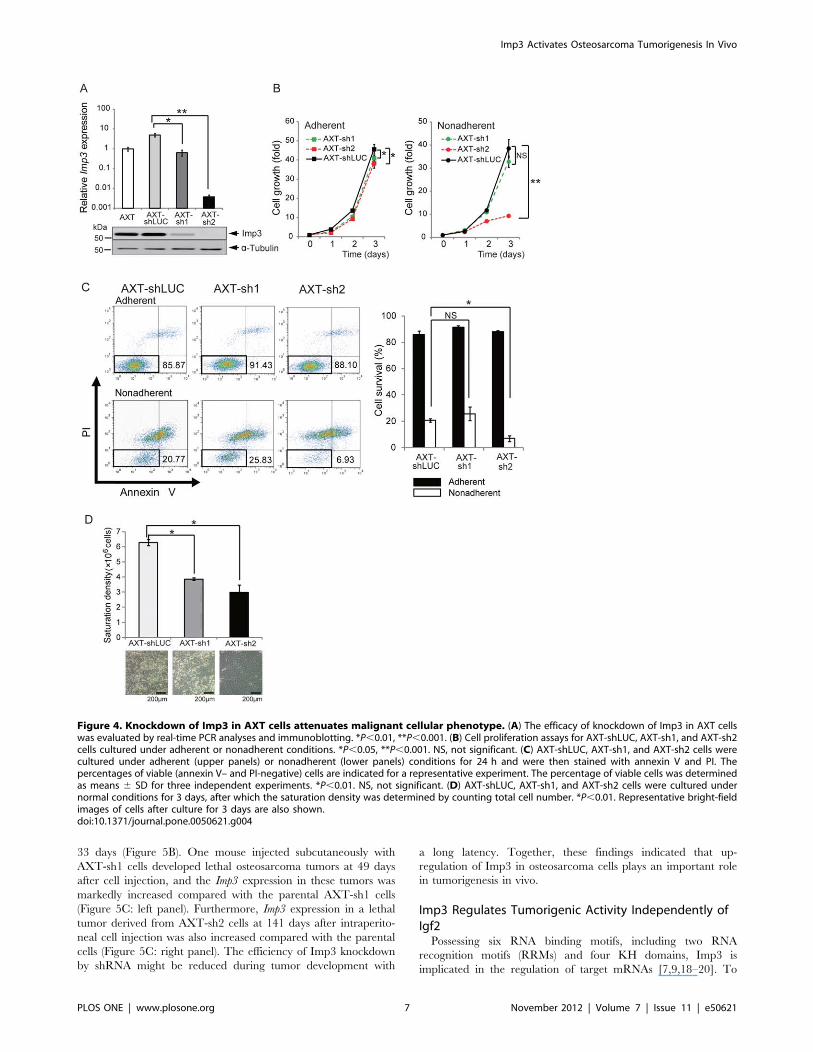
Knockdown of Imp3 Attenuates the MalignantPhenotype of AXT Cells in vitroTo evaluate the relation between Imp3 expression and
tumorigenic activity, we depleted AXT cells of Imp3 by shRNAs
targeting two different coding sequences (yielding AXT-sh1 and
AXT-sh2 cells). The amount of Imp3 mRNA was reduced by
a factor of ,1000 or ,7 in AXT-sh2 and AXT-sh1 cells,
respectively, compared with cells expressing control shRNA
(AXT-shLUC cells), with similar changes also being apparent for
Imp3 protein (Figure 4A). Whereas knockdown of Imp3 resulted
in only a small reduction in the rate of AXT cell proliferation
under normal culture conditions, the growth rate of AXT-sh2 cells
was greatly reduced compared with that of AXT-shLUC cells
under non-adherent conditions (Figure 4B). AXT-sh2 cells thus
showed growth characteristics similar to those of AX cells
(Figure 1B). In contrast, the growth rate of AXT-sh1 cells did
Imp3 Activates Osteosarcoma Tumorigenesis In Vivo
PLOS ONE | www.plosone.org 4 November 2012 | Volume 7 | Issue 11 | e50621
not differ significantly from the control cells under non-adherent
conditions (Figure 4B), likely as a result of the limited knockdown
of Imp3 in AXT-sh1 cells.
We next investigated anchorage-independent survival of AXT-
shLUC and Imp3-depleted AXT cells. Flow cytometric analysis of
cells stained with annexin V and propidium iodide (PI) revealed
that the size of the double-negative (viable) population was equally
large for each cell line under adherent conditions (Figure 4C).
However, under non-adherent conditions, the size of the viable
population was significantly smaller for AXT-sh2 than for AXT-
shLUC cells. Again, similar to the cell growth pattern, the
anchorage-independent survival of AXT-sh1 cells did not differ
significantly from the control cells. These findings suggested that
the up-regulation of Imp3 expression in tumor cells might
contribute to their escape from anoikis.
Loss of contact inhibition and consequent overgrowth to a high
density is key malignant properties of transformed cells [1,17]. We
cultured AXT-shLUC and Imp3-depleted AXT cells to conflu-
ence and determined the saturation density. Whereas AXT-
shLUC cells continued to grow past confluence, resulting in the
formation of large piles and high saturation density, AXT-sh1 and
AXT-sh2 cells manifested contact inhibition and lower saturation
density (Figure 4D). Aberrant Imp3 expression in tumor cells may
thus promote anchorage-independent growth and loss of contact
inhibition.
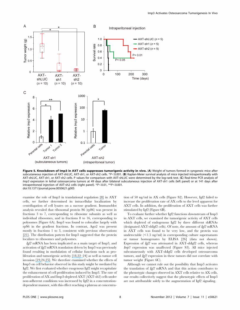
Knockdown of Imp3 in AXT Cells SuppressesTumorigenic Activity in vivoWe next examined whether knockdown of Imp3 in AXT cells
might affect tumorigenic activity in vivo. Whereas all mice
injected subcutaneously with Imp3-depleted AXT cells developed
palpable tumors, the weight of these tumors was significantly
smaller than those derived from AXT-shLUC cells (Figure 5A).
None of the mice injected with Imp3-depleted AXT cells
manifested lung or liver metastasis, whereas all mice injected with
AXT-shLUC cells developed metastases at both sites (Table S3).
We also evaluated tumorigenic activity after intraperitoneal
injection of osteosarcoma cells, which resulted in earlier death
from primary tumors. Whereas AXT-sh2 cells did not generate
lethal tumors within .140 days, AXT-shLUC cells did so within
Figure 2. Tumorigenic activity of osteosarcoma cells correlates with Imp3. (A) Immunofluorescence analysis of Imp3 expression in AX cells.The boxed region is shown at higher magnification in the lower panel. (B) Real-time PCR analysis of Imp3 expression in AX cells and in an AX subclone(designated AX-low) obtained by single-cell cloning. *P,0.01. (C) Weight of tumors derived from subcutaneously injected AX-low cells. (D) Real-timePCR analysis of Imp3 expression in AX-low cells as well as in the AX-low cell-derived tumors. *P,0.05, **P,0.01, ***P,0.001. NS, not significant. (E)Immunofluorescence analysis of Imp3 expression in AXT cells. The boxed region is shown at higher magnification in the lower panel. (F) AXTsubclones (designated AXT-high and AXT-low) isolated by single-cell cloning were subjected to real-time PCR analysis of Imp3 as well as toimmunofluorescence analysis of Imp3 protein. (G) Weight of tumors derived from subcutaneously injected AXT-high or AXT-low cells. *P,0.001.doi:10.1371/journal.pone.0050621.g002
Imp3 Activates Osteosarcoma Tumorigenesis In Vivo
PLOS ONE | www.plosone.org 5 November 2012 | Volume 7 | Issue 11 | e50621
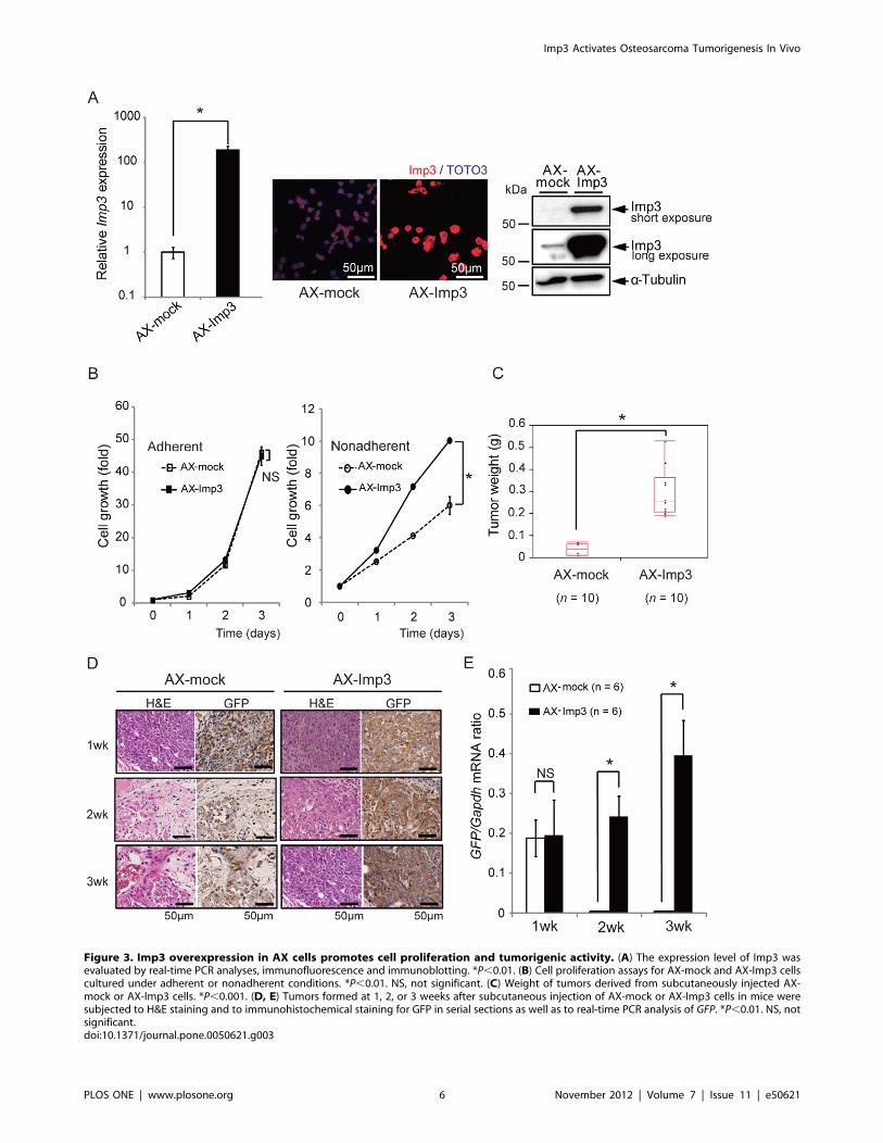
Figure 3. Imp3 overexpression in AX cells promotes cell proliferation and tumorigenic activity. (A) The expression level of Imp3 wasevaluated by real-time PCR analyses, immunofluorescence and immunoblotting. *P,0.01. (B) Cell proliferation assays for AX-mock and AX-Imp3 cellscultured under adherent or nonadherent conditions. *P,0.01. NS, not significant. (C) Weight of tumors derived from subcutaneously injected AX-mock or AX-Imp3 cells. *P,0.001. (D, E) Tumors formed at 1, 2, or 3 weeks after subcutaneous injection of AX-mock or AX-Imp3 cells in mice weresubjected to H&E staining and to immunohistochemical staining for GFP in serial sections as well as to real-time PCR analysis of GFP. *P,0.01. NS, notsignificant.doi:10.1371/journal.pone.0050621.g003
Imp3 Activates Osteosarcoma Tumorigenesis In Vivo
PLOS ONE | www.plosone.org 6 November 2012 | Volume 7 | Issue 11 | e50621
33 days (Figure 5B). One mouse injected subcutaneously with
AXT-sh1 cells developed lethal osteosarcoma tumors at 49 days
after cell injection, and the Imp3 expression in these tumors was
markedly increased compared with the parental AXT-sh1 cells
(Figure 5C: left panel). Furthermore, Imp3 expression in a lethal
tumor derived from AXT-sh2 cells at 141 days after intraperito-
neal cell injection was also increased compared with the parental
cells (Figure 5C: right panel). The efficiency of Imp3 knockdown
by shRNA might be reduced during tumor development with
a long latency. Together, these findings indicated that up-
regulation of Imp3 in osteosarcoma cells plays an important role
in tumorigenesis in vivo.
Imp3 Regulates Tumorigenic Activity Independently ofIgf2Possessing six RNA binding motifs, including two RNA
recognition motifs (RRMs) and four KH domains, Imp3 is
implicated in the regulation of target mRNAs [7,9,18–20]. To
Figure 4. Knockdown of Imp3 in AXT cells attenuates malignant cellular phenotype. (A) The efficacy of knockdown of Imp3 in AXT cellswas evaluated by real-time PCR analyses and immunoblotting. *P,0.01, **P,0.001. (B) Cell proliferation assays for AXT-shLUC, AXT-sh1, and AXT-sh2cells cultured under adherent or nonadherent conditions. *P,0.05, **P,0.001. NS, not significant. (C) AXT-shLUC, AXT-sh1, and AXT-sh2 cells werecultured under adherent (upper panels) or nonadherent (lower panels) conditions for 24 h and were then stained with annexin V and PI. Thepercentages of viable (annexin V– and PI-negative) cells are indicated for a representative experiment. The percentage of viable cells was determinedas means 6 SD for three independent experiments. *P,0.01. NS, not significant. (D) AXT-shLUC, AXT-sh1, and AXT-sh2 cells were cultured undernormal conditions for 3 days, after which the saturation density was determined by counting total cell number. *P,0.01. Representative bright-fieldimages of cells after culture for 3 days are also shown.doi:10.1371/journal.pone.0050621.g004
Imp3 Activates Osteosarcoma Tumorigenesis In Vivo
PLOS ONE | www.plosone.org 7 November 2012 | Volume 7 | Issue 11 | e50621
examine the role of Imp3 in translational regulation [8] in AXT
cells, we further determined its intracellular localization by
centrifugation of cell lysates on a sucrose gradient. Immunoblot
analysis revealed that ribosomal protein S6 (rpS6) was present in
fractions 3 to 7, corresponding to ribosome subunits as well as
individual ribosomes, and in fractions 8 to 16, corresponding to
polysomes (Figure 6A). Imp3 was found to colocalize largely with
rpS6 in the gradient fractions. In contrast, Ago2 was present
mostly in fractions 1 to 3, consistent with previous observations
[21]. The distribution pattern for Imp3 suggested that the protein
localizes to ribosomes and polysomes.
Igf2 mRNA has been implicated as a main target of Imp3, and
activation of Igf2mRNA translation driven by Imp3 was previously
found resulting in modulation of cellular functions such as pro-
liferation and tumorigenic activity [18,22–24] as well as tumor cell
invasion [19,24,25]. We therefore examined whether the effects of
Imp3 on cell behavior observed in this study might be mediated by
Igf2. We first evaluated whether exogenous Igf2 might recapitulate
the enhancement of cell proliferation induced by Imp3. The rate of
proliferation of AX and Imp3-depleted AXT (AXT-sh2) cells under
non-adherent conditions was increased by Igf2 in a concentration-
dependent manner, with this effect reaching a plateau at concentra-
tion of 50 ng/ml in AX cells (Figure S2). However, Igf2 failed to
increase the proliferation rate of AX cells to the level apparent for
AXT cells. In addition, the proliferation of AXT cells was further
stimulated by Igf2 (Figure 6B).
To evaluate further whether Igf2 functions downstream of Imp3
in AXT cells, we examined the tumorigenic activity of AXT cells
which depleted of endogenous Igf2 by three different shRNAs
(designated AXT-shIgf2 cells). Of note, the amount of Igf2 mRNA
in AXT cells was found to be very low, and the protein was
undetectable (,1.5 ng/ml) in corresponding culture supernatants
or tumor homogenates by ELISA [26] (data not shown).
Expression of Igf2 was attenuated in AXT-shIgf2 cells, whereas
Imp3 expression was unaffected (Figure S3). All mice injected
subcutaneously with AXT-shIgf2 cells developed osteosarcoma
tumors, and Igf2 expression in these tumors did not correlate with
tumor weight (Figure 6C).
Although we cannot rule out the possibility that Imp3 activates
the translation of Igf2 mRNA and that this action contributes to
the phenotypic changes observed in AXT cells relative to AX cells,
our results collectively suggest that the phenotypic effects of Imp3
are not attributable solely to the augmentation of Igf2 signaling.
Figure 5. Knockdown of Imp3 in AXT cells suppresses tumorigenic activity in vivo. (A) Weight of tumors formed in syngeneic mice aftersubcutaneous injection of AXT-shLUC, AXT-sh1, or AXT-sh2 cells. *P,0.001. (B) Kaplan-Meier survival analysis of mice injected intraperitoneally withAXT-shLUC, AXT-sh1, or AXT-sh2 cells. P values for comparison with AXT-shLUC were determined by the log-rank test. (C) Real-time PCR analysis ofImp3 expression in lethal osteosarcoma tumors at 49 days after bilateral subcutaneous injection of AXT-sh1 cells (left panel) or at 141 days afterintraperitoneal injection of AXT-sh2 cells (right panel). *P,0.01, **P,0.001.doi:10.1371/journal.pone.0050621.g005
Imp3 Activates Osteosarcoma Tumorigenesis In Vivo
PLOS ONE | www.plosone.org 8 November 2012 | Volume 7 | Issue 11 | e50621
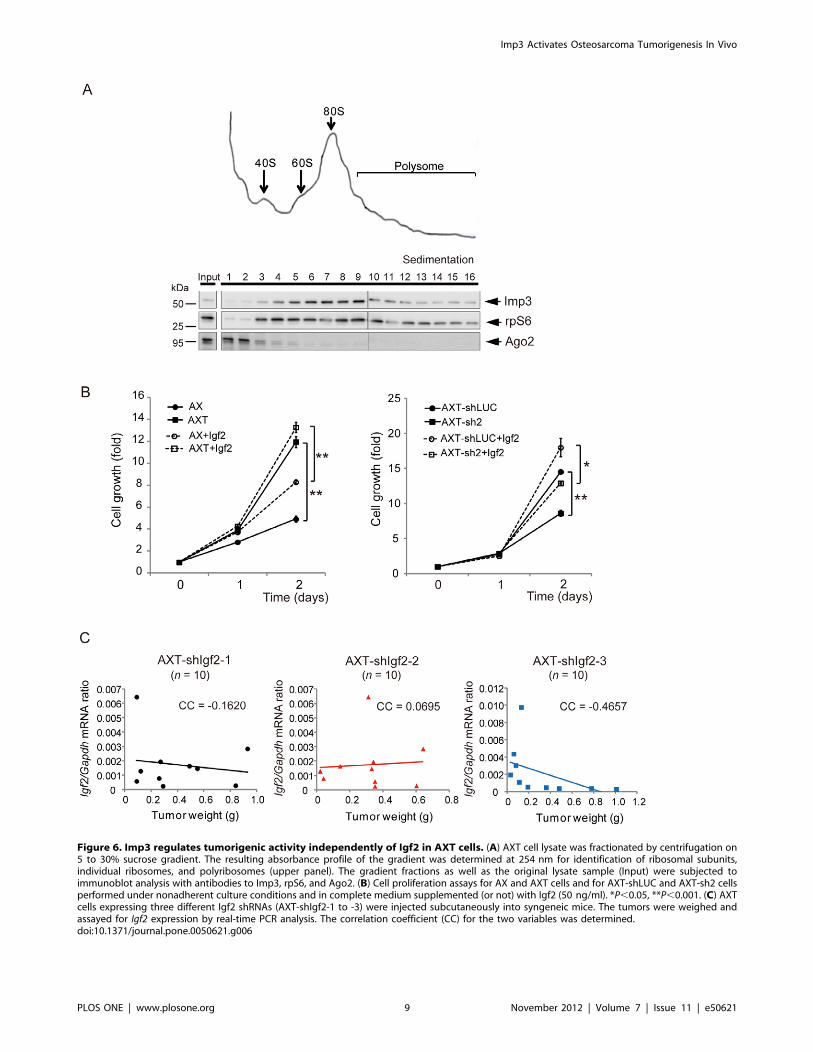
Figure 6. Imp3 regulates tumorigenic activity independently of Igf2 in AXT cells. (A) AXT cell lysate was fractionated by centrifugation on5 to 30% sucrose gradient. The resulting absorbance profile of the gradient was determined at 254 nm for identification of ribosomal subunits,individual ribosomes, and polyribosomes (upper panel). The gradient fractions as well as the original lysate sample (Input) were subjected toimmunoblot analysis with antibodies to Imp3, rpS6, and Ago2. (B) Cell proliferation assays for AX and AXT cells and for AXT-shLUC and AXT-sh2 cellsperformed under nonadherent culture conditions and in complete medium supplemented (or not) with Igf2 (50 ng/ml). *P,0.05, **P,0.001. (C) AXTcells expressing three different Igf2 shRNAs (AXT-shIgf2-1 to -3) were injected subcutaneously into syngeneic mice. The tumors were weighed andassayed for Igf2 expression by real-time PCR analysis. The correlation coefficient (CC) for the two variables was determined.doi:10.1371/journal.pone.0050621.g006
Imp3 Activates Osteosarcoma Tumorigenesis In Vivo
PLOS ONE | www.plosone.org 9 November 2012 | Volume 7 | Issue 11 | e50621
Discussion
Cancer cells acquire various malignant properties during the
disease course. The aim of this study was to identify molecules that
contribute to such changes in cancer cells with the use of two
newly established osteosarcoma cell lines, AX and AXT. AXT
cells, which were isolated from osteosarcoma tumors formed by
AX cells, manifest a tumorigenic activity in vivo greater than that
of AX cells. We identified Imp3 as a key molecule that contributes
to the acquisition of malignant properties by AX cells and their
associated conversion into AXT cells.
The up-regulation of Imp3 expression in tumors formed by AX
cells in vivo was found not to be attributable simply to the
expansion of the small population of cells that initially expresses
Imp3 at high level, but was instead due to the induction of Imp3
expression during tumorigenesis. This result has important
implications with regard to the evaluation of cellular tumorige-
nicity and the notion of cancer stem cells, in that it shows that the
properties of cancer cells can change markedly in vivo.
The molecular mechanisms of re-emergence of oncofetal
proteins in cancer cells remain to be fully elucidated. Our findings
suggest that the up-regulation of Imp3 expression during osteosar-
coma formations could be partially attributable to epigenitic
modifications (Figure S1). Previous reports indicated that IMP3
expression could be regulated by growth factor signaling in breast
cancer cells [27] or miRNA in Drosophila [28]. Treatment of
epigenetic modification drugs with AX cells could not fully
recapitulate the high expression level of Imp3 in AXT cells
(Figure 1C and Figure S1), therefore, other upstream mechanisms
could be involved during tumorigenesis in AX cells. In contrast,
Ink4a/Arf knockout stromal cells, which are parental cells for AX
cells, did not exhibit as much response to the epigenetic modifiers
as AX cells (data not shown), which might reflect the differential
plasticity in epigenetic regulation between normal cells and cancer
cells [29].
Both gain and loss of function of Imp3 in osteosarcoma cells
revealed that Imp3 confers the ability to undergo anchorage-
independent growth, loss of contact inhibition, and resistance to
anoikis in vitro, all of which contribute to the development of
tumorgenic potential. Previous studies reported that Imp3
enhances cell proliferation and invasion [14,18,22,24,25,27].
Collectively Imp3 might contribute to the regulation of molecules
involved in cell cycle and remodeling of cytoskeleton.
We found that Imp3 was associated with individual ribosomes,
ribosome subunits, and polysomes in AXT cells, consistent with
the proposed role for Imp3 in the regulation of translation
[7,8,18]. The oncogenic effects of Imp3 have been suggested to be
mediated through Igf2, the mRNA for which is translationally
activated by Imp3 [18,22–24]. However, our findings indicate that
the malignant properties conferred by Imp3 are not attributable to
the action of Igf2 alone. The Igf2 expression in AXT cells was thus
found to be extremely low, and the encoded protein in tumors was
not detectable with ELISA. Furthermore, exogenous Igf2 did not
provide growth advantage for AX cells as great as that conferred
by Imp3 expression, and shRNA-mediated suppression of
endogenous Igf2 expression did not affect the tumorigenic activity
of AXT cells. Translational regulation of several molecules such as
CD44, CD164, and MMP9 has been suggested to underlie
changes in cellular phenotype induced by Imp3, without being
accompanied by modification of Igf2 [25,27]. Collectively, our
results suggest that deregulation of Imp3 expression in AXT cells
might affect the expression of key molecules other than Igf2, as has
been suggested previously [20].
We found that knockdown of Imp3 in AXT cells resulted in
a marked reduction in tumorigenic activity in vivo. Moreover,
90% of the human osteosarcoma specimens analyzed were positive
for Imp3 expression. IMP3 has previously been suggested as
a prognostic marker for metastatic or angiogenic potential in
human osteosarcoma [30,31]. Our results implicate Imp3 as
a molecule capable of conferring critical properties to transformed
cells for tumorigenic ability in vivo, which is indispensable for
tumor-initiating cells, often consistent with cancer stem cells
[32,33]. Thus Imp3 might be a key regulator of cancer stem-like
characteristics in cancer cells, in which case it may also be
a potential therapeutic target for osteosarcoma as well as other
tumor types.
Supporting Information
Figure S1 Effects of epigenetic modifiers on Imp3expression. Real-time PCR analysis of Imp3 expression in AX
cells after treatment with DNMT1 inhibitor; 5AzaD and HDAC
inhibitors; TSA, VPA or SAHA at the indicated concentration.
*P,0.05, **P,0.01, ***P,0.001. NS, not significant.
(TIF)
Figure S2 Effect of Igf2 on osteosarcoma cell prolifer-ation in vitro. The proliferation of AX and AXT-sh2 cells was
assayed under nonadherent culture conditions supplemented with
the indicated concentrations (0 to 500 ng/ml) of Igf2.
(TIF)
Figure S3 Depletion of Igf2 mRNA in AXT cells. The
expression levels of Igf2 and Imp3 in AXT-shIgf2 cells were
evaluated by real-time PCR analysis.
(TIF)
Table S1 Sequences of PCR primers, predicted PCRproduct sizes, and target sequences for shRNAs.
(DOCX)
Table S2 Genes whose expression is up-regulated inAXT cells compared with AX cells.
(DOCX)
Table S3 Knockdown of Imp3 in AXT cells suppressestumorigenic activity in vivo.
(DOCX)
Acknowledgments
We thank Prof. H. Siomi and Prof. M. C. Siomi for helpful advice, I.
Ishimatsu for technical assistance and K. Arai for secretarial assistance.
Author Contributions
Conceived and designed the experiments: AU TS TI ES NO K. Miyoshi
HS. Performed the experiments: AU TS K. Masuda SIY TI KMi.
Analyzed the data: AU TS K. Miyoshi HS. Contributed reagents/
materials/analysis tools: TI ES NO SK AM YT KB DA HS. Wrote the
paper: AU TS HS.
References
1. Hanahan D, Weinberg RA (2000) The hallmarks of cancer. Cell 100: 57–70.
2. Simpson CD, Anyiwe K, Schimmer AD (2008) Anoikis resistance and tumor
metastasis. Cancer Lett 272: 177–185.
3. Shimizu T, Ishikawa T, Sugihara E, Kuninaka S, Miyamoto T, et al. (2010) c-MYC overexpression with loss of Ink4a/Arf transforms bone marrow stromal
cells into osteosarcoma accompanied by loss of adipogenesis. Oncogene 29:
5687–5699.
Imp3 Activates Osteosarcoma Tumorigenesis In Vivo
PLOS ONE | www.plosone.org 10 November 2012 | Volume 7 | Issue 11 | e50621
4. Thullberg M, Stromblad S (2008) Anchorage-independent cytokinesis as part of
oncogenic transformation? Cell Cycle 7: 984–988.5. Fujino RS, Tanaka K, Morimatsu M, Tamura K, Kogo H, et al. (2006)
Spermatogonial cell-mediated activation of an IkappaBzeta-independent nuclear
factor-kappaB pathway in Sertoli cells induces transcription of the lipocalin-2gene. Mol Endocrinol 20: 904–915.
6. Kitamura T, Koshino Y, Shibata F, Oki T, Nakajima H, et al. (2003)Retrovirus-mediated gene transfer and expression cloning: powerful tools in
functional genomics. Exp Hematol 31: 1007–1014.
7. Nielsen FC, Nielsen J, Christiansen J (2001) A family of IGF-II mRNA bindingproteins (IMP) involved in RNA trafficking. Scand J Clin Lab Invest Suppl 234:
93–99.8. Nielsen J, Christiansen J, Lykke-Andersen J, Johnsen AH, Wewer UM, et al.
(1999) A family of insulin-like growth factor II mRNA-binding proteins repressestranslation in late development. Mol Cell Biol 19: 1262–1270.
9. Mori H, Sakakibara S, Imai T, Nakamura Y, Iijima T, et al. (2001) Expression
of mouse igf2 mRNA-binding protein 3 and its implications for the developingcentral nervous system. J Neurosci Res 64: 132–143.
10. Mueller-Pillasch F, Lacher U, Wallrapp C, Micha A, Zimmerhackl F, et al.(1997) Cloning of a gene highly overexpressed in cancer coding for a novel KH-
domain containing protein. Oncogene 14: 2729–2733.
11. Jiang Z, Chu PG, Woda BA, Rock KL, Liu Q, et al. (2006) Analysis of RNA-binding protein IMP3 to predict metastasis and prognosis of renal-cell
carcinoma: a retrospective study. Lancet Oncol 7: 556–564.12. Chen ST, Jeng YM, Chang CC, Chang HH, Huang MC, et al. (2011) Insulin-
like growth factor II mRNA-binding protein 3 expression predicts unfavorableprognosis in patients with neuroblastoma. Cancer Sci 102: 2191–2198.
13. Jeng YM, Chang CC, Hu FC, Chou HY, Kao HL, et al. (2008) RNA-binding
protein insulin-like growth factor II mRNA-binding protein 3 expressionpromotes tumor invasion and predicts early recurrence and poor prognosis in
hepatocellular carcinoma. Hepatology 48: 1118–1127.14. Kabbarah O, Nogueira C, Feng B, Nazarian RM, Bosenberg M, et al. (2010)
Integrative genome comparison of primary and metastatic melanomas. PLoS
One 5: e10770.15. Yaniv K, Yisraeli JK (2002) The involvement of a conserved family of RNA
binding proteins in embryonic development and carcinogenesis. Gene 287: 49–54.
16. Mueller F, Bommer M, Lacher U, Ruhland C, Stagge V, et al. (2003) KOC isa novel molecular indicator of malignancy. Br J Cancer 88: 699–701.
17. Wang JB, Erickson JW, Fuji R, Ramachandran S, Gao P, et al. (2010) Targeting
mitochondrial glutaminase activity inhibits oncogenic transformation. CancerCell 18: 207–219.
18. Liao B, Hu Y, Herrick DJ, Brewer G (2005) The RNA-binding protein IMP-3 isa translational activator of insulin-like growth factor II leader-3 mRNA during
proliferation of human K562 leukemia cells. J Biol Chem 280: 18517–18524.
19. Nielsen FC, Nielsen J, Kristensen MA, Koch G, Christiansen J (2002)
Cytoplasmic trafficking of IGF-II mRNA-binding protein by conserved KHdomains. J Cell Sci 115: 2087–2097.
20. Hafner M, Landthaler M, Burger L, Khorshid M, Hausser J, et al. (2010)
Transcriptome-wide identification of RNA-binding protein and microRNAtarget sites by PAR-CLIP. Cell 141: 129–141.
21. Hock J, Weinmann L, Ender C, Rudel S, Kremmer E, et al. (2007) Proteomicand functional analysis of Argonaute-containing mRNA-protein complexes in
human cells. EMBO Rep 8: 1052–1060.
22. Liao B, Hu Y, Brewer G (2011) RNA-binding protein insulin-like growth factormRNA-binding protein 3 (IMP-3) promotes cell survival via insulin-like growth
factor II signaling after ionizing radiation. J Biol Chem 286: 31145–31152.23. Liao B, Patel M, Hu Y, Charles S, Herrick DJ, et al. (2004) Targeted knockdown
of the RNA-binding protein CRD-BP promotes cell proliferation via an insulin-like growth factor II-dependent pathway in human K562 leukemia cells. J Biol
Chem 279: 48716–48724.
24. Suvasini R, Shruti B, Thota B, Shinde SV, Friedmann-Morvinski D, et al. (2011)Insulin Growth Factor-2 Binding Protein 3 (IGF2BP3) Is a Glioblastoma-specific
Marker That Activates Phosphatidylinositol 3-Kinase/Mitogen-activated Pro-tein Kinase (PI3K/MAPK) Pathways by Modulating IGF-2. J Biol Chem 286:
25882–25890.
25. Vikesaa J, Hansen TV, Jonson L, Borup R, Wewer UM, et al. (2006) RNA-binding IMPs promote cell adhesion and invadopodia formation. EMBO J 25:
1456–1468.26. Shimizu T, Ishikawa T, Iwai S, Ueki A, Sugihara E, et al. (2012) Fibroblast
Growth Factor-2 Is an Important Factor that Maintains Cellular Immaturityand Contributes to Aggressiveness of Osteosarcoma. Mol Cancer Res 10: 454–
468.
27. Samanta S, Sharma VM, Khan A, Mercurio AM (2012) Regulation of IMP3 byEGFR signaling and repression by ERbeta: implications for triple-negative
breast cancer. Oncogene.28. Toledano H, D’Alterio C, Czech B, Levine E, Jones DL (2012) The let-7-Imp
axis regulates ageing of the Drosophila testis stem-cell niche. Nature 485: 605–
610.29. Berdasco M, Esteller M (2010) Aberrant epigenetic landscape in cancer: how
cellular identity goes awry. Dev Cell 19: 698–711.30. Do SI, Kim YW, Park HR, Park YK (2008) Expression of insulin-like growth
factor-II mRNA binding protein 3 (IMP3) in osteosarcoma. Oncol Res 17: 269–272.
31. Chen P, Wang SJ, Wang HB, Ren P, Wang XQ, et al. (2012) The distribution of
IGF2 and IMP3 in osteosarcoma and its relationship with angiogenesis. J MolHistol 43: 63–70.
32. Visvader JE (2011) Cells of origin in cancer. Nature 469: 314–322.33. Magee JA, Piskounova E, Morrison SJ (2012) Cancer stem cells: impact,
heterogeneity, and uncertainty. Cancer Cell 21: 283–296.
Imp3 Activates Osteosarcoma Tumorigenesis In Vivo
PLOS ONE | www.plosone.org 11 November 2012 | Volume 7 | Issue 11 | e50621
Related Documents