J Med Genet 1996;33:469-474 Unusual molecular findings in autosomal recessive spinal muscular atrophy Gert Matthijs, Els Schollen, Eric Legius, Koen Devriendt, Nathalie Goemans, Hulya Kayserili, Memnune Yiiksel Apak, Jean-Jacques Cassiman Abstract All three types of autosomal recessive spinal muscular atrophy map to chro- mosome 5q11.2-q13.3 and are associated with deletions or mutations of the SMN (survival motor neurone) gene. The avail- ability of a test to distinguish between the SMN gene and its nearly identical cen- tromeric copy cBCD541 allows molecular diagnosis. We have analysed patients from 24 Belgian and 34 Turkish families for the presence or absence of a deletion in the SMN gene. A homozygous deletion in the SMN gene was seen in 90% of unrelated SMA patients. A non-radioactive SSCP assay allows for a semiquantitative anal- ysis of the copy number of the centromeric and SMN genes. Hence, direct carrier de- tection has become feasible under certain conditions. We observed a phenotypically normal male, father of an SMA type I patient, presenting with only a single copy of the SMN gene and lacking both copies of the cBCD541 gene. This illustrates that a reduction of the total number of SMN and cBCD541 genes to a single SMN copy is compatible with normal life. In another SMA type I family, there is evidence for a de novo deletion of the centromeric gene in a normal sib. This observation il- lustrates the susceptibility of the SMA locus to de novo deletions and re- arrangements. (J7 Med Genet 1996;33:469-474) Key words: spinal muscular atrophy; SMN gene; non- radioactive SSCP. Spinal muscular atrophy (SMA) is a lethal autosomal recessive disease common in white populations (1 in 6000 newborns).' SMA is characterised by degeneration of the anterior horn cells of the spinal cord, leading to sym- metrical paralysis of voluntary muscles with muscular atrophy. Three different clinical syn- dromes can be defined on the basis of severity. The acute form of SMA, SMA type I (Werdnig- Hoffmann disease) starts with hypotonia and weakness in utero or within the first few months of life. These children are never able to sit unsupported. The majority of these children die within the first three years of life because of recurrent respiratory infections. In the inter- mediate type of SMA (Dubowitz disease or SMA type II) onset is usually between 3 and 15 months of age. These children learn to sit without support but never manage to stand or walk unaided. They survive beyond 4 years of age until adolescence or later. The onset of the chronic form of SMA, SMA type III (Kugelberg-Welander disease) is after the age of 2 years and is characterised by proximal muscle weakness, predominantly of the legs. These patients manage to stand and walk un- supported, but have problems with running, jumping, and climbing. Their walking distance is limited. The long term survival is usually good, depending on respiratory function. All three types map to chromosome region 5ql1.2-q13.3. Recently, Lefebvre et a12 and Roy et al3 have described the genomic structure of the SMA locus, although both groups identified a different gene. The region is characterised by an inverted duplication of ap- proximately 500 kb. Patients with SMA have deletions in the telomeric repeating unit. Lefebvre et al2 identified a gene in this region, which is called SMN (Survival Motor Neur- one, TBCD54 1), and contains eight exons extending over approximately 20 kb. There is an almost identical copy of this gene in the centromeric repeating unit (CBCD541). In the series of Lefebvre et al,2 the vast majority (98&6%) of patients have a deletion in the SMN gene, whereas point mutations have been identified in the remaining three of 229 patients, indicating that mutations in this gene may indeed cause SMA. In the same region, Roy et al' identified the NAIP gene (Neuronal Apoptosis Inhibitory Protein) with 16 exons spanning 60 kb. They showed that 45% of SMA type I and 18% of SMA type II and III patients have a partial or complete homozygous deletion of the NAIP gene.3 However, further analysis showed that other loci are probably involved in the pathogenesis. In summary, it is hypothesised that mutations in the SMN gene are the major determinant of the SMA pheno- type, whereas the extent of the deletions, which may include the NAIP gene, may correlate with the severity of the disease.2 3 We report on the presence or absence of a deletion in the SMN gene in 58 SMA patients, using a non-radioactive SSCP (single stranded conformation polymorphism) assay. This test discriminates between the centromeric gene CBCD541 and telomeric SMN gene and allows for the quantification of the different copies. In some cases it can be used for carrier testing. Together with the overall data, two unusual cases are described. Materials and methods SUBJECTS AND FAMILY DATA Twenty-four families are of Belgian origin. All samples from these SMA families had pre- Center for Human Genetics, University of Leuven, Gasthuisberg O&N6, B-3000 Leuven, Belgium G Matthijs E Schollen E Legius K Devriendt J-J Cassiman Department of Paediatrics, University Hospital Leuven, Gasthuisberg, B-3000 Leuven, Belgium N Goemans Division of Medical Genetics, Institute of Child Health, University of Istanbul, Millet Caddesi Capa, 34390 Istanbul, Turkey H Kayserili M Yuiksel Apak Correspondence to: Dr Matthijs. Received 15 September 1995 Revised version accepted for publication 16 January 1996 469 group.bmj.com on July 14, 2011 - Published by jmg.bmj.com Downloaded from

Welcome message from author
This document is posted to help you gain knowledge. Please leave a comment to let me know what you think about it! Share it to your friends and learn new things together.
Transcript
J Med Genet 1996;33:469-474
Unusual molecular findings in autosomalrecessive spinal muscular atrophy
Gert Matthijs, Els Schollen, Eric Legius, Koen Devriendt, Nathalie Goemans,Hulya Kayserili, Memnune Yiiksel Apak, Jean-Jacques Cassiman
AbstractAll three types of autosomal recessivespinal muscular atrophy map to chro-mosome 5q11.2-q13.3 and are associatedwith deletions or mutations of the SMN(survival motor neurone) gene. The avail-ability of a test to distinguish between theSMN gene and its nearly identical cen-tromeric copy cBCD541 allows moleculardiagnosis. We have analysed patients from24 Belgian and 34 Turkish families for thepresence or absence of a deletion in theSMN gene. A homozygous deletion in theSMN gene was seen in 90% of unrelatedSMA patients. A non-radioactive SSCPassay allows for a semiquantitative anal-ysis ofthe copy number ofthe centromericand SMN genes. Hence, direct carrier de-tection has become feasible under certainconditions. We observed a phenotypicallynormal male, father of an SMA type Ipatient, presenting with only a single copyof the SMN gene and lacking both copiesof the cBCD541 gene. This illustrates thata reduction of the total number of SMNand cBCD541 genes to a single SMN copyis compatible with normal life. In anotherSMA type I family, there is evidence for ade novo deletion of the centromeric genein a normal sib. This observation il-lustrates the susceptibility of the SMAlocus to de novo deletions and re-arrangements.(J7 Med Genet 1996;33:469-474)
Key words: spinal muscular atrophy; SMN gene; non-radioactive SSCP.
Spinal muscular atrophy (SMA) is a lethalautosomal recessive disease common in whitepopulations (1 in 6000 newborns).' SMA ischaracterised by degeneration of the anteriorhorn cells of the spinal cord, leading to sym-metrical paralysis of voluntary muscles withmuscular atrophy. Three different clinical syn-dromes can be defined on the basis of severity.The acute form ofSMA, SMA type I (Werdnig-Hoffmann disease) starts with hypotonia andweakness in utero or within the first few monthsof life. These children are never able to situnsupported. The majority of these childrendie within the first three years of life becauseof recurrent respiratory infections. In the inter-mediate type of SMA (Dubowitz disease orSMA type II) onset is usually between 3 and15 months of age. These children learn to sitwithout support but never manage to stand orwalk unaided. They survive beyond 4 years of
age until adolescence or later. The onsetof the chronic form of SMA, SMA type III(Kugelberg-Welander disease) is after the ageof 2 years and is characterised by proximalmuscle weakness, predominantly of the legs.These patients manage to stand and walk un-supported, but have problems with running,jumping, and climbing. Their walking distanceis limited. The long term survival is usuallygood, depending on respiratory function.
All three types map to chromosome region5ql1.2-q13.3. Recently, Lefebvre et a12 andRoy et al3 have described the genomic structureof the SMA locus, although both groupsidentified a different gene. The region ischaracterised by an inverted duplication of ap-proximately 500 kb. Patients with SMA havedeletions in the telomeric repeating unit.Lefebvre et al2 identified a gene in this region,which is called SMN (Survival Motor Neur-one, TBCD54 1), and contains eight exonsextending over approximately 20 kb. There isan almost identical copy of this gene in thecentromeric repeating unit (CBCD541). In theseries of Lefebvre et al,2 the vast majority(98&6%) of patients have a deletion in theSMN gene, whereas point mutations have beenidentified in the remaining three of 229patients, indicating that mutations in this genemay indeed cause SMA. In the same region,Roy et al' identified the NAIP gene (NeuronalApoptosis Inhibitory Protein) with 16 exonsspanning 60 kb. They showed that 45% ofSMA type I and 18% of SMA type II and IIIpatients have a partial or complete homozygousdeletion of the NAIP gene.3 However, furtheranalysis showed that other loci are probablyinvolved in the pathogenesis. In summary, it ishypothesised that mutations in the SMN geneare the major determinant of the SMA pheno-type, whereas the extent of the deletions, whichmay include the NAIP gene, may correlate withthe severity of the disease.2 3We report on the presence or absence of a
deletion in the SMN gene in 58 SMA patients,using a non-radioactive SSCP (single strandedconformation polymorphism) assay. This testdiscriminates between the centromeric geneCBCD541 and telomeric SMN gene and allowsfor the quantification of the different copies.In some cases it can be used for carrier testing.Together with the overall data, two unusualcases are described.
Materials and methodsSUBJECTS AND FAMILY DATATwenty-four families are of Belgian origin. Allsamples from these SMA families had pre-
Center for HumanGenetics, University ofLeuven, GasthuisbergO&N6, B-3000 Leuven,BelgiumG MatthijsE SchollenE LegiusK DevriendtJ-J Cassiman
Department ofPaediatrics, UniversityHospital Leuven,Gasthuisberg, B-3000Leuven, BelgiumN Goemans
Division of MedicalGenetics, Institute ofChild Health,University of Istanbul,Millet Caddesi Capa,34390 Istanbul, TurkeyH KayseriliM Yuiksel ApakCorrespondence to:Dr Matthijs.Received 15 September1995Revised version accepted forpublication 16 January 1996
469
group.bmj.com on July 14, 2011 - Published by jmg.bmj.comDownloaded from
Matthzis, Schollen, Legius, Devriendt, Goemans, Kayserili, Apdk, Cassiman
i r1i jy;
i
A~~
112-*~ I" ii
a.1
Exon 7
.XOfl 8
FL x oI.11
xrn 8
2
Figure 1 SSCP analysis for the direct detection of the absence of exons 7 and 8 of the SMN gene in SMA patients. In (A), the patient in family 7(lane 4) had a homozygous deletion of exons 7 and 8 of the SMN gene on both chromosomes (C= carmier, N= normal, S=SMA patient, * = confirmedwith genetic markers). In (B), family 21, the deletion on one of the disease chromosomes does not include the telomeric exon 8, whereas in (C), infamily 4, both chromosomes show deletions in the SMN gene that do not include exon 8. Tel and Cen indicate bands of the SSCP pattern thatcorrespond to exons 7 and 8 of the telomeric (SMN) and centromeric (cBCD541) genes respectively.
viously been referred to our centre for mo-
lecular analysis. Another 34 patients were ofTurkish origin. In the Belgian series, 11 patientshad been diagnosed as SMA type I, seven as
SMA type II, and six as SMA type III. Allpatients in the Turkish group were seen byHK: 27 type I, five type II, and two type IIIpatients.
MICROSATELLITE MARKERS
Families were typed for three dinucleotide re-
peats: cVS19 (D5S435), proximal to the SMAlocus and two repeats in the 5' and 3' regionof the MAP-1B gene (D5S112, MAP5' andMAP3' respectively) located distal to the SMAlocus."6 One PCR primer of each pair was
fluorescently labelled (FITC, fluorescein-iso-thiocyanate). All PCR products were elec-trophoresed on an ALF DNA Sequencer(Pharmacia) and analysed using FragmentManager software (Pharmacia).
SMN AND NAIP DELETION DETECTION
The centromeric gene CBCD541 and the te-lomeric SMN gene differ by only five basesubstitutions. Lefebvre et al developed a radio-active SSCP analysis which distinguishes thecentromeric gene and the SMN gene in exons
7 and 8. A modified SSCP method was usedin this study. Fragments encompassing exons
7 and 8 were amplified from genomic DNAusing primers RLll (SMA7A), 541C960(SMA8A), and 541C 1120 (SMA8B) andprimer SMA7B (5'-TCCTTAATTTAAG-GAATGTGAGCA-3'). The primers SMA7Band SMA8B were FITC labelled. DNA fromperipheral leucocytes (300ng) was amplifiedfor 30 cycles in 50 gl of PCR mix containing200 jtmol/l dNTPs, 0 5 jmol/l each primer,and 1 U Taq polymerase (Perkin-Elmer). Cyc-ling conditions were one minute at 94°C, one
minute at 60'C,-and one minute at 72°C. Tento 15 RI of the PCR products were mixed withan equal volume of formamide, denatured forfive minutes at 95°C, loaded onto a non-de-
naturing polyacrylamide gel (0 5 x HydrolinkMDE (J T Baker) in 0 6 x TBE (10 x TBE is1 mol/l Tris, 0 82 mol/l boric acid, 10 mmol/lEDTA)), and electrophoresed at 4°C for 10hours at 400 V. The gels were directly scannedon a Fluorimager (Vistra) and the signals were
analysed with the ImagequaNT software (Mo-lecular Dynamics).Roy et al3 described a PCR assay for the
detection of homozygous NAIP deletions byamplification of exon 5 ofthe gene. Two primersets were used, one for exon 5 and one forexon 13, the latter as an amplification control.3
ResultsMOLECULAR DIAGNOSIS
In 34 of 38 type I patients, in 11 of 12 type IIpatients, and in seven of eight type III patients,a homozygous deletion of exon 7 of the SMNgene was detected (an example of a type Ifamily is given in fig 1A). Of these, two un-
related patients with SMA type I, one patientwith SMA type II and one patient with SMAtype III, were found to have a single copy ofexon 8 ofSMN (fig 1B, a type II family). Thesepatients are thus compound heterozygotes fora deletion including exon 8 and a smaller de-letion of exon 7 but not exon 8. In a singletype I patient, both copies of exon 8 of theSMN gene were present, whereas both copiesof exon 7 of the SMN gene were missing (fig1C). This patient is thus homozygous for thesmaller deletion. Therefore, of the 104 deletionchromosomes observed in these patients, 98were also lacking exon 8 (94%).
In the six remaining, unrelated patients, fourtype I, one type II, and one type III patients,at least one copy of the exons 7 and 8 of theSMN gene was present on SSCP analysis. ABelgian type I patient's presentation was clin-ically and histopathologically indistinguishablefrom Werdnig-Hoffmann disease. The dia-gnosis of Kugelberg-Welander type in the re-
maining Belgian patient has been confirmed byseveral specialists in the field, including Dr VDubowitz in London. The clinical diagnosis of
,~~~~~~~~~* 1" '. '.."
:I... W...I...
470
group.bmj.com on July 14, 2011 - Published by jmg.bmj.comDownloaded from

Unusual molecular findings in autosomal recessive spinal muscular atrophy
.4" *...... N
4.~amv-* 0 0
1 2 3 4 5 6
Figure 2 PCR analysis for the directNAIP The amplified fragments are 43respectively. Lanes 1, 2, and 3: probanLanes 4 to 8: members offamily 11 (s4(same order as in fig 4B).
N O
C) C) C)r. U (J)CN N N
Exon 7 O-_ _
Exon 8
1 2 3
Figure 3 Analysis of the copy numbeiA panel of samples was selected from Xdifferent combinations of centromeric a?The genotypes were quantified and conj(Cen and Tel as in fig 1.) The weak bincomplete separation of the alleles in ti
SMATurki;produanalysnot sJhbut dcto chr4featurslowinwhichcasesleculai
In sishowecases (The
in fanmentelificaticopy c4. Onhomo0patiengene cfamilyfig 2.
!.i- Exon 5 and the intensities of the bands corresponded.!:.Exon 13 to the number of alleles (fig 3). Carriers of a
deletion of the SMN gene with two copies of
7 8 9 10 11 12 the centromeric gene (2Cen/lTel, being SMAcarriers) and normal subjects lacking an exon
detection of the homozygous absence of exon 5 of of the centromeric gene (1Cen/2Tel, being{5 bp and 241 bp for exon 5 and exon 13zds offamilies 7, 21, and 4 respectively (see fig 1). neutral in terms of SMA) could easily be iden-ame order as in fig 4A). Lanes 9 to 12: family 12 tified. However, it was not possible to dis-
tinguish between carriers of an SMN deletionthat also lacked a copy of an exon of thecentromeric gene (1lCen/lTel) and normal
H H2 H H H (2Cen/2Tel) subjects. Also, the distinction be-tween SMA patients with 2Cen/OTel or 1 Cen/
c U 0 e OTel could not be made based on SSCP resultsalone (see below), nor could people lacking
_ Cen both copies of the exons of the centromericgene be directly typed as OCen/ITel or OCen/
Tel 2Tel (see below). Addition of an internal stand-ard during PCR did not improve the test (notshown). The SSCP pattern obtained for exon7 was more complex, and the reproducibility
_ _ Tel of the pattern in a routine setting was not as
_ Cen good, but it could be interpreted in most cases.A reference set of samples with different com-
4 5 6 7 8 binations of deletions was included in everyr of the centromeric and telomeric genes by SSCP run (fig 3), and was used for comparison.,zembers of different families understudy. The Densitometric scans of the bands were per-nd telomeric genes are indicated under each lane. formed routinely (not shown).firmed by analysis of the corresponding families.7and at Cen in lanes 7 and 8 is because of the The carrier status of the parents was testedhe SSCPfor exon 7. in 13 cases in which homozygous deletions of
exon 8 of the SMN gene had been found inthe affected sib (13 pairs of parents tested). In
type I was confirmed in three ,patients of four patients in four different families, thesh origin. Two of these patients are the SSCP results indicated equal doses of exon 8 ofict of first cousin marriages. However, the centromeric and telomeric genes (subjects;is of the CVS and MAP-1B markers did typed as 1Cen/lTel or 2Cen/2Tel). However,iow homozygosity, which might suggest by determining the deletion status of sibs in)es not prove that the disease is not linked these families and by following the segregationomosome 5q in these patients. Diagnostic of the dinucleotide repeat alleles, these subjectses in a Turkish type II patient include a were typed as lCen/lTel (the families wereig nerve conduction velocity on EMG, analysed as those shown in fig 4). Thus, nomay take it beyond classical SMA. These evidence for de novo deletions in any of thesewill be further investigated at the mo- patients was withheld. The SSCP method hasr level by sequence analysis. also been used in five new cases for prenatalummary, SSCP analysis ofthe SMN gene diagnosis, in combination with flankingd a homozygous deletion in 52 of 58 markers. The results of the SSCP analysis were(90%). consistent with the results of the MAP-i B andhomozygous deletion excluding exon 8 cVS19 markers (results not shown). Similarly,
aily 4 (fig 1C) has been further docu- the SSCP assay confirmed the results of eightd by looking at NAIP.3 PCR amp- previous prenatal diagnoses with linked markersion showed the presence of at least one in six families. In one case, a weak signal fromf exon 5 ofNAIP in the patient in family the SMN gene was observed with material fromthe other hand, there is evidence for a an affected fetus. Quantification of the SSCPzygous deletion of exon 5 ofNAIP in the results and analysis ofthe flanking marker allelestin family 7 (fig lA). Exon 5 of the NAIP confirmed that the signal was the result of-ould also be amplified in the proband of contamination with maternalDNA and that the21 (fig 1B). The results are shown in pattern should not be interpreted as that of an
unaffected carrier (data not shown).Two familial cases are described in further
detail because of interesting observations.CARRIER TESTING AND PRENATAL DIAGNOSISThe use of a single, fluorescently labelledprimer in the PCR resulted in simple SSCPpatterns which could easily be interpreted(compared to the radioactive methods used byLefebvre et al' and Rodrigues et alt8). TheSSCP results could be used in a quantitativeway. The gel system reproducibly generatedsingle bands for the exon 8 fragments of thetelomeric and centromeric genes respectively,
A PHENOTYPICALLY NORMAL PARENT WITH ASINGLE COPY OF THE SMN GENE AND AHOMOZYGOUS DELETION OF THE CENTROMERICCBCD541 GENEThe proband in family 11 was a girl who diedof SMA type I. The family had been seekinggenetic counselling and prenatal testing fora second pregnancy. Genetic marker analysis
471
group.bmj.com on July 14, 2011 - Published by jmg.bmj.comDownloaded from

Matthijs, Schollen, Legius, Devriendt, Goemans, Kayserili, Apdk, Cassiman
A Family 11t B Pamily 12
VS',5MAP5'MAP3
5.7 117 -7 7 717 7111
2|2 113 2 3 2i3 211
3,3 3L3 3 3 33 'i3
Lty_?-s
9
;2 3o6 3 2!r12 32
Eli-c
Exori 7 ..
} .. ~ e
Tel
Exon 8
*O iw 0 C'e r-!I~~ ~ ~ ~ ~ ~ ~ ~
..~Q o
_N
3 '
1 2 3 4
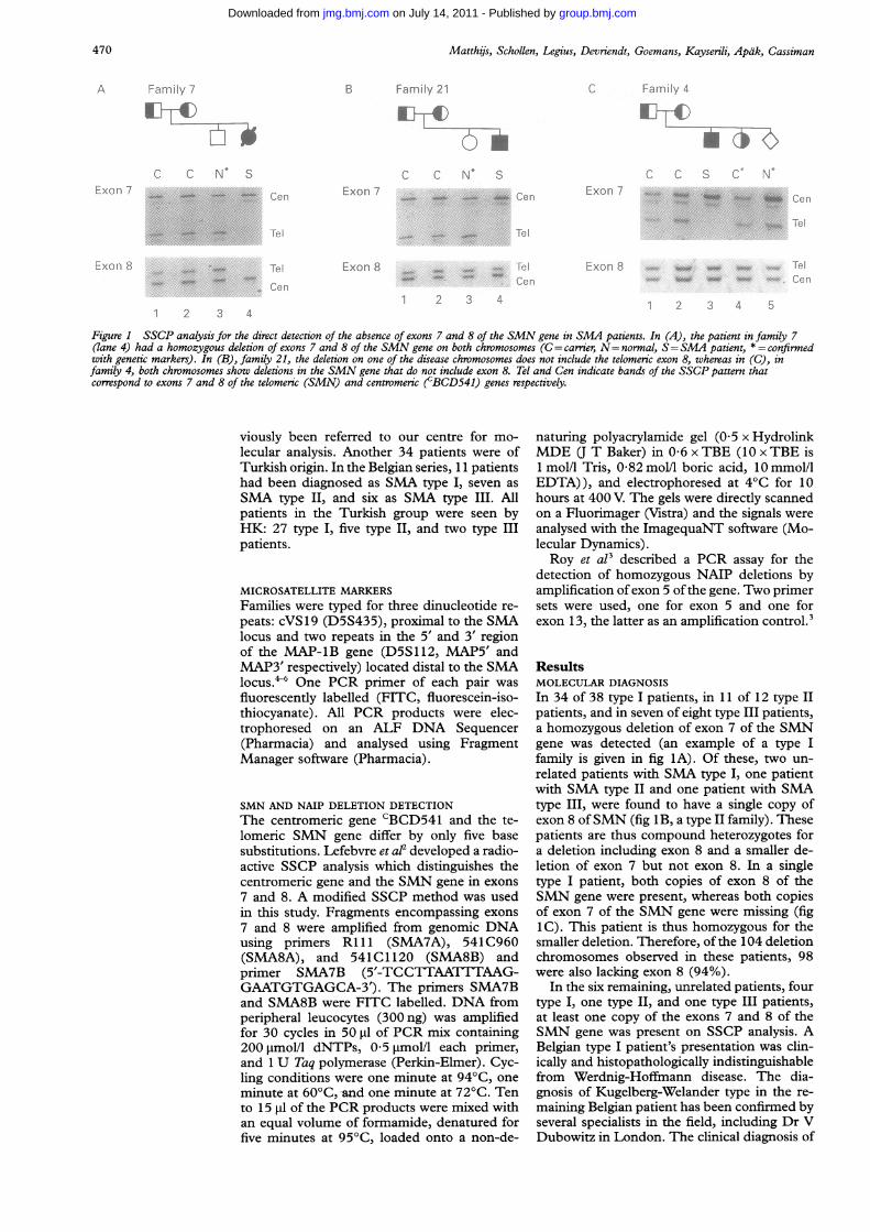
Figure 4 Family structure and genotype offamilies with (A) a parent with a single copy of the SMN gene and a homozygous deletion of thecBCD541 gene (family 11) and (B) a de novo deletion of the centromeric gene (family 12). The SSCP results are shown (Cen and Tel as in fig 1)and the presence and absence of the centromeric and telomeric genes are drawn schematically for each patient. The legend to the icon representing thechromosomes is given in (C). Open boxes represent deletions in the gene (absence of the corresponding exon), filled boxes represent the presence of thegene. Above the pedigree, the haplotypes for the different chromosomes are shown: marker c VSJ 9 is located proximal and markers MAPS' and MAP3'are distal to the SMA locus.
showed that the fetus had identical haplotypesto the dead patient and the pregnancy was
terminated. Marker analysis of a third preg-
nancy showed a fetus with a carrier phenotype.In this case, the presence of at least one copy
of the SMN gene was shown by SSCP analysis(fig 4A). SSCP analysis also showed that thefather lacked the centromeric CBCD541 gene
on both chromosomes. Since he is a carrier ofan SMN deletion, he can have only one normalcopy of the SMN gene. The mother was a
normal carrier, that is, two centromeric genes
and one normal copy of the SMN gene were
present as evidenced by SSCP analysis of exons7 and 8. The fetus in the third pregnancy was
typed as either lCen/lTel, in which case hewas a carrier, or 2Cen/2Tel (see above for thelimitations of this assay). Because analysis withflanking markers had shown that this fetus hadindeed received a haplotype associated with thedisease from his father and a normal chro-mosome from his mother, only the lCen/lTelgenotype is plausible for the fetus in the thirdpregnancy. The fetus received the normal cen-
tromeric and telomeric copies from the mother,and a chromosome deleted in the SMN andCBCD541 genes from the father. As a con-
sequence, the genotype of the proband and thefetus of the second pregnancy is lCen/OTel,which is compatible with the SSCP results.Thus, the father, who is phenotypically normal,is probably OCen/lTel and passed an "empty"chromosome to all sibs in this family (fig 4A).Owing to the limitations of the SSCP assay,we cannot exclude that the father has twoSMN genes on his normal chromosome, or thenormal SMN gene on one chromosome and a
centromeric to telomeric gene conversion on
the other chromosome. The first possibility
cannot be checked because the normal chro-mosome has not been passed to the children.The latter possibility is not compatible with theSSCP data obtained for the children, unlesstwo independent de novo deletion events are
invoked. Only interphase FISH could indicatethe number of SMN genes in the father's gen-
ome, but is not available yet.
DE NOVO DELETION OF THE CENTROMERICcBCD541 GENE IN A PATERNAL MEIOSISIn family 12, the proband was diagnosed withSMA type I at 6 months of age. Her sister wasprenatally typed as normal, based on geneticmarkers. The results were later confirmed clin-ically. Direct analysis of the SMN gene bySSCP confirmed the diagnosis in the proband(absence of exons 7 and 8 of the SMN gene,fig 4B). In the sister a homozygous absence ofexons 7 and 8 of the centromeric gene was
found. The father is a normal carrier (2Cen/1 Tel) whereas the mother was typed as lCen/lTel. Analysis with cVS 19 and MAP5' markersshowed the segregation ofthe two normal chro-mosomes 5 in the second child, and there isno evidence for loss of a chromosome, nor
for non-paternity. Because the father has twocentromeric copies and the second child hasnone, a de novo deletion must have occurredon the paternal chromosome in the secondchild. The analysis also allows the phase of thealleles on the maternal chromosomes 5 to bededuced: one lacks the centromeric gene, theother the SMN gene.
DiscussionLefebvre et al2 showed that most cases ofSMA,irrespective of their being typed as either
(:1S19MAP5MAP3
Exon 7
Exon 8
C~rint r.or-,ier
n xcx\)riJ. BBLD541 (en-).r
RxRrl% ] SMri; enre
T
;.7/- r -,1 e
472
C:
."
group.bmj.com on July 14, 2011 - Published by jmg.bmj.comDownloaded from

Unusual molecular findings in autosomal recessive spinal muscular atrophy
Werdnig-Hoffmann, Dubowitz, or Kugelberg-Welander disease, are the result of deletions inthe chromosomal region 5q11.2-q13.3. Theseauthors succeeded in pinpointing the geneticcause of the disease to the telomeric copy of aduplicated gene, the SMN gene. To show thatpart of this gene was deleted in 98-5% of theirSMA patients, they used an SSCP method todiscriminate between the SMN gene and thehighly homologous, centromeric copy of thatgene, cBCD541. This method has allowed usand others7 to confirm directly the diagnosis ofSMA in patients.The results of this report concerning a series
of Belgian and Turkish families confirm theprevious observations.2 The deletion detectionrate of 90% is below the 98-5% obtained bythe French group.' The assay only looks at thepresence or absence of two exons in the 3' endof the SMN gene. Interestingly, four of 38SMA type I cases escaped elucidation, whileone of 12 SMA type II cases, and one of eightSMA type III cases also need further in-vestigation or re-evaluation of the clinical data.These cases will be further investigated bysequencing the SMN gene. On the other hand,a direct test is now available for the con-firmation of clinical diagnosis without the needfor a muscle biopsy and its use can be extendedto atypical cases. Deletions in the SMN genehave already been identified in patients withinfantile SMA and congenital heart defects9and in a patient with the fetal hypokinesiasequence and signs of SMA.10 The use of theSSCP method in prenatal diagnosis has beenpreviously reported.8 We have also successfullyused the described non-radioactive SSCPmethod for prenatal diagnosis of SMN de-letions, and the test allows diagnosis in casesin which no DNA of an affected sib is available.For families in which no evidence of a deletionwas obtained, prenatal testing will have to bebased on linked polymorphic markers alone, asbefore, and one should be aware of the factthat the disease may not necessarily be linkedto chromosome 5q in such families.A PCR method with restriction digestion,
based on the polymorphic differences betweenthe CBCD541 and SMN genes, has been de-scribed by van der Steege et al. " In our hands,the SSCP based test has proved reliable. It hasthe advantages of a non-radioactive methodand it also allowed for a limited quantificationofthe different fragments and for carrier testingin some cases. However, the carrier status ofthe parents or sibs could o'nly be reliably de-termined whenever a dose difference was ob-served between the fragments of the SMN geneand of the centromeric gene. In practice, thedifference between two centromeric exon frag-ments plus two SMN exon fragments (2Cen/2Tel) and one copy of each exon (1 Cen/lTel),the latter representing an SMA carrier, couldnot reliably be made. Given the number ofpeople that lack both copies of the centromericgene (4 4%, data from Lefebvre et al'), thefrequency of carriers of a deletion of thecBCD541 gene must be high: 0 33 (basedon Hardy-Weinberg equilibrium). This wouldsimply mean that 33% of the SMA carriers also
lack a copy of the centromeric gene, beinglCen/lTel. Thus, every third "normal" SSCPresult would be misinterpreted. The differencebetween 1Cen/OTel and 2Cen/OTel can alsonot be shown. This, however, would not affectthe molecular diagnosis. The opposite result,OCen/lTel versus OCen/2Tel, again results inan uncertain carrier determination. These lim-itations diminish the value of this assay forcarrier detection in general and screening forSMA is not yet possible.
It is recommended to check for both exon 7and exon 8. The exon 8 assay detected only94% of the deletion chromosomes identifiedby the exon 7 assay. The existence of this raredeletion, including exon 7 but not exon 8, hasalso been previously reported.'7 In practicalterms, the combination of both assays offersmore reliable genotyping. It would also detectpatients in which a gene conversion may bepresent (see Lefebvre et a!2). Similarly, theavailability of the closely linked genetic markerscVS19 and MAP-lB gives confirmatory in-formation for determination of the phase andinterpretation of the SSCP data in equivocalcases, and, whenever possible, a combinationofboth techniques is recommended for prenataldiagnosis.There is no evidence for a de novo deletion
of the SMN gene in 13 cases in which theparents have been genotyped. However, wedescribe a de novo deletion event in the cen-tromeric gene in a sib ofan SMA type I patient.It could be suggested that the absence of acopy of the telomeric (SMN) gene in the fatherhas facilitated the loss of the centromeric geneon the homologous chromosome during mei-osis. If so, the opposite event might occur aswell: the absence ofthe centromeric gene mightfacilitate the loss of the telomeric gene on thehomologous chromosome, generating a diseasechromosome. This would imply an increasedrisk for de novo deletions in carriers of a cen-tromeric deletion. While the CBCD54 1 deletioncarrier frequency is very high,' this could ex-plain the high frequency of SMA carriers inthe population. It would be interesting to usethe semiquantitative SSCP assay in familieswith de novo SMA cases to substantiate thishypothesis. Alternatively, the accumulation ofcentromeric deletions may represent the silentcounterpart of random telomeric or cen-tromeric deletions, in which case the telomericdeletion frequency in the population has beenreduced because of the lethal phenotype. Alltogether, this observation stresses the instabilityof this chromosomal region. Other data ob-tained by Lefebvre et al' and by our groupl'also suggest that complex rearrangements canoccur on this chromosome.Nothing is settled yet as to the size of the
deletion in SMA cases. Four patients, of typesI, II, and III, have been found to carry acombination of a deletion including exon 7only on one allele, and a larger deletion, in-cluding both exon 7 and exon 8 of the SMNgene, on the other allele. Moreover, an SMAtype I patient was identified, in whom exon8 of the SMN gene was conserved on bothchromosomes (fig 1C). In this patient, there is
473
group.bmj.com on July 14, 2011 - Published by jmg.bmj.comDownloaded from
Matthijs, Schollen, Legius, Devriendt, Goemans, Kayserili, Apdk, Cassiman
no evidence for a homozygous deletion ofNAIP. Deletions leading to the most severe
phenotype may thus not include exon 8 of theSMN gene and its 3' region (see also Lefebvreet al2), nor NAIP. This indicates that, if othergenes play a role in the full phenotype ofSMA,they probably are located centromeric to theSMN gene.The observation of a homozygous deletion
in the centromeric gene in a phenotypicallynormal carrier of the SMN deletion (fig 4A)shows that one functional copy of the SMNgene in the absence of a functional CBCD541gene is sufficient for normal development.Lefebvre et al2 have shown that both thecentromeric gene and the SMN gene are tran-scribed and their predicted amino acid se-quence is identical. Moreover, the centromericgene but not the SMN gene itself is alternativelyspliced. The physiological role of these genesand transcripts and their specific role in thepathogenesis of the disease thus remain un-
known.In conclusion, direct (prenatal) molecular
diagnosis ofSMA has become feasible by look-ing at the presence or absence of a single geneon chromosome 5q. However, it has becomeevident that this region of chromosome 5qshows some special characteristics whichshould lead to caution in DNA diagnosis andprenatal diagnosis.
G-M is a Postdoctoral Researcher of the National Foundationfor Scientific Research (NFWO, Belgium). The technical as-sistance of S Hermans and K Van den Bosch is gratefullyacknowledged. This investigation was supported by grant S2/5-ID. E103 of the NFWO and by the Inter-University Networkfor Fundamental Research sponsored by the Belgian Gov-ernment (1991-1996).
1 Czeizel A, Hamula J. A Hungarian study on Werdnig-Hoffmann disease. J Med Genet 1989;26:761-3.
2 Lefebvre S, Burglen L, Reboullet S, et al. Identification andcharacterization of a spinal muscular atrophy-determininggene. Cell 1995;80:155-65.
3 Roy N, Mahadevan MS, McLean M, et al. The gene forNAIP, a novel protein with homology to baculoviral in-hibitors of apoptosis, is partially deleted in individualswith spinal muscular atrophy. Cell 1995;80:167-78.
4 Soares VM, Brzustowicz LM, Kleyn PW, et al. Refinementof the spinal muscular atrophy locus to an interval betweenD5S435 and MAP-lB. Genomics 1993;15:365-71.
5 Lien LL, Boyce FM, Kleyn P, et al. Mapping of humanmicrotubule-associated protein 1B in proximity to thespinal muscular atrophy locus at 5q1 3. Proc Natl Acad SciUSA 1991;88:7873-6.
6 Clermont 0, Burlet P, Burglen L, et al. Use of genetic andphysical mapping to locate the spinal muscular atrophylocus between two new highly polymorphic DNA markers.Am J Hum Genet 1994;54:687-94.
7 Rodrigues NR, Owen 0, Talbot K, Ignatius J, Dubowitz V,Davies KE. Deletions in the survival motor neuron geneon 5ql 3 in autosomal recessive spinal muscular atrophy.Hum Mol Genet 1995;4:631-4.
8 Rodrigues NR, Campbell L, Owen 0, Rodeck CH, DaviesKE. Prenatal diagnosis of spinal muscular atrophy by genedeletion analysis. Lancet 1995;345:1049.
9 Burglen L, Spiegel R, Ignatius J, et al. SMN gene deletionin variant of infantile spinal muscular atrophy. Lancet1995;346:316-7.
10 Devriendt K, Lammens M, Matthijs G, et al. Homozygousdeletion in the four survival motor neuron genes on 5q13causes severe congenital Werdnig-Hoffman disease. AnnNeurol (submitted).
11 van der Steege G, Grootscholten PM, van der Vlies, et al.PCR-based DNA test to confirm clinical diagnosis ofautosomal recessive spinal muscular atrophy. Lancet 1995;345:985-6.
474
group.bmj.com on July 14, 2011 - Published by jmg.bmj.comDownloaded from
doi: 10.1136/jmg.33.6.469 1996 33: 469-474J Med Genet
G Matthijs, E Schollen, E Legius, et al. recessive spinal muscular atrophy.Unusual molecular findings in autosomal
http://jmg.bmj.com/content/33/6/469Updated information and services can be found at:
These include:
References http://jmg.bmj.com/content/33/6/469#related-urls
Article cited in:
serviceEmail alerting
the box at the top right corner of the online article.Receive free email alerts when new articles cite this article. Sign up in
Notes
http://group.bmj.com/group/rights-licensing/permissionsTo request permissions go to:
http://journals.bmj.com/cgi/reprintformTo order reprints go to:
http://group.bmj.com/subscribe/To subscribe to BMJ go to:
group.bmj.com on July 14, 2011 - Published by jmg.bmj.comDownloaded from
Related Documents