UNSW Embryology Outflow Tract Heart (cut) Week 5, right oblique view

Welcome message from author
This document is posted to help you gain knowledge. Please leave a comment to let me know what you think about it! Share it to your friends and learn new things together.
Transcript

UNSW Embryology
Outflow TractHeart (cut)Week 5, right oblique view

atrioventricular canals
right and left
atriaright and left
conotruncus lumen
Heart (cut)Week 5, right oblique view

ventricular septum (muscular)
endocardial cushions
ventriclesright and left
Heart (cut)Week 5, right oblique view
conotruncus lumen


Endocardial cushion development
(truncal and bulbar)

bulbar ridges
truncal ridges
Endocardial cushion development
(truncal and bulbar)

bulbar ridges
truncal ridges
Truncal mesenchyme expands due to migration from: ventral pharyngeal area, pharyngeal arches and endoderm

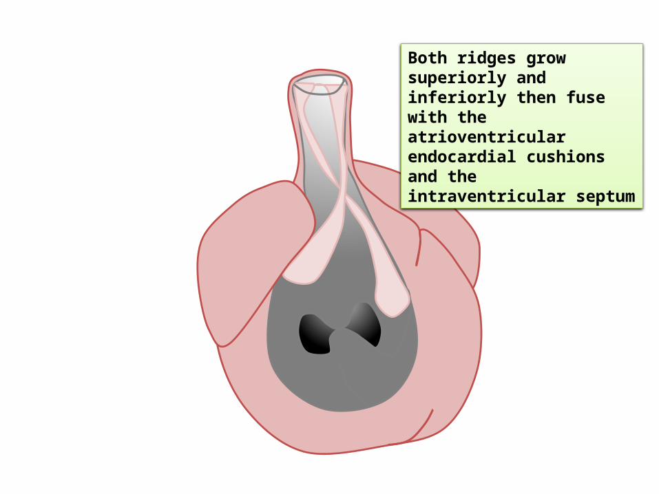
Both ridges grow superiorly and inferiorly then fuse with the atrioventricular endocardial cushions and the intraventricular septum
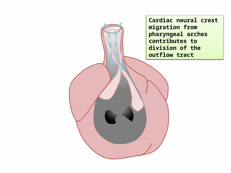
Cardiac neural crest migration from pharyngeal arches contributes to division of the outflow tract

Both ridges grow superiorly and inferiorly then fuse with the atrioventricular endocardial cushions and the intraventricular septum

Both ridges grow superiorly and inferiorly then fuse with the atrioventricular endocardial cushions and the intraventricular septum

Both ridges grow superiorly and inferiorly then fuse with the atrioventricular endocardial cushions and the intraventricular septum

Both ridges grow superiorly and inferiorly then fuse with the atrioventricular endocardial cushions and the intraventricular septum


Aorta and pulmonary trunk are then initially established from this septation
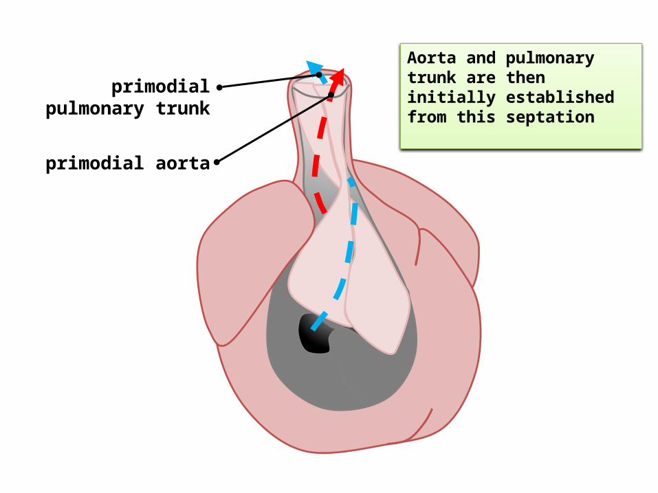
primodial pulmonary trunk
primodial aorta
Aorta and pulmonary trunk are then initially established from this septation

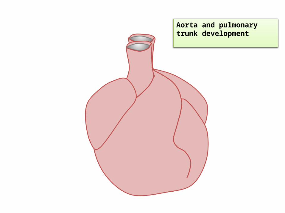
Aorta and pulmonary trunk development
Aorta and pulmonary trunk development

Aorta and pulmonary trunk development

Aorta and pulmonary trunk development
pulmonary trunkaorta
Related Documents