J. exp. Biol. 136, 311-332 (1988) 311 Printed in Great Britain © The Company of Biologists Limited 1988 UNPAIRED MEDIAN NEURONES IN A LEPIDOPTERAN LARVA (ANTHERAEA PERNYl) I. ANATOMY AND PHYSIOLOGY BY S. J. H. BROOKES* AND R. DE G. WEEVERS Department of Zoology, University of Bristol, UK Accepted 5 January 1988 Summary The anatomy and physiology of two unpaired median neurones (MCI and MC2) with bilaterally symmetrical axons in abdominal ganglia 3, 4, 5 and 6 olAntheraea pernyi larvae were studied. Intracellular dye filling of MCI and MC2 revealed that they were distinguishable from all other neurones in the ganglia and that they both had axons projecting out of the ganglia in right and left nerves 1. The two cells were identical in their central anatomy and physiology, but could be distinguished from one another by their peripheral branching patterns. The significance of these patterns was investigated by detailed study of the neural and muscular anatomy of the proleg-bearing segments 3, 4, 5 and 6. The peripheral axons of MCI and MC2 were exclusively associated with nerve trunks that could be traced to blocks of muscle. Intracellular recordings of the two median cells characteristically showed overshooting soma action potentials that were followed by a long afterhyperpolar- ization lasting many seconds. Simultaneous recordings from median cells in the same ganglion revealed that MCI and MC2 shared an excitatory synaptic drive that largely determined their patterns of firing. Recordings from median cells in different ganglia showed that the common synaptic drive was also shared by median cells in different segments. Selective lesions of the ventral nerve cord indicated that the synaptic drive to MCI and MC2 originated in the suboeso- phageal ganglion. These cells were similar in anatomy and physiology to the median cells in several other insects. Introduction Bilaterally symmetrical nerve cells with bifurcating axons have been described in a number of insect species, mostly after backfilling dye into nerves that innervate muscle blocks (locusts and cockroaches, see Hoyle, 1978; crickets, Davis & Alanis, 1979; Hemiptera, Davis, 1977; Coleoptera, Christensen & Carlson, 'Present address: Department of Human Physiology, Flinders Medical Centre, Bedford Park, South Australia 5042. Key words: unpaired median neurones, Lepidoptera, neurophysiology, anatomy.

Welcome message from author
This document is posted to help you gain knowledge. Please leave a comment to let me know what you think about it! Share it to your friends and learn new things together.
Transcript

J. exp. Biol. 136, 311-332 (1988) 3 1 1Printed in Great Britain © The Company of Biologists Limited 1988
UNPAIRED MEDIAN NEURONES IN A LEPIDOPTERANLARVA (ANTHERAEA PERNYl)
I. ANATOMY AND PHYSIOLOGY
BY S. J. H. BROOKES* AND R. DE G. WEEVERS
Department of Zoology, University of Bristol, UK
Accepted 5 January 1988
Summary
The anatomy and physiology of two unpaired median neurones (MCI and MC2)with bilaterally symmetrical axons in abdominal ganglia 3, 4, 5 and 6 olAntheraeapernyi larvae were studied. Intracellular dye filling of MCI and MC2 revealed thatthey were distinguishable from all other neurones in the ganglia and that they bothhad axons projecting out of the ganglia in right and left nerves 1. The two cellswere identical in their central anatomy and physiology, but could be distinguishedfrom one another by their peripheral branching patterns. The significance of thesepatterns was investigated by detailed study of the neural and muscular anatomy ofthe proleg-bearing segments 3, 4, 5 and 6. The peripheral axons of MCI and MC2were exclusively associated with nerve trunks that could be traced to blocks ofmuscle.
Intracellular recordings of the two median cells characteristically showedovershooting soma action potentials that were followed by a long afterhyperpolar-ization lasting many seconds. Simultaneous recordings from median cells in thesame ganglion revealed that MCI and MC2 shared an excitatory synaptic drivethat largely determined their patterns of firing. Recordings from median cells indifferent ganglia showed that the common synaptic drive was also shared bymedian cells in different segments. Selective lesions of the ventral nerve cordindicated that the synaptic drive to MCI and MC2 originated in the suboeso-phageal ganglion. These cells were similar in anatomy and physiology to themedian cells in several other insects.
Introduction
Bilaterally symmetrical nerve cells with bifurcating axons have been describedin a number of insect species, mostly after backfilling dye into nerves thatinnervate muscle blocks (locusts and cockroaches, see Hoyle, 1978; crickets, Davis& Alanis, 1979; Hemiptera, Davis, 1977; Coleoptera, Christensen & Carlson,
'Present address: Department of Human Physiology, Flinders Medical Centre, BedfordPark, South Australia 5042.
Key words: unpaired median neurones, Lepidoptera, neurophysiology, anatomy.
312 S. J. H. BROOKES AND R. DE G. WEEVERS
1981, 1982; Lepidoptera, Taylor & Truman, 1974; Casaday & Camhi, 1976;Rheuben & Kammer, 1980; Kondoh & Obara, 1982). The association of these cellswith nerve trunks that innervate muscles (Hoyle, Dagan, Moberly & Colquhoun,1974; Davis, 1977; Davis & Alanis, 1979; Kondoh & Obara, 1982; and the presentstudy) suggests that this class of neurones may be involved in the control of themusculature in these phylogenetically diverse groups of insects. In the locust andcockroach many bilaterally symmetrical neurones have been shown to haveovershooting action potentials in their somata (Crossman, Kerkut, Pitman &Walker, 1971). In contrast, most insect neurones appear to lack soma spikes(Orthoptera, Hoyle & Burrows, 1973; Dictyoptera, Fourtner & Pearson, 1977;Odonata, Simmons, 1977; Lepidoptera, Rind, 1983).
The present study investigates the anatomy and physiology of two unpairedmedian neurones (MCI and MC2) in the abdominal segments of A. pernyi, andincludes a detailed description of the muscular anatomy of the segment as noprevious accounts in the literature were adequate. Results indicated that the twomedian cells investigated in the present study were uniquely identifiable neurones.Several physiological and anatomical similarities between MCI and MC2 and thedorsal unpaired median (DUM) neurones of locusts and cockroaches were noted.
Materials and methods
Rearing methods
Caterpillars of the Chinese oak silkmoth Antheraea pernyi were raised fromhatching to their third instar in batches of 50-100 in constant light conditions at18-28°C, depending on the rate of growth required. The animals were fed onbeech, hawthorn, oak or sweet chestnut leaves during the summer, and onevergreen oak leaves during the winter. Nutritional deficiencies of evergreen oakwere overcome by spraying freshly cut leaves with the water-soluble componentsof the diet of David & Gardiner (1966). To reduce the spread of infectiousdiseases, the third-instar animals were transferred to individual polystyrenecontainers in which a small piece of moistened 'oasis' was placed. Animals werefed daily until spinning started at the end of the fifth instar. Five days afterspinning, pupae were removed from their cocoons to monitor their development,and were kept at 10—28°C, depending on when adults were required. Followingeclosion, moths were kept in the dark and observed until they had mated. Eggswere surface-sterilized in 5 % sodium hypochlorite solution in dilute detergent,rinsed in distilled water then allowed to develop at 18-28°C until hatching. Usingthese methods a continuous culture of Antheraea pernyi was maintained through-out the year.
The preparation
The dorsal surface of fifth-instar Antheraea pernyi larvae was smeared withpetroleum jelly, sharpened tungsten hooks were inserted into the head capsuleand anal skin flap and the animal was chilled at 0°C until just immobilized

Unpaired lepidopteran neurones 313
(normally 8-12 min). It was then suspended in a dissection chamber, a dorsalincision was made from the tail to the head capsule and the resulting flaps of skinwere fixed onto swivelling pins at the side of the chamber. The gut and silk glandswere ligatured and removed, care being taken not to disrupt the tracheal supply tothe ganglion under study. Saline modified from Weevers (1966) (in mmoll"1:NaCl, 20; KC1, 30; MgCl2, 20; CaCl2, 3; sucrose, 160; NaH2PO4, 10-9; Na2HPO4,4-1; pH6-6) was then superfused through the body cavity at a rate of 1-2 ml min"1.Abdominal ganglion 3, 4, 5 or 6 was then lifted onto a vinyl-covered stainless-steelplatform and pinned out with small insect pins approximately 2 mm long. Up to 20such pins were inserted into connective tissue and large tracheae to immobilize theganglion for recording. The connective tissue sheath covering the ganglion wasfocally softened by applying a 1 % protease solution (Sigma Type VI) in a flat-bottomed pipette for 8-12 min. Microelectrodes pulled on a Brown & Flamingpipette puller (Sutter Instrument Co.), filled with 3moll"1 KC1 or 2 m o i r 'potassium acetate solution, with resistances of 20-50 MQ were used to makeintracellular recordings. Penetration of median cells was aided by the use of apiezoelectric prodder (Weevers, 1980) driven by square wave pulses from astimulator. Signals from the microelectrode were passed through a home-builtelectrometer with internal bridge balance circuitry, displayed on an oscilloscope(Tektronix 5111 A) and simultaneously recorded on a Racal Store 4FM taperecorder. Intracellular recordings of muscle activity were made in a similar wayexcept that electrodes were of lower resistance (1-10 MQ) and were mounted on aflexible silver wire (Woodbury & Brady, 1956).
Extracellular recordings of neural activity were made en passant from nervetrunks with a chloride-coated silver wire hook electrode which could be retractedinto a paraffin-filled glass sleeve. Recordings were made between the hook and anindifferent electrode placed in the body cavity via an Isleworth A103 pre-amplifier. Nerve trunks were stimulated by a fine pair of chloride-coated silverwires insulated except at their tips. Recordings were played into a DigitimerDS900 transient recorder and plotted on a Gould x,y plotter.
Dye filling of neurones
For intracellular dye filling, microelectrodes were filled with 100 mmoll"1
hexammine cobaltic chloride, 3 % Lucifer Yellow (Stewart, 1981) or 4 % horse-radish peroxidase (Boehringer Mannheim, grade 1) in 0-5moll~l potassiumacetate solution. Dye was injected into physiologically identified cells by passing1-10nA constant-current pulses, 500ms long at lHz for 5-30min. Cobalt-filledcells were then treated with dilute ammonium sulphide solution (two drops in10 ml of phosphate buffer, pH 70) for up to 5 min, rinsed in fresh phosphate bufferand fixed in 5 % formaldehyde in buffer for 30 min. After washing overnight inrunning tap water, ganglia were rinsed twice in distilled water and then intensifiedusing the method of Bacon & Altman (1977). Ganglia were then dehydrated inethanol, cleared in methyl benzoate and xylene, and mounted in neutral Canadabalsam.
314 S. J. H . BROOKES AND R. DE G. WEEVERS
Cells filled with horseradish peroxidase were processed according to the methodof Roberts & Clarke (1982). Lucifer Yellow fills were fixed in 5 % formaldehyde inlOOmmoir1 phosphate buffer pH70, dehydrated in ethanol and mounted inFluoromount (Edwin Gurr Ltd).
Vital staining of caterpillars
The general neuroanatomy of fifth-instar caterpillars was revealed by intra vitaminjection of 1-2 ml of 0-17% Methylene Blue in saline, reduced with Rongalite(see Pantin, 1946, for exact details). After 15min the animal was dissected in thenormal way from the dorsal midline, either under phosphate buffer (pH70) orunder saturated ammonium molybdate solution. Six preparations were studied indetail in this way.
Results
Of the many neurones with cell bodies in the midline of the abdominal ganglia oflarval Antheraea pernyi, only two were recorded that had axons projectingbilaterally and symmetrically in both nerves 1 of the ganglion. These neuroneswere named median cells 1 and 2 (MCI and MC2), and were found to be uniquelyidentifiable. The anatomy of the segment in fifth-instar larvae is described first toprovide a frame of reference for the description of the morphology of the twomedian cells.
The peripheral nervous system of abdominal segment 5
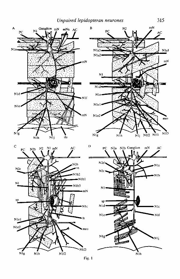
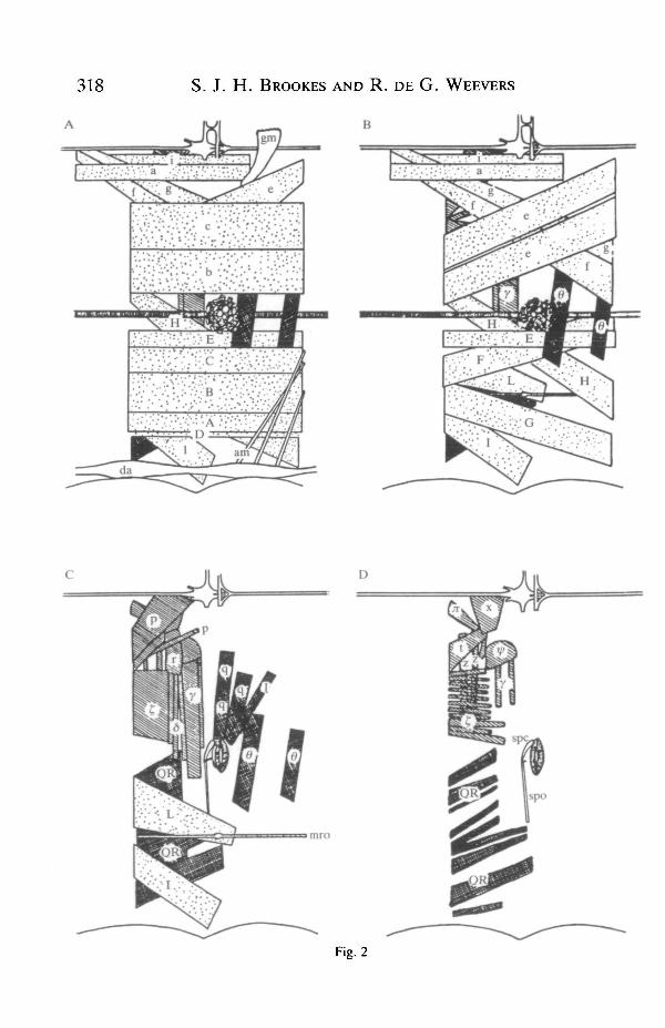
Methylene Blue staining of nerve trunks in the abdomen revealed fine branchesthat were only seen with great difficulty in living material. Fig. 1 shows datacollected from six preparations using this technique and subsequently confirmed inmany in vivo preparations. The diagrams show the structure of nerves at fourstages of dissection starting after the removal of the gut, silk glands, gonads and fatbody. The four stages progress from the innermost layer of longitudinal musclesoutwards to the oblique muscles directly below the cuticle. The nomenclature ofnerve branches was based on the system of Pipa & Cook (1959) as it was felt thatthe system of Beckel (1958) was too inflexible to allow for the variability ofbranching seen in different preparations.
Fig. 1. Four stages in the dissection of the nervous system of the fifth abdominalsegment of fifth-instar larval Antheraea pernyi are. shown, starting with the innermostlayer and progressing outwards towards the cuticle. For the sake of clarity the gut, silkglands, fat body and most of the tracheae have been omitted. The nomenclature isbased on the system of Pipa & Cook (1959). Each diagram shows half of an abdominalbody segment, with the ventral midline at the top and the dorsal midline at the bottom,the segment being flattened for the purposes of dissection. AC, anterior connective;PC, posterior connective; vs, ventral scolopale organ; Is, lateral scolopale organ;mro, muscle receptor organ; sp, spiracle; da, dorsal aorta.
PC
Unpaired lepidopteran neurones
Ganglion ^ mNa AC B N 2 mN
VJdL/
315
AC
Nlal
mN
NigNlh Nlj da
PC N2b N2 NlmN AC
\A \hi Z
Nlf
mN Nle2
D
Nlh Nlj Nlf2 Nlfl
PC N2a N2b Ganglion mN AC
\
N1D
Nig Nlh Nlh
316 S. J. H. BROOKES AND R. DE G. WEEVERS
The named nerve trunks were all consistently identifiable in different animals;finer branching was extremely variable but that represented in Fig. 1 is typical. Thepattern of branching was basically similar to that described in the earlier studies ofLibby (1959) and Beckel (1958) (in Hyalophora cecropia) with the followingexceptions. Beckel's nerve A l l which was shown to run from Nlb3 near thespiracle and anastamose with N2d in the next anterior segment was not seen inAntheraea pernyi (see Fig. 1C). Libby (1959) also failed to record this branch.Second, the long branch of N2b (see Fig. 1C) was shown by Beckel as beingdistally attached to a branch of Nlc; this connection was never seen. Fouranastamoses were seen; Nl joined mN both near the ganglion and near thespiracle. The small branch mNa (see Fig. 1A) which anastamosed with N2c of thenext anterior ganglion was that shown by Taghert & Truman (1982), in Manducasexta, to contain the axons of three identified bursicon-containing cells. The fourthanastamosis was again with mN where Nlb3 joined it just ventral to the spiracle.
The targets of each of the named branches are summarized in Table 1. NervesNlb l , Nlb2, Nlcl , Nlc2, Nlfl, Nlf2, Nlf3 and N2b were never seen to have anyconnections with muscle fibres and appeared to innervate either the cuticle oridentified sense organs. Nerve trunks Nlal, Nla2, Nla3, Nlb3, Nld, Nle, Nig,Nlh and Nlj could only be traced to bundles of muscle fibres; no branches couldbe traced to run to the cuticle or to identified sense organs.
The motor units of abdominal segment 5
Motor units were characterized in the following way. By placing a finestimulating electrode on a muscle fibre it was possible to stimulate a singlemotoneurone and all of its collaterals, and by using a movable intracellularrecording electrode the extent of the motor unit could be defined. An extracellularrecording electrode was placed on the proximal root of the nerve and used tocheck that only a single antidromic spike was elicited by stimulation.
The results obtained using these methods are shown in Fig. 2 (for the sake ofsimplicity the gut, silk glands, fat body, nervous system and tracheae have beenomitted). The nomenclature used is that of Lyonet (1762) and Forbes (1914),except for muscles n and ty, which were not identified by these authors. Theshading of the muscles reflects the source of their innervation, the 19 stippledmotor units were supplied by motoneurones with somata in the next anteriorsegmental ganglion vie Nl. Four motor units (crosshatched) were supplied bymotoneurones with cell bodies in the same segment's ganglion and axons in Nl.Ten muscles (striped) were supplied by motoneurones with axons in N2. Thespiracle opener (spo) is a non-muscular ligament (Beckel, 1958) and the alarymuscle, heart and spiracle closer were innervated by the median nerve (mN).
Of all the muscles tested, only one (y) was shown to be innervated by more thanone axon. All fibres in this muscle showed two sizes of EJP in spontaneous activityand in response to graded stimulation of N2. All other motor units had only onesize of spontaneous or evoked EJP. No evidence for inhibitory input was seen inany muscles in the abdomen of Antheraea pernyi larvae.

Unpaired lepidopteran neurones 317
Table 1. Targets of peripheral nerve trunks and their innervation by axons of MCIand MC2
Name ofnerve
mNmNaNlNlalNla2Nla3NlblNlb2Nlb3NlclNlc2NldNlelNle2NlflNlf2N1ONigNlhNljN2N2aN2bN2cN2d
Traced tosomaticmuscles?
9
?
yesyesyesyesnonoyesnonoyesyesyesnononoyesyesyesyesyesno?
yes
Innervation by the median cellsthe nerve
MCI orMC2
—-
bothbothbothboth
--
MCI—-
MC2MC2MC2
-——
MC2MC2MC2
----—
indicatestrunk on one or more occasions.
Peripheral target(s) traced
Heart, spiracle, neurohaemal organAnastamoses with N2c of anterior segmentDorsal and ventral muscles and sense organsMuscles b,c,d,eMuscles d,e,f,gMuscles f,g,iVentral and lateral cuticleVentral scolopale organMuscles 9,q and 1, anastamosis with mNAnterior lateral cuticleDorsolateral scolopale organMuscles E,H,FMuscles C,B,LMuscle of muscle receptor organNucleus of muscle receptor organPosterior dorsal cuticleAnterior dorsal cuticleMuscle QRMuscles QR,A,I,D,GMuscles A,D,GMuscles of proleg, ventral cuticleMuscles x,p,t,;z:Ventral and proleg cuticleAnastomosis with median nerveMuscles r,z,£,<5,y,i/>
that an axon branch was physiologically traced into
The central morphology of the median cells
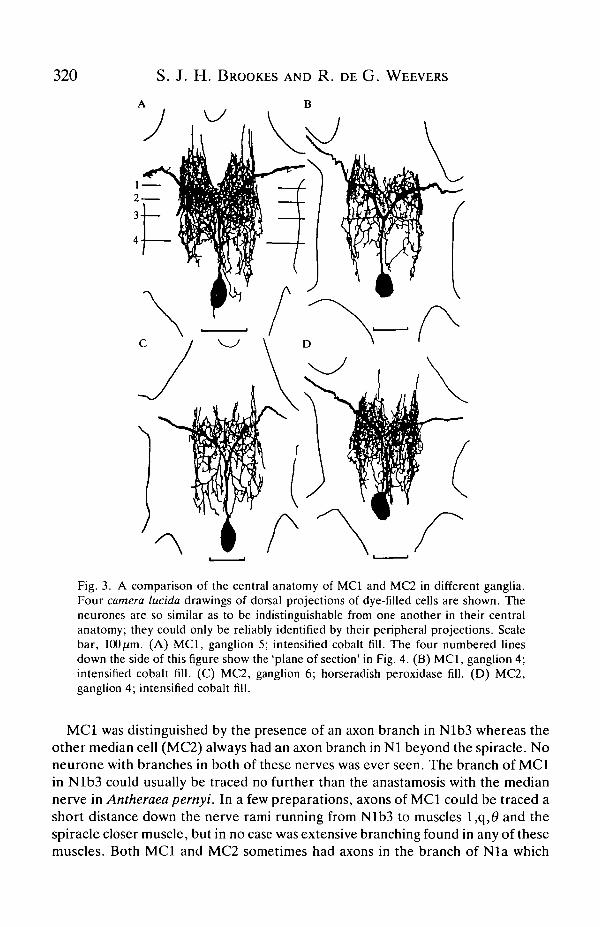
Two neurones, MCI and MC2, were recorded that had axons in both right andleft nerves 1 of abdominal ganglia and were distinguishable from all otherneurones by this criterion and by their characteristic intracellular physiology. Itwas of interest, therefore, to examine their central morphology to determinewhether these two cells were uniquely identifiable, to see how they differed fromone another and to compare their anatomy with that of DUM (dorsal unpairedmedian) cells in other insects. Intracellular dye fills of the two median cells wereobtained on many occasions; typical examples are shown in Fig. 3. The moststriking feature of the cell was the symmetry of its axons, dendrite distribution andbranching at least to the level of secondary branching. The posteriorly sited somagave rise to a single neurite that ran forward without branching quite deep withinthe neuropile. At a level just behind the roots of nerve 1 the neurite rose sharply,
318 S. J. H. BROOKES AND R. DE G. WEEVERS
B
•'•••" i:--\'-•.;•:•• :'•'••'••'.••'•' :'•:•'•{:':
' • . ' ! - : • • • • • . ' : ' - . • . • • b • . ' • • V . : !: : •:':'.•• •••
_ da
Fig. 2

Unpaired lepidopteran neurones 319
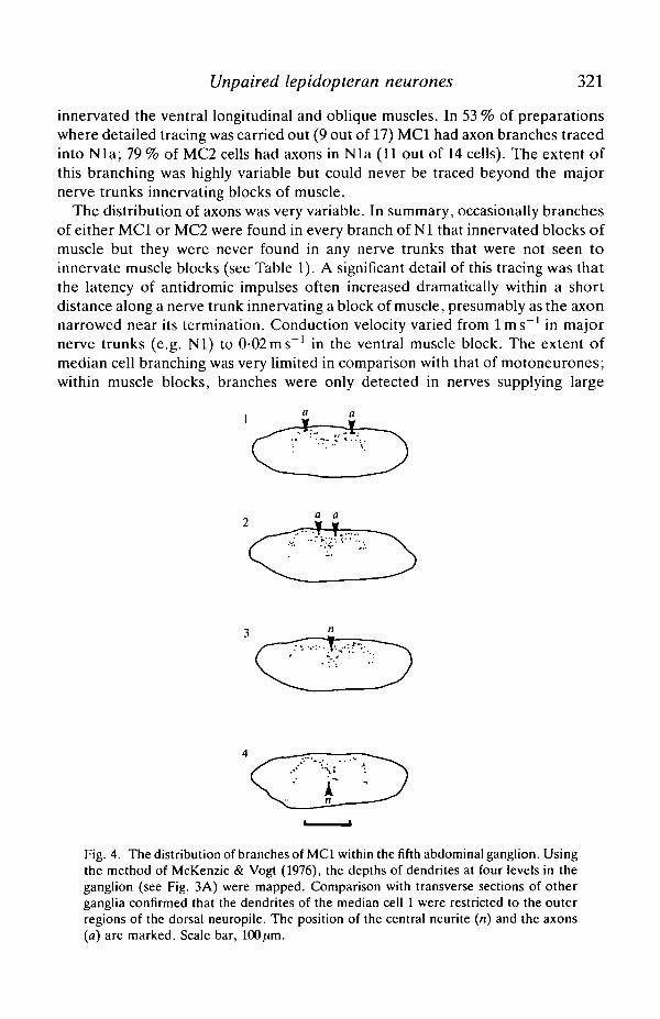
and bifurcated into two axons which ran over the dorsal surface of the neuropileand then out of right and left nerves 1 of the ganglion. Using the method ofMcKenzie & Vogt (1976) the vertical distribution of dendrites was mapped for fourregions of the neuropile (see Fig. 4) from the fine focus control on a microscope.This indicated that dendritic branching was largely restricted to the dorsal,dorsomedial and dorsolateral regions of the neuropile.
Intracellular dye filling indicated that there were no significant differencesbetween the central morphology of MCI and MC2; in fact, they could only bedistinguished by their peripheral branching pattern. This is shown by the dye fillsdrawn in Fig. 3. Apart from characteristic differences in peripheral axonbranching, the only difference between MCI and MC2 was that MCI tended tohave the more dorsal cell body. However, this was not a reliable distinguishingcharacteristic. In 78 % of preparations where measurements were recorded (32 outof 41) MCI had its cell body within 50 nm of the dorsal ganglion surface (measuredwith the fine advance of the micromanipulator). MC2 had a dorsally located somain only 38 % of preparations (10 out of 36). There was no significant tendency forone cell body to be more posteriorly located than the other.
Peripheral branching of the median cells
Peripheral axons of the median cells were traced with three methods. First,antidromic impulses were evoked by stimulation of peripheral nerve trunks andwere recorded in the cell soma; this indicated the presence of an axon branch atthe point of stimulation. The paucity of synaptic input from nerve 1 made thismethod reliable and quick. Orthodromic tracing used the presence of extracellularpotentials recorded in peripheral nerve trunks and time-locked to intracellularlyrecorded soma spikes. This method was slow, it often required signal averagingand could damage peripheral nerves. The third method was to inject dye into thesoma of the median cell and let it diffuse into the axons. This method was rathercapricious; however, it did give unequivocal confirmation of the basic branchingpatterns.
Fig. 2. Four stages in the dissection of the fifth abdominal segment of fifth-instar larvalAntheraea pernyi are shown to reveal the structure of the somatic musculature and itssource of innervation. The figures start with the innermost layer of fast longitudinalmuscles and progress outwards towards the cuticle. For the sake of clarity the gut, silkglands, fat body, tracheae and nervous system have been omitted. The 19 stippledmuscles were innervated by motoneurones with somata in the next anterior ganglionand axons in nerve 1. The four crosshatched muscles were innervated by motoneuroneswith somata in the ganglion of segment 5 and axons in nerve 1. The ten striped motorunits were supplied by motoneurones with axons in nerve 2. The nomenclature is thatof Lyonet (1762) and Forbes (1914). gm, strap-like muscle connected to the side of thegut. This muscle had a very different appearance from the other somatic muscles anddid not appear to be innervated by any nerve branches arising from the abdominalnerve cord, spc, spiracle closer muscle; mro, muscle receptor organ; spo, spiracleopener ligament (non-muscular); am, alary muscles; da, dorsal aorta.
320 S. J. H. BROOKES AND R. DE G. WEEVERS
A B
Fig. 3. A comparison of the central anatomy of MCI and MC2 in different ganglia.Four camera lucida drawings of dorsal projections of dye-filled cells are shown. Theneurones are so similar as to be indistinguishable from one another in their centralanatomy; they could only be reliably identified by their peripheral projections. Scalebar, 100ftm. (A) MCI, ganglion 5; intensified cobalt fill. The four numbered linesdown the side of this figure show the 'plane of section' in Fig. 4. (B) MCI, ganglion 4;intensified cobalt fill. (C) MC2, ganglion 6; horseradish peroxidase fill. (D) MC2,ganglion 4; intensified cobalt fill.
MCI was distinguished by the presence of an axon branch in Nlb3 whereas theother median cell (MC2) always had an axon branch in Nl beyond the spiracle. Noneurone with branches in both of these nerves was ever seen. The branch of MCIin Nlb3 could usually be traced no further than the anastamosis with the mediannerve in Antheraea pernyi. In a few preparations, axons of MCI could be traced ashort distance down the nerve rami running from Nlb3 to muscles l,q,# and thespiracle closer muscle, but in no case was extensive branching found in any of thesemuscles. Both MCI and MC2 sometimes had axons in the branch of Nla which
Unpaired lepidopteran neurones 321
innervated the ventral longitudinal and oblique muscles. In 53 % of preparationswhere detailed tracing was carried out (9 out of 17) MCI had axon branches tracedinto Nla; 79 % of MC2 cells had axons in Nla (11 out of 14 cells). The extent ofthis branching was highly variable but could never be traced beyond the majornerve trunks innervating blocks of muscle.
The distribution of axons was very variable. In summary, occasionally branchesof either MCI or MC2 were found in every branch of Nl that innervated blocks ofmuscle but they were never found in any nerve trunks that were not seen toinnervate muscle blocks (see Table 1). A significant detail of this tracing was thatthe latency of antidromic impulses often increased dramatically within a shortdistance along a nerve trunk innervating a block of muscle, presumably as the axonnarrowed near its termination. Conduction velocity varied from l m s " 1 in majornerve trunks (e.g. Nl) to 0-02ms~' in the ventral muscle block. The extent ofmedian cell branching was very limited in comparison with that of motoneurones;within muscle blocks, branches were only detected in nerves supplying large
Fig. 4. The distribution of branches of MCI within the fifth abdominal ganglion. Usingthe method of McKenzie & Vogt (1976), the depths of dendrites at four levels in theganglion (see Fig. 3A) were mapped. Comparison with transverse sections of otherganglia confirmed that the dendrites of the median cell 1 were restricted to the outerregions of the dorsal neuropile. The position of the central neurite (n) and the axons(a) are marked. Scale bar, 100fim.

322 S. J. H. BROOKES AND R. DE G. WEEVERS
Nlh
Fig. 5. Peripheral branching of MCI and MC2. (A,B) Schematic diagrams of axonbranching patterns typical of MCI and MC2, respectively, traced physiologically inganglion 5 of the same preparation. The main nerve trunks are labelled as in Fig. 1.
bundles of fibres, never in the fine branches that ramified over single muscle fibres.Typical patterns of branching of MCI and MC2 from a single preparation areshown schematically in Fig. 5.
The neurophysiology of MCI and MC2
The apparent resting potential of the two median cells compared with thebaseline potential when the microelectrode was outside the ganglion was— 19-2 ±4-2 mV (N= 10). It is likely, however, that this does not represent thetrue difference in potential between the inside of the cell and the surroundingextracellular fluid. Whilst advancing the microelectrode through a ganglion, acomplex series of potentials was often recorded before a median cell waspenetrated, even when the cell was clearly visible as the most dorsal neural soma.Typically, a negative potential of up to -70 mV (probably as a perineurial glial cellwas penetrated) was the first event recorded; this was typically followed by anapparent positive potential averaging +33-1 ± 4-0 mV (N = 10), which was pre-sumed to represent a transperineurial potential, as has been reported in thecockroach (Treherne & Schofield, 1981). Thus a better estimate of the real resting
Unpaired lepidopteran neurones
B
323
D
JIs
20 mV
Fig. 6. (A,B) Soma action potentials of MCI and MC2, respectively, had acharacteristic shape with a pronounced inflection on the falling phase (arrowheads).Impulses were of very slow time course; at half peak amplitude they lasted from 10 to25 ms. (C,D) MCI and MC2 impulses are shown on a slower time scale. Immediatelyfollowing each soma action potential was an afterhyperpolarization up to 20 mV belowresting potential and up to 400 ms long at half peak amplitude. This was followed inmost median cells by a long afterhyperpolarization which profoundly depressed theexcitability of the neurone for up to 20 s.
potential of the median cells is given by the difference between the apparentresting potential and the transperineurial potential, i.e. —52-3 ± 8-2 mV (TV = 10).
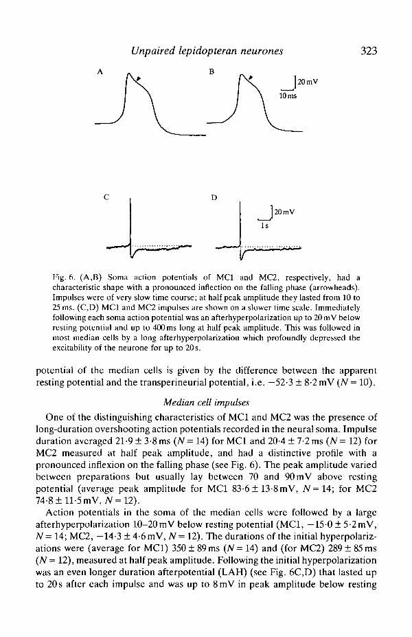
Median cell impulses
One of the distinguishing characteristics of MCI and MC2 was the presence oflong-duration overshooting action potentials recorded in the neural soma. Impulseduration averaged 21-9 ± 3-8 ms (N= 14) for MCI and 20-4 ± 7-2 ms (N= 12) forMC2 measured at half peak amplitude, and had a distinctive profile with apronounced inflexion on the falling phase (see Fig. 6). The peak amplitude variedbetween preparations but usually lay between 70 and 90 mV above restingpotential (average peak amplitude for MCI 83-6 ± 13-8 mV, N=14; for MC274-8 ± 11-5 mV, A'=12).
Action potentials in the soma of the median cells were followed by a largeafterhyperpolarization 10-20mV below resting potential (MCI, —15-0 ± 5-2 mV,N = 14; MC2, —14-3 ±4-6mV, N = 12). The durations of the initial hyperpolariz-ations were (average for MCI) 350 ± 89 ms (N= 14) and (for MC2) 289 ± 85 ms(N= 12), measured at half peak amplitude. Following the initial hyperpolarizationwas an even longer duration afterpotential (LAH) (see Fig. 6C,D) that lasted upto 20 s after each impulse and was up to 8mV in peak amplitude below resting
324 S. J. H. BROOKES AND R. DE G. WEEVERS
5mV
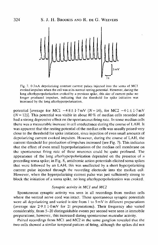
Fig. 7. 0-3 nA depolarizing constant current pulses injected into the soma of MCIevoked impulses when the cell was at its normal resting potential. However, during thelong afterhyperpolarization evoked by a previous spike, this size of current pulse nolonger produced impulses, indicating that the threshold for spike initiation wasincreased by the long afterhyperpolarization.
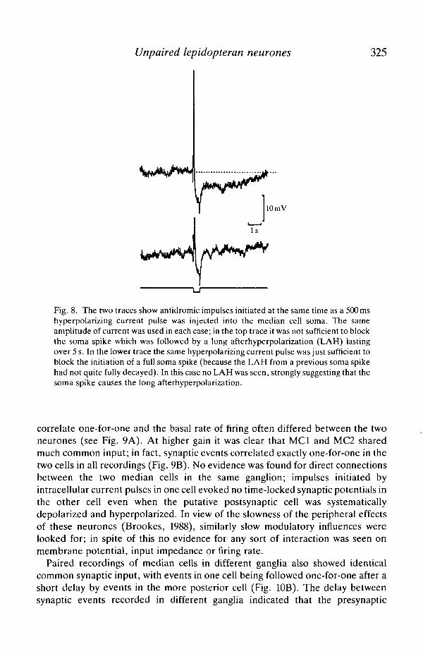
potential [average for MCI -4-8±l -7mV (N=U), for MC2 - 4 - l ± l - 7 m V(N = 12)]. This potential was visible in about 80% of median cells recorded andhad a strong depressive effect on the spontaneous firing rate. In some median cellsthere was a measurable increase in cell conductance during the course of LAH. Itwas apparent that the resting potential of the median cells was usually poised veryclose to the threshold for spike initiation, since injection of even small amounts ofdepolarizing current evoked impulses. However, during the course of LAH, thecurrent threshold for production of impulses increased (see Fig. 7). This indicatesthat the effect of even small hyperpolarizations of the median cell membrane onthe spontaneous firing rate of these neurones could be quite profound. Theappearance of the long afterhyperpolarization depended on the presence of apreceding soma spike; in Fig. 8, antidromic action potentials elicited soma spikesthat were followed by an LAH; this was unaffected by a short hyperpolarizingcurrent pulse injected through the recording electrode into the median cell.However, when the hyperpolarizing current pulse was just sufficiently strong toblock the initiation of a soma spike, no long afterhyperpolarization was evoked.
Synaptic activity in MCI and MC2
Spontaneous synaptic activity was seen in all recordings from median cellswhere the ventral nerve cord was intact. These spontaneous synaptic potentialswere all depolarizing and varied in size from 1 to 5 mV in different preparations(average size 2 - 9 ± 1 0 m V for 21 preparations). Their frequency also variedconsiderably, from 5-20 distinguishable events per second were seen in immobilepreparations; however, this increased during spontaneous muscular activity.
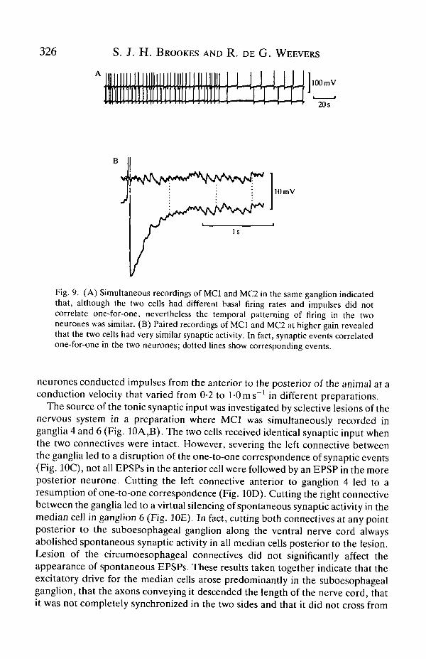
Paired recordings from MCI and MC2 in the same ganglion revealed that thetwo cells showed a similar temporal pattern of firing, although the spikes did not
Unpaired lepidopteran neurones 325
10 mV
Fig. 8. The two traces show antidromic impulses initiated at the same time as a 500 mshyperpolarizing current pulse was injected into the median cell soma. The sameamplitude of current was used in each case; in the top trace it was not sufficient to blockthe soma spike which was followed by a long afterhyperpolarization (LAH) lastingover 5 s. In the lower trace the same hyperpolarizing current pulse was just sufficient toblock the initiation of a full soma spike (because the LAH from a previous soma spikehad not quite fully decayed). In this case no LAH was seen, strongly suggesting that thesoma spike causes the long afterhyperpolarization.
correlate one-for-one and the basal rate of firing often differed between the twoneurones (see Fig. 9A). At higher gain it was clear that MCI and MC2 sharedmuch common input; in fact, synaptic events correlated exactly one-for-one in thetwo cells in all recordings (Fig. 9B). No evidence was found for direct connectionsbetween the two median cells in the same ganglion; impulses initiated byintracellular current pulses in one cell evoked no time-locked synaptic potentials inthe other cell even when the putative postsynaptic cell was systematicallydepolarized and hyperpolarized. In view of the slowness of the peripheral effectsof these neurones (Brookes, 1988), similarly slow modulatory influences werelooked for; in spite of this no evidence for any sort of interaction was seen onmembrane potential, input impedance or firing rate.
Paired recordings of median cells in different ganglia also showed identicalcommon synaptic input, with events in one cell being followed one-for-one after ashort delay by events in the more posterior cell (Fig. 10B). The delay betweensynaptic events recorded in different ganglia indicated that the presynaptic
326 S. J. H. BROOKES AND R. DE G. WEEVERS
A I,
100 mV
20s
10 mV
Fig. 9. (A) Simultaneous recordings of MCI and MC2 in the same ganglion indicatedthat, although the two cells had different basal firing rates and impulses did notcorrelate one-for-one, nevertheless the temporal patterning of firing in the twoneurones was similar. (B) Paired recordings of MCI and MC2 at higher gain revealedthat the two cells had very similar synaptic activity. In fact, synaptic events correlatedone-for-one in the two neurones; dotted lines show corresponding events.
neurones conducted impulses from the anterior to the posterior of the animal at aconduction velocity that varied from 0-2 to 1-0 ms"1 in different preparations.
The source of the tonic synaptic input was investigated by selective lesions of thenervous system in a preparation where MCI was simultaneously recorded inganglia 4 and 6 (Fig. 10A,B). The two cells received identical synaptic input whenthe two connectives were intact. However, severing the left connective betweenthe ganglia led to a disruption of the one-to-one correspondence of synaptic events(Fig. 10C), not all EPSPs in the anterior cell were followed by an EPSP in the moreposterior neurone. Cutting the left connective anterior to ganglion 4 led to aresumption of one-to-one correspondence (Fig. 10D). Cutting the right connectivebetween the ganglia led to a virtual silencing of spontaneous synaptic activity in themedian cell in ganglion 6 (Fig. 10E). In fact, cutting both connectives at any pointposterior to the suboesophageal ganglion along the ventral nerve cord alwaysabolished spontaneous synaptic activity in all median cells posterior to the lesion.Lesion of the circumoesophageal connectives did not significantly affect theappearance of spontaneous EPSPs. These results taken together indicate that theexcitatory drive for the median cells arose predominantly in the suboesophagealganglion, that the axons conveying it descended the length of the nerve cord, thatit was not completely synchronized in the two sides and that it did not cross from

Unpaired lepidopteran neurones
Lesion 1
327
Lesion 2
10 mV
Fig. 10. (A) A diagram showing the sites of the lesions that produced the activitydisplayed in B-E. (B) Control. Both connectives were intact along the length of theanimal. Synaptic events correlate one-for-one in the two median cells in ganglia 4 and6. Dotted lines indicate corresponding synaptic events in the two cells. (C) Cutting theleft connective between ganglia 4 and 6 disrupted one-for-one correspondence ofsynaptic events. Each EPSP in ganglion 6 was preceded by an event in ganglion 4.However, some events in ganglion 4 were no longer followed by an EPSP in the mediancell in ganglion 6. The solid lines indicate some synaptic events which appeared in themore anterior ganglion that were not followed by a corresponding event in the moreposterior ganglion. (D) Severing the left connective anterior to ganglion 4 caused aresumption of one-for-one correspondence of potentials, since both cells now onlyreceived input from the right connective. (E) Cutting the right connective anterior toganglion 6 virtually silenced the spontaneous activity recorded in the more posteriormedian cell.
one side to the other within the abdominal ganglia. Electrical stimulation of the cutends of connectives evoked synaptic activity similar to the spontaneous activityseen in preparations with an intact nerve cord. Interestingly, stimulation of the
328 S. J. H . BROOKES AND R. DE G. WEEVERS
peripheral nerves 1 and 2 also evoked EPSPs in most preparations, indicating thatthere were inputs other than those descending from the suboesophageal ganglion.
Discussion
The anatomy of abdominal segments
The muscles of several families of lepidopteran larvae have been describedelsewhere (Cossidae, Lyonet, 1762; Notodontidae, Lubbock, 1859; Lasiocampi-dae, Noctuidae and Sphingidae, Forbes, 1914; Saturniidae, Beckel, 1958; Libby,1959; Galleria mellonella, Randall, 1968; Manduca sexta, Rheuben & Kammer,1980). However, the description given here is based on both anatomical andphysiological evidence and thus describes for the first time the muscular anatomyin terms of motor units. The most striking feature of the segmental anatomy wasthe extreme simplicity of the innervation and musculature. The association ofMCI and MC2 axons with nerve trunks that innervated bundles of muscle fibresrather than with nerve trunks running to identified sense organs or to the cuticlewas very striking.
In none of the Methylene Blue or in vivo preparations were peripheral nerve cellbodies seen on segmental nerves 1 and 2, although such neurones have beendescribed in the stick insect and in the blowfly larva (Finlayson & Osborne, 1968),and in the median nerve of three families of lepidopteran larvae (Hinks, 1975;Griffiths & Finlayson, 1982). In the noctuid, arctiid and sphingid caterpillarsexamined by these authors, peripheral neural somata were confined to the distalend of the median nerve near the alary muscles, and were never seen on branchessupplying somatic muscles. Since axons of the median cells were never traced tothe distal median nerve it is unlikely that they synapse onto or interact directlywith any peripheral neurosecretory cells.
The morphology of median cells 1 and 2
The consistent central anatomy of the median cells revealed by intracellular dyefilling in the present study indicates that these cells are uniquely identifiable. Theyprobably correspond to the bilaterally symmetrical neurones described by Taylor& Truman (1974) in abdominal ganglion 4 of larval Manduca sexta. MCI and MC2have also been recorded and dye-filled in abdominal segments 3, 4, 5, and 6 inA. pernyi.
MCI and MC2 share a number of features with DUM cells that have beenstudied extensively in locusts and cockroaches. First, they share the characteristicbilateral symmetry of their branching (Crossman et al. 1971). Second, they haveaction potentials of relatively long duration that invade their cell bodies. Third,they have axons that can be traced into nerves supplying blocks of muscle, afeature of many of the unpaired neurones stained by backfilling from peripheralnerve trunks in cockroach and locust ganglia (Hoyle, 1978), in a cricket (Davis &Alanis, 1979), in a hemipteran (Davis, 1977), in fireflies (Christensen & Carlson,1981, 1982) and in silkmoth larvae and adults (Taylor & Truman, 1974; Casaday &
Unpaired lepidopteran neurones 329
Camhi, 1976; Rheuben & Kammer, 1980; Kondoh & Obara, 1982). Lastly, MCIand MC2 have been shown to have modulatory effects on a number of somaticmuscles (Brookes, 1988) similar to those described for DUMETi in the locust(Evans & O'Shea, 1977; O'Shea & Evans, 1979). However, there are also anumber of differences between MCI and MC2 in Antheraea pernyi larvae andDUM cells in locusts and cockroaches. Both cells in this study had extensiveperipheral branching. Several DUM cells with exclusively central branching havebeen described (Crossman etal. 1971), but no such cells were recorded in A. pernyilarvae. The long afterhyperpolarization seen after impulses in MCI and MC2 hasnot been reported in DUM cells in other insects. The one-for-one correspondenceof synaptic events in MCI and MC2 was not reported for DUM cells in the locustmetathoracic ganglion, although common synaptic inputs were seen (Hoyle &Dagan, 1978).
The significance of soma action potentials
MCI and MC2 both had overshooting soma action potentials which dis-tinguished them from the majority of insect neurones. It appears that thebiophysical basis of the difference between spiking and non-spiking neural somatain insects is due to the presence of a stronger repolarizing outward potassiumcurrent in non-spiking somata which prevents the initiation of overshooting actionpotentials (Goodman & Heitler, 1979; Miyazaki, 1980). Goodman & Heitler(1979) speculated that DUMETi had a reduced outward potassium current as amechanism for prolonging the axon spike, thus potentiating transmitter release.Such a mechanism might be expected to cause increased soma excitability. Theysupport their reasoning with the observation that the soma spike of DUMETi isunlikely to play an important physiological role as it often failed when recordingswere made at 32°C, the temperature at which the locusts were raised.
Although the suggestions of Goodman & Heitler (1979) are attractive forexplaining the presence of a soma spike in DUMETi, it has been shown that thesoma spike of MCI and MC2 correlates with the appearance of a longafterhyperpolarizing potential (LAH) that strongly limits their excitability. Theeffect of this potential was to maintain a low basal rate of discharge which could beoverridden by increased descending synaptic drive during periods of excitability.As such, it would appear that the soma spikes of MCI and MC2 and the associatedLAHs probably play a significant role in determining the firing of the cells.Another possible role for the soma spikes in these cells could be to coordinateimpulse initiation in the two axons. During prolonged periods of recording, somaspikes always evoked impulses in axons that could be detected peripherally.Consideration of the symmetry of the cells suggests that there may be two spikeinitiation sites, as has been reported for DUMETi in the locust (Heitler &Goodman, 1978); in spite of this, axon spikes were extremely rarely seen duringintracellular recording. Thus, bilateral coordination of firing in the axons may beanother role for the soma spike. This is of obvious adaptive significance
330 S. J. H. BROOKES AND R. DE G. WEEVERS
considering the widespread peripheral effects of the median cells (see Brookes,1988).
The suboesophageal ganglion has classically been associated with the initiationand maintenance of movement in insects (see, for example, Roeder, 1963; Kien,1983). In the light of the involvement of MCI and MC2 with the modulatorycontrol of somatic muscles (see Brookes, 1988) it is perhaps not surprising thatmuch of their synaptic input should arise in this ganglion. The sharing of synapticinput between median cells in different ganglia is likely to have the effect ofensuring that the entire somatic musculature supplied by these cells receives asimilar pattern of modulatory input.
ReferencesBACON, J. P. & ALTMAN, J. S. (1977). A silver intensification method for cobalt filled neurones in
wholemount preparations. Brain Res. 138, 359-363.BECKEL, W. E. (1958). The morphology, histology and physiology of the spiracular regulatory
apparatus of Hyalophora cecropia (L) (Lepidoptera). Proc. Tenth int. Cong. Entomol., vol. 2,pp. 87-115.
BROOKES, S. J. H. (1988). Unpaired median neurones in a lepidopteran larva (Antheraeapernyi). II. Peripheral effects and pharmacology. J. exp. Biol. 136, 333-350.
CASADAY, G. B. & CAMHI, J. M. (1976). Metamorphosis of flight motor neurons in the mothManduca sexta. J. comp. Physiol. 112,143-158.
CHRISTENSEN, T. A. J. & CARLSON, A. D. (1981). Symmetrically organised dorsal unpairedmedian (DUM) neurones and flash control in the male firefly Photuris versicolor. J. exp. Biol.93, 133-147.
CHRISTENSEN, T. A. & CARLSEN, A. D. (1982). The neurophysiology of larval fireflyluminescence; direct activation through four bifurcating (DUM) neurons. J. comp. Physiol.148, 503-514.
CROSSMAN, A. R., KERKUT, G. A., PITMAN, R. M. & WALKER, R. J. (1971). Electrically excitablenerve cell bodies in the central ganglia of two insect species Periplaneta americana andSchistocerca gregaria. Investigation of cell geometry and morphology by intracellular dyeinjection. Comp. Biochem. Physiol. 40A, 579-596.
DAVID, G. & GARDINER, B. O. C. (1966). Artificial diet for Pieris brassica. Bull. ent. Res. 56,581-593.
DAVIS, N. T. (1977). Motor neurons of the indirect flight muscles of Dysdercus fulvoniger. Ann.ent. Soc. Am. 70, 377-386.
DAVIS, N. T. & ALANIS, J. (1979). Morphological and electrophysiological characteristics of adorsal unpaired median neuron of the cricket Acheta domesticus. Comp. Biochem. Physiol.62A, 777-788.
EVANS, P. D. & O'SHEA, M. (1977). The identification of an octopamine neurone whichmodulates neuromuscular transmission in the locust. Nature, Lond. 270, 257-259.
FINLAYSON, L. H. & OSBORNE, M. P. (1968). Peripheral neurosecretory cells in the stick insectCarausius morosus and the blowfly larva Phormia terrae-nova. J. Insect Physiol. 14,1793-1801.
FORBES, W. T. M. (1914). A structural study of caterpillars. III. The somatic muscles. Ann. ent.Soc. Am. 7, 109-134.
FOURTNER, C. R. & PEARSON, K. G. (1977). Morphological and physiological properties ofmotoneurons innervating insect leg muscles. In Identified Neurons and the Behaviour ofArthropods (ed. G. Hoyle), pp. 87-99. New York: Plenum Press.
GOODMAN, C. S. & HEITLER, W. J. (1979). Electrical properties of insect neurones with spikingand non spiking somata; normal, axotomized and colchicine treated neurones. J. exp. Biol.83, 95-121.

Unpaired lepidopteran neurones 331
GRIFFITHS, A. C. & FINLAYSON, L. H. (1982). Extraganglionic neurosecretory and nonneurosecretory neurons in the larva of the moth Agrotis segetum. Int. J. Insect Morph.Embryol. 11,341-349.
HEITLER,W. J. & GOODMAN, C. S. (1978). Multiple sites of spike initiation in a bifurcating locustneurone. J. exp. Biol. 76, 63-84.
HINKS, C. F. (1975). Peripheral neurosecretory cells in some Lepidoptera. Can. J. Zool. 53,1035-1038.
HOYLE, G. (1978). The dorsal unpaired median neurones of the locust metathoracic ganglion./. Neurobiol. 9, 43-57.
HOYLE, G. & BURROWS, M. (1973). Neural mechanisms underlying behaviour in the locustSchistocerca gregaria. I. Physiology of identified motoneurons in the metathoracic ganglion./. Neurobiol. 4, 3-41.
HOYLE, G. & DAGAN, D. (1978). Physiological characteristics and reflex activation of DUM(octopaminergic) neurons of locust metathoracic ganglion. J. Neurobiol. 1, 59-79.
HOYLE, G., DAGAN, D., MOBERLEY, B. & COLQUHOUN, W. (1974). Dorsal unpaired medianinsect neurones make neurosecretory endings on skeletal muscle. /. exp. Zool. 187, 159-165.
KIEN, J. (1983). The initiation and maintenance of walking in the locust; an alternative to thecommand concept. Proc. R. Soc. Ser. B 219, 137-174.
KONDOH, Y. & OBARA, Y. (1982). Anatomy of motoneurones innervating mesothoracic indirectflight muscles in the silkmoth Bombyx mori. J. exp. Biol. 98, 23-38.
LIBBY, J. L. (1959). The nervous system of certain abdominal segments of the cecropia larva(Lepidoptera; Saturniidae). Ann. ent. Soc. Am. 52, 469-480.
LUBBOCK, J. (1859). Arrangement of cutaneous muscles of the larvae of Pygaera bucephala.Family Notodontidae. Trans. Linn. Soc. Lond. 22, 173-191.
LYONET, P. (1762). Traite Anatomique de la Chenille qui Ronge le Bois de Saule. Amsterdam: LaHaye.
MCKENZIE, J. D. & VOGT, B. A. (1976). An instrument for light microscopic analysis of threedimensional neuronal morphology. Brain Res. I l l , 411-415.
MIYAZAH, S. (1980). The ionic mechanism of action potentials in neurosecretory cells and nonneurosecretory cells of the silkworm. J. comp. Physiol. 140, 43-52.
O'SHEA, M. & EVANS, P. D. (1979). Potentiation of neuromuscular transmission by anoctopaminergic neurone in the locust. J. exp. Biol. 79, 169-190.
PANTIN, C. F. A. (1946). Notes on Microscopical Technique for Zoologists. Cambridge:Cambridge University Press.
PIPA, R. L. & COOK, E. F. (1959). Studies on the hexapod nervous system. I. Ann. ent. Soc. Am.52, 695-724.
RANDALL, W. C. (1968). Anatomical changes in the neuromuscular complex of the proleg ofGalleria mellonella. Lepidoptera; Pyralididae. J. Morph. 125, 105-127.
RHEUBEN, M. B. & KAMMER, A. E. (1980). Comparison of slow larval and fast adult muscleinnervated by the same motoneuron. J. exp. Biol. 84, 103-118.
RIND, F. C. (1983). The organization of flight motoneurones in the moth, Manducasexta. J. exp.Biol. 102, 239-251.
ROBERTS, A. & CLARKE, J. D. W. (1982). The neuroanatomy of an amphibian embryo spinalcord. Phil. Trans. R. Soc. Ser. B 296, 195-212.
ROEDER, K. R. (1963). Nerve Cells and Insect Behaviour. Harvard University Press.SIMMONS, P. J. (1977). The neural control of dragonfly flight. II. Physiology. J. exp. Biol. 71,
141-156.STEWART, W. W. (1981). "Lucifer" dyes - highly fluorescent dyes for biological tracing. Nature,
Lond. 292, 17.TAGHERT, P. H. & TRUMAN, J. W. (1982). Identification of the bursicon-containing neurones in
the abdominal ganglia of the tobacco hornworm, Manduca sexta. J. exp. Biol. 98, 385-401.TAYLOR, H. M. & TRUMAN, J. W. (1974). Metamorphosis of the abdominal ganglion of the
tobacco hornworm Manduca sexta. J. comp. Physiol. 90, 367-388.TREHERNE, J. E. & SCHOFIELD, P. K. (1981). Mechanisms of ionic homeostasis in the central
nervous system of an insect. J. exp. Biol. 95, 61-73.

332 S. J. H. BROOKES AND R. DE G. WEEVERS
WEEVERS, R. DE G. (1966). A lepidopteran saline; the effects of inorganic cation concentrationson sensory reflex and motor responses in a herbivorous insect. J. exp. Biol. 44, 163-176.
WEEVERS, R. DE G. (1980). A simple and compact piezoelectric vibrator for microelectrodeinsertion./. Physioi, Lond. 303, 22P.
WOODBURY, J. W. & BRADY, A. J. (1956). Intracellular recordings from moving tissues with aflexibly mounted microelectrode. Science 123, 100-101.
Related Documents


