UNIVERSITY OF GONDAR FACULTY OF VETERINARY MEDICINE PREVALENCE OF BOVINE FASCIOLOSIS IN AND AROUND DEBIREBIRHAN CITY ,……..East/West….Ethiopia??? BY BINIAM ESAYAS ADVISOR: BIRHANU AYELE (DVM, MSVE, Ass. Prof.) NOVEMBER, 2014 DEBREBRHAN, ETHIOPIA

Welcome message from author
This document is posted to help you gain knowledge. Please leave a comment to let me know what you think about it! Share it to your friends and learn new things together.
Transcript
UNIVERSITY OF GONDAR
FACULTY OF VETERINARY MEDICINE
PREVALENCE OF BOVINE FASCIOLOSIS IN AND AROUND DEBIREBIRHAN CITY
,……..East/West….Ethiopia???
BY BINIAM ESAYAS
ADVISOR: BIRHANU AYELE (DVM, MSVE,
Ass. Prof.)
NOVEMBER, 2014
DEBREBRHAN, ETHIOPIA
Table of Contents
LIST OF ABBRVIATIONS..........................................II1. INTRODUCTION................................................12. LITERATURE REVIEW...........................................32.1General overview of bovine fasciolosis.....................32.2.1 Etiology..............................................32.2.2 Taxonomic classification..............................42.2.3 Morphology............................................4
2.3 Epidemiology..............................................42.4 Life cycle................................................62.5 Pathogenesis..............................................62.6 Clinical signs............................................72.7 Diagnosis.................................................82.8 Treatment.................................................82.9 Prevention and control....................................9
2.2.
3. MATERIAL AND METHODS.......................................113.1 Study area...............................................113.2 Study population.........................................113.3 Study Design and Sample size determination...............113.4 Data collection..........................................123.5 Feacal sample collection.................................123.6 Examination of fecal samples.............................123.7 Identification of Fasciola species.......................133.8 Data Management and Analysis.............................13
4. Work plan..................................................145. REFERENCES.................................................15
LIST OF ABBRVIATIONSELISA Enzyme Linked Sorbet Assay
IH
Intermediate host
GLDH
Glutamate dehydrogenase
GGT Gamma
glutamate transferase
CDC
Center for Disease Control and Prevention
LDH
Lactate dehydrogenase

1.INTRODUCTION
Bovine fasciolosis is an economically important parasitic disease
of cattle caused by Fasciolidae trematodes of the genus Fasciola.
The two most important species of this genus, F.hepatica and
F.gigantica, are commonly known as liver flukes Fasciolagigantica
and Fasciola hepatica can infect a wide variety of domesticated
animals, wildlife and people(Howell et al., 2012).
The disease is caused by the trematode Fasciola hepatica, which has a
cosmopolitan distribution (Radostits et al., 2007). Infection with
Fasciola gigantica is regarded as one of the most common single
helminth infection of ruminants in Asia and Africa. Its economic
importance is mostly obvious when the disease causes mortality,
but even subclinical infections have been shown to cause high
losses from reduced feed efficiency, weight gains, milk
production, reproductive performance, carcass quality and work
output in draught animals, and from condemnation of livers at
slaughter (Pfukenyi1, 2006).
Active infections of F. gigantica in cattle are common in lower
altitude settings but appear to diminish with increasing
elevation. This is likely due to a growing paucity of
intermediate hosts, specifically populations of Lymneae natalensis
for which a natural boundary of 1800 meters appeared. Although F.
hepatica was not encountered, the presence of several populations
1
of Lymneae truncatula at elevations over 3000 m point towards a
potential transmission zone (Howell et al., 2012).
The liver fluke infection occurs in ruminants, resulting in poor
productivity, reduced milk yields and condemned livers at
slaughter (Van Dijket al., 2010). It is an economically significant
parasite in livestock and is emerging as an important zoonotic
infection in human.The incidence of the disease in bovines has
increased worldwide in recent years as a possible consequence of
global climate changes (Fairweather, 2011).
Fasciolosis can present as subclinical, acute, subacute, or
chronic based on the number of metacercariae ingested. The acute
and subacute forms of the disease are primarily due to mechanical
damage caused by simultaneous migration of immature flukes in the
hepatic parenchyma (Müller, 2007). Chronic fasciolosis develops
when the adult parasites migrate to the bile ducts and cause
cholangitis, biliary obstruction, and fibrosis (Radostits et al.,
2007).
Fasciolosis outbreaks with a high mortality rate occur in sheep
(Fiss et al., 2012). However, fasciolosis is not lethal in cattle,
and bovines rarely acquire the acute form of the parasitosis
(Müller, 2007).
The complex nature of the lifecycle and epidemiologyof this
snail-borne disease presents challenges for predictive mapping at
the herd-level, as well as disease managementand animal husbandry
at the individual-level (Howell et al.,2012).2
Infection of domestic ruminants with F. hepatica (temperate liver
fluke) and F. gigantica(tropical liverfluke) causes significant
economic loss. Recently world wide losses in animal productivity
due to fasciolosis were conservatively estimated at over US$3.2
billion perannum.In addition fasciolosis is now recognized as
anemerging human disease. The World Health Organization (WHO) has
estimated that 2.4 million people are infected with Fasciola and
180 million are at risk of infection. Among the animal diseases
that hinder the animal health are fasciolosis have great economic
impact, especially in developing countries. Ethiopia owns huge
number of ruminants having high contribution for meat consumption
and generatescash income from export of live animals, meat,
edible organs and skin. In spite of the presence of huge ruminant
population, Ethiopia fails to optimally exploit these resources
due to a number of factors such as recurrent drought,
infrastructures problem, rampant animal diseases, poor nutrition,
poor husbandry practices, shortage of trained man power and lack
of government policies for disease prevention and control
(Mekonnen, 2012).Therefore the objective of the study will be:-To
determine the prevalence of fasciolosis infection in cattle in
and around. Debre Brhan city.
3
2. LITERATURE REVIEW
2.1General overview of bovine fasciolosis
Fasciola parasites are large hermaphrodites worm with leaf shaped
body and spiny cuticle (Hansen and perry, 2005). Fasciola hepatica is
a trematode (fluke) parasite that infests humans and many species
of animals. F. hepatica is the usual cause of fascioliasis. It is
one of the largest flukes, measuring up to 3.5 cm by 1.5 cm. The
parasite lives in the liver and bile duct. Its hosts include
herbivorous mammals and it is found in 46 species of domestic and
wild animals as well as man. The intermediate host is the Lymnaea
genus of snail which lives in marshy areas and
standingwater(Tayloretal.,2007).
Fasciolagigantica may also cause similar human disease, and several
other species cause disease in animals. Fasciolahalli and
Fasciolacalifornica infest sheep and cattle in the USA and may be
synonymous with Fasciolajacksoni which infests elephants in Africa
and India, Fasciolanyanzae whose host is the hippopotamus, and
Fasciola magna which infests mostly deer, but also cattle and sheep
(http://www.patient.co.uk/doctor/Fasciola-Hepatica.htm#, 2013.,
Fiss et al., 2012).
4
2.2 Etiology and its general feature
2.2.1 Etiology
Fasciolosis is caused by parasitic flat worms of liver fluke
which infest the liver of various animals specially sheep and
cattle (Taylor et al., 2007).Infection of domestic ruminants with F.
hepatica (temperate liver fluke) and F. gigantic (tropical liverfluke)
are most common causes of fascoilosis(Walker et al., 2008).
2.2.2 Taxonomic classification
Taxonomic classification of fasciola presented in the phylum
platy helminthes, class trematoda sub class digenea, super family
fasciolidea, genus fasaciola and species F. hepatica and F.gigantica
(Dunn ,1998; soulsby, 1982).
2.2.3 Morphology
Grossly, the young flukeat the time of entry in to the liver is
1. 2mm in length and lancet like in appreance. When it becomes
fully mature in the bile ducts, it is leaf shape, grey brown in
color and round. It is around 3.5 cm in length and one cm in
width. The anterior end is conical and marked off by distinct
shoulder from the body (Urquhart et al., 1996). Adult F.hepatica has
flat leaf like body typical of flukes and measures 20-30 mm in
length and 8-15 cm in diameter wide (Dunn, 1998). It has anterior
elongation (a cephalic cone) on which oral and ventral suckers
5
are approximately of equal size are located. The intestine of
adult parasites highly branched with numerous diverticulae
extending from anterior to posterior end of body. A pair of
testes also highly branched and located in the posterior half of
the body. The relative compact, ovary located just above the
testes and linked to short convoluted uterus opening to genital
pore above the ventral sucker. The vitelleria are highly diffuse
and branched in the lateral and posterior region of the body.
F.gigantica is a parasite very similar to F. hepatica and its length
may vary from 25-75mm long and 15mm wide. In addition cephalic
cone is proportionally shorter than that of F.hepatica and its body
even more leaf like in shape (soulsby, 1982). The young fluke at
the time of entry in to the liver is 1-2mm in length, lancet like
and undifferentiated from that of F.hepatica (Taylor et al., 2007).
Microscopically the tegument is backwardly projecting spines. An
oral and ventral sucker may be readily seen. The egg is oval,
operculated, yellow and large (150 by 90micro meter), and about
twice the size of trichostrogyle egg (Urquhart et al., 1996).
2.3 Epidemiology
Flukes found all over the world being totally depends on water as
the medium for infection of both the intermediate and definitive
hosts (kassai, 1999).They are very discriminating in their choice
of snail hosts(Bowman, 2003).
6
F. hepatica is a temperate species and found in all continents
including highlands of Ethiopia and Kenya except Antarctica. F.
hepatica is the major cause of liver fluke diseases in Ethiopia
and its tropical counterpart, infects various animal species, but
mostly herbivores. It affects ruminants much more than man (Yilma
and Malone, 1998).
Fascioliasis is one of the most economically important parasitic
diseases of livestock, causing disease in sheep and other
domestic animals in Latin America, Africa, Europe, and China. Of
the 750 million people who live in endemic areas, over 40 million
are thought to be infected in total by food-borne trematodes.
Specific figures for F. hepatica are estimated at 2.4 million in 61
countries and the number at risk is more than 180 million
throughout the world.It is most common in Bolivia, Ecuador, Egypt
and Peru, but is also found in European countries, including
France, the UK, Spain and Portugal. The incidence has apparently
increased over the last 20 years (Fisset al., 2012 or available at
http://www.patient.co.uk/doctor/Fasciola-Hepatica.htm#, 2o14).
F.gigantica on the other hand is widely distributed in tropical
countries of Africa and Asia. In Ethiopia, it is found at
altitude below 1200-2560mean above sea level. Mixed infection by
two species can be encountered at 1200-1800 meters mean above sea
level (Yilma and Malone, 1998).
Human and animal fasciolosis occurs worldwide. While animal
fasciolosis is distributed in countries with high cattle and7
sheep production, human fasciolosis occurs, excepting Western
Europe, in developing countries. Fasciolosis occurs only in areas
where suitable conditions for intermediate hosts exist (Torgerson
and Claxton, 1999). The IH is a fresh water snail of the genus
lymnae. L.natalensis (truely aquatic) and L.truncatula(amphibious)
(Taylor et al., 2007) is the most important im host for F.hepatica
in different parts of the world (Kassai, et al., 1999).and in
ethiopia (Grabber, 1978). It is an amphibious or mid dwelling
snail which prefers moist temperature conditions (15-22°C) though
it appears that variants found in the tropics have adaptation to
higher temperature mostly in the lowland areas and can breed and
survive at 26°C with sufficient moisture. The intermediate host
of F.gigantica is L.natalensis and L.acuminate (Elmahdi et al; 2004).
L.natalensis is recognized intermediate hosts for F.gigantica.
L.acuminate is a strictly aquatic snail often found in Africa
(yilma and Malone, 1998).
2.4 Life cycle
Fasciola eggs are passed in the feces or immature eggs are
discharged in the biliary ducts and in the stool. The eggs
release miracidia, which invade a suitable snail intermediate
host. In the snail the parasites develop into cercariae, which
are released from the snail and encyst as metacercariae on
aquatic vegetation or other surfaces. Mammals become infected by
eating contaminated vegetation. After ingestion, the
metacercariae encyst in the duodenum and migrate through the
8
intestinal wall, the peritoneal cavity, and the liver parenchyma
into the biliary ducts, where they develop into adults. The adult
flukes live in the large biliary ducts of the mammalian host.
Humans become infected by ingesting contaminated freshwater
plants, especially watercress. Human infection by consumption of
raw liver from infected sheep, goats, and cows has also been
reported (Merk, 2007).
2.5 Pathogenesis
The development of infection in definitive host is divided into
two phases: the parenchymal (migratory) phase and the biliary
phase(Dubinský, 1993; Andrew, 1999).The parenchymal phase begins
when excysted juvenile flukes penetrate the intestinal wall.
After the penetration of the intestine, flukes migrate within the
abdominal cavity and penetrate the liver or other organs. F.
hepatica has a strong predilection for the tissues of the liver.
Occasionally, ectopic locations of flukes such as the lungs,
diaphragm, intestinal wall, kidneys, and subcutaneous tissue can
occur. During the migration of flukes, tissues are mechanically
destroyed and inflammation appears around migratory tracks of
flukes. The second phase (the biliary phase) begins when
parasites enter the biliary ducts of the liver. In biliary ducts,
flukes mature, feed on blood, and produce eggs. Hypertrophy of
biliar ducts associated with obstruction of the lumen occurs as a
result of tissue damage (Behm and Sangster, 1999). The fibrotic
response of liver to fluke induced damage varies with host. The
9
sever reaction in cattle, which induce calcification of the bile
ducts, appear to hider the establishments and feeding of
challenge infections there by re- enforcing immune response
(Radiostits et al; 2007). It must be emphasized that diarrhea is
not a feature of bovine fasciolosis unless complicated by the
presence of ostertagiosis (Taylor et al; 2007).
2.6 Clinical signsClinical signs of fasciolosis are always closely associated with
infectious dose (amount of ingested metacercariae). In sheep and
cattle are, as the most common definitive host, clinical
presentation is divided into 4 types:Acute Type I Fasciolosis:
infectious dose is more than 5000 ingested metacercariae. Sheep
suddenly die without any previous clinical signs. Ascites,
abdominal haemorrhage , icterus, pallor of membranes, weakness may
be observed in sheep.Acute Type II Fasciolosis: infectious dose
is 1000-5000 ingested metacercariae. As above, sheep die but
briefly show pallor, loss of condition and
ascites.SubacuteFasciolosis: infectious dose is 800-1000 ingested
metacercariae. Sheep are lethargic, anemic and may die. Weight
loss is dominant feature.Chronic Fasciolosis: infectious dose is
200-800 ingested metacercariae. It is the most clinical syndrome
in sheep and cattle.It occur when the parasite reaches hepatic
bile duct Asymptomatic or gradual development of bottle jaw and
10
ascites (ventral edema), emaciation, weight loss (Behm and
Sangster, 1999; Taylor et al; 2007)
In blood, anemia, hypoalbuminemia, and eosinophilia may be
observed in all types of fasciolosis.Elevation of liver enzyme
activities, such a glutamate dehydrogenase (GLDH), gamma-
glutamyltransferase (GGT), and lactate dehydrogenase (LDH), is
detected in subacute or chronic fasciolosis from 12-15 week after
ingestion of metacercariae. Economical effect of fasciolosis in
sheep consists in sudden deaths of animals as well as in
reduction of weight gain and wool production. In goats and
cattle, the clinical manifestation is similar to sheep. However,
acquired resistance to F. hepatica infection is well known in adult
cattle. Calves are susceptible to disease but in excess of 1000
metacercariae are usually required to cause clinical fasciolosis.
In this case the disease is similar to sheep and is characterized
by weight loss, anemia, hypoalbuminemia and (after infection with
10,000 metacercariae) death. Importance of cattle fasciolosis
consist in economic losses caused by condemnation of livers at
slaughter and production losses especially due to reduced weight
gain (Phiri et al., 2006).
In sheep and sometimes cattle, the damaged liver tissue may
become infected by the Clostridium bacteria C. novyitype B. The
bacteria will release toxins into the bloodstream resulting in
what is known as black disease. There is no cure and death
follows quickly. As C. novyi is common in the environment, black
11
disease is found wherever populations of liver flukes and sheep
overlap (Taylor et al; 2007).
2.7 Diagnosis
A tentative dx of fasciolosis is may be established basedon prior
knowledge of the epidemiology of the disease in a given
environment; observation of clinical signs, information on
grazing history and seasonal occurrence. Confirmatory diagnosis,
however, is based on demonstration of fasciola eggs through
standard examination of feces in the
laboratory;postmortemexamination of infected animals and
demonstration of immature and mature flukes in the liver (Khan,
2005).
Oval, operculated, goden brown egg, 130-150 by 65-90 micro
meters, must be distinguished from those of paraphistomosis,
which are larger and clear egg of F.hepatica,cannot be
demonstrated in feces during acute fasciolosis. In Sub acute
/chronic cases in cattle, the number varies from day to day, and
repeated feacalexamination may be required. Diagnosis can be
aided by an ELISA (commercially available in Europe) that enables
diagnosis 2-3 weeks after infection and well before the prepatent
period. Plasma concentration of gamaglutamate trasferase, which
are increased the bile duct damage are also helpful during the
late maturation period when flukes are in the bile ducts (Merk,
2007).
12
The simple sedimentation technique using tape water (cup
sedimentation) has shown to be superior to other concentration
techniques (merthiolate iodine formaldehyde or formaldehyde ether
concentration) and also katokatz thick smear method. Since eggs
are not shed continuously, repeated examination are necessary and
examination may fail despite the presence of flukes. A single
positive result is not proof either, since eggs of liver flukes
with in contaminated beef liver pass the intestinal tract of the
host without morphological alteration (Krauss et al., 2003).
2.8 Treatment
A number of drugs have been used in control fasciolosis in
animals. Drugs differ in their efficacy, mode of action, price,
and viability (Fairweather, 1998). Triclabendazole (TCBZ) is the
most widely used drug for the control of fasciolosis in
ruminants. It is highly effective against immature and adult
stages of Fasciolaspp. and frequent treatments within the prepatent
period can reduce the fluke infection to a negligible level (Moll
et al,2000).
Triclabendazole (Fasinex) is considered as the most common drug
due to its high efficacy against adult as well as juvenile
flukes. Triclabendazole is used in control of fasciolosis of
livestock in many countries. Nevertheless, long-term veterinary
use of triclabendazole has caused appearance of resistance in F.
hepatica. In animals, triclabendazole resistance was first
described in Australia,later in Irelandand Scotlandand more
13
recently in the Netherlands (Mitchelletal, 1998). Considering this
fact, scientists have started to work on the development of new
drug. Recently, a new fasciolicide was successfully tested in
naturally and experimentally infected cattle in Mexico. This new
drug is called 'Compound Alpha' and is chemically very similar to
triclabendazole (Molletal., 2000; Ibarra etal, 2004).
Albendazole is braodspecrum drug especially for use against
cestodes and flukes. Closentel will kill the majoriy of flukes
greater than 4 weeks old. Clorsulon is applied in combination
with ivermectin for combined flukes and round worm control both
immature and adult (Kahn, 2005).
Nitroxynil and oxyclosanide are less effective against immature
flukes and should be used in the treatment of chronic fasciolosis
(adult flukes). Treated cattle should be moved to clean pastures
wherever possible (NADIS, 2007).
2.9 Prevention and control
Generally control of fasciolosis can be achieved by reducing the
IH (snail) , by chemical or biological, strategic application of
antihelmethics and reduction of the number of snail by drainage ,
fencing and other management practice and reduction in the risk
of infection by planned grazing management (Hendrix, 1998).
Water-grown vegetables should be washed with 6% vinegar or
potassium permanganate for 5-10 minutes, which kills the encysted
metacercariae. This approach is more successful than attempts to
14
halt the consumption of raw vegetables. Cook water-grown
vegetables thoroughly before eating. Avoid sewage contamination
of growing areas. Use of molluscicides is the most frequent
public health intervention, as it prevents the transmission of
many other trematodes, including Schistosoma species (Kahn, 2005).
Treatment of animals to reduce the reservoir and reduce stock
losses has been used. Until the introduction of single-dose
triclabendazole, bithionol was the only available treatment, much
limited by expense and treatment duration.In some areas of the
world where fascioliasis is found (endemic), special control
programs are in place or are planned. The types of control
measures depend on the setting (such as epidemiologic, ecologic,
and cultural factors). Strict control of the growth and sale of
watercress and other edible water plants is important.Individual
people can protect themselves by not eating raw watercress and
other water plants, especially from endemic grazing areas. As
always, travelers to areas with poor sanitation should avoid food
and water that might be contaminated (tainted). Vegetables grown
in fields that might have been irrigated with polluted water
should be thoroughly cooked, as should viscera from potentially
infected animals.No vaccine is available to protect people
against Fasciolainfection.For the future, vaccination would seem to
be a feasible option (CDC, 2013).
Control measures for fasciola species ideally should involve
removal of flukes in affected animals,reduction of the
15
intermediate host snail population and prevention of livestock
access to snail infected pasture(Merk, 2007). In prophylactic
uses of fluke antihelmenthics is aimed at; reducing pasture
contamination by fluke egg at atime most suitable for their
development; removing fluke population at atime of heavy burdens
or at a period of nutritional and pregnancy stressto the animal
(Urquhart, 1996).
3. MATERIAL AND METHODS
3.1 Study area
16
The study will be conducted at DebreBrihan town and its
surrounding from October 2015 to April 2016.Which is found
Amihara regional state situated at 130 km northeast of Addis
Ababa. The town DebireBirhan geographically located at lat. 090
31/ N and long 390 28/ E with an altitude of 2780m a.s.l. This
area is mountainous with large plane grazing lands and dissected
by two rivers, namely Dalicha and Beriesa. The rainy season of
this area extends from February to April, June to September while
the dry season extends from November to January. The mean annual
temperature of DebireBirhan is 12.9 where the minimum and maximum
temperature is 6.10c and 19.90c respectively. The average annual
rain fall is 905.4mm and relative humidity is 62.3%. The minimum
(1.70c) and maximum (21.6oc) temperature are registered in
November and July respectively. The livestock population in the
area comprises of cattle (2984), goat (115), sheep (5912), horse
(169) and poultry (5190) (Zerihune, A., 2006).
3.2 Study population
Study will be conduct from October 2014 to April 2015 in and
around Debre Brhan. A total number of 384 animals will randomly
select to determine the prevalence of bovine fasciolosis which
mostly include (all cattle types) adult, young, males,
females,indigenous or cross breed and/ pure Holstein Friesian
cattle.
3.3 Study Design and Sample size determination
17
The study will be cross-sectional study from October 2013 to
april 2014 whereby the study animals will selecte around Debre
Brebrhan city using simple random sampling in such a way that by
taking the list of animals diagnose in around the city.
To calculate the total sample size, the following parameters were
used: 95% level of confidence (CL), 5% desired level of precision
and with the assumption of 50% expected prevalence of
fasciolosis, the sample sizes was determined using the formula
given in (Thrusfield , 2005).
N= 1.96 2 Pexp 1-Pexp
d2
n = required sample size, Pexp = expected prevalence, d =
Desired absolute precision
Therefore, based on the above formula the total sample sizes will
be calculating 384 each.
3.4 Data collection
While collecting fecal samples from study animals, all data is
recording with pre-designed format and enter in to computer using
microsft excel spread sheet. The individual animal details such
as animal, sex, age, place and breed are also registere together.
18
3.5 Feacal sample collection
The initial identification of the animal detail will be taking
and recording. Feacal samples are collecting directly from the
rectum of the animal. The faecal sample is then putting 10%
formalin filled universal sampling bottle. After labeling with
specific identification number each sample will be taking and
diagnosis into Debre Brebrhan vet. Clinic parasitology laboratory
for coprological examination.
3.6 Examination of fecal samples
Sedimentation technique is employing to assess the presences of
fasciola egg through repeating dilution of the fecal suspension
and sedimentation of the eggs, which are heavier than most of the
fecal particles (Hansen and pretty, 1994). Allcollecting fecal
samples will be examined at Debre Brhan vet. clinic
parasitology laborator
3.7 Identification of Fasciola species
Fluke sample were collected from liver affected by fasciolosis
during pm inspection and added to universal bottles labeled,
preserved by 10% formalin and then taking in to Debre Brebrhan
veterinary clinic parasitology laboratory. Fluke samples were
added on sieve and water was poured on it to get fluke clean.
19
Finally flukes get on petridish for identification purpose under
stereo MicroScope. Representative Sample will take from both spp
and measured to determine their lengh versus width on slide.
Investigation and Identification of Fasciolawas done according to
their distinct morphological characteristics following the
standard guidelines given by Urquhart et al.
Liver Examination: The liver of each study animal will carefully
examine for presence of lesions suggestive of Fasciola infection
externally and sliced for confirmation. Liver flukes will recover
for differential count by cutting the infected liver into fine,
approximately 1cm, slices with a sharp knife according to Hansen
and Perry. Each mature fluke will identify to species level
according to its shape and size.
3.8 Data Management and Analysis
Data will be store in a Microsoft Excel spread sheet and analyze
using statistical software and Prevalence rate is b/n the
parameters like age , breed, and sex will be analize.A 95%
confidence interval and 5% significance level are using to
determine whether there is significant difference in the measured
parameters. Statistical association of Fasciolaprevalence with
body condition of the animals was analyze using logistic
regression analysis. All animals (adult, young, male, female,
exotic or cross) including in this study.
20
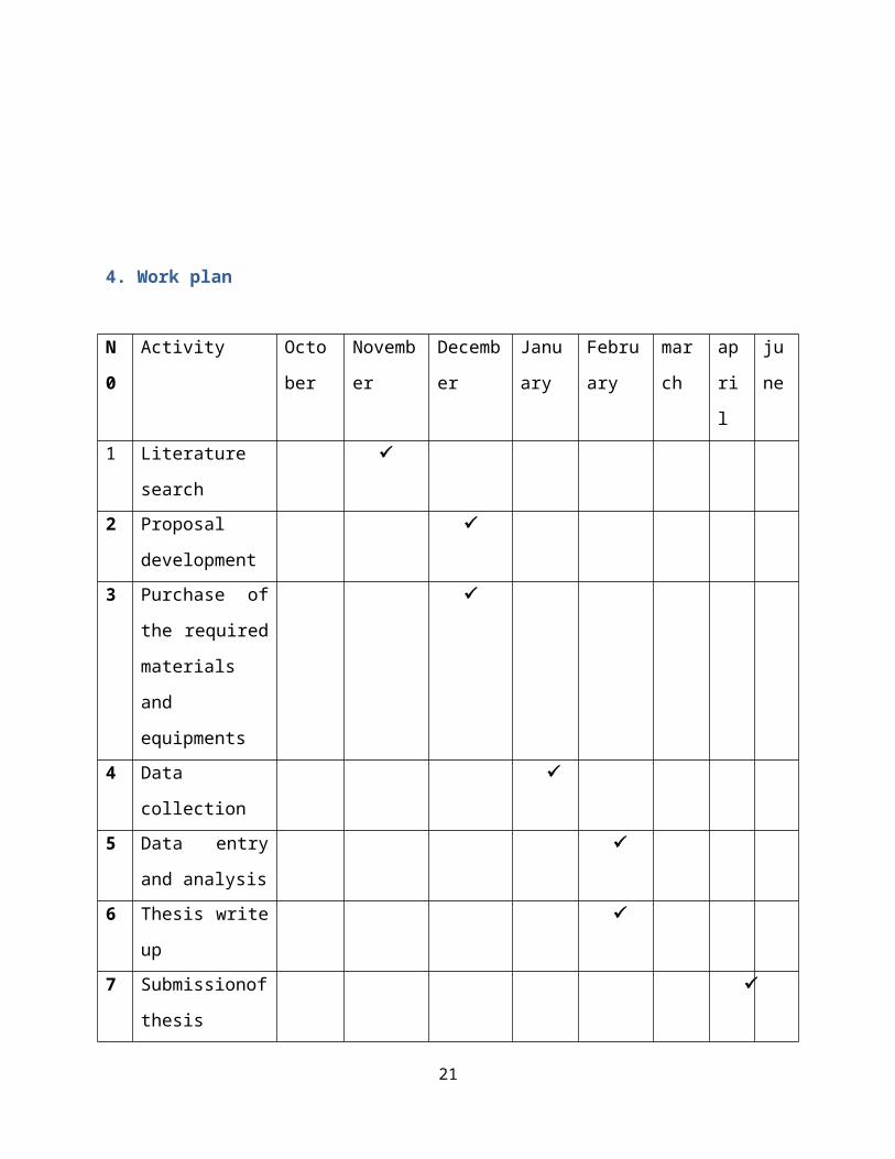
4. Work plan
N
0
Activity Octo
ber
Novemb
er
Decemb
er
Janu
ary
Febru
ary
mar
ch
ap
ri
l
ju
ne
1 Literature
search
2 Proposal
development
3 Purchase of
the required
materials
and
equipments
4 Data
collection
5 Data entry
and analysis
6 Thesis write
up
7 Submissionof
thesis
21
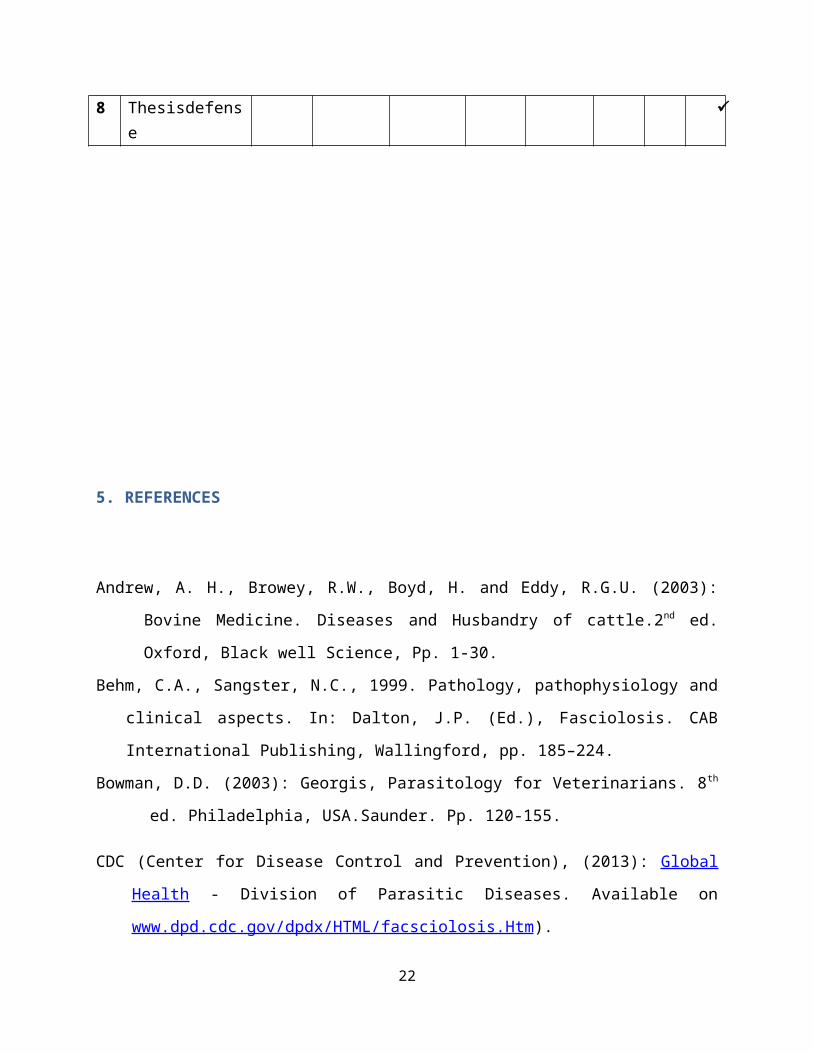
8 Thesisdefense
5. REFERENCES
Andrew, A. H., Browey, R.W., Boyd, H. and Eddy, R.G.U. (2003):
Bovine Medicine. Diseases and Husbandry of cattle.2nd ed.
Oxford, Black well Science, Pp. 1-30.
Behm, C.A., Sangster, N.C., 1999. Pathology, pathophysiology and
clinical aspects. In: Dalton, J.P. (Ed.), Fasciolosis. CAB
International Publishing, Wallingford, pp. 185–224.
Bowman, D.D. (2003): Georgis, Parasitology for Veterinarians. 8th
ed. Philadelphia, USA.Saunder. Pp. 120-155.
CDC (Center for Disease Control and Prevention), (2013): Global
Health - Division of Parasitic Diseases. Available on
www.dpd.cdc.gov/dpdx/HTML/facsciolosis.Htm).
22
CSA, Central Statistics Agency, Federal Democratic Republic ofEthiopia (2006): Agricultural Sample Survey 2006/07, volume II, Report on livestock and
livestock characteristics. Statistical Bulletin 388. Addis Ababa, Ethiopia. Pp. 9-10,25- 27.
Dubinský, P., 1993. Trematódy a trematodózy. In: Jurášek, V.,
Dubinský, P. a kolektív, Veterinárnaparazitológia.
Prírodaa.s., Bratislava, 158–187. in Slovakian
Fairweather I. 2011. Reducing the future threat from (liver)
fluke: realistic prospect or quixotic fantasy Vet.
Parasitol.180:133-143.
Fiss L., Adrien M.L., Marcolongo-Pereira C., Assis-Brasil N.D.,
Ruas J.L., Sallis E.S.V., Riet-Correa F. &Schild A.L.
2012. Subacute and acute fasciolosis in sheep in Southern
Brasil.Parasitol. Res. DOI 10.1007/s00436- 012-3096-2.
http://www.patient.co.uk/doctor/Fasciola-Hepatica.htm#,
2013).
Grebber, M., 1978.Helminthes and helmenthossis of domestic and
wild animals of Ethiopia.bulletin.animal health and
production in Africa. 23:86
Hansen, J., and perry, B., 2005.Tthe epidemiology diagnosis and
control of helminthes parasite of ruminants. A hand book of
23
rome, Foodand Agricultural organization of United
Nation .Pp.72-78.
Hendrix, C.M. (1998): Diagnostic Veterinary Parasitology. 2nd ed.
United state of America; Mousby. Pp. 151-178.
Ibarra F, Vera Y, Quiroz H, et al. (February 2004). "Determination
of the effective dose of an experimental fasciolicide in
naturally and experimentally infected cattle". Vet. Parasitol
Kassai, T. M., Carderodel, C. J., Evzeby, S., Heipe, Th. and
Himenas (1999): Standardize nomenclature of animal parasite
disease (SNOAPAD).Veterinary Parasitology, 29: 299-326.
Kaufmann, J. (1996): Parasitic infection of domestic animals. A
diagnostic manual. Basel, Boston, Birkhauser. Pp. 91-96.
Khan, E.M., 2005.The merk veterinary manual. 9th ed. USA: Merk
and co.,inc. Pp.269-278.
Krauss, H., Weber, A., Appel, M., Endris, B., Isberg, H.D., Schiefer, H.G., Slenka, W., Alexander, V. and Graevenitz, H.(2003): Zoonoses. Infectious disease transmissible from animals to human.3rd ed. Washington, D.C. ASM press. Pp. 317-356.
Mekonnen Addis, Microbiology and Veterinary Public Health Team,
School of Veterinary Medicine, College of Agriculture and
Veterinary Medicine, Jimma University, Jimma, Ethiopia
24
Merck, C.K. (2005): The Merck Veterinary Manual. CNS Disease by
helminthes and arthropods.USA, White house station. Merck and
co., INK. Pp. 274-290.
Mitchell GB, Maris L, Bonniwell MA (October 1998).
"Triclabendazole-resistant liver fluke in Scottish
sheep".Vet. Rec.143 (14): 399. PMID 980220
Moll L, Gaasenbeek CP, Vellema P, Borgsteede FH (July 2000).
"Resistance of Fasciola hepatica against triclabendazole in
cattle and sheep in Thenetherlands". Vet. Parasitol.91 (1–2): 153–
8.
Moll;_L, Gaasenbeek,P.H ,Vellema , P, Borgsteede,H. M, 200O.
Resistance of Fasciola hepatica against triclabendazole in cattle,
Veterinary Parasitology 91 , 153–158
Müller G. 2007. Fasciolose, p.639-650 In: Riet-Correa F., Schild
A.L., Lemos R.A.A. & Borges J.R.J. (Eds), Doenças de
Ruminantes e Equídeos. Vol.1. 3ª ed. Pallotti, Santa Maria.
NADIS, 2007.health bluttin, knowledge transfer to
farmers.Fascioliasis (liver fluke) in cattle(NADIS Parasite
Forecast – see latest update at www.nadis.org.uk )
Pfukenyi, D.M., Mukaratirwa, S., Willingham, A.L. &Monrad, J.
2006. Epidemiological studies of Fasciolagigantica infections
in cattle in the highveld and lowveld communal grazing areas
of Zimbabwe. Onderstepoort Journal of Veterinary Research,
73:37–51
25
Phiri I.K, Phiri A.M, Harrison,L.J 2006. "Serum antibody isotype
responses of Fasciola-infected sheep and cattle to excretory
and secretory products of Fasciola species".Vet. Parasitol.141 (3–
4): 234–42. doi:10.1016/j.vetpar.2006.05.019. PMID 16797844.
Radostits O.M., Gay C.C., Hinchcliff K.W. & Constable P.D. 2007.
Veterinary Medicine: a textbook of the diseases of cattle,
horses, sheep, pigs and goats. 10th ed. W.B. Saunders,
Edinburgh. Pp.2156
Taylor, M.A., Coop, R.L., Wall, R.L. (2007): Veterinary
Parasitology. 3rd ed. UK, Black Well Publisher. Pp. 343-345.
Torgerson, P; and Claxton J (1999). "Epidemiology and
control.".In Dalton, JP.Fasciolosis. Wallingford, Oxon, UK: CABI
Pub. pp. 113–49. ISBN 0-85199-260.
Thrusfield, M., 2005. Survey in Veterinary Epidemiology. 2nd ed. USA:
Blackwell Science, Limited, Cambridge.
Urquhart, G.M., J. Armour, J.L. Duncan, A.M. Dunn and F.W.
Jennings, 1996. Veterinary parasitology, 2 nded University of
Oxford, Long man scientific. The high and technical press,
UK, pp: 112-120.
Van Dijk J., Sargison N.D., Kenyon F. &Skuce P.J. 2010. Climate
change and infectious disease: helminthological challenges to
farmed ruminants in temperate regions. Anim. 4:377-392.
26
Walker, S.M., A.E. Makundi, F.V. Namuba,A.A. Kassuku, J. Keyyu,
E.M. Hoey, P. ProdohlJ.R. Stothard and A. Trudgett, 2008. The
distributionofFasciola hepatica and
Fasciolagiganticawithinsouthern Tanzania-constraints
associated with the intermediate host. Parasitol., 135: 495-
503
Yilma,J. and Malone, J.B.,1998. A geological information system
forecast model for strategic control of fasciolosis in
Ethiopia. Vet. Parasitology.Pp.103-127.
ISPUB.com / IJVM/3/2/9672 Author/Editor Login Registration
Facebook Google Plus
ISPUB.com
27
INTERNETSCIENTIFICPUBLICATIONS
Home Journals Latest Articles Disclaimers Article Submissions Contact Help
The Internet Journal of Veterinary Medicine Volume 3 Number 2
The Internet Journal of Veterinary Medicine Volume 3 Number 2
Original Article
The Prevalence and Economic Significance of Bovine Fasciolosis at Jimma, Abattoir, Ethiopia
T Tolosa, W Tigre
Keywordseconomic significance, ethiopia, fasciolosis, jimma municipality abattoir, local adult cattle, prevalence
CitationT Tolosa, W Tigre. The Prevalence and Economic Significance of Bovine Fasciolosis at Jimma, Abattoir, Ethiopia. The Internet Journal of Veterinary Medicine. 2006
28
Volume 3 Number 2.
AbstractA study was conducted to determine the prevalence rate and the economic significance of bovine Fasciolosis in Jimma municipality abattoir by using post-mortem examination of liver of each slaughteredanimal in particular and secondary data analysis. The objectives of the study were to determine the overall prevalence rate and economic significance of bovine fasciolosis in Jimma municipality abattoir and to determine the most prevalent species of liver fluke in indigenous adult cattle slaughtered in the abattoir and thus, in the localities from where these food animals were provided for slaughtering. From thetotal number of cattle slaughtered (468) during the study period 46.58% (218) of them were found to be positive for Fasciolosis. F. hepatica was found to the most liver fluke species affecting cattle slaughtered in the study area. 63.89 % of the total livers found to bepositive for bovine Fasciolosis were infected by F. hepatica whereas F. gigantica and unidentified or immature forms of fasciola species recovered were 24.07 % and 12.04 % respectively. In line to the economic importance of bovine Fasciolosis in the study area, the problem caused loss of an average of 148.12 and 54,063.34 Ethiopian birr per day and annum, respectively and thus found to have significant economic importance.
Introduction
Bovine fasciolosis is an economically important parasitic disease of cattle caused by Fasciolidae of the genus Fasciola. The two most important species of this genus, , are commonly known as liver flukes.
Generally, the distributioin of fasciolosis is worldwide, however, thedistribution of F.hepatica, is limited to temperate areas and highlands of tropical and sub-tropical regions (Soulsby 1986). The definitive hosts for are most mammals among which sheep and cattle arethe most important once. The geographic distribution of trematode
29
species is dependent on the distribution of suitable species of snails. The genus Lymnaea in general and L.trancatula in particular isthe most common intermediate hosts for . This species of snail was reported to have a worldwide distribution (Urquhart el al 1996).
The presence of fasciolosis due to and in Ethiopia has long been knownand its prevalence and economic significance has been reported by several workers; different works so far conducted in Ethiopia reportedvariable prevalence rates of bovine fascioosis in different localitiesof the country (Getu 1987; Abebe 1988; Mulugeta 1993; Dagne 1994; Wondwosen 1990; Yosef 1993; Adem 1994; Mezgebu 1995). In Ethiopia, theprevalence of bovine fasciolosis has shown to range from 11.5% to 87% (Malone et al 1998). The study conducted at Dire Dawa revealed that out of 2224 cattle slaughtered in the abattoir, the prevalence of fasciolosis has been found to be 14.4% in which Fasciola hepatica was observed to be the most commonly recovered fluke species (Daniel 1995). F. hepatica was shown to be the most important fluke species inEthiopian livestock with distribution over three quarter of the nationexcept in the arid north-east and east of the county. The distributionof F. gigantica was mainly localized in the western humid zone of the country that encompasses approximately one fourth of the nation (Malone et al 1998). Moreover, the studies also showed that fasciolosis has higher economic significance on animal production and productivity. The economic losses due to fasciolosis throughout the world are enormous and these losses are associated with mortality, morbidity, reduced growth rate, condemnation of fluky, liver, increased susceptibility to secondary infections and expense due to control measures (Malone et al 1998). A rough estimate of the economicloss due to decreased productivity caused by bovine fasciolosis is about 350 million birr per annual (Bahiru and Ephrem 1979). According to the study conducted by Abdul (1992) and Daniel (1995) a total economic loss of about154, 188 and 215,000 Ethiopian birr per annum incattle were reported due to fasciolosis at Ziway and Dire Dawa municipal slaughterhouses, respectively.
Diagnosis is based primarily on clinical signs, seasonal occurrence, previous history of fasciolosis on the farm or the identification of
30
snail habitats; postmortem examination, haematological tests and examination of faeces for fluke eggs. Even though, it is impossible todetect fasciola in live animals, liver examination at slaughter or necropsy was found to be the most direct, reliable, and cost effectivetechnique for the diagnosis of fasciolosis (Urquhart et al 1996).
Therefore, the objectives of this study were to determine the prevalence and the economic significance of bovine fasciolosis due to organ condemnation in Jimma municipality abattoir.
Materials and Methods
Description of the Study Area
The study was conducted in Jimma zone, Southwestern part of Ethiopia at Jimma municipality abattoir. Jimma town, the capital of Jimma zone is located in Oromia Regional Administration, 346 km Southwest of Addis Ababa at latitude of about 7013'-8056' N and longitude of about 35052'-37037' E, and at an elevation ranging from 880 m to 3360 m above sea level. The study area receives a mean annual rainfall of about 1530 millimeters which comes from the long and short rainy seasons. The annual mean minimum and maximum temperature during the study period were 14.4 and 26.7 degree Celsius respectively .
Study population
In the study 468 adult male indigenous cattle provided for slaughter from different localities in the southwestern part of Ethiopia were included. Cattle slaughtered in the abattoir were brought from different markets which in turn are provided from different livestock markets in their vicinity.
Study design
A cross sectional study type was conducted to determine the prevalencerate and the economic significance of bovine Fasciolosis by using post-mortem examination of different organs in general and liver of each slaughtered animal in particular and also secondary data analysis. During the study special attention was given to the livers
31
of the animals and liver of each slaughtered was carefully examined byvisualization and palpation of the entire organ that was followed by transverse incision of the organ across the thin left lob in order to confirm the case or the problem (Urquhart 1996; Soulsby 1986,). Species identification of the recovered fasciola was also performed (based on the morphological features of the agents) and classified in to F. hepatica, F. gigantica and unidentified or immature forms of liver fluke. (Urquhart 1996; Soulsby 1986,)
Data Collection
Appropriate data were collected by using post-mortem examination of the organs so far claimed to be infected by fasciolosis and secondary data analysis. An interview was made with retailers of offal produced at Jimma municipality abattoir to obtain information on the average price of a liver in the study area during the study time. According tothe response of the retailers the price of a liver was found to be tenEthiopian birr on average.
Statistical Analysis
Prevalence of fasciolosis was calculated as the number of cattle foundto be infected with Fasciola, expressed as a percentage of the total number of cattle slaughtered (Thrusfield 1995). The economic significance of the problem was analyzed based on the information obtained during interview and calculated on daily and annual basis.
Results
Postmortem examination inspection
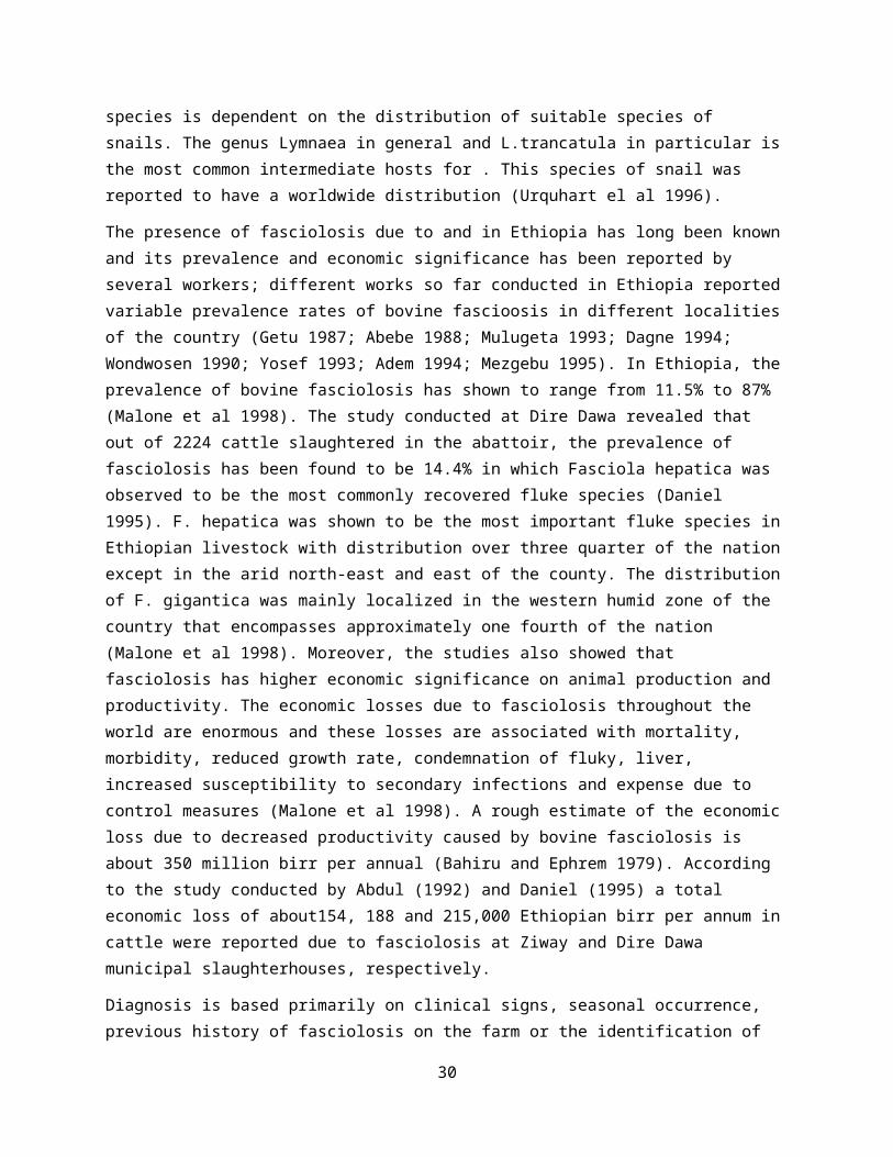
A total of 468 adult indigenous cattle were slaughtered at Jimma abattoir and examined for fasciolosis. Of the total cattle slaughteredand examined (N=468), 46.15% (n= 216) of them were found to be positive for lesion of fasciolosis (Table 1).
Figure 1Table 1: Prevalence of bovine fasciolosis and the different fasciola species recovered during the study period.
32
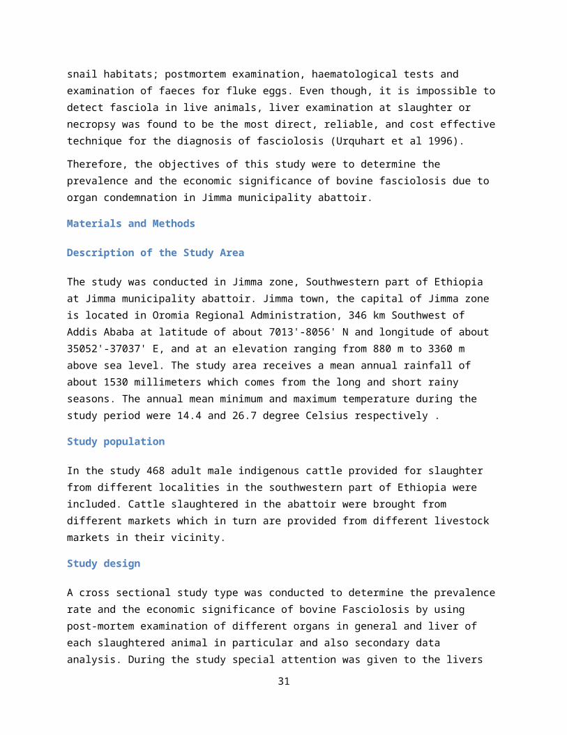
Fasciola species identification
From a total of 216 livers found positive for fluke infection during post mortem inspection of slaughtered animals, 138 livers (63.89 %) harboured F. hepatica , 52 livers (24.07 %) F. gigantica and 26 livers(12.04 %) infected with un identified species due to immature fluke (Table 2).
Figure 2Table 2: species of fasciola encountered in affected livers during post mort examination of slaughtered animals
From the result of the study, the economic loss due to liver condemnation was estimated to be an amount of 145.33 Ethiopian birr daily and 53,046.67 Ethiopian birr annually.
Analysis of abattoir data
Analyses were made on one year meat inspection records obtained from Jimma municipality abattoir. A total of 11349 adult male indigenous cattle were slaughtered by the time from June 2005 to May 2006 in the aforementioned slaughter house. Of the total livers (N= 11,349) inspected for liver fluke, 48.53% (n=5,508) of them were found to be positive for fasciola and this resulted an average daily and annual economic loss of about 150.90 and 55,080.00 Ethiopian birr, respectively. An overall prevalence of 48.53% of bovine fasciolosis was recorded from data obtained in which the highest value (5.29%) and
33
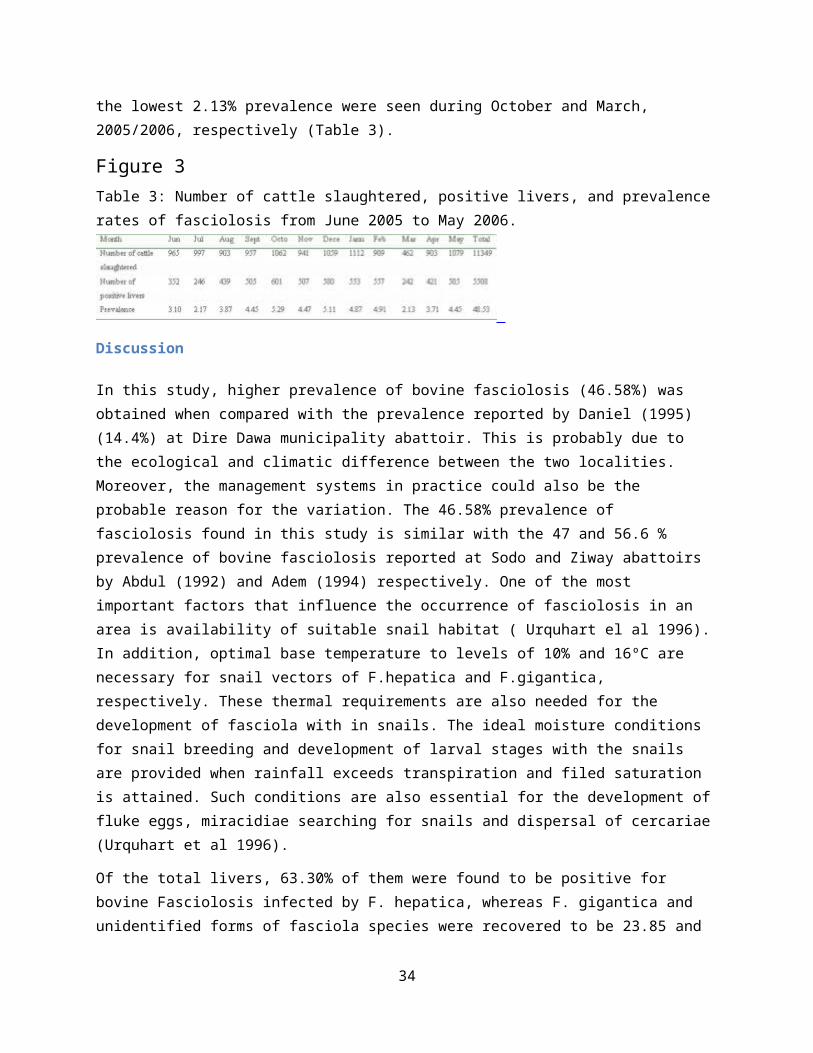
the lowest 2.13% prevalence were seen during October and March, 2005/2006, respectively (Table 3).
Figure 3Table 3: Number of cattle slaughtered, positive livers, and prevalencerates of fasciolosis from June 2005 to May 2006.
Discussion
In this study, higher prevalence of bovine fasciolosis (46.58%) was obtained when compared with the prevalence reported by Daniel (1995) (14.4%) at Dire Dawa municipality abattoir. This is probably due to the ecological and climatic difference between the two localities. Moreover, the management systems in practice could also be the probable reason for the variation. The 46.58% prevalence of fasciolosis found in this study is similar with the 47 and 56.6 % prevalence of bovine fasciolosis reported at Sodo and Ziway abattoirs by Abdul (1992) and Adem (1994) respectively. One of the most important factors that influence the occurrence of fasciolosis in an area is availability of suitable snail habitat ( Urquhart el al 1996).In addition, optimal base temperature to levels of 10% and 16ºC are necessary for snail vectors of F.hepatica and F.gigantica, respectively. These thermal requirements are also needed for the development of fasciola with in snails. The ideal moisture conditions for snail breeding and development of larval stages with the snails are provided when rainfall exceeds transpiration and filed saturation is attained. Such conditions are also essential for the development offluke eggs, miracidiae searching for snails and dispersal of cercariae(Urquhart et al 1996).
Of the total livers, 63.30% of them were found to be positive for bovine Fasciolosis infected by F. hepatica, whereas F. gigantica and unidentified forms of fasciola species were recovered to be 23.85 and
34

11.93% diagnosed as positive for Fasciolosis. Similar study conducted at Zeway abattoir reported 60.3% 0f the liver harbored F.hepatica, 10.2% F. gigantica and 29.5% infested by both species (Adem 1994). Theprevalence and the species involved vary significantly with locality. This is atributed mainly to the variation in the climatic and ecological conditions such as altitude, rainfall, temperature and livestock management system(Yilma & Malone 1998). Moreover, Garber & Daynes reported that; in Ethiopia F. hepatica and F. gigantica infections occur in areas above 1800 m.a.s.l. and below 1200m.a.s.l. respectively. The high prevalence rate of F. hepatica may be associated with the existence of favourable ecological biotops for L. truncatula. Relatively small proportion of cattle were found infected with F. gigantica alone or mixed infection with both spp. This may be explained by cattle coming for slaughter from highland and middle altitude zone flood prone areas, drainage ditches are favourable habitat to natalensis (Urquhart el al 1996).
The highest prevalence rate was analyzed during October, when the wet-ecological conditions still prevailed. It has been described that the bionomic requirements for breeding of the Lymnaea snails and development of the intramolascan stages of the flukes often reach the optimum threshold during the wet months of the year. During the dry periods, breeding of the snails and development of the larval flukes slow down or stops completely and snails undergo a state of aestivation (Yilma and Malone 1998).
Although a decreasing trend was analyzed along with the advancement ofthe dry season, relatively high prevalence of fasciola infection was analyzed from the data recorded by the abattoir. This may be attributed to infections acquired during previous peak snail activity season. In addition, the existence of permanent suitable ecological conditions in areas like slow flowing rivers, streams and low lying marshy areas may contribute to persistent but relatively low grade infection during the dry season.
The total economic loss encountered due to condemnation of infected liver from one year data recorded from abattoir in this study was 55,080.00 birr per annum. There was also similar economic loss due to
35
infected liver condemnation of post mortem inspection result. This finding is by far lower than the results reported by Adem (1994) and Daniel (1995) a total economic loss of about154, 188 and 215,000 Ethiopian birr per annum in cattle due to fasciolosis at Ziway and Dire Dawa municipal slaughterhouses, respectively. This is probably due to the ecological and climatic difference between the two localities.
Conclusions
This study demonstrated that bovine fascvolosis prevalent in cattle inthe area, it causes great economic losses as a result of condemnation of infected livers and its prevalence is relatively high throughout the year in the study area, due to the fact that the area is very suitable for the intermediate host snails and the parasite.
Acknowledgements
The authors would like to thank student Abiyot Mengesha, Adamu Yimer, Endalkachew Sisay, Gebeyaw Ezezew and Terefe Taye for data collection and Jimma Municipality abattoir workers for their cooperation during the study period.
Referencesr-0. Abdul J R 1992 Economic Significance of Bovine Fasciolosis and Hydatidiosis. In soddo, DVM thesis, Faculty of Veterinary Medicine, Addis Ababa University Debre Zeit, Ethiopia.r-1. Abebe M 1988 Prevalence of Bovine Fasciolosis and its Economic Significance at Nekempte, DVM thesis, Faculty of Veterinary Medicine Addis Ababa University Debre Zeit, Ethiopia.r-2. Adem A 1994 Prevalence of Bovine and Ovine Fasciolosis: A Preliminary Survey Around Ziway Region (Shewa), DVM Thesis, Faculty ofVeterinary Medicine Addis Ababa University Debre Zeit, Ethiopia.r-3. Bahiru G and Ephrem M 1979 Preliminary survey of bovine fasciolosis. Ethiopian. Control rh. Agil. Science. 1 1 50-127.r-4. Blood D C, Henderson J A and Radostits O M 1979 A Text Book of The disease of Cattle, Sheep, Pigs and Horses in USA, 5th ed.
36
Philadelphia, USA 756-760,.r-5. Dagne M 1994 Survey on Prevalence and Economic Significance of Bovine Fasciolosis in Debre Berhan region, DVM Thesis, Faculty of Veterinary Medicine, Addis Ababa University. Debre Zeit, Ethiopia.r-6. Daniel F 1995 Economic Importance of organs condemnation due to Fasciolosis and Hydatidosis in cattle and sheep slaughtered at Dire Dawa Abattoir, DVM thesis, Faculty of Veterinary Medicine, Addis AbabaUniversity Debre Zeit, Ethiopia.r-7. Getu D 1987 A study on the Incidence and Economic Significance ofFasciolasis in Woliata Awraja, DVM Thesis, Faculty of Veterinary Medicine, Addis Ababa University. Debre Zeit, Ethiopia.r-8. Malone J B., Gommes R., Hansen J., Yilma J M., Slingenberg J., Snijders F., Nachet O F and Ataman E 1998 A Geographic Information System on the potential Distribution and abundance of Fasciola hepatica and F. gigantica in East Africa based on food and agricultureorganization databases, Elev, Vet Parasitol, 78 87-101.r-9. Mezgebu B 1995 A survey on Ovine Fasciolosis and Lung worm Infestation in Addis Ababa and the Surrounding highland areas, DVM thesis, Faculty of Veterinary Medicine, Addis Ababa University. Debre Zeit, Ethiopia.r-10. Mulugeta T 1993 Prevalence and Economic Significance of Bovine fasciolosis at the Sopral Kombolcha meat factory, DVM thesis, Faculty of Veterinary Medicine, Addis Ababa University. Debre Zeit, Ethiopia.r-11. Soulsby E J L 1982 Helminthes, Arthropods and Protozoa of Domesticated Animals, Seventh Edition. Balliere Tindall, London, UK .40-52.r-12. Thrusfield M 1995 Veterinary Epidemiology second edition, University of Edinburgh, Black well science 180-188.r-13. Urquhart G M., Duncan J L., Armour J., Dunn A M., Jenning 1996 Veterinary Parasitology. Second Edition. Blacwell Scince, UK. 103-113.r-14. Wondwosen A 1990 Prevalence of Bovine Fasciolosis in Arsi Administrative Region, DVM Thesis, Faculty of Veterinary Medicine, Addis Ababa University. Debre Zeit, Ethiopia.r-15. Yilma J M and Malone J B 1998 A Geographic Information System forecast model for Strategic control of fasciolasis in Ethiopia, Faculty of Veterinary Medicine, Addis Ababa University. Ethiopia Elev.
37
Vet Parasitol 78 103-123.r-16. Yosef S 1993 Prevalence of Ovine Gastrointestinal Helminthes in and around Asella, DVM Thesis, Faculty of Veterinary Medicine, Addis Ababa University. Debre Zeit Ethiopia. 47
{full_citation}
Author InformationTadele Tolosa, DVM, MVScCollege of Agriculture and Veterinary Medicine, Jimma University
Worku Tigre, DVMCollege of Agriculture and Veterinary Medicine, Jimma University
Share This ArticleYour free access to ISPUB is funded by the following advertisements:
BACK TO TOP
Facebook Google Plus
© 2013 Internet Scientific Publications, LLC. All rights reserved. UBM Medica Network Privacy Poli
38

39
Related Documents