UNIT IV DISEASES OF HONEY BEE BACTERIAL DISEASES American foulbrood disease (AFB) Beekeepers in temperate and sub-tropical regions around the world generally regard American foulbrood (AFB) as possibly the most destructive microbial disease affecting bee brood. The disease did not originate in, nor is it confined to, the Americas. It is widely distributed wherever colonies of Apis mellifera are kept. In tropical Asia, where sunlight is abundant and temperatures are relatively high throughout the year, the disease seldom causes severe damage the beekeeping operations. The disease is contagious and the pathogenic bacterium can remain dormant for as much as and more than 50 years. Therefore, beekeepers and extension specialists throughout Asia should be acquainted with the symptoms of this disease and know how to cope with it should the need arise. Cause American foulbrood disease is caused by a spore-forming bacterium, Paenibacillus larvae, which only affects bee brood; adult bees are safe from infection. At the initial stage of colony infection, only a few dead older larvae or pupae will be observed. Subsequently, if remedial action is not taken, the disease will spread within the colony and can quickly spread to other colonies in the apiary as a result of robbing, drifting workers, or contamination caused by the beekeeper's hive manipulations. In the same way the pathogen agent can spread to other apiaries. Natural transfer mainly takes place within a radius of 1 km around the apiary. Often spores enter the bee colonies via foreign honey. Commercially available honey may be highly contaminated; therefore, special attention should be paid near honey processing enterprisesand waste disposal sites. Symptoms At the initial stage of AFB infection, isolated capped cells from which brood has not emerged can be seen on the comb. The caps of these deadbrood cells are usually darker than the caps of healthy cells, sunken, and often punctured. On the other hand the caps of healthy brood cells are slightly protruding and fully closed. As the disease spreads within the colony, a scattered, irregular pattern of sealed and unsealed brood cells (see Plate 1) can be easily distinguished from the normal, compact pattern of healthy brood cells observed in healthy colonies. The bee brood affected by AFB is usually at the stage of older sealed larvae or young pupae, upright in the cells. Often therefore, a protruding tongue can be found with the rest of the body already decayed. At first the dead brood is dull white in colour, but it gradually changes to light brown, coffee brown, and finally dark brown or almost black. The consistency of the decaying brood is soft. Once the dead brood have dried into scales, the test cannot be used. The dry brood lies flat on the lower side of the cell wall, adhering closely to it – in contrast to sacbrood. This scale is usually black or dark brown and brittle. Often, a fine, threadlike proboscis or tongue of the dead pupa can be seen protruding from the scale, angling toward the upper cell wall. The pathogen bacteria may be identified using Plate 1 Irregular pattern of sealed brood with sunken and punctured caps, typifying American foulbrood infestation. 4 Honey bee diseases and pests: a practical guide a microscopic preparation or, more frequently, by cultivation on selective culture media. The Columbia slant culture has proved to be most effective for this purpose. The result is controlled by biochemical or serological tests and more often by means of the Polymerase Chain Reaction (PCR). As PCR is very sensitive its suitability is restricted regarding the direct evidence in comb samples (see OIE Manual of Diagnostics, 2004). Commercially available ‘AFB diagnosing kits’ are based on serological evidence of the pathogenagent. In general, they are appropriate for field use. But if there are clinically indifferent cases, misinterpretations may occur. The examination of samples from stored food of sealed brood combs has become important in diagnosing AFB, although it is not effective in detecting evidence of an outbreak of AFB. However, it is suitable for population screenings in apiaries and in determining the pathogen pressure in the individual colonies. The diagnostic reliability of the samples from the food wreath depends on the quality of sample extraction. If samples are taken from newly gathered food or from other areas than the sealed brood combs, wrong diagnoses might be made resulting in false negative results. Control In several countries, where apiculture includes large commercial operations, frequent, efficient

Welcome message from author
This document is posted to help you gain knowledge. Please leave a comment to let me know what you think about it! Share it to your friends and learn new things together.
Transcript
UNIT IV DISEASES OF HONEY BEE BACTERIAL DISEASES
American foulbrood disease (AFB) Beekeepers in temperate and sub-tropical regions around the world generally regard American foulbrood
(AFB) as possibly the most destructive microbial disease affecting bee brood. The disease did not originate in,
nor is it confined to, the Americas. It is widely distributed wherever colonies of Apis mellifera are kept. In
tropical Asia, where sunlight is abundant and temperatures are relatively high throughout the year, the disease
seldom causes severe damage the beekeeping operations. The disease is contagious and the pathogenic
bacterium can remain dormant for as much as and more than 50 years. Therefore, beekeepers and extension
specialists throughout Asia should be acquainted with the symptoms of this disease and know how to cope
with it should the need arise.
Cause
American foulbrood disease is caused by a spore-forming bacterium, Paenibacillus larvae, which only affects
bee brood; adult bees are safe from infection. At the initial stage of colony infection, only a few dead older
larvae or pupae will be observed. Subsequently, if remedial action is not taken, the disease will spread within
the colony and can quickly spread to other colonies in the apiary as a result of robbing, drifting workers, or
contamination caused by the beekeeper's hive manipulations. In the same way the pathogen agent can spread
to other apiaries. Natural transfer mainly takes place within a radius of 1 km around the apiary. Often spores
enter the bee colonies via foreign honey. Commercially available honey may be highly contaminated;
therefore, special attention should be paid near honey processing enterprisesand waste disposal sites.
Symptoms
At the initial stage of AFB infection, isolated capped cells from which brood has not emerged
can be seen on the comb. The caps of these deadbrood cells are usually darker than the caps of healthy cells,
sunken, and often punctured. On the other hand the caps of healthy brood cells are slightly protruding and fully
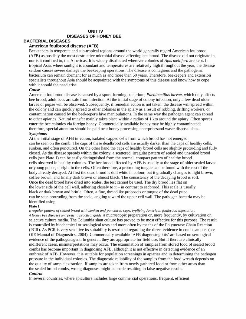
closed. As the disease spreads within the colony, a scattered, irregular pattern of sealed and unsealed brood
cells (see Plate 1) can be easily distinguished from the normal, compact pattern of healthy brood
cells observed in healthy colonies. The bee brood affected by AFB is usually at the stage of older sealed larvae
or young pupae, upright in the cells. Often therefore, a protruding tongue can be found with the rest of the
body already decayed. At first the dead brood is dull white in colour, but it gradually changes to light brown,
coffee brown, and finally dark brown or almost black. The consistency of the decaying brood is soft.
Once the dead brood have dried into scales, the test cannot be used. The dry brood lies flat on
the lower side of the cell wall, adhering closely to it – in contrast to sacbrood. This scale is usually
black or dark brown and brittle. Often, a fine, threadlike proboscis or tongue of the dead pupa
can be seen protruding from the scale, angling toward the upper cell wall. The pathogen bacteria may be
identified using Plate 1
Irregular pattern of sealed brood with sunken and punctured caps, typifying American foulbrood infestation.
4 Honey bee diseases and pests: a practical guide a microscopic preparation or, more frequently, by cultivation on
selective culture media. The Columbia slant culture has proved to be most effective for this purpose. The result
is controlled by biochemical or serological tests and more often by means of the Polymerase Chain Reaction
(PCR). As PCR is very sensitive its suitability is restricted regarding the direct evidence in comb samples (see
OIE Manual of Diagnostics, 2004). Commercially available ‘AFB diagnosing kits’ are based on serological
evidence of the pathogenagent. In general, they are appropriate for field use. But if there are clinically
indifferent cases, misinterpretations may occur. The examination of samples from stored food of sealed brood
combs has become important in diagnosing AFB, although it is not effective in detecting evidence of an
outbreak of AFB. However, it is suitable for population screenings in apiaries and in determining the pathogen
pressure in the individual colonies. The diagnostic reliability of the samples from the food wreath depends on
the quality of sample extraction. If samples are taken from newly gathered food or from other areas than
the sealed brood combs, wrong diagnoses might be made resulting in false negative results.
Control
In several countries, where apiculture includes large commercial operations, frequent, efficient

inspection services are particularly advanced and a ‘search and destroy’ strategy may be adopted in an attempt
to minimize damage to apiaries caused by this serious honey bee disease. The procedure involves hive
inspections by qualified apiary inspectors. The entire honeybee population that is infected by American
foulbrood is killed and hive materials belonging to the colony, are disinfected or destroyed by burning. The
bees are usually killed by poisonous gas such as the burning of sulphur powder. All the dead bees, the frames,
the supers, the honey and the contaminated equipment are thrown into a 1m x 1m x 1m hole in the ground.
Kerosene is poured over the pile and set alight. When all the material has been completely burned, the hole is
carefully filled in, to prevent worker bees belonging to healthy colonies from robbing any remaining
contaminated honey. Although the above-mentioned method has proven effective, the practice of burning
AFB infected colonies and equipment is costly, especially taking into account the high cost of
beekeeping equipment. The destruction of brood combs and food combs is absolutely necessary
as, apart from the bees, they are the main carriers of spores. Dry combs, without brood, can be preserved if an
examination of wax samples in the laboratory does not reveal Paenibacillus spores. In
which case the dry combs must also be destroyed.Old hives should be burned. Well conserved hives,
however, should be disinfected. The inner part of a hive, once carefully cleaned, can quickly be singed
out with the flame of a gas burner. The wooden surface should look slightly brownish. When this is not
possible, e.g. if the hive is made from plastic, they should be cleaned and brushed with 3 to 5 percent sodium
hydroxide. Before using other substances for disinfection you should make sure that no residues remain that
could be dangerous to bees or the consumer of the processed honey. The killing of the bees can be avoided if
the BOX 1 Stretch test A simple way of determining whether AFB caused the death of the brood is the ‘stretch test’ (see Plate 2). A small stick,
match or toothpickis inserted into the body of the decayed larva and then gently and slowly, withdrawn. If the disease is
present, the dead larva will adhere to the tip of the stick, stretching for up to 2.5 cm before breaking and snapping back in
a somewhat elastic way. This symptom called ‘ropiness’, typifies American foulbrood disease, but it can be
observed in decaying brood only.
Plate 2
Stretch test for American foulbrood disease.n* Irregular pattern of sealed brood with sunken and punctured caps,
typifying American Foulbrood infestation. W.RITTER Chapter 2 - Microbial diseases 5 artificial swarm method is applied.
A traditional method is to keep the bee colony in a dark environment for several days. The bees are pushed
into a decontaminated hive with new combs, the bee entrance is closed and they are placed in a dark preferably
quite cool room. Within two days, the bees have used up the contaminated food. The colonies can then be
placed either at their former stand or within a distance of at least 3 km away. If the bees are kept in the dark
for three days they forget their old stand and can be placed anywhere.
On the third day, however, some food shortage may occur. Therefore, the colonies should be fed.
The direct artificial swarm method is less complicated. First, a clean, decontaminated hive is
prepared. Instead of combs it contains three to six wooden bars, depending on the colony’s strength,
provided with a wax strip as a starter for further comb construction. Using a queen excluder fixed
at the entrance or above the bottom of the hive should prevent disappearance of the queen. The
prepared hive is placed at the colony’s old stand subject to sanitation. Now the bees are pushed
or brushed into the empty hive. Three days later, the combs that have been partially constructed by
the bees are removed again and burned. Combs with midribs later replace these. Now sanitation
is finished. The combs and the hive of the old colony are burned or decontaminated. In some countries,
beekeepers who destroy their AFB-infected colonies receive compensation, either directly from the
government or from beekeepers’ organizations. Chemotherapeutic methods of controlling
AFB involve the administration of antibiotics or sodium sulfathiazole, in various formulations, fed mixed with
powdered sugar or sugar syrup. Antibiotics and sulfonamides prevent multiplication of the agent, though it will
not kill the spores. Therefore, multiplication may begin again shortly after treatment, which is why
treatment must be repeated in shorter and shorter intervals. Over time the inner part of the hive,
the food and honey become increasingly contaminated by spores. Stopping treatment without
simultaneous disinfection leads irrevocably to a relapse. However, detectable residues remain even after a
period of time has elapsed between treatment and honey extraction. European foulbrood disease (EFB) As with American foulbrood disease, the name of this bacterial bee brood disease is inappropriate.

The range of distribution of European foulbrood disease is not confined to Europe alone and the
disease is found in all continents where Apis mellifera colonies are kept. Reports from India indicate that A. cerana colonies are also subject to EFB infection. The damage inflicted on honey bee
colonies by the disease is variable. EFB is generally considered less virulent than AFB; although
greater losses in commercial colonies have been recorded in some areas resulting from EFB.
Cause
The pathogenic bacterium of EFB is Mellissococcus pluton. It is lanceolate in shape and
occurs singly, in chains of varying lengths, or in clusters. The bacterium is Gram-positive and does
not form spores. While many strains of M. pluton are known, all are closely related.
Symptoms
Honey bee larvae killed by EFB are younger than those killed by AFB. Generally speaking,
the diseased larvae die when they are four to five days old, or in the coiled stage. The colour of the
larva changes at it decays from shiny white to pale yellow and then to brown. When dry, the scales of
larvae killed by EFB, in contrast to AFB scales, donot adhere to the cell walls and can be removed
with ease. The texture of the scales is rubbery rather than brittle, as with AFB. A sour odour can
be detected from the decayed larvae. The clinicalpicture and the odour can vary depending on the
kind of other bacteria involved (Bacillus alvei, Streptococcus faecalis, Achromobacter eurydice).Another symptom that is characteristic of EFB
is that most of the affected larvae die before their cells are capped. The sick larvae appear somewhat
displaced in the cells (see Plate 3). Plate 3
Larvae in coiled stage, killed by European foulbrood disease.W.RITTER 6 Honey bee diseases and pests: a practical guide
When a scattered pattern of sealed and unsealed brood is observed in a diseased colony, this is
normally an indication that the colony has reached
a serious stage of infection and may be significantly weakened. However, this is the case with all brood
diseases. EFB is transferred in the same way as
AFB. Melissococcus pluton as a permanent form, does not form spores but capsules which are less
resistant than the spores of P. larvae.
The detection of M.pluton is normally carried out microbiologically. Selective culture media (OIE,
2004; Bailey and Ball, 1991) are most appropriate.
For further verification biochemical tests or the PCR can be applied. The gene technological test
is very sensitive and is therefore less suitable for
the detection of M. pluton in suspicious brood. A single-use test set is commercially available based
on a serological proof like the AFB test set (see OIE Manual of Diagnostics, 2004).
Control
The choice of an EFB control method depends on the strength of the infection, i.e. how many brood
cells and combs are infested. If the infection is weak, it is often sufficient to stimulate the
hygiene behaviour of the bees. Either they are placed at a good foraging site or they are fed
with honey or sugar water. An even better result is achieved if the individual combs are sprayed
with a thinned honey solution. If the infestation is stronger it makes sense to reduce the number
of pathogens in the colony by removing the most infested brood combs. Empty combs or healthy
brood combs then replace these. Since the bees’ hygiene behaviour is also genetically determined,
replacement of the queen is also possible. Requeening can strengthen the colony by giving
it a better egg-laying queen, thus increasing its resistance to the disease and interrupting the
ongoing brood cycle giving the house bees enough time to remove infected larvae from the hive. In
serious cases, the same methods can be used as for AFB. Sometimes chemotherapeutic measures
such as antibiotics are called for, however, their application, always risks the danger of residues.
2.2 FUNGAL DISEASE Chalkbrood disease (Ascosphaerosis) In Asia, chalkbrood is rarely considered to be a serious honey bee disease, although in
Japan the disease has been reported to cause problems to beekeepers. In temperate America
and Europe, however, cases have occurred in which chalkbrood has caused serious damage to
beekeeping; therefore, Asian beekeepers should be aware of this problem.

Cause
Chalkbrood is a disease caused by the fungus Ascosphaera apis. As its name implies, it affects
honey bee brood. This fungus only forms spores during sexual reproduction. Infection by spores
of the fungus is usually observed in larvae that is three to four days old. The spores are absorbed
either via food or the body surface.
Symptoms
Initially, the dead larvae swell to the size of the cell and are covered with the whitish mycelia of the
fungus. Subsequently, the dead larvae mummify, harden, shrink and appear chalklike. The colour
of the dead larvae varies with the stage of growth of the mycelia: first white, then grey and finally,
when the fruiting bodies are formed, black (see Plate 4). When infestation is heavy, much of the
sealed brood dies and dries out within their cells.When such combs are shaken the mummified
larvae make a rattling sound. In the laboratory the fungus can be identified by its morphology (see
OIE Manual of Diagnostics, 2004). Plate 4
Brood killed by chalkbrood: white and black mummies. W.RITTERChapter 2 - Microbial diseases 7
Control
As with other brood diseases, the bees remove the infested brood with their hygiene behaviour
(see European foulbrood), which is especially effective for white mummies. Though as soon
as the fruit bodies of A. apis have developed, cleaning honey bees spread the spores within
the colony by this behaviour. During the white mummy stage the fungus continues to develop at
the hive bottom. If the mummies are not removed quickly, the spores may enter the brood cells
carried there by circulating air. The beekeeper can stimulate the hygiene
behaviour of the bees by changing the broodrearing conditions. In this respect, it is most
important to adapt the size of the hive to the strength of the bee colony. In this way the bees have
a chance to inspect and clean the many brood cells.Therefore, in most cases, the method of
stimulating hygiene behaviour, already described under European foulbrood control, is sufficient
for chalkbrood control. The beekeeper should ensure that the colony has a strong worker
population, and that the hive is well ventilated and free from accumulated moisture. At early stages of
chalkbrood infection, adding young adult workers and hatching brood, combined with sugar-syrup
feeding, often proves to be helpful. Currently there is no known successful
chemical control against chalkbrood. It means that chemical treatment shows a little
effect to control chalkbrood. In most cases, commercialised substances only show a positive
effect because they are sprayed, or fed with sugar water as described above.
2.3 VIRAL DISEASES Over the past years at least 18 virus types and strains have been recorded as disease pathogens
of adult bees and bee brood, nearly all are RNA viruses. Laboratory examination for virus diseases
is difficult, calling for sophisticated equipment and procedures, since particles of the virus are too small
to be observed with ordinary light microscopes. However, they can rarely be differentiated with
an electron microscope. Apart from serological methods, most of the known viruses can now be
identified by genetic technologies (PCR).The damage caused to colonies by viral infection
varies considerably according to a number of factors, which include the type and strain of virus involved,
the strength of the colony, weather conditions, the season and food availability. Basically, bees are
well-protected against infection with their chitin body shell and gut coating. Parasitic mites sucking
the blood of the bees, however, can penetrate this protection. Therefore, increased infestation by
parasites is often accompanied by increased virus infection. Little known viruses such as Acute
Paralyses Bee Virus (APBV), and Deformed Wing Virus (DWV) may become increasingly destructive
in the future. As not much is known about the life cycle and pathogenity of most virus diseases, there
are only a few ways to control them. Therefore, reflecting this situation, only the most widespread
sacbrood is described.
Sacbrood disease Sacbrood disease (caused by Morator aetotulas) is perhaps the most common viral disease of honey bees. In Asia, at least two major typeshave been recorded. Sacbrood disease that affects
the common honey bee Apis mellifera and the sacbrood disease of the Asian hive bee A. cerana.

A new type of sacbrood virus has recently been reported in Asian colonies of A. cerana. It is
highly probable that the virus is native to the continent and that it has been with the Asian hive
bees over the long period of its evolution. Since its first discovery in Thailand in 1981, it has been
found in association with A. cerana in India, Plate 5
Honey bee larvae killed by sacbrood disease. W.RITTER 8 Honey bee diseases and pests: a practical guide
Pakistan, Nepal, and perhaps all other countries in Asia within the honey bee’s range of distribution.
Several reports indicate that nurse bees are the vectors of the disease. Larvae are infected via
brood-food gland secretions of worker bees.
Symptoms
Field inspection to determine whether the pathogenic virus has infected a colonycan be
easily carried out following symptomology. Diseased larvae fail to pupate after four
days; they remain stretched out on their backs within their cells (distinct from the mostly
twisted position of larvae affected by European foulbrood. The anterior section of the larva,
consisting of its head and thorax, is the first part of its body to change colour, changing from white
to pale yellow and finally to dark brown and black (see Plate 5). On removing the larvae from
their cell the inspector can easily observe that their skin is quite tough and that its contents are
watery; the infected larva thus has the appearance of a small, watery sac. Dead larvae remaining
within their cells eventually dry out to flat scales that adhere loosely to the cell floor.
Control
No chemotherapeutic agent is effective in preventing or controlling sacbrood disease.
Colonies often recover from the infection without the beekeeper's intervention, particularly if the
infection is not new to the geographic area. This mainly depends on the hygiene behaviour of the
bees, which may be stimulated as with other brood diseases (see European foulbrood). Since the disease
usually occurs when the colony is under stress (shortage of food, food-storage space, unfavourable
climatic conditions such as damp during the rainy or cold season, unhygienic hive interior, poor
queen, infestation with other diseases, etc.), the beekeeper should deal with severe cases by requeening
the colony, removing infected brood combs and taking other management measures to restore colony strength,
such as providing food and adding worker population. If there is an extremely
strong infestation it may be convenient to apply the artificial swarm method as for American foulbrood.
2.4 PROTOZOAN DISEASE Nosema disease (Nosemosis) Nosema disease is generally regarded as one of the most destructive diseases of adult bees, affecting
workers, queens and drones alike. Seriously affected worker bees are unable to fly and may
crawl about at the hive entrance or stand trembling on top of the frames. The bees appear to age
physiologically: their life-span is much shortened and their hypopharyngeal glands deteriorate, the
result is a rapid dwindling of colony strength. Other important effects are abnormally high rates
of winter losses and queen supersedures.
In climates with pronounced long periods of flight restrictions, i.e. no flight opportunities
even for a day, the infection easily reaches a severe stage that visibly affects the strength of
the colony. Less obvious infection levels in other climates often go undetected.
The damage caused by Nosema disease should not be judged by its effect on individual colonies
alone as collectively it can cause great losses in apiary productivity.
Cause
The disease is caused by the protozoan Nosema apis, whose 5 to 7 mm spores infest the bees,
are absorbed with the food and germinate in the midgut. After penetration into the gut wall the
cells multiply forming new spores that infect new gut cells or can be defecated. The nutrition of the
bees is impaired, particularly protein metabolism.
Symptoms
Unfortunately, there is no reliable field diagnostic symptom enabling a diseased worker bee to be
identified without killing it, nor can the beekeeper recognize an infected queen. However, in severe cases of infection, it is sometimes possible to separate healthy from diseased bees, the abdomen
of an infected worker often being swollen and shiny in appearance. On dissection, the individual Plate 6

Nosema apis spores (magnification factor 400 x). W.RITTER Chapter 2 - Microbial diseases 9
circular constrictions in the alimentary canals of uninfected bees are clearly visible, while the
constrictions cannot be seen clearly in diseased bees. Easy separation, after killing, of first
abdominal segments with intestines attached, which shows white if strongly infected, versus a
normal transparent, darker grey/ochre colour if there is no or only a low infection.
The most reliable method of detecting Nosema disease involves laboratory procedures using a
microscope for diagnosis. A simple diagnostic method used for adult workers is to use a sample
of 20 suspected workers. The bees are killed, and their abdomens are removed and ground in water
(2 to 3 ml per sample). A drop of the suspension of pulverized bee abdomens is then viewed under
a microscope. If the disease is present, reasonably large individual bacilliform spores with bright,
queen’s egg-laying capacity fluorescent edges (see Plate 6) will be observed. In the visual field of the
microscope, at a 400 fold magnification, up to 20 spores indicate a weak, 20 to 100 a medium and
100 and more a severe infestation. In productive beekeeping, a healthy queen with
a good egg-laying capability is always required, and Nosema disease in queens is therefore critical.
The queen’s egg laying ability can be reduced possibly inducing her supersedure. She may also
become the major cause of spreading the diseasewithin the colony. On the other hand, beekeepers
are naturally reluctant to destroy queens in the uncertain possibility that they are infected. The
microscopic inspection of her faeces makes it possible to verify the presence or absence of the
disease in the queen. Placed alone in a Petri dish, the queen will defecate in about an hour, the faeces
appearing as colourless drops of clear liquid. This liquid can be examined under the microscope forthe
presence of spores, without further preparation (see OIE Manual of Diagnostics, 2004).
Control
Nosema can best be controlled by keeping colonies as strong as possible and removing
possible causes of stress. Colonies and apiariesshould receive adequate ventilation and
protection from the cold and from humidity. The bees should have the possibility of foraging
regularly in order to defecate. This prevents spreading of the spores within the colony.
Beekeepers should also ensure that their colonies and queens come from disease-free stock.
Hive equipment that is suspected of being contaminated by Nosema apis spores should be
thoroughly decontaminated, preferably by heat and fumigation.
The best prevention is to change the combs once every two years. During normal wax
processing the Nosema spores are killed. The only effective chemotherapeutic method currently available for
treating Nosema is to feed the colony with fumagillin (25 mg active ingredient per litre of sugar syrup),
preferably at a time when the colony is likely to encounter stress conditions, such as during a long winter or
rainy season. Fumagillin can repress and prevent infection in bee packages, in queens in mating nuclei and in
wintering colonies. The active ingredient of fumagillin is an antibiotic. It is of the utmost importance that no
medication be administered to colonies when there is a chance of contaminating the honey crop.BOX 2 Heat treatment and fumigation Heat treatment
Infected equipment is maintained at 49°C (120°F) for 24-hours, ensuring that hot air passes through all stacked combs
during the entire period of treatment. The temperature must however be carefully regulated, because heat at levels higher
than that specified will melt wax.
Fumigation
A pad of cotton or other absorbent material, soaked with 80 percent acetic acid, is placed over the top-bars of the comb in
each hive. The hive bodies are stacked together, the entrance is closed, all cracks are sealed, and the stacks are placed in
an open shed for about a week. After this period, the hives are opened and the pads of acetic acid are removed. The combs
must then be allowed to air for 48 hours to rid them of acetic acid residue so that they can be used again. The spores in the
food cannot be killed. Therefore, the food combs have to be centrifuged before decontamination. The food should not be
used anymore for bees.

UNIT 111
DISEASES OF SILK WORM
1. GRASSERIE:
Causative agent: Bombyx mori Nuclear Polyhedrosis Virus
Occurrence: The disease prevails all through the year but its severity is more during
Summer and Rainy seasons.
Source of infection: Silkworm gets infected when it feed on contaminated mulberry
leaves. The milky white fluid released by the grasserie larvae, contaminated
silkworm rearing house and appliances are the sources of infection.
Predisposing factors: High temperature, low humidity and poor quality mulberry
leaves.
Symptoms:
The skin of infected larvae becomes shining before moult and fails to moult.
Inter segmental swelling appears and the colour of the body becomes
yellowish.
The infected larvae move restlessly in the rearing bed/ along the rim of the
trays.
Infected larval body ruptures easily and turbid white haemolymph oozes out.
Management:
Practice thorough disinfection of rearing house, its surroundings and
appliances with any recommended disinfectant.
Conduct an optional disinfection with 0.3% slaked lime solution when high
incidence of disease noticed in the previous crop.
Practice personal and rearing hygiene.
Collect the diseased larvae and ensure its proper disposal.
Maintain optimum temperature and humidity in the rearing house.
Feed quality mulberry leaf and avoid overcrowding.
Apply recommended bed disinfectant as per schedule and quantity.
Feed Amruth as per schedule to control grasserie disease.

2. FLACHERIE:
Causative agent: Bombyx mori Infectious flacherie virus/Bombyx mori
Densonucleosis virus or different pathogenic bacteria viz., Streptococcus
sp./Staphylococcus sp./Bacillus thuringiensis/Serratia marscesence individually or
in combination of bacteria and viruses.
Occurrence: The disease is common during Summer and Rainy seasons.
Source Infection: Silkworm gets infected by eating contaminated mulberry leaf.
Dead diseased silkworm, its faecal matter, gut juice, body fluid are the sources of
pathogen contamination. The infection can also takes place through
injuries/cuts/wounds.
Predisposing factors:Fluctuation in temperature, high humidity and poor quality of
leaves.
Symptoms:
The larvae become soft and flaccid.
The growth of infected larvae retarded, becomes inactive and vomit gut juice.
The faeces become soft with high moisture content. Sometimes chain type
excreta and rectal protrusion also observed.
Larval head and thorax become translucent.
When infected with Bacillus thuringiensis symptoms of toxicity such as
paralysis and sudden death are observed. After death, larvae turn black in
color and gives foul smell.
Some times, the dead larvae turn red when infected with Serratia sp.
Management:
Disinfect the rearing house, its surroundings and equipments with
recommended disinfectant mentioned above.
Pick up diseased larvae and dispose them by burning.
Provide good quality leaf grown under good Sunlight and recommended
inputs. Do not provide over matured/over stored /dirty leaf to the silkworms
Avoid starvation, overcrowding and accumulation of faeces in the rearing
bed.
Rear silkworms under optimum temperature and humidity.
Avoid injury to the larvae.
Apply recommended bed disinfectant as per schedule and quantity.
Feed Amruth as per schedule to control flacherie disease.

3. MUSCARDINE:
Causative agent : Among fungal diseases, White Muscardine is common. The
disease is caused by Beauveria bassiana.
Occurrence: The disease is common during Rainy and winter seasons.
Source of Infection: The infection starts when conidia come in contact with
silkworm body. Mummified silkworms / alternate hosts (most are lepidopteron
pests), contaminated rearing house and appliances are sources of infection.
Predisposing factors : Low temperature with high humidity.
Symptoms:
The larvae loose appetite and become inactive.
Presence of moist specks on the skin.
The larva vomits and turns flaccid.
After death, larva gradually becomes hard followed by mummification due to
growth of aerial mycelia and conidia over the body and body turns chalky
white.
Management:
Disinfect the rearing house, its surroundings and equipments with
recommended disinfectant as mentioned above.
Control mulberry pests in the mulberry garden.
Pick up diseased larvae before mummification and dispose them by burning
Avoid Low temperature and high humidity in the rearing house. If required
use heater/stove to raise the temperature.
Regulate bed humidity during rainy season by dusting slaked lime powder
during moult.
Apply bed disinfectant, Vijetha and Vijetha supplement/Ankush/any
recommended bed disinfectant as per schedule and quantity.
4. PEBRINE:
Causative agent: Nosema bombycis / different strains of microsporidia.
Occurrence: Non-seasonal
Sources of Infection: Silkworm gets infected through eggs
(Transovarian/Transovum transmission) or by eating contaminated mulberry leaf.
Infected silkworms, faecal matter, contaminated rearing house and appliances and
alternate hosts (mulberry pest) are the sources of infection.

Symptoms:
Irregular hatching of silkworm eggs.
Irregular size of the larval body and moulting.
The infected larva looses its appetite and becomes inactive with wrinkled skin.
Black pepper-like spots appear on the body of the infected worms.
White postules appear on the silkgland when examined under microscope
with presence of shining oval spores.
Management:
Disinfect the rearing house, surroundings and with recommended disinfectant
as mentioned above.
Conduct strict mother moth examination and surface disinfection of silkworm
eggs to produce and rear disease free layings.
Follow strict hygiene maintenance during rearing.
Control mulberry pests in and around the mulberry garden.
Apply recommended bed disinfectant, Vijetha/Ankush as per schedule and
quantity.
Monitor seed crops constantly to eliminate the microspodian infection.
Disinfection of rearing house, its surroundings and appliances:
Select any recommended disinfectant for disinfection purpose. CSR&TI, Mysore
has recommended the following disinfectants:
0.05% Asthra solution (Add 50g Asthra powder in 100 liters of water and stir
thoroughly and keep for 2 hours for dissolution of the powder).
2.5 % Sanitech/Serichlor in 0.5% Slaked lime solution (To prepare 100 liters
of solution, take 250g of activator in to a basin/bucket and add 2.5 liters of
Sanitech/Serichlor solution. Keep it for 10 minutes. Add activated solution to
the rest of water. To this solution, add 500 g slaked lime powder and mix
thoroughly).
2% Bleaching powder in 0.3% slaked lime solution (To prepare 100 liters of
solution, add little water to 2 kg bleaching powder and 300g slaked lime
powder and make a paste. Add this paste to the rest of water and stir
thoroughly. Keep for 10 minutes and use the supernatant).
0.3 % Slaked lime solution (optional disinfection if viral diseases noticed in
previous crop – Add 300g of slaked lime to 100 liters of water and stir
thoroughly. Keep for 10 minutes and use supernatant).

The total requirement of disinfectant solution for disinfection is estimated
based on the rearing house floor area (Length × Breadth of floor).
The quantity of disinfectant solution required is 1.5 lt./sq. m or 140 ml/sq. ft.
floor area of rearing house (height 3 m /10 ft.) + 10% of total quantity of
disinfectant solution.
Disinfect the rearing house, appliances and surroundings by spraying the
solution with power sprayer. Two times disinfection recommended for each
crop (once 3days before initiation of rearing and after completion of rearing).
UNIT IV
ECONOMIC IMPORTANCE OF HONEY
Honey has been used since prehistoric time. People were very much dependent upon honey as medicine
and essential nutritive diet.Honey is prepared by honeybees that belong to genus Apis. Four species of
honeybees are reported, Apis dorsata, A. indica, A. florea and A. mellifera.
"The culture of honeybees for the production of honey and bee wax is known as apiculture."
Honey is plant product of high nutritive value. It contains about 80% sugar. It is not a direct plant product
because the nectar, pollen grains and cane sugar is collected by the bees in their crops where it gets mixed
with saliva, containing certain enzymes and undergoes chemical changes due to enzyme action. At this stage
cane sugar (sucrose) is converted into dextrose and levulose and some ingredients of bees also added to the
mixture to reduce the water content. The whole mixture is then collected in the honey sac (crop) until it
reaches the hive. As the honeybee reaches the hive this compound is regurgitated in the hive cell and is
known as honey.
Chemical Composition of honey
Honey is very sweet in taste. It is a sugar rich compound having following ingredients :
(1) Levulose - 38%
(2) Dextrose - 21%
(3) Maltose and other sugars - 9%
(4) Enzymes and pigments - 2.3%
(5) Ash - 10%
(6) Water - 17%
Economic Importance of Honey
Honey is used by human beings as food and medicine.
1. Food value : 100 gm of honey provides as much nourishment as 6 litres of milk or 800 gm
cream or 170 gm meat. 2.1 gm of honey provides 67 Kcal of energy. Sugar, minerals, vitamins
and other vital elements of honey are easily absorbed by systems. Honey can be taken by
healthy as well as sick persons at any time in any season. It is used in the preparation of
candies, cakes and bread.

2. Medicinal value : Honey is mild laxative, antiseptic and sedative. It is generally used in
Ayurvedic and Unani systems of medicines. Honey is quiet helpful in building up of the
haemoglobin of blood and also used as preventive against fever, cough and cold. It is also used
to treat ulcers of tongue and alimentary canal. Germs of typhoid are killed by honey within 48
hours.
UNIT 111
ECONOMIC IMPORTANCE OF SILK
The raw silk is used in the manufacture of woven materials and the knitted fabrics for the
preparation of garments, parachutes, parachute cords, fishing lines sieve for flour mills, insulation
coil for telephones and wireless receivers and tyres of racing cars. Fabrics for garments in various
waves plaint will, salt, stockings, dyed and printed ornamented fabrics for saris, jackets, shawls and
wrappers are made out of this material.
Status of sericulture industry in India: At the root of the social, economic, cultural and political
progress of India, there are 6.5 lacs villages where 75% of the population of the country lives. The
real progress of our country is definitely on the development of these villages.
For the economic independence of the villages it is most important to check the flow of ingenious
people from the villages towards cities so that the villages also may get a chance for advancement
and progress. Even today the main source of earning livelihood in villages is agriculture and
agriculture based industry on which 70% of the population depends.
Majority of the village population even today lives below poverty line. It is believed that small scale
and cottage industries are only in small and under developed counties but this assumption is only
partially correct because in developed countries like USA, Germany, France, Britain, Russia,
Switzerland and Denmark cottage industries occupy a special importance in their economy.
In such countries where there is dearth of capital but ample labour power, small scale and cottage
industries also have great importance. When we discuss the economic policy of India, one doubt
always, haunts our mind, whether on the strength of small and cottage industries only would it be
possible for us to face the challenges in the international economic competition.
Gandhi’s economic philosophy provides solution to this doubt that small cottage industries and
modern and large industrial units have to be free from mutual competition, then only all round
development of the nation can be possible.
A. In business:
1. Sericulture is a small scale, agro based cottage industry.
2. In India, the sericulture is carried out in 27500 villages.
3. It provides self employment.
B. As source of Employment:
1. In different states of India more than 4 million people are employed in sericulture.
2. Thus, sericulture jobs and is a source of income.
C. Clothing:
1. Sericulture substitutes the cotton textile industry.
2. Fine delicate clothes are prepared form silk threads.
3. Silk threads are also exported. India gets large foreign exchange by silk experts.
D. Importance of silk:
There are many other uses of silk:
1. Silk is used in the preparation of garments.
2. Silk is used in the preparation of parachute cords.
3. Preparation of fishing line and elastic webs.
4. In preparation of race tyres of cars and two wheelers.
5. In insulation coils for telephone and wireless receivers.
METHODS OF BEE KEEPING
It can be studied under two heads
Traditional method:
Fixed comb hives
A fixed comb hive is a hive in which the combs cannot be removed or manipulated for
management or harvesting without permanently damaging the comb. Almost any hollow structure
can be used for this purpose, such as a log gum, skep, wooden box, or a clay pot or tube. Fixed
comb hives are no longer in common use in industrialized countries, and are illegal in places that
require movable combs to inspect for problems such as varroa and American foulbrood. In many
developing countries fixed comb hives are widely used and, because they can be made from any
locally available material, are very inexpensive.
Beekeeping using fixed comb hives is an essential part of the livelihoods of many communities in
poor countries. The charity Bees for Developmentrecognizes that local skills to manage bees in
fixed comb hives[24]
are widespread in Africa, Asia, and South America. Internal size of fixed comb
hives range from 32.7 liters (2000 cubic inches) typical of the clay tube hives used in Egypt to 282
liters (17209 cubic inches) for the Perone hive. Strawskeps, bee gums, and unframed box hives are
unlawful in most US states, as the comb and brood cannot be inspected for diseases. However,
skeps are still used for collecting swarms by hobbyists in the UK, before moving them into
standard hives. Quinby used box hives to produce so much honey that he saturated the New York
market in the 1860s. His writings contain excellent advice for management of bees in fixed comb
hives.

MODERN METHODS OF BEE KEEPING:


Langstroth hive
In modern beekeeping, a Langstroth hive is any vertically modular bee hive that accepts frames that are
locally referred to as "Langstroth" frames. The actual dimensions of so-called Langstroth frames differ by
region or manufacturer. These modern Langstroth hives have little in common with Rev LL Langstroth's
bee hive that was originally patented in 1852 and manufactured until approximately 1920.
Historically, a "Langstroth hive" is the hive that was designed by Rev LL Langstroth in 1852. The historical
Langstroth hive had a portico entrance, integrated floor and non-removable brood box, a single removable
honey box (using the same frame size as the brood box) that sat inside an outer box that extended from the
brood box, and a hinged roof. LL Langstroth's famous book on beekeeping went through several editions
until about 1900, but in all of them the hive that is illustrated is the same as the original design. The original
Langstroth frame dimensions is no longer in use.
Similar designs is the standard beehive used in many parts of the world forbeekeeping. The advantage of
this hive is that the bees build honeycomb intoframes, which can be moved with ease. The frames are
designed to prevent bees from attaching honeycombs where they would either connect adjacent frames, or

connect frames to the walls of the hive. The movable frames allow the beekeeper to manage the bees in a
way which was formerly impossible.
Other inventors, notably François Huber in 1789, had designed hives with frames (the so-called leafe or
book hive), but Langstroth's hive was a practical movable frame hive, which overcame the tendency of the
bees to fill empty spaces with comb and to cement smaller spaces together withpropolis. In contrast to
August von Berlepsch's frame-movable side-opened hive (May 1852, Germany), Langstroth's hive was top-
opened, as was the Bevan top-bar hive (1848, UK). These combined adaptations led to the Langstroth hive
design being preferred by beekeepers over all others, and his hive is used throughout the world.
Design
The Langstroth bee hive is made up from top to bottom of:
telescoping cover or migratory cover
inner cover
one or more hive bodies or honey supers made of wood, polystyrene, or other plastic
(optional) queen excluder between brood box and honey supers
eight to ten frames, made of wood or plastic, per hive body or honey super
(optional) foundation made of wax and wires or plastic
bottom board, with optional entrance reducer
Outer cover
This is a wooden or polystyrene cover that fits on the top of the hive. In higher latitudes (further north in
the Northern Hemisphere; further south in the Southern Hemisphere), a cover which telescopes down
around the inner cover and an inch or so down over the top super, called a telescoping cover, is usually
used. Many commercial beekeepers use what is known as a migratory cover, a solid cover which does not
extend beyond the sides of a hive body.
Inner cover
The inner cover provides a barrier between the telescoping cover and the bees. In more temperate climates,
a plastic foil may be used as an inner cover. Plastic foil should not be used to winter bees under, as trapped
condensation would cause the hive to become wet, and bees can be lost due to freezing when temperatures
fall during the night. In areas with a hot summer, a solid inner cover with a communication hole provides
dead-air space for insulation against both heat and cold. This prevents the bees from gluing the top cover to
the top bars of the super under it. When an inner cover is used, the top cover is more easily removed from
the hive. Notches in the frame of the solid inner cover and telescoping cover can serve as an upper entrance
for the bees. A communication hole in the middle allows bees to reach emergency food placed above by
the beekeeper if it becomes required. Granulated sugar can be poured onto the inner cover near the hole,
and the bees will be able to access it during even the coldest of days.
Hive body and hive super
Hive bodies and hive supers are four-sided boxes with standardized inside dimensions. There are generally
four different sizes. Outside box dimensions vary depending on the type of material used. Polystyrene
boxes have much larger outside dimensions than boxes made out of wood. Deep and medium hive bodies
are provided to serve as the brood chamber, the part of the hive where the queen lays eggs and the bees
care for the larvae. Medium, shallow and comb honey supers are used for honey stores and to harvest the
honey. The inside width is 14–11/16 inches (373 mm) and the inside length is 18–5/16 inches (465 mm).
The frames rest on a rabbeted side along both ends of each box.
The deep hive body is normally used only for brood, as it becomes too heavy to handle manually if it
becomes filled with honey. Commercial operations usually use one- or two-deep hive bodies for brood, and

additional shallow hive components for honey supers. Some hobbyists prefer to standardize on all
mediums. Shallow supers are not ideal for the brood chamber of the hive because the bees need to form a
single compact sphere during the cold winter months — a sphere that can expand and contract without being
divided by a horizontal plane in the middle caused by the gaps between combs in multiple hive bodies.
The hive body or hive super holds 8 to 10 frames that are standardized in length. The frames hold the
foundation and the honeycomb that is built on it.
Hive equipment manufacturers will often produce bodies and supers that vary from other manufacturers
though the differences are generally 1/8th of an inch or less. The following table includes interior
dimensions and volumes for the 5, 8, and 10 frame shallow, medium, and deep bodies/supers.
Specialty parts
The Cloake board, also known as the "bottom-without-a-bottom", is a specialty piece of hive equipment that
is installed between two hive bodies of the brood nest. It allows the beekeeper to insert a sliding metal or
wood panel, which will split the hive into two parts without having to lift the hive boxes, the objective being
to split a single hive into two independent hives.
The queen excluder is a mesh grid, usually made of wire or plastic, sized such that worker bees can pass
through, but queens (generally) cannot. When used, it is generally placed between the hive body and the
honey supers. The purpose of the queen excluder is to keep the queen from laying eggs in the honey
supers, which can lead to darker honey and can also complicate extraction. Many beekeepers reject the use
of queen excluders, however, claiming that they create a barrier for workers and result in lower levels of
honey collection and storage.
A feeder is most often used to feed granulated sugar or sugar syrup at times of the year when no, or not
enough, nectar flow is available from natural sources to meet the hive's needs. There are various styles.
Division board feeders have a shape similar to that of the frame, and hang inside the hive body in the same
manner as a frame. Entrance feeders are wedged into the hive entrance on the bottom board with an
inverted container of feed. Hive-top feeders have the same footprint as the hive body and are placed on top
of the hive, but underneath the telescoping cover. Other hive-top feeders consist of an inverted container
with small holes in the lid, which are placed either directly on top of the frames, or on top of the hole in the
inner cover.
An "escape board" is placed between the brood boxes and the supers to clear the supers of most of the bees.
The escape board lets bees exit the supers into other areas of the hive, but makes it difficult for the bees to
re-enter the supers. There are several different designs.
UNIT I11
SERICULTURE REARING METHODS
1. Rearing of chawki worms : Chawki rearing has a great influence on the cocoon crop production. Chawki
worms must be reared under good hygienic conditions and feeding them with quality mulberry leaf rich
in nutrients and moisture content. Young worms are more resistant to high temperature, feed actively
and grow vigourously. It also helps in improved survival rate and cocoon characters. The amount of
ingestion and digestion is increased with rise in relative humidity during young stage.
Rearing schedule for chawki worms
Factor Instar
1st
stage 2nd
stage
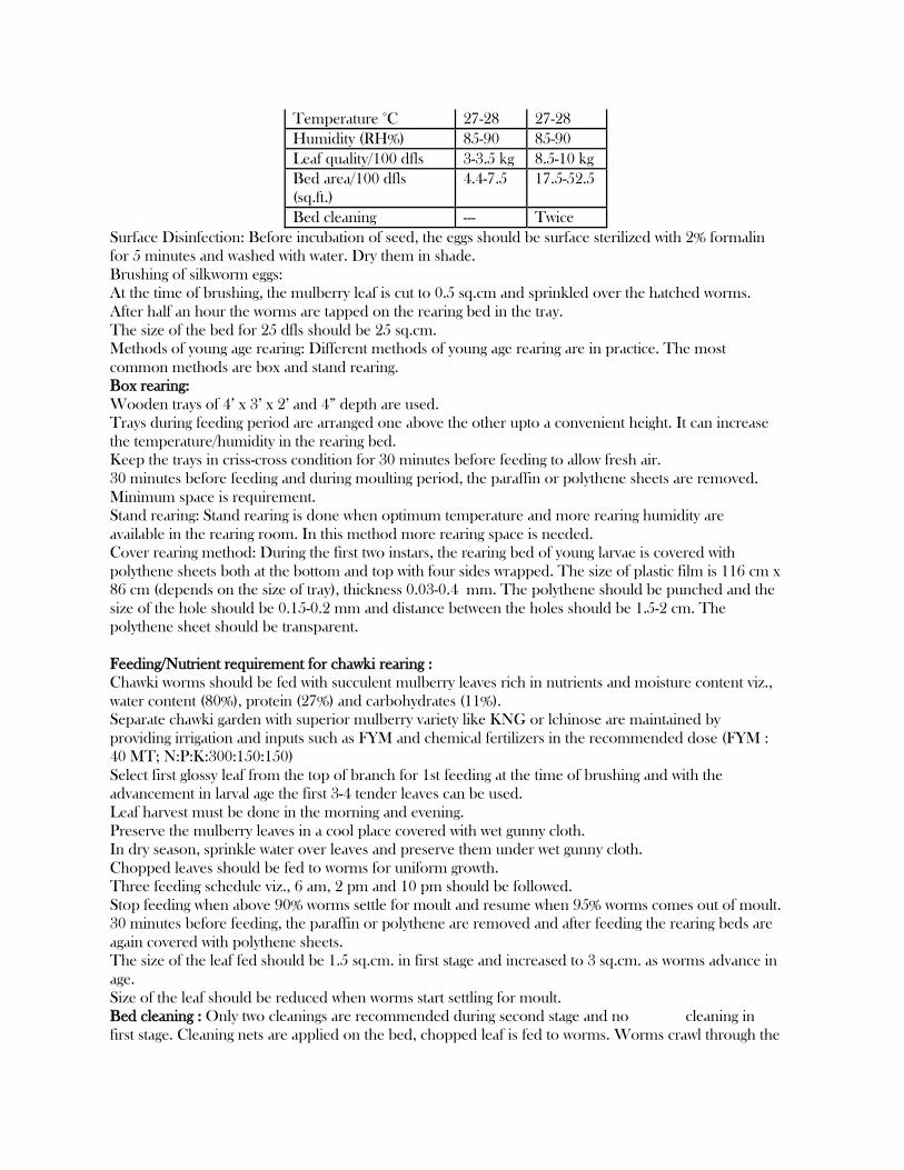
Temperature °C 27-28 27-28
Humidity (RH%) 85-90 85-90
Leaf quality/100 dfls 3-3.5 kg 8.5-10 kg
Bed area/100 dfls
(sq.ft.)
4.4-7.5 17.5-52.5
Bed cleaning --- Twice
Surface Disinfection: Before incubation of seed, the eggs should be surface sterilized with 2% formalin
for 5 minutes and washed with water. Dry them in shade.
Brushing of silkworm eggs:
At the time of brushing, the mulberry leaf is cut to 0.5 sq.cm and sprinkled over the hatched worms.
After half an hour the worms are tapped on the rearing bed in the tray.
The size of the bed for 25 dfls should be 25 sq.cm.
Methods of young age rearing: Different methods of young age rearing are in practice. The most
common methods are box and stand rearing.
Box rearing:
Wooden trays of 4’ x 3’ x 2’ and 4‖ depth are used.
Trays during feeding period are arranged one above the other upto a convenient height. It can increase
the temperature/humidity in the rearing bed.
Keep the trays in criss-cross condition for 30 minutes before feeding to allow fresh air.
30 minutes before feeding and during moulting period, the paraffin or polythene sheets are removed.
Minimum space is requirement.
Stand rearing: Stand rearing is done when optimum temperature and more rearing humidity are
available in the rearing room. In this method more rearing space is needed.
Cover rearing method: During the first two instars, the rearing bed of young larvae is covered with
polythene sheets both at the bottom and top with four sides wrapped. The size of plastic film is 116 cm x
86 cm (depends on the size of tray), thickness 0.03-0.4 mm. The polythene should be punched and the
size of the hole should be 0.15-0.2 mm and distance between the holes should be 1.5-2 cm. The
polythene sheet should be transparent.
Feeding/Nutrient requirement for chawki rearing :
Chawki worms should be fed with succulent mulberry leaves rich in nutrients and moisture content viz.,
water content (80%), protein (27%) and carbohydrates (11%).
Separate chawki garden with superior mulberry variety like KNG or lchinose are maintained by
providing irrigation and inputs such as FYM and chemical fertilizers in the recommended dose (FYM :
40 MT; N:P:K:300:150:150)
Select first glossy leaf from the top of branch for 1st feeding at the time of brushing and with the
advancement in larval age the first 3-4 tender leaves can be used.
Leaf harvest must be done in the morning and evening.
Preserve the mulberry leaves in a cool place covered with wet gunny cloth.
In dry season, sprinkle water over leaves and preserve them under wet gunny cloth.
Chopped leaves should be fed to worms for uniform growth.
Three feeding schedule viz., 6 am, 2 pm and 10 pm should be followed.
Stop feeding when above 90% worms settle for moult and resume when 95% worms comes out of moult.
30 minutes before feeding, the paraffin or polythene are removed and after feeding the rearing beds are
again covered with polythene sheets.
The size of the leaf fed should be 1.5 sq.cm. in first stage and increased to 3 sq.cm. as worms advance in
age.
Size of the leaf should be reduced when worms start settling for moult.
Bed cleaning : Only two cleanings are recommended during second stage and no cleaning in
first stage. Cleaning nets are applied on the bed, chopped leaf is fed to worms. Worms crawl through the
net. After two hours, worms are transferred to another tray. If cleaning nets are not available, the
topmost layer with worms must be taken with a feather.
Spacing : Overcrowding of the silkworms in the early stage leads to sizing and poor growth. Regulate the
spacing for the healthy growth of the silkworms. There should be uniform distribution of the larvae in
the bed.
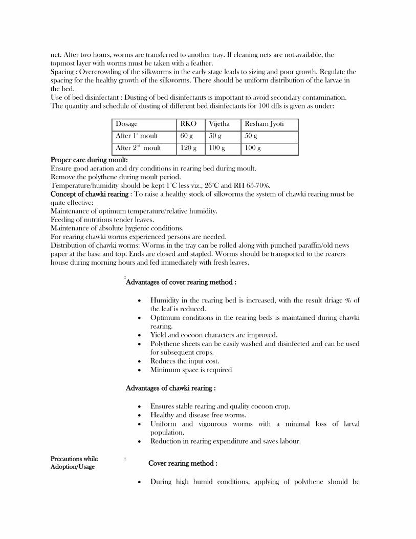
Use of bed disinfectant : Dusting of bed disinfectants is important to avoid secondary contamination.
The quantity and schedule of dusting of different bed disinfectants for 100 dfls is given as under:
Dosage RKO Vijetha Resham Jyoti
After 1st
moult 60 g 50 g 50 g
After 2nd
moult 120 g 100 g 100 g
Proper care during moult:
Ensure good aeration and dry conditions in rearing bed during moult.
Remove the polythene during moult period.
Temperature/humidity should be kept 1°C less viz., 26°C and RH 65-70%.
Concept of chawki rearing : To raise a healthy stock of silkworms the system of chawki rearing must be
quite effective:
Maintenance of optimum temperature/relative humidity.
Feeding of nutritious tender leaves.
Maintenance of absolute hygienic conditions.
For rearing chawki worms experienced persons are needed.
Distribution of chawki worms: Worms in the tray can be rolled along with punched paraffin/old news
paper at the base and top. Ends are closed and stapled. Worms should be transported to the rearers
house during morning hours and fed immediately with fresh leaves.
: Advantages of cover rearing method :
Humidity in the rearing bed is increased, with the result driage % of
the leaf is reduced.
Optimum conditions in the rearing beds is maintained during chawki
rearing.
Yield and cocoon characters are improved.
Polythene sheets can be easily washed and disinfected and can be used
for subsequent crops.
Reduces the input cost.
Minimum space is required
Advantages of chawki rearing :
Ensures stable rearing and quality cocoon crop.
Healthy and disease free worms.
Uniform and vigourous worms with a minimal loss of larval
population.
Reduction in rearing expenditure and saves labour.
Precautions while
Adoption/Usage
: Cover rearing method :
During high humid conditions, applying of polythene should be

avoided.
During moulting time the polythene sheets should be removed.
Use of thicker gauge polythene sheet should be avoided.
Drying should be avoided from sun.
Precautionary measures during the rearing of young larvae(Chawki):
Before entering the rearing room, where chawki worms are reared,
hands should be washed.
Separate footwear should be used inside the rearing rooms.
Silkworm litter should not be thrown in the rearing room.
Rearing rooms should be kept clean and tidy.
Avoid touching the worms.
LATE AGE SILKWORM REARING
The third, fourth and fifth instar larvae are considered as late age worms. They are reared in bamboo trays. Newspapers are spread over the trays to absorb excess moisture in leaves and faecal pellets.
The temperature and humidity requirement gradually comes down as the stage advances. Leaves of medium maturity (6th leaf onwards) are fed in the third and fourth age and coarse
leaves are fed in the fifth age. Over matured and yellow leaves should be rejected, since they may induce disease outbreak.
Bed disinfectants

Apply bed disinfectants like TNAU Seridust, Resham Jyothi, Vijetha or Sajeevini @ 4 kgs/100 dfls.
Stage (before feeding) Bed disinfectant (Qty/100 dfls)
(g)
After 1st moult 50
After 2nd moult 150
After 3rd moult 800
After 4th moult 1000
On fourth day of final
instar
2000
Total 4000
Moulting
Remove the paraffin papers Evenly spread the larvae in the rearing bed 6-8 h before settling for moult. Provide air circulation to avoid excess humidity inside the room. Provide charcoal stove/heaters to raise the room temperature during winter. Apply lime powder at 60 minutes before resumption of feeding daily during rainy/winter seasons
to reduce the dampness in bamboo trays.
Mounting
Apply Sampoorna @ 20 ml (dissolved in 4 l of water) per 100 dfls over the leaves for early and uniform spinning of cocoons.
After attaining full growth in the final instar, the worms cease to feed and are ready to spin. Such worms are slightly translucent and raise their heads to find a place for spinning. These worms have to be picked up and transferred to a mountage for spinning cocoons. Mounting of worms should not be delayed as the ripened worms will waste silk. About 800-900 worms per m2 are to be kept on a mountage. For 100 dfls, about 30 to 40
chandrakis are required. Mountages should be kept under shade in well ventilated place.
Care during spinning
Quality of silk depends on the care taken at the time of spinning. Mature worms are sensitive to temperature, humidity, light, etc., at the time of spinning. The ripe worm requires space equal in area to square of the length of its body for spinning. Proper spacing avoids wastage of silk for forming preliminary web and avoids double cocoons. To prevent staining of cocoons, keep mountage in an inclined position so that the urine may drop
to the ground.
Maintenance of humidity
Fluctuation of humidity causes abrupt thinning and thickening of silk filament. A relative humidity of 60-70% is ideal for spinning. Provide proper ventilation and straw mats below the mountage to quid excreta. Provide even and moderate lighting. Improper lighting (bright light or dark shadow) causes
crowding of larvae to shaded area leading to double cocoons. Remove dead worms and non-spinners on the 2nd day of spinning. To protect the silkworm from predatory ants, apply malathion 5% dust/lakshman rekha at the
base of mountage stand.

Harvesting
The silk worms complete spinning in 2 to 3 days but the cocoons should not be harvested at this time as the worms inside are still in the prepupal stage.
Harvesting should be done on the fifth day (7th day for bivoltine hybrids) when pupae are fully formed and hard.
Do not harvest when the pupa is in amber colour. Dead and diseased worms on the mountages should be removed before harvest. Marketing of cocoons should be done on the sixth day (8th day for bivoltine hybrids).
Shoot rearing for late age worms Silkworm larvae consume 85% of their food requirement during fifth instar. Fifty per cent of the labour input is utilized during the last seven days of rearing. Rearing house
Provide separate rearing house for shoot rearing in shady areas. Separate room should be provided for young age worm rearing, leaf storing and hall for late age worm rearing.
Shoot rearing rack
A rearing rack of 1.2m x 11m size is sufficient to rear 50 dfls. Provide 15 cm border on all sides of the shelf to prevent the migration of the larvae. Arrange the shelves in three tier system with 50 cm space between the tiers. Fabricate the rack stand with wood, or steel and the rearing seat with wire mesh/bamboo mat.
Shoot harvesting
Harvest the shoots at 1 m height from ground level at 60 to 70 days after pruning. Store the shoots vertically upwards in dark cooler room. Provide thin layer of water (3 cm) in one corner of storage room and place the cut of shoots in the
water for moisture retention.
Feeding
Provide a layer of newspaper in rearing shelf. Disinfect the bed, spread the shoot in perpendicular to width of the bed. Place top and bottom ends of the shoots alternatively to ensure equal mixing of different qualities
of leaves. Transfer the third instar larvae to shoots immediately after moulting. Watch for feeding rate from 4th day of fourth instar. If 90% of larvae have not settled for
moulting, provide one or two extra feedings. Provide 3 feedings during rainy/winter months and 4 feedings during summer rearing.
Spacing
18-36 m2/100 dfls.
Bed cleaning
Bed cleaning is done once during second day of fifth instar following rope (or) net method. In rope method, spread 2 m length of rope (two numbers) at parallel row leaving 0.5m on other
side. After 2 to 3 feedings, ends of the ropes are pulled to the centre to make it into a bundle.

In net cleaning method, spread 1.5 cm2 size net across the bed. After 2 or 3 feedings, the nets are lifted and the old bed is cleaned and disinfected. Transfer the net to newer shelf, spread the net over the shoots; larvae will migrate to lower layer.
Advantages
1. Labour saving upto 70% when compared on hour to hour basis with leaf feeding method. 2. Leaf saving upto 15-20%. Hence, leaf cocoon ratio is less by 2-3 kg and extra cocoon production. 3. Better cocoon characters and effective rate of rearing (ERR). 4. Better preservation of leaf quality both during storing and on the bed. 5. More organic matter production (upto 18 tonnes per ha per year). 6. Better hygienic conditions can be maintained. 7. Handling of silkworms minimised. Hence, contamination and spreading of disease reduced. 8. Bed cleaning only once after IV moult. 9. Worms and leaves are kept away from the litter. Hence, chances of secondary contamination are minimised. 10. Labour dependent risk is reduced.
Disadvantages
1. Required rearing room floor area is more (by 30%) 2. Bed refusals will not be available as a cattle feed. 3. Planting materials (cuttings) will not be available.
UNIT 111
STATUS OF SERICULTURE IN JAMMU AND KASHMIR
Silk industry has occupied a prime place in the industrial structure of the
state. Its significance in the state can be understood in terms of position
it occupied during different periods of history and the interest different
rulers took in developing this industry, as they observed that this industry
had very flourishing and prosperous future for the development of the state
economy. During 15th century, during the reign of king Zain-ul-Abbidin,
Kashmir attained great progress in this industry. During Mughal period also,
the industry flourished because they were the great lovers of silken cloths.
The industry however passed through many ups and downs, during its long
history, but the same could not dampen the enthusiasm of the rulers as from
time to time steps were taken to promote the growth of the industry.
Organizational changes on modern lines were undertaken in tune with the
available factory inputs. Separation of processes was initiated and the
quality of seed was improved to increase production. The combined effect of
all these measures was that the employment in the industry increased along
with the remuneration for those who are directly employed by the industry
also witnessed an un-presidential rise. Thus the industry has witnessed

various changes in respect of performance, organization, diversification and
modernization that have made the industry economically viable during the pre-
1947 period. However, in the post 1947 period, changes in the structure of
land ownership which affected the incentive structure in agriculture,
introduction of new avenues of income and consequent improvement in the
economic condition of the people, reduced the interest of farmers towards
silk worm rearing. In addition, the state monopoly control which has once
helped to organize the industry on modern lines became an obstacle in its
development. Consequently during 1988-89, silk industry was demonopolised and
mulberry tree was declared as farmer’s property. Once the monopoly system was
dispended with, there was a tremendous change in the overall transformation
of sericulture development in the state. People started showing enormous
interest to adopt this craft as a means of their livelihood. But the
unfavorable conditions created in the valley, immediately after
demonopolisation act was enforced, and the constraints such as non-
availability of quality mulberry leaves, un-scientific rearing techniques,
poor quality of seed, lack of proper supervision, competition from other
crops and handicrafts, lack of proper extension activities and also the
marketing, financial and other constraints again stood in the way of
development of sericulture in the state. Cocoon which is an intermediate
product/input in the production of silk has a direct bearing on the
quantitative and qualitative variations in silk production. This can be
proved by looking at the data pertaining to cocoon production and silk
production during the last ten years. The production of cocoons has witnessed
cyclical trends during the last three decades and no firm trend is traceable.
These ups and downs in cocoon production are also visible in the production
of silk in the Valley and also in respect of the performance of the industry
as a whole. Unfortunately, no study has been made so far, in the state to
analyse the relationship between cocoon production and silk production and to
identify the factors responsible for poor performance of the industry, As per
Government of Jammu & Kashmir the data of production is as under

UNIT IV
Tools of Apiculture
The Hive and Its Parts
Honeybees can live in hollow trees, wall voids in buildings, attics, or any other
protected place. Several types of hives have been designed to manage honeybees. Old-
fashioned hives were simple devices, such as plain boxes, short sections of hollow
logs called gums, or straw baskets called skeps. These hive styles have many
disadvantages and are rarely used now. Combs in them were usually irregular and
braced together with bur comb. Individual combs could not be removed from the hive
without damaging other pieces or even injuring or killing the queen. It was also
difficult to inspect the hives for diseases and other problems.
Modern hives with movable frames allow easy inspection and honey removal. Hive
design is efficacious for other management practices and for the bees. The inner
dimensions of the hive and its parts are very precise. They are based on a dimension
called the "bee space," which is about 5/16-inch wide or deep. Proper spacing is

important. If gaps are too wide, bees build brace comb and glue down movable
frames. The modern hive consists of several parts.
A hive stand keeps the hive off the ground so it is less likely to rot, flood, or be
attacked by termites. It can be as simple as a few bricks stacked under each hive
corner, or it might be a wood frame with an alighting board. The alighting board
allows heavily loaded field bees to land more easily before crawling into the hive.
The hive rests on the three rails of the bottom board. The open side is the hive
entrance. This opening can be closed or narrowed with an entrance cleat when
necessary. Reducing the entrance opening in the fall keeps out field mice looking for
shelter. The standard hive body or brood chamber holds 10 frames of comb. Besides
being the nursery, it is also pantry, kitchen, living room, dining room, bedroom, and
workshop for the bees. If it becomes too crowded, the bees might begin rearing brood
in the supers. If colonies get very large, provide extra hive bodies for the brood
chamber.
A queen excluder is sometimes placed above the brood chamber to keep the queen in
the brood chamber. Slots in the excluder are wide enough workers can go back and
forth but too narrow for the queen to pass through. Beekeepers who produce extracted
honey do not use excluders because they reduce the bees' efficiency. For comb honey
and chunk honey production, the excluder assures that brood are not in the honey
product.
Chambers above the brood chamber are called supers. They are the same size as the
brood chamber and are used for storage of surplus honey. Deep supers are used by
those who primarily produce extracted honey. Larger boxes require less handling but
are heavy when full of honey. Shallow supers are easier to lift and convenient for
harvesting small honey yields from a particular nectar source.
The inner cover is a flat piece with an oblong hole in the center. A bee escape can be
put in the hole when needed. The hole provides ventilation and a place to puff smoke
when opening the hive. The edges of the inner cover have railings on both faces. The
railing on one side is higher than the other. The tall railing should be on the outside. If
the tall rail is on the inside, the bees build wax between it and the tops of the frames.
This buildup is a mess to clean.
The top cover is a waterproof lid that rests on the edges of the top super. Bees do not
glue down the top cover, so it can be lifted from the hive without prying or jarring.
Frames are the inside parts that hold the comb. They consist of a top and bottom and
two end bars. The wide part of an end bar is keeled on one edge. Place frames in the
hive so that the keeled edge of one frame abuts the flat edge of the next one. Frames
help keep comb-building regular and allow easy inspection and honey removal. All
frames are the same length, but there are different depths and styles.
Carefully put together unassembled frames. Fit the frame together so that the keel on
the left end bar is toward you and the keel on the right end bar is away from you. If
you rotate the frame, the keel is still toward you on the left side and away from you on
the right. Use plenty of nails when fastening the frame together. Otherwise, it can pull
apart when the comb is full of honey.
To ensure that the comb is regular, frames are fitted with thin sheets of embossed wax
called foundation. Foundation for brood frames and extracted honey frames has
embedded wires for extra strength. This prevents the comb from sagging when the
wax gets soft during hot weather or from tearing apart during extraction.
Foundation can be purchased with wires in it, or wire can be embedded after the
foundation is fitted into the frame. It is more convenient for the beginner to buy wired
foundation for brood frames. Use unwired foundation in the honey frames if you are
going to produce chunk honey. The size of foundation sheets varies with frame size.
Bee supply catalogs specify the frame styles different foundation sheets fit.
Other Beekeeping Equipment
A hive tool is the most useful piece of beekeeping equipment. It can be used to pry up
the inner cover, pry apart frames, scrape and clean hive parts, and do many other jobs.
Examining a hive is much easier when you use a smoker. It is used to puff smoke into
the entrance before opening the hive and blow smoke over the frames after the hive is
opened. Smoke causes the bees to gorge themselves with honey; then they are much
gentler. Smoke must be used carefully. Too much can drive bees from the hive. They
will be slow to settle down after the hive is closed again. Burlap, rotted wood,
shavings, excelsior, cardboard, or cotton rags are good smoker fuels.
Even the most experienced beekeepers wear veils to protect their face and eyes. Wire
veils keep bees farther away from the face than those made of cloth. Black veiling is
geneally easier to see through. Some beekeepers prefer to wear a bee suit. Whether or
not a suit is used, a beekeeper's clothing should be light in color. Bees generally do
not like dark colors and will attack dark objects.
Bee gloves protect the hands and arms from stings, but it is hard to do some jobs while
wearing them. It is a good idea to wear gloves until you feel more comfortable
working your bees.
A brush gently removes bees from frames. A leafy twig or bunch of grass can do the
same job and gives you fewer tools to carry around.
A bee escape is used to clear bees out of supers. Place the escape in the center hole of
the inner cover below the super to be cleared. Bees can pass through in only one
direction. It usually takes about a day to get the bees out of a super.
Wire embedders are handy for wiring your own foundation. Various jigs for
assembling frames and supers save time and assure square fitting. These can be
purchased or made. Most beekeepers are creative with tools and make various gadgets
to do special jobs.
UNIT 111 TYPES OF SILK
There are four types of natural silk which are commercially known and produced in the world. Among
them mulberry silk is the most important and contributes as much as 90 per cent of world production,
therefore, the term "silk" in general refers to the silk of the mulberry silkworm. Three other commercially
important types fall into the category of non-mulberry silks namely: Eri silk; Tasar silk; and Muga silk.
There are also other types of non-mulberry silk, which are mostly wild and exploited in Africa and Asia,
are Anaphe silk, Fagara silk, Coan silk, Mussel silk and Spider silk.
Bulk of the commercial silk produced in the world comes from this variety and often generally refers
to mulberry silk. Mulberry silk comes from the silkworm, Bombyx mori L which solely feeds on the
leaves of mulberry plant. These silkworms are completely domesticated and reared indoors.
Mulberry silk contributes to around 90 percent of the world silk production.
Tasar silk
The tasar silkworms belong to the genus Antheraea and they are all wild silkworms. There are many
varieties such as the Chinese tasar silkworm Antherae pernyi Guerin which produces the largest quantity
of non-mulberry silk in the world, the Indian tasar silkworm Antheraea mylitte Dury, next in importance,
and the Japanese tasar silkworm Antheraea yamamai Querin which is peculiar to Japan and produces
green silk thread.
The Chinese and Japanese tasar worms feed on oak leaves and other allied species. The Indian tasar
worms feeds on leaves of Terminalia and several other minor host plants. The worms are either uni-
or bivoltine and their cocoons like the mulberry silkworm cocoons can be reeled into raw silk.
Eri silk

These belong to either of two species namely Samia riciniand Philosamia ricini. P.ricini (also called as
castor silkworm) is a domesticated one reared on castor oil plant leaves to produce a white or brick-red
silk popularly known as Eri silk.
Since the filament of the cocoons spun by these worms is neither continuous nor uniform in thickness,
the cocoons cannot be reeled and, therefore, the moths are allowed to emerge and the pierced
cocoons are used for spinning to produce the Eri silk yarn.
Muga silk
The muga silkworms (Antheraea assamensis) also belong to the same genus as tasar worms, but produce
an unusual golden-yellow silk thread which is very attractive and strong. These are found only in the state
of Assam, India and feed on Persea bombycina and Litsaea monopetala leaves and those of other
species.
The quantity of muga silk produced is quite small and is mostly used for the making of traditional
dresses in the State of Assam (India) itself.
Anaphe silk
This silk of southern and central Africa is produced by silkworms of
the genus Anaphe: A. moloneyi Druce, A. panda Boisduval, A. reticulate Walker, A. ambrizia Butler, A.
carteri Walsingham, A. venata Butler and A. infracta Walsingham. They spin cocoons in communes, all
enclosed by a thin layer of silk.
The tribal people collect them from the forest and spin the fluff into a raw silk that is soft and fairly
lustrous. The silk obtained from A. infracta is known locally as "book", and those from A. moleneyi as
"Trisnian-tsamia" and "koko" (Tt). The fabric is elastic and stronger than that of mulberry silk. Anaphe
silk is used, for example, in velvet and plush.
Fagara silk
Fagara silk is obtained from the giant silk moth Attacus atlas L. and a few other related species or races
inhabiting the Indo-Australian bio-geographic region, China and Sudan. They spin light-brown cocoons
nearly 6 cm long with peduncles of varying lengths (2-10 cm).
Coan silk
The larvae of Pachypasa atus D., from the Mediterranean bio-geographic region (southern Italy, Greece,
Romania, Turkey, etc.), feed primarily on trees such as pine, ash cypress, juniper and oak.
They spin white cocoons measuring about 8.9 cm x 7.6 cm. In ancient times, this silk was used to make the
crimson-dyed apparel worn by the dignitaries of Rome; however, commercial production came to an end
long ago because of the limited output and the emergence of superior varieties of silk.
Mussel silk
Whereas the non-mulberry silks previously described are of insect origin, mussel silk is obtained from a
bivalve, Pinna squamosa, found in the shallow waters along the Italina and Dalmatian shores of the Adriatic.
The strong brown filament, or byssus, is secreted by the mussel to anchor it to a rock or other surface. The
byssus is combed and then spun into a silk popularly known as ―fish wool‖. Its production is largely
confined to Taranto, Italy.

Spider silk
Spider silk – another non-insect variety – is soft and fine, but also strong and elastic. The commercial
production of this silk comes from certain Madagascan species, including Nephila madagascarensis, Miranda aurentia and Epeira. As the spinning tubes (spinne-rules) are in the fourth and fifth abdominal
segments, about a dozen individuals are confined by their abdominal part to a frame from which the
accumulated fibre is reeled out four or five times a month. Because of the high cost of production, spider
silk is not used in the textile industry; however, durability and resistance to extreme temperature and
humidity make it indispensable for cross hairs in optical instruments.
Related Documents