Unit 7A Medical Biotechnology I

Unit 7A Medical Biotechnology I. Lesson 1 Disease Detection Lecture- Model organisms, biomarkers, Human Genome Project contribution to disease detection.
Dec 26, 2015
Welcome message from author
This document is posted to help you gain knowledge. Please leave a comment to let me know what you think about it! Share it to your friends and learn new things together.
Transcript

Unit 7A
Medical Biotechnology I

Lesson 1
• Disease Detection• Lecture- Model organisms, biomarkers,
Human Genome Project contribution to disease detection.
• Create a concept map demonstrating how designated terms and concepts are related.

Disease Detection
• Models of Human Disease• Many medical biotechnology
treatments in disease are made possible because of model organisms.
• We share a large number of genes with other organisms.
• Genes in other organisms that have sequence similarities to humans are called homologues
• A number of genetic diseases occur in model organisms.
Disease Detection• When researchers study
homologues for diseases, they are interested in two things.
1. What does the gene do? i.e. proteins and molecules that contribute to the disease.
2. What happens if gene transcription is disrupted.? i.e the disease trait can be eliminated from the organism.
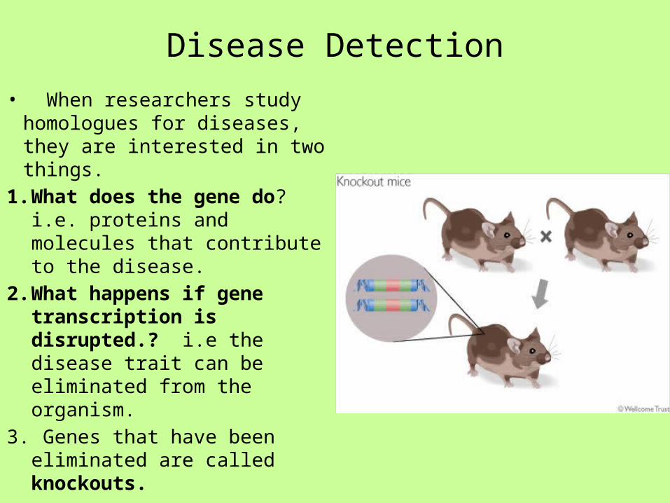
3. Genes that have been eliminated are called knockouts.
Disease Detection
• Knockouts• Knockouts are genetically engineered.• The active gene is either replaced or disrupted
with an inactive DNA sequence.• Depending on where the inactive DNA
sequence is inserted into the gene, there can be a variety of outcomes.
• Most often, the trait expression is eliminated.

Disease Detection• Knockouts• Engineered genes are inserted into a blastocyst and it is implanted into a female mouse.•Off spring are bred through 2-3 generations until a knockout mouse, homozygous for the knockout genes, is produced.•Often drugs are tested on the knockout mice. The expectation is that the drug would have an effect on a diseased mouse and no effect on a knockout mouse.•If the knockout mouse is effected, it can indicate there would be side effects in humans.

Disease Detection
• http://learn.genetics.utah.edu/content/tech/transgenic/
• Knockout Mice

Disease Detection
• Examples of model organisms in detection.
• Ob gene is linked to obesity. Mice without the Ob gene become obese. Ob codes for leptin, which regulates hunger telling the body when it is full.
• This discovery led to treating obese human children with leptin and they have responded well in preliminary studies.

Disease Detection• Examples of model organisms in
detection.• In developing embryos, some cells
must die to make room for others (apoptosis). How is this determined?
• A study of C. elegans, a roundworm, allowed scientists to determine the fate or lineage of all of its embryonic cells. Understanding programmed cell death has application to Alzheimer disease, Huntington disease, and Parkinson disease.

Disease Detection• Biomarkers• For many diseases, early detection is critical.• One detection approach is to look for
biomarkers as indicators of disease.• Biomarkers are proteins whose production is
increased in diseased tissues.• Many biomarkers are released into blood and
urine as a product of cell damage.• EX. A protein called prostate specific antigen
(PSA) is released into the blood when the prostate gland is inflamed.
• Elevated PSA levels indicate inflammation and even cancer.
• Many companies are working on a variety of biomarkers that can be used in disease detection.
Disease Detection
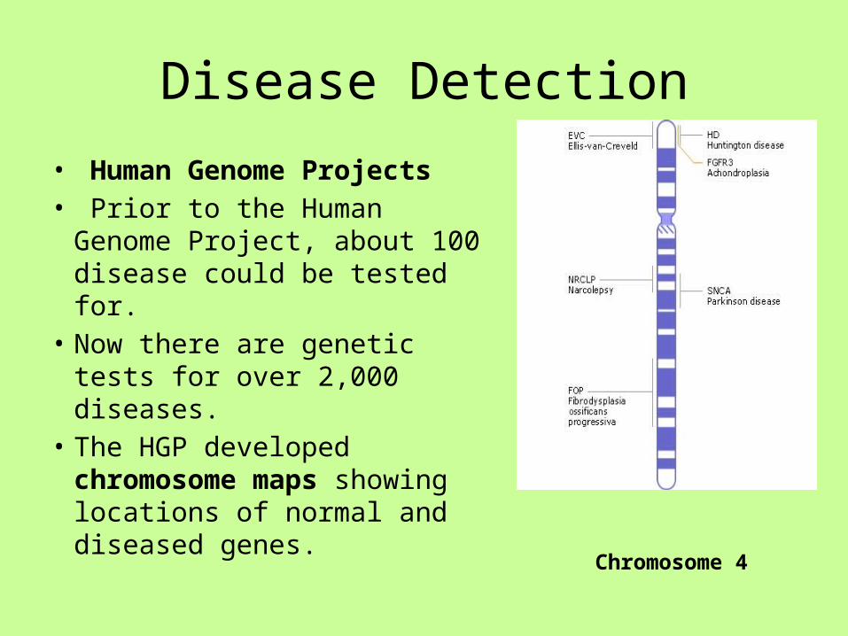
• Human Genome Projects• Prior to the Human Genome
Project, about 100 disease could be tested for.
• Now there are genetic tests for over 2,000 diseases.
• The HGP developed chromosome maps showing locations of normal and diseased genes. Chromosome 4

Lesson 2
• Disease Detection: Testing• Work in groups of 4. Read powerpoint on
amniocentesis, RFLP analysis, SNPs, and microarray. • Discuss content with your group and respond to
questions.• Watch animation for Amniocentesis , RFLP analysis,
SNPs • Complete Questions• Complete SNP activity.• Complete Microarray Simulation

Genetic TestingAmniocentesis• Until recently, most genetic testing occurred on fetuses to identify
gender and genetic diseases.• Amniocentesis is one technique used to collect genetic material for
genetic testing.• When the developing fetus is around 16 weeks of age, a needle is
inserted into the mother’s abdomen into a pocket of amniotic fluid that surrounds and cushions the fetus. Amniotic fluid is removed.
• The fluid contains cells from the fetus, such as skin cells.• Skin cells are cultured to increase their number.• Mitotic chromosomes are removed and stained to create a
karyotype
• http://www.youtube.com/watch?v=bZcGpjyOXt0
Genetic Testing
• Chorionic Villi Sampling• Chorionic villi Sampling (CVS) can also be done to
diagnose genetic disease in fetuses who are 8 -10 weeks in age.
• A suction tube removes a layer of cells called the chorionic villus, tissue that helps make up the placenta.
• CVS collects enough cells so a karyotype can be made from the cells retrieved.
• http://video.about.com/pregnancy/Chorionic-Villus-Sampling.htm
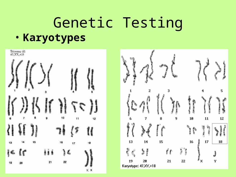
Genetic Testing• Karyotypes

Genetic Testing• Karyotyping can be
carried out with adults.• Typically blood is drawn
and white blood cells are used.
• Fluorescence in situ hybridization(FISH) is used.
• Chromosomes are hybridized with fluorescent probes.

Genetic Testing
• Karyotypes• FISH can be performed with
probes that fluoresce different colors.
• This is called spectral karyotyping.
• It is very useful in identifying missing parts of chromosomes, extra chromosomes, and translocation mutations.
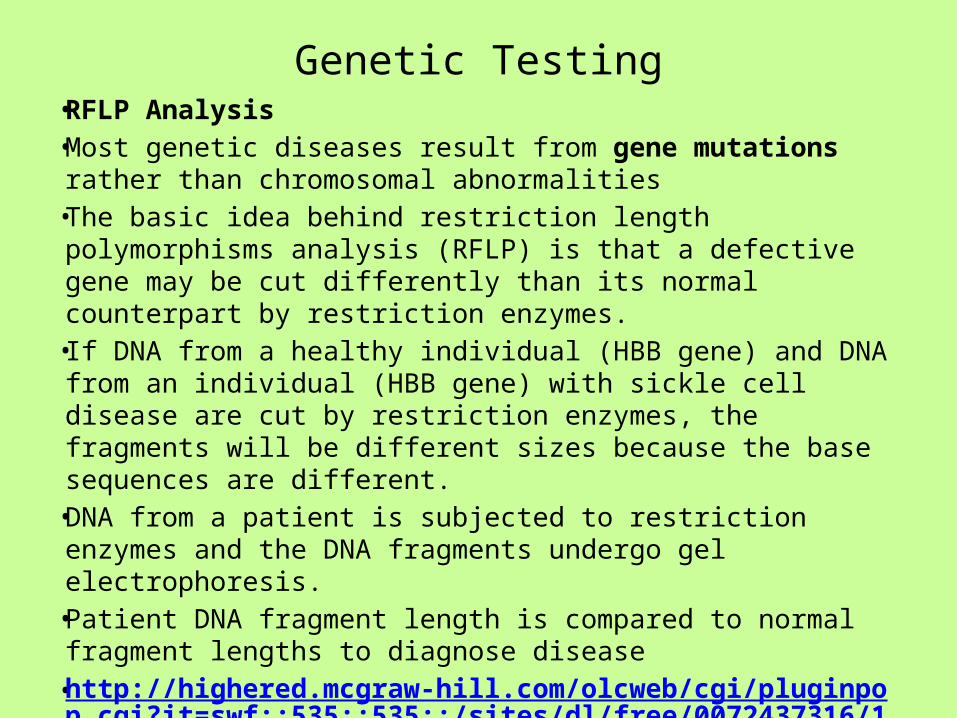
Genetic Testing• RFLP Analysis• Most genetic diseases result from gene mutations rather
than chromosomal abnormalities• The basic idea behind restriction length polymorphisms
analysis (RFLP) is that a defective gene may be cut differently than its normal counterpart by restriction enzymes.
• If DNA from a healthy individual (HBB gene) and DNA from an individual (HBB gene) with sickle cell disease are cut by restriction enzymes, the fragments will be different sizes because the base sequences are different.
• DNA from a patient is subjected to restriction enzymes and the DNA fragments undergo gel electrophoresis.
• Patient DNA fragment length is compared to normal fragment lengths to diagnose disease
• http://highered.mcgraw-hill.com/olcweb/cgi/pluginpop.cgi?it=swf::535::535::/sites/dl/free/0072437316/120078/bio20.swf::Restriction%20Fragment%20Length%20Polymorphisms

Genetic Testing
• RFLP Analysis
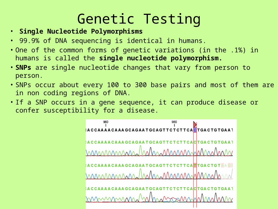
Genetic Testing• Single Nucleotide Polymorphisms• 99.9% of DNA sequencing is identical in humans.• One of the common forms of genetic variations (in the .1%) in humans is called
the single nucleotide polymorphism.• SNPs are single nucleotide changes that vary from person to person.• SNPs occur about every 100 to 300 base pairs and most of them are in non
coding regions of DNA.• If a SNP occurs in a gene sequence, it can produce disease or confer susceptibility
for a disease.

Genetic Testing• SNPs• Because SNPs occur frequently throughout the genome, they are
valuable markers to identifying disease related genes.• SNPs are being used to predict stroke, cancer, heart disease, and
behavioral illnesses.• Many groups of SNPs on the same chromosome are called a
haplotype.• The HapMap project is identifying and cataloguing the
chromosomal location of over 1.4 million SNPs present in 3 billion base pairs of the human genome.
• Complete the SNP activity. http://www.pbs.org/wgbh/nova/teachers/activities/0302_01_nsn.html
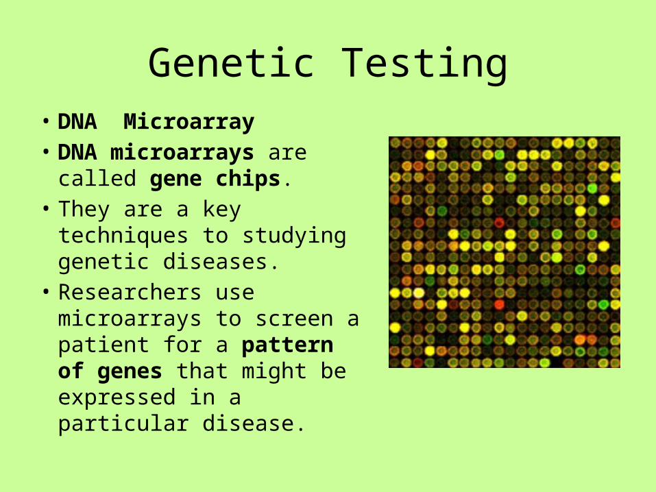
Genetic Testing• DNA Microarray• DNA microarrays are called
gene chips.• They are a key techniques to
studying genetic diseases.• Researchers use microarrays
to screen a patient for a pattern of genes that might be expressed in a particular disease.
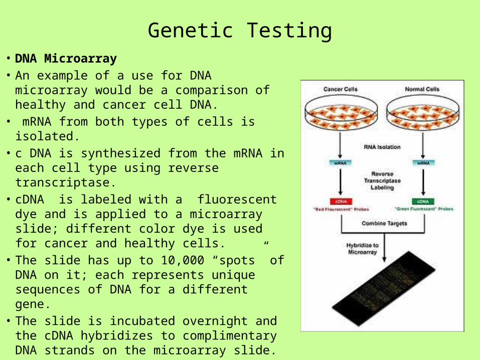
Genetic Testing• DNA Microarray• An example of a use for DNA microarray
would be a comparison of healthy and cancer cell DNA.
• mRNA from both types of cells is isolated.• c DNA is synthesized from the mRNA in
each cell type using reverse transcriptase.• cDNA is labeled with a fluorescent dye and
is applied to a microarray slide; different color dye is used for cancer and healthy cells.
• The slide has up to 10,000 “spots” of DNA on it; each represents unique sequences of DNA for a different gene.
• The slide is incubated overnight and the cDNA hybridizes to complimentary DNA strands on the microarray slide.

Genetic Testing

Genetic Testing• DNA Microarray• The slide is scanned by a laser that
causes the dye to fluoresce when cDNA binds to gene DNA on the slide.
• The fluorescent spots indicate which genes are expressed in the cells of interest.
• Gene expression patterns from each of the cell types is compared to see which genes are active in a healthy cell and which are active in a cancer cell.
• Results of microarray studies can be used to develop new drugs to combat cancer and other diseases.

Genetic Testing
• http://learn.genetics.utah.edu/content/labs/microarray/
Visit the virtual DNA microarray simulation for a detailed description of the procedure.

Lesson 3
• Case Study: Pharmacogenetics.• Powerpoint introduction• Work in groups of 4 to read and discuss each
section of the pharmacogenetics case study.• Respond to case study questions.• Whole class discussion of responses.
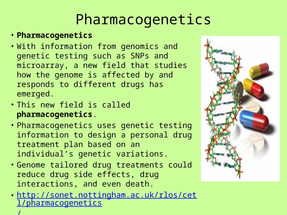
Pharmacogenetics• Pharmacogenetics• With information from genomics and genetic
testing such as SNPs and microarray, a new field that studies how the genome is affected by and responds to different drugs has emerged.
• This new field is called pharmacogenetics.• Pharmacogenetics uses genetic testing
information to design a personal drug treatment plan based on an individual’s genetic variations.
• Genome tailored drug treatments could reduce drug side effects, drug interactions, and even death.
• http://sonet.nottingham.ac.uk/rlos/cetl/pharmacogenetics/

Lesson 4
• Treatments for Disease• Lecture- Nanotechnology, Artificial Blood, and
Monoclonal Antibodies.• Powerpoint presentation of content.
Nanotechnology
• For homework:• Visit the following website and respond to
questions.• http://www.nano.gov/nanotech-101

Nanotechnology• Nanotechnology :
Understanding and controlling of matter at the nanoscale; dimensions between approximately 1 and 100 nanometers, where unique phenomena enable novel applications.
• Encompassing nanoscale science, engineering, and technology, nanotechnology involves imaging, measuring, modeling, and manipulating matter at this length scale.

Nanotechnology• Matter such as gases, liquids, and solids
can exhibit unusual physical, chemical, and biological properties at the nanoscale, differing in important ways from the properties of bulk materials and single atoms or molecules.
• Some nanostructured materials are stronger or have different magnetic properties compared to other forms or sizes or the same material.
• Others are better at conducting heat or electricity. They may become more chemically reactive or reflect light better or change color as their size or structure is altered.

Nanotechnology
• Nanoparticles such as• Iron• Gold• Liquid crystals• And others • Are nanoparticles that can be used in medical
applications.• Some of these compounds can be inert at the
“macro” level but become catalysts at the nanoscale. In addition, they easily penetrate cells (soluable) and interact with cellular molecules.

Nanotechnology• A nanoparticle called a microsphere is of
particular interest in medicine.• It is composed of a phospholipid bilayer to
which drugs can be attached.• The microspheres can target specific cells and
deliver needed drugs. • Advantage: They can dissolve in the body.• Examples • Researchers are investigating ways to implant
microspheres holding anticancer drugs next to tumors.
• Researchers are working on ways to attach microspheres to wafers for pain anesthetics
• Microspherse called liposomes are being used in gene therapy.

Nanotechnology• View the animations about nanotechnology• http://nano.cancer.gov/learn/understanding/video_j
ourney.asp
• http://www.azonano.com/nanotechnology-video-details.aspx?VidID=437
• http://www.azonano.com/nanotechnology-video-details.aspx?VidID=480
• http://www.azonano.com/nanotechnology-video-details.aspx?VidID=469
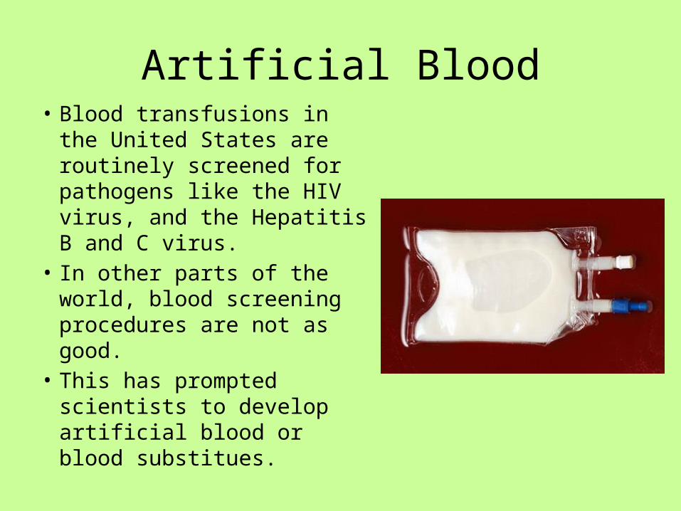
Artificial Blood• Blood transfusions in the
United States are routinely screened for pathogens like the HIV virus, and the Hepatitis B and C virus.
• In other parts of the world, blood screening procedures are not as good.
• This has prompted scientists to develop artificial blood or blood substitues.

Artificial Blood
• Major Advantages of Artificial Blood1. It is a disease free alternative.2. It is in constant supply without shortages.3. Available for emergency situations.4. Can be stored for a long period of time.
(Blood needs to be refrigerated and lasts 42 days. Artificial blood can last up to 3 months unrefrigerated.
5. There would be no recipient rejection as there are no antigenic molecules in artificial blood.

Artificial Blood
• Major Disadvantage1. Artificial Blood serves one primary
purpose; it is designed to carry oxygen.
• Normal red blood cells have other functions. In addition to carrying oxygen, they are a source of iron and have a role in eliminating carbon dioxide from the blood.

Artificial Blood• Currently there are 2 major types of
artificial blood:1. Hemoglobin based: Made from
cow or human blood. Blood is process and hemoglobin is purified.
2. Fluorocarbon based: Fluorocarbon emulsions are made with particles about 1/40 size the red cells. The fluorcarbon binds to oxygen in a fashion similar to hemoglobin.
• There is some newer research which is combining the oxygen carrying portion of the hemoglobin molecule with a polymer shell!

Monoclonal Antibodies
• Monoclonal antibodies are specific antibodies targeted towards the specific molecular structure on an antigen (epitope) that causes the immune response
• Treating patients with monoclonal antibodies can be effective in transplant rejection, cardiovascular disease, some allergies, and certain cancers like breast cancer.
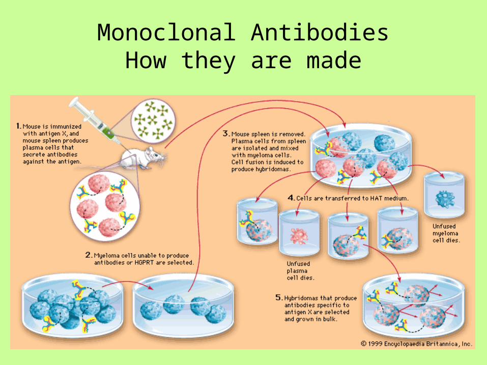
Monoclonal AntibodiesHow they are made

Monoclonal Antibodies
• A mouse is injected with an antigen and B cells (plasma cells) produce antibody.
• The spleen of the mouse is removed and the B cells are mixed with myeloma cells (cancerous). Myeloma cells won’t stop dividing.
• B cells and myeloma cells merge and become hybridomas.
• Hybridomas are antibody manufacturing factories.• Antibodies are isolated and given to patients. • http://highered.mcgraw-hill.com/sites/0072556781/st
udent_view0/chapter32/animation_quiz_3.html

Lesson 5
• Webquest: Gene Therapy• We will be visiting the website listed below:• http://learn.genetics.utah.edu/content/tech/g
enetherapy/• Research University of Utah Genetics website
to study multiple aspects of gene therapy.• Respond to questions on your handout.

Lesson 6
• Gene Therapy Project – Market a Vector• Using information from gene therapy
webquest, you will work with a partner and design a powerpoint and brochure to market a gene therapy vector to research scientists.
• You will present your powerpoint to class and distribute brochures.
• Refer to your handout for details.
Lesson 7
• Group work – Stem Cells• The topic of stem cells has been addressed in introductory
biology . This lesson is a review and refresher for previously learned content.
• Work in groups of 4 to review the powerpoint on assigned section of content. Develop review questions for content.
• Teacher will approve all review material.• Reassign one “expert” from each group assignment to a new
grouping.• New group will review powerpoint together, discuss content and
review questions.• Teacher will provide a written quiz at the end of the assignment.
Stem cells
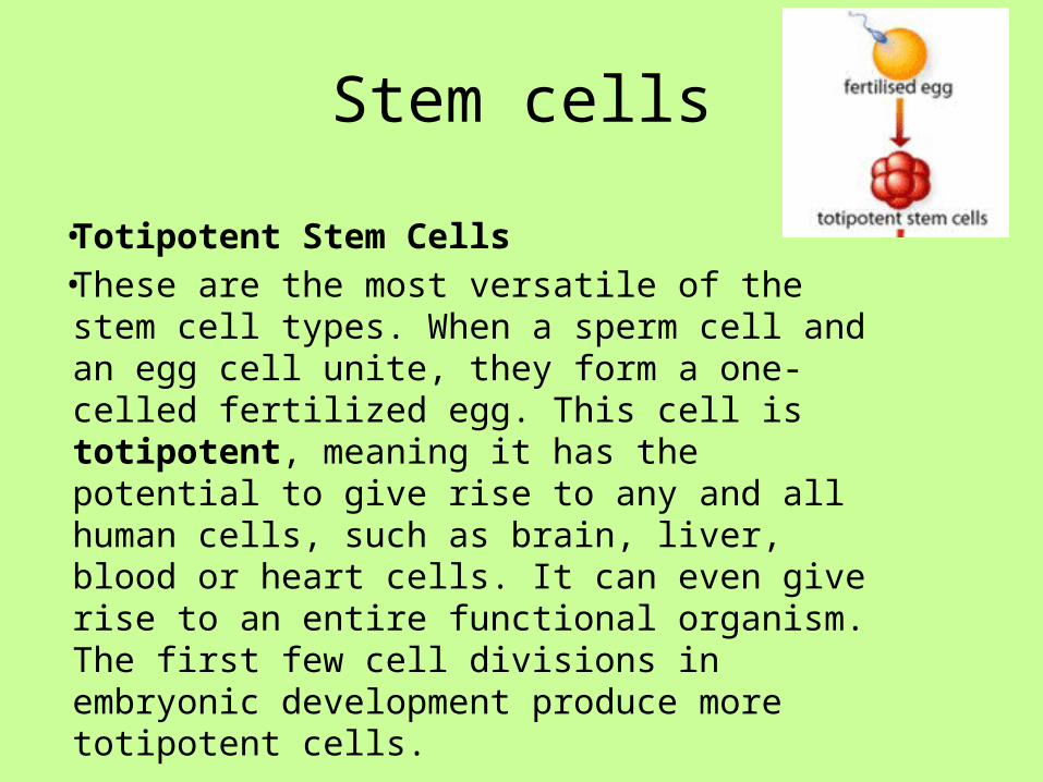
• Totipotent Stem Cells• These are the most versatile of the stem cell
types. When a sperm cell and an egg cell unite, they form a one-celled fertilized egg. This cell is totipotent, meaning it has the potential to give rise to any and all human cells, such as brain, liver, blood or heart cells. It can even give rise to an entire functional organism. The first few cell divisions in embryonic development produce more totipotent cells.

Stem Cells
• Pluripotent Stem Cells (Embryonic Stem Cells)• These cells are like totipotent stem cells in that they can
give rise to all tissue types. Unlike totipotent stem cells, however, they cannot give rise to an entire organism. On the fourth day of development, the embryo forms into two layers, an an outer layer which will become the placenta, and an inner mass which will form the tissues of the developing human body. These inner cells, though they can form nearly any human tissue, cannot do so without the outer layer; so are not totipotent, but pluripotent. As these pluripotent stem cells continue to divide, they begin to specialize further.

Stem Cells
• Multipotent Stem Cells• These are less plastic and more
differentiated stem cells. They give rise to a limited range of cells within a tissue type. The offspring of the pluripotent cells become the progenitors of such cell lines as blood cells, skin cells and nerve cells. At this stage, they are multipotent. They can become one of several types of cells within a given organ. For example, multipotent blood stem cells can develop into red blood cells, white blood cells or platelets

Stem Cells• Adult Stem Cells• An adult stem cell is a multipotent
stem cell in adult humans that is used to replace cells that have died or lost function. It is an undifferentiated cell present in differentiated tissue. It renews itself and can specialize to yield all cell types present in the tissue from which it originated.

Stem Cells• Induced Pluripotent Stem
Cells (IPsCs)• IPSCs are differentiated
cells that have been reprogrammed back to pluripotent stem cells.
• The introduction of 4 genes OCT3/4, SOX2, c-MYC, and KLF4 by a retrovirus into cells reprograms the cells into an earlier stage of differentiation similar to embryonic stem cells.
• These genes encode transcription factors involved in cell development.

Stem Cells - IPSCs

Stem Cells
• IPSCs• IPSCs can be used for patient specific
therapies without the risk of cell rejection.• Cells could be taken from a patient,
reprogrammed into an IPC, and then differentiated into a cell that could combat disease in the patient.
• There would be no need for embryonic stem cells.

Stem Cells• IPSCs• Scientists still do not fully understand
how to control induced pluripotent stem cells.
1. They do not understand the degree of pluripotency in these cells.
2. Producing them is inefficient. 1 in 1000 cells exposed to a reprogramming approach becomes an IPSC.
3. The cells require constant feeding in cell culture.
4. The cells have low viability after they have been frozen for storage.
5. The cells are prone to forming tumors.6. Occasionally, IPSCs spontaneously
revert to differentiated cells.
Stem Cells
• Stem Cell Therapies• Potential and promise are two words frequently
used to describe stem cell therapies.• The most promising application to date has been
for leukemia.1. Patients receive chemotherapy or radiation to
destroy cancerous white blood cells.2. Patients receive WBC stem cells which
proliferate to normal cells.

Stem Cells• Stem Cell Therapy• Researchers have injected
stem cells from different sources into damaged heart tissue of mice (heart tissue does not repair itself well). The stem cells developed into cardiac muscle and improved heart function by 35%. This work shows promise for human heart attack victims.

Stem Cells
• Stem Cell Therapy• Researchers have demonstrated that embryonic stem
cells can be differentiated to form neurons in mice to show improvement in spinal cord injuries. The FDA has approved the first clinical trial for the use of embryonic stem cells to treat humans with spinal cord injuries.

Stem Cells
• Challenges for stem cell therapy1. Controlling differentiation –When stem cells are injected
scientists cannot control the spread of cells to other places in the body nor can they control the differentiation of stem cells into tissues other than those that were intended.
2. Injected human embryonic stem cells tend for form tumors.3. Chromosomal abnormalities – Abnormality in
chromosome number( trisomy) occurs frequently when stem cells differentiate
• The most promising therapy appears to be differentiating the stem cells in vitro and them injecting them.
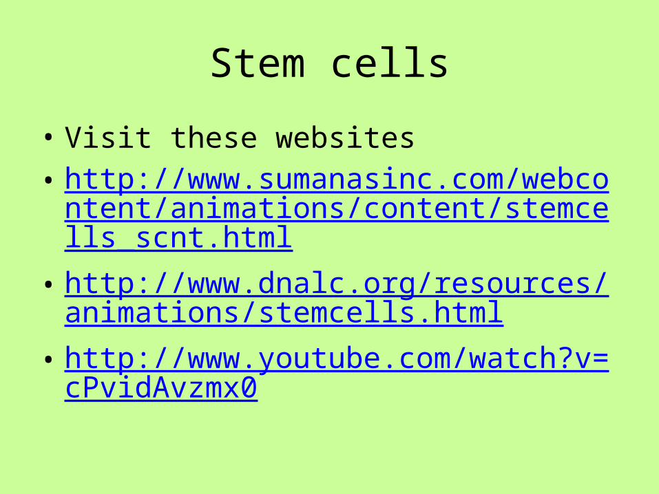
Stem cells
• Visit these websites• http://www.sumanasinc.com/webcontent/ani
mations/content/stemcells_scnt.html• http://www.dnalc.org/resources/animations/s
temcells.html• http://www.youtube.com/watch?v=cPvidAvz
mx0

Lesson 8
• Leukemia Webquest• Research leukemia website.
https://sites.google.com/site/stemcellsinaction/home/stem-cell-webquest-directions
• Respond to questions.• Write one paragraph: Why has leukemia stem
cell therapy been successful while other types of stem cell therapies have failed?
Lesson 9
• Debate: Should embryonic stem cells be used as research tools?
• Work with a partner and read research articles on stem cell social policy. Discuss the pros and cons of the argument with partner.
• Work in groups of 4 on assigned topic. Research on computer additional information to support your topic. Develop a 5 minute argument defending your position.
• Debate: One person from each group will present pro or con argument. Instead of rebuttal, members of the audience will each have to speak about their opinion on stem cell social policy. Class will vote at end of debate.

Unit 7B
Medical Biotechnology II

Lesson 1
• Introduction: movie “Contagion” • Discussion: Is this movie realistic with regards
to how diseases in spread, how governments respond, and the availability of vaccines?
• http://www.movie2k.to/Contagion-watch-movie-1033665.html
Lesson 2
• Infectious• Classification, transmission, prevention• Lecture; Read and study powerpoint• Work with a partner and develop review
questions • Whole class discussion of review questions.• Gram Stain Lab: Normal Flora• Case Study: Childbed fever
Infectious Disease Classification
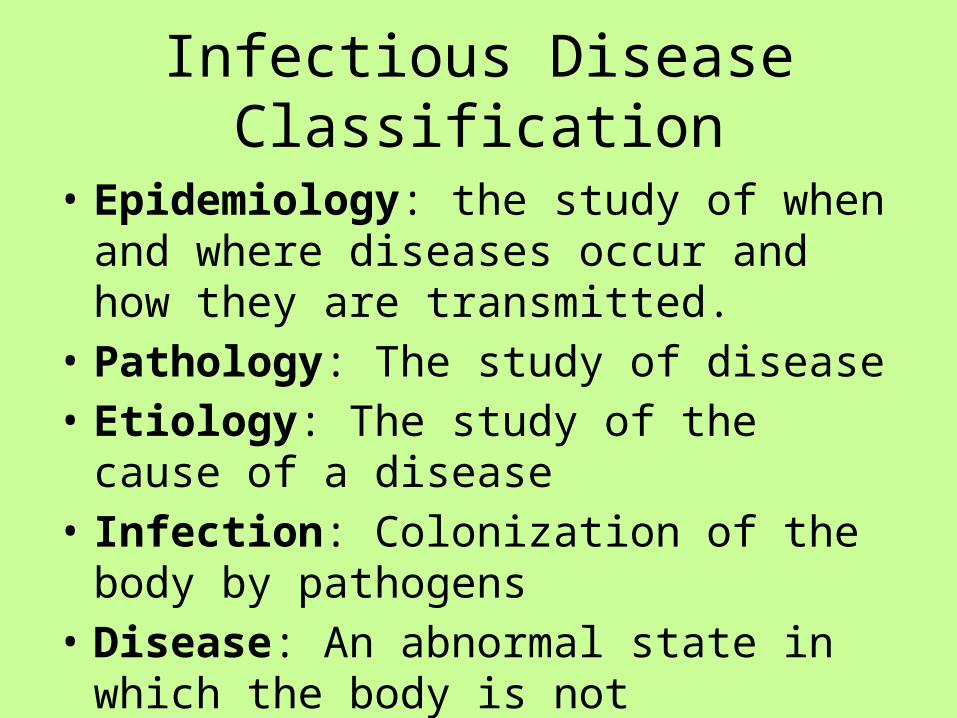
• Epidemiology: the study of when and where diseases occur and how they are transmitted.
• Pathology: The study of disease• Etiology: The study of the cause of a disease• Infection: Colonization of the body by
pathogens• Disease: An abnormal state in which the body
is not functioning normally.
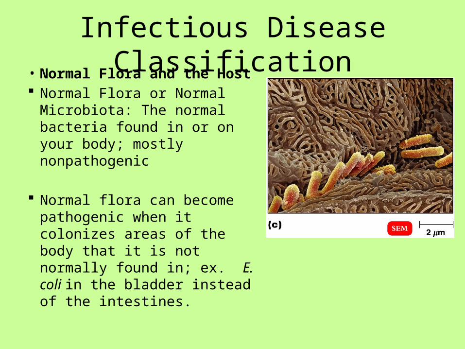
Infectious Disease Classification• Normal Flora and the Host Normal Flora or Normal
Microbiota: The normal bacteria found in or on your body; mostly nonpathogenic
Normal flora can become pathogenic when it colonizes areas of the body that it is not normally found in; ex. E. coli in the bladder instead of the intestines.
Infectious Disease Classification
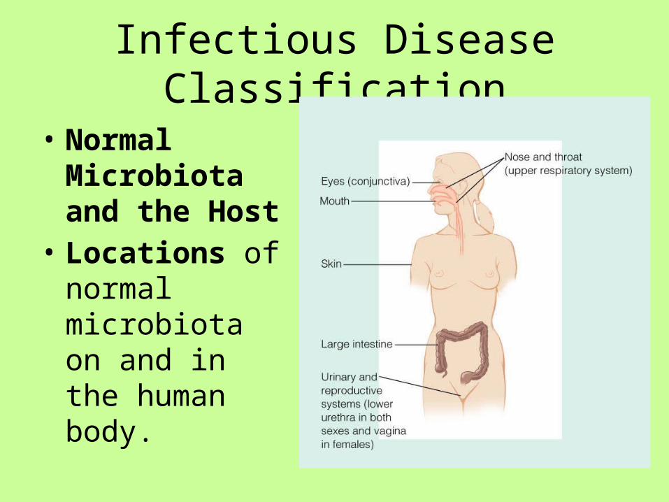
• Normal Microbiota and the Host
• Locations of normal microbiota on and in the human body.
Infectious Disease Classification
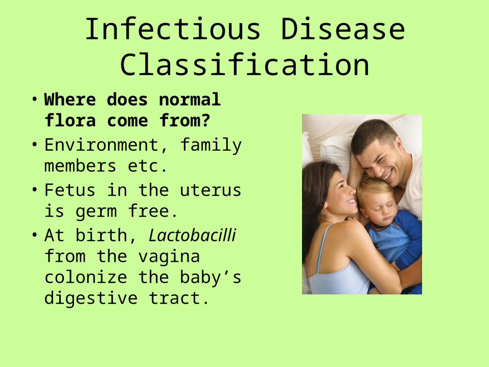
• Where does normal flora come from?
• Environment, family members etc.
• Fetus in the uterus is germ free.
• At birth, Lactobacilli from the vagina colonize the baby’s digestive tract.

Infectious Disease Classification
• Transient Flora and the Host
• Transient Flora: Bacterial changes of normal flora due to seasonal changes (temperatures, etc.) or age and activity.

Infectious Disease Classification Can normal flora benefit the host? Microbial antagonism: how microbes
inhibit the growth of other microbes, usually by competition (e.g. bacteriocins).
Bacteriocins: chemicals produced by bacteria to inhibit the growth of other bacteria (normal flora produce a lot of this).
Probiotics are live microbes applied to or ingested into the body, intended to exert a beneficial effect.

Infectious Disease Classification• Symbiosis• Symbiotic relationship: organisms
living in a close, intimate relationship with each other.– Commensalism, one organism is
benefited and the other is unaffected. (Normal flora).
– Mutualism, both organisms benefit. (Normal flora).
– Parasitism, one organism benefits at the expense of the other. (Infectious disease)

Infectious Disease Classification• Pathogen• A pathogen or infectious agent is a
biological agent that causes disease or illness to its host.
• The term is used for agents that disrupt the normal physiology of a cell, fungus, animal or plant.
• A pathogen can be viral, bacterial, fungal, or a prion.
• A “primary pathogen” is defined as an organism capable of causing disease in a healthy person with a normal immune response.
• A “secondary pathogen” is an infectious agent that causes a disease that follows the initial infections.
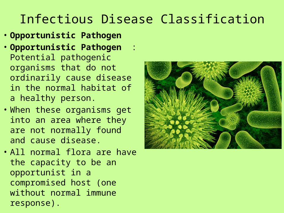
Infectious Disease Classification• Opportunistic Pathogen• Opportunistic Pathogen :
Potential pathogenic organisms that do not ordinarily cause disease in the normal habitat of a healthy person.
• When these organisms get into an area where they are not normally found and cause disease.
• All normal flora are have the capacity to be an opportunist in a compromised host (one without normal immune response).

Transmission Infectious Disease
• Reservoirs of Infection• For a disease to perpetuate itself, there must
be a continual source of disease. This continual source is referred to as the reservoir.
• Reservoirs are classified as either human, animal or nonliving.
Transmission Infectious Disease
• Reservoirs of Infection Reservoirs of infection are continual sources
of infection. Human — AIDS, gonorrhea
Carriers may have inapparent infections or latent diseases.
Animal Zoonoses. Diseases that occur primarily in animals. Example
Rabies, Lyme disease, toxoplasmosis, influenza Nonliving —
Soil: Botulism, tetanus Water: Cholera
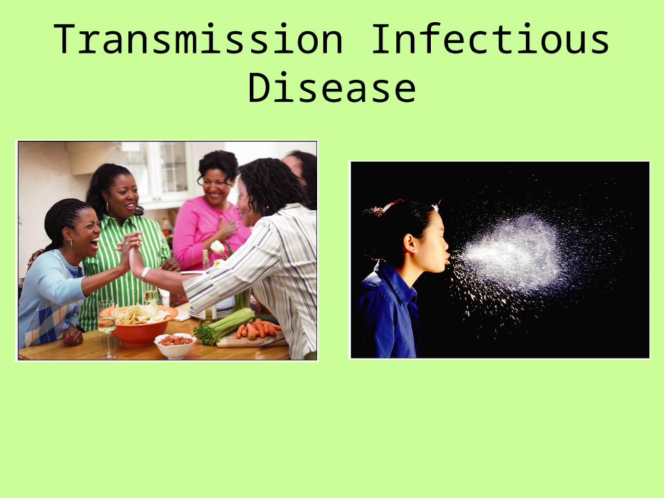
Transmission Infectious DiseaseContact transmission.
1. Direct contact: Person to person transmission by physical contact. This includes touching, kissing and sexual intercourse.
2. Indirect contact Disease is transmitted from a nonliving object (fomite) to a host.
- Fomites may include eating utensils, toys, towels, door knobs, etc.
3. Droplet transmission. Mucous droplets from coughs sneezes laughing or talking. Droplet travels less than one meter from the reservoir to host.
- Example Whooping cough, Influenza, the Common Cold.
Transmission Infectious Disease

Transmission Infectious Disease
• Vehicle: Transmission by an inanimate reservoir
- Food: E. coli gastroenteritis (fecal/oral) - Water: Cholera (fecal/oral) - Airborne: Anthrax• Vectors: Arthropods, especially fleas, ticks,
and mosquitoes. - Plague, Lyme disease

Transmission Infectious Disease

Prevention
• Vaccines• Antimicrobial drugs• Handwashing • Sanitation of fomites and water
supplies• Prepare and store food properly• Control pests (insects and
rodents)• Quarantine

Lesson 3• Part 1 – Stages of Infection• Read powerpoint online• Work with a partner to develop review questions.• Whole class discussion questions.• Read article about stages of infection, traditional medical tests, and molecular
biology tests used to diagnose.• Part 2 – Epidemiology• Read SARS time line (Refer to handout)• What types of activities occur during an epidemic• Read powerpoint online.• Write a one paragraph description of the important elements in an
epidemiological investigation.• View video –SARS The True Story• Class discussion

Stages of Infection• How Infectious Agents
Cause Disease• Production of poisons,
such as toxins and enzymes, that destroy cells and tissues.
• Direct invasion and destruction of host cells.
• Triggering responses from the host’s immune system leading to disease signs and symptoms.

Stages of Infection
1. Entry of Pathogen – Portal of Entry
2. Colonization – Usually at the site of entry
3. Incubation Period– Asymptomatic period – Between the initial
contact with the microbe and the appearance of the first symptoms

Stages of Infection
4. Prodromal Symptoms– Initial Symptoms
5. Invasive period– Increasing Severity of
Symptoms – Fever – Inflammation and Swelling – Tissue Damage – Infection May Spread to Other
Sites – Acme
Stages of Infection
6. Decline of Infection - Improvement in
symptoms
7. Convalescence

Diagnostic Tests- Traditional
• Isolation of Pathogens from Clinical Specimens
• If a physician suspects a bacterial infection, samples of infected body fluids or tissues are collected from the patient.
• Samples may include blood, spinal fluid, pus, sputum, urine, or feces.
• A swab may be used to sample the infected area.

Diagnostic Tests - Traditional
• Isolation of Pathogens from Clinical Specimens
• The swab is then inoculated onto the surface of an agar plate or put into a tube of liquid medium.
• The bacteria is grown and isolated.

Diagnostic Tests-Traditional
• Isolation of Pathogens from Clinical Specimens
• The bacteria is identified by growth dependent rapid identification systems.
• These systems contain a battery of biochemical tests.

Diagnostic Tests-Traditional• Isolation of Pathogen from
Clinical Specimen• Identified pathogens are then
tested for sensitivity to antimicrobial agents.
• Drug sensitivity testing guides antimicrobial therapy for the patient.
• Small wafers with antibiotics are placed on a plate of bacteria. Large zones of no bacterial growth indicated antimicrobial sensitivity.

Diagnostic Tests- Traditional• Serology Tests for Antibody or
Antigen (Bacterial & Viral)• The agglutination of antigen coated
or antibody coated latex beads with a complimentary antibody or antigen is a typical method of rapid diagnosis.
• Blood serum from the patient is used in this test.
• If , for example, a patient has antibody to a particular infectious agent, the antibody will bind to the antigen coated latex beads.
• The suspension becomes visibly clumped. http://www.youtube.com/watch?v=hRzOwSTkF0s
• (first 3 min)
Diagnostic Tests- ELISA
• Enzyme Linked Immunoassay (viruses)-ELISA• A target antigen is bound to a solid phase such as the
plastic on a microplate.• A patient’s blood serum is added to the microplate.• Antibody in the serum will bind to the antigen.• The well in the plate is washed with an enzyme tagged
antihuman antibody which binds to the patient antibody.
• A substrate for the enzyme is added and a color reaction occurs .
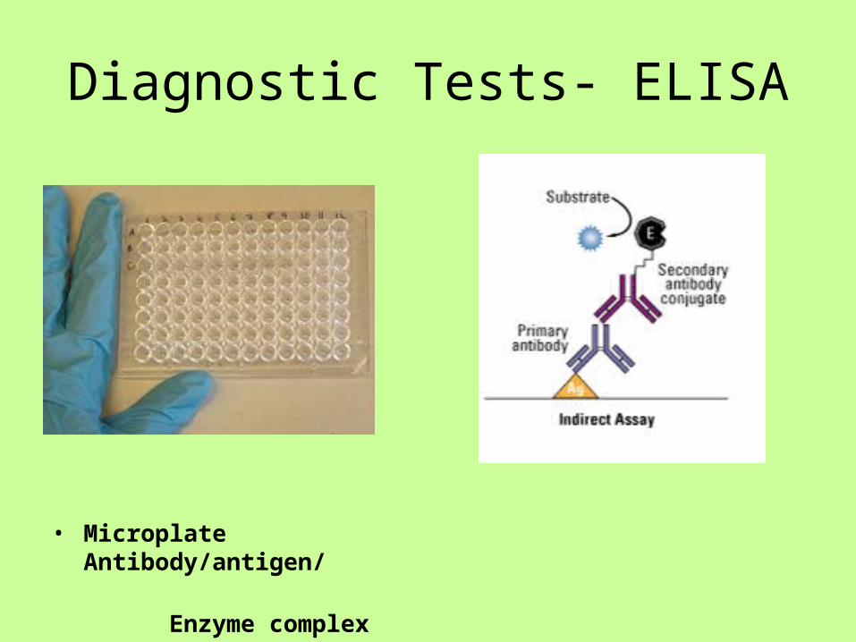
Diagnostic Tests- ELISA
• Microplate Antibody/antigen/ Enzyme complex

Molecular Diagnostic Tests• Nucleic Acid Hybridization• To identify a bacteria or virus, a
species specific nucleic acid probe is needed.
• Probes are a single strand of DNA with a sequence unique and complimentary to the gene of interest.
• If a clinical specimen contains the microorganism of interest, the probe will bind to the microorganism’s gene DNA sequence.
• The double stranded DNA is detected because the probe is labeled with a radioactive, fluorescent, or enzyme tag.

Molecular Diagnostic Tests• PCR• There are PCR tests available to extract
DNA or RNA from bacteria or viruses.• The PCR method uses species specific
primers for targeted DNA or cDNA.• The DNA is then amplified.• Detection of the gene sequence can be
done by gel electrophoresis.• An alternative to electrophoresis is the use
of PCR machines with precision optics monitors.
• The primer has a fluorescent label and the monitor plots the uptake of the primer. If the primer has been used, this indicates the presence of the microorganism.
Epidemiology• In investigating an outbreak, speed is essential, but getting the right answer is
essential, too. To satisfy both requirements, epidemiologists approach investigations systematically, using the following 10 steps:
• Prepare for field work• Establish the existence of an outbreak• Verify the diagnosis• Define and identify cases• Describe and orient the data in terms of time, place, and person• Develop hypotheses• Evaluate hypotheses• Refine hypotheses and carry out additional studies• Implement control and prevention measures• Communicate findings• The steps are presented here in conceptual order. In practice, however, several
may be done at the same time, or they may be done in a different order. For example, control measures should be implemented as soon as the source and mode of transmission are known, which may be early or late in any particular outbreak investigation.

Epidemiology
• Step 1: Prepare for Field Work• Before leaving for the field:• Research the disease and gather the supplies/
equipment needed• Make necessary administrative and personal
arrangements for such things as travel. • Consult with all parties to determine your role in the
investigation and who your local contacts will be.

Epidemiology
• Step 2: Establish the Existence of an Outbreak• Verify that a suspected outbreak is indeed a real
outbreak. • Some of the cases will be associated with a true outbreak
with a common cause, some will be unrelated cases of the same disease, and others will turn out to be unrelated cases of similar but unrelated diseases.
• Before you can decide whether an outbreak exists (i.e., whether the observed number of cases exceeds the expected number), you must first determine the expected number of cases for the area in the given time frame.

Epidemiology
Epidemiology• Step 3: Verify the Diagnosis• In addition to verifying the existence of an outbreak early in
the investigation, you must also identify as accurately as possible the specific nature of the disease.
• Goals in verifying the diagnosis are two-fold. - First, ensure that the problem has been properly diagnosed—that it really is what it has been reported to be. - Second, for outbreaks involving infectious or toxic-chemical agents, be certain that the increase in diagnosed cases is not the result of a mistake in the laboratory.
Dx

Epidemiology
• Step 4: Define and Identify Cases• Establish a case definition. Your next task as an
investigator is to establish a case definition, or a standard set of criteria for deciding whether, in this investigation, a person should be classified as having the disease or health condition under study. A case definition usually includes four components:
• clinical information about the disease, • characteristics about the people who are affected, • information about the location or place, and • a specification of time during which the outbreak
occurred.
Epidemiology• Step 5: Describe and Orient the Data in Terms of Time, Place, and
Person• After data collection, characterize an outbreak by time, place, and
person. This step may be performed several times during the course of an outbreak. Characterizing an outbreak by these variables is called descriptive epidemiology,
• This step is critical- First, by becoming familiar with the data, you can learn what information is reliable and what is not. - Second, you provide a comprehensive description of an outbreak by showing its trend over time, its geographic extent (place), and the populations (people) affected by the disease. This description lets you begin to assess the outbreak in light of what is known about the disease and to develop causal hypotheses.

Epidemiology (descriptive)

Epidemiology• Step 6: Develop Hypotheses• In real life, we begin to generate hypotheses to
explain why and how the outbreak occurred when we first learn about the problem. But at this point in an investigation, after you have interviewed some affected people, spoken with other health officials in the community, and characterized the outbreak by time, place, and person, your hypotheses will be sharpened and more accurately focused.
• The hypotheses should address the source of the agent, the mode (vehicle or vector) of transmission, and the exposures that caused the disease. Also, the hypotheses should be proposed in a way that can be tested.
Epidemiology
• Step 7: Evaluate Hypotheses• The next step is to evaluate the credibility of
your hypotheses. There are two approaches you can use, depending on the nature of your data: 1) comparison of the hypotheses with the established facts and 2) analytic epidemiology, which allows you to test your hypotheses with cohort and case control studies.

Epidemiology (cohort study)

Epidemiology• Step 8: Refine Hypotheses and Carry Out Additional Studies• Additional epidemiological studies
When analytic epidemiological studies do not confirm your hypotheses, you need to reconsider your hypotheses and look for new vehicles or modes of transmission. This is the time to meet with case-patients to look for common links and to visit their homes to look at the products on their shelves.
• Also, confirmation from laboratory findings can be valuable.

Epidemiology
Step 9: Implementing Control and Prevention Measures
• Even though implementing control and prevention measures is listed as Step 9, in a real investigation you should do this as soon as possible.
• Control measures, which can be implemented early if you know the source of an outbreak, should be aimed at specific links in the chain of infection, the agent, the source, or the reservoir.

Epidemiology
• Step 10: Communicate Findings• Your final task in an investigation is to
communicate your findings to others who need to know. This communication usually takes two forms: 1) an oral briefing for local health authorities and 2) a written report

Epidemiology
• SARS – The True Story http://www.youtube.com/watch?v=MXPaee0uEQM
• Case study: SARS

Lesson 4
• Origin of SARs and evolution of the virus• Work in groups of 4. Read powerpoint and
web articles about origin of SARS virus and viral evolution.
• Discuss and respond to questions.• Write a short essay explaining how natural
selection occurred with the SARS virus.

Origin of SARS & Evolution
• Visit the following websites for the origin of SARS:
• http://www.abc.net.au/science/features/sars/default.htm
• http://learn.genetics.utah.edu/archive/sars/index.html

Origin of SARS and Evolution
• Visit the following websites for the evolution of SARS:
• http://www.smartplanet.com/blog/science-scope/study-shows-how-swiftly-infectious-viruses-evolve/12136
• http://www.scientificamerican.com/article.cfm?id=sars-evolution-traced
• http://learn.genetics.utah.edu/archive/sars/index.html
Identification of SARS virus
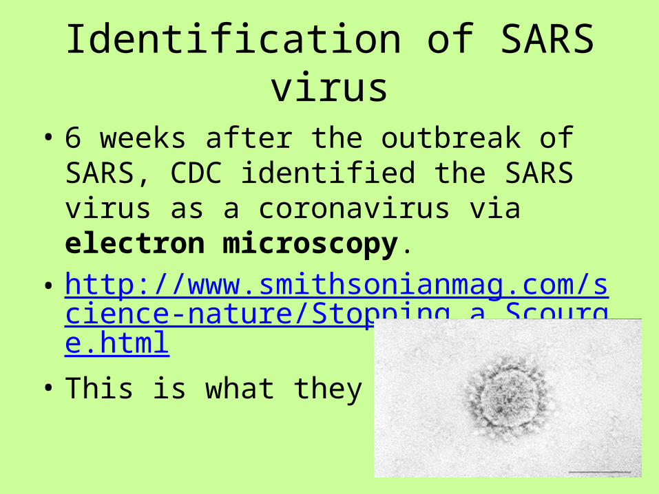
• 6 weeks after the outbreak of SARS, CDC identified the SARS virus as a coronavirus via electron microscopy.
• http://www.smithsonianmag.com/science-nature/Stopping_a_Scourge.html
• This is what they saw:

Identification of SARS virus
• Coronavirus• Are single stranded RNA viruses• Replicate in the cytoplasm of the
host cell.• Spherical 60- 220 nm in size.• Contain club shaped
glycoproteins on their surfaces. The virus looks like it has a crown.
• Largest genome of any RNA viruses; 29,700 base pairs.
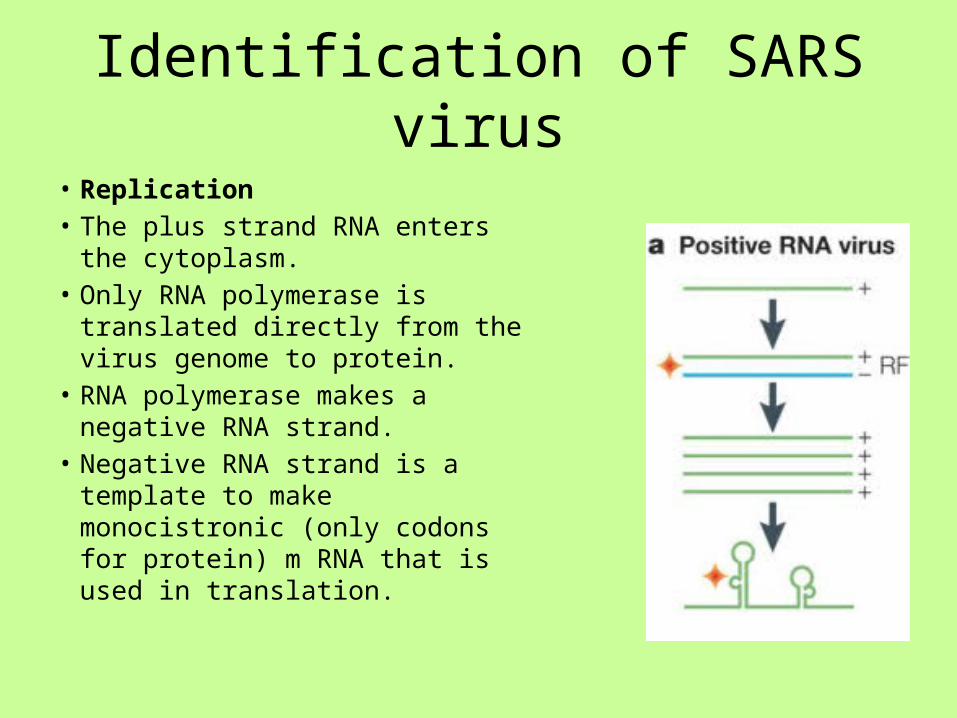
Identification of SARS virus
• Replication• The plus strand RNA enters the
cytoplasm.• Only RNA polymerase is translated
directly from the virus genome to protein.
• RNA polymerase makes a negative RNA strand.
• Negative RNA strand is a template to make monocistronic (only codons for protein) m RNA that is used in translation.


Identification of SARS virus• The second step in identification
of the virus was to discover the DNA sequence of the virus.
• Three months after the outbreak, Canada’s Michael Smith Genome Sciences Center in British Columbia sequenced the SARS coronavirus.
• The DNA sequence is located in GenBank.
• http://www.msfhr.org/news/features/2009/03/speed_demons

Identification of SARS virus• Bioinformatics• DNA isolates were taken from SARS
virus and compared to DNA isolates from other coronaviruses and other strains of SARS viruses.
• By comparing SNP (single nucleotide polymorphism) among the DNA isolates, researchers were able to classify the virus and create a phylogenetic tree.
• Researchers also discovered that four proteins were responsible for pathogenesis of SARS: spike (S) protein; small envelope (E) protein; membrane (M) protein; and nucleocaspid (N) protein.

Patient Diagnostics -SARS
• The DNA sequencing and research led to two frequently used tests to help diagnose SARS in patients.
1. RTPCR – Real Time Reverse Transcriptase PCR
2. ELISA – Enzyme linked immunoassay

Patient Diagnostics• RTPCR• Enables researchers to quantify amplification reactions in real time.• A specialized thermo cycler with a laser scan beams light through the PCR tube.• The PCR product is labeled with a fluorescent tag.• The amount of fluorescent light produced after each cycle is printed on a computer
readout.• This allows for quantification of the number of PCR products produced after each
cycle.• For diagnosis of SARs, patient tissues and body fluids can be used directly. The
primers used in the test are specific for cDNA made from the viral genome.• http://www.youtube.com/watch?v=kvQWKcMdyS4

Patient Diagnosis• ELISA• The SARS antigen is bound to a
microplate.• The patient’s blood serum with SARS
antibodies is introduced.• The patient SARs antibody binds to the
SARS antigen.• A second antibody with an enzyme
attached combines with the SARS antibody/antigen complex.
• When a substrate is added,t he well in the microplate turns color with a positive reaction .
• http://highered.mcgraw-hill.com/sites/0072556781/student_view0/chapter33/animation_quiz_1.html.

Patient Diagnostics• ViroChip• A DNA microarray test system called ViroChip enables doctors to
screen for the type of virus present in a patient, if they do not know the virus type.
• A ViroChip has 22,000 DNA sequences on it and identifies a variety of different viruses.
• http://www.nytimes.com/2008/10/07/health/research/07conv.html
Lesson 5
• Immunology• Work in groups of 4. • Read powerpoint, discuss, and respond to
questions.• Complete chart of immune responses.• Whole class discussion of responses.• Write a 5 minute commercial or skit about the
immune system.• Present skit or commercial.

Immunology• Two types of immunity• Innate or Non-specific Immunity• Adaptive or Acquired Immunity

Immunology
• 3 Lines of Defense in immunity
• Barriers at body surfaces (innate)
1. Intact skin and mucous membranes
2. Infection fighting tears and saliva.
3. Normal bacterial flora outcompete pathogens.
4. Flushing effects of tears, urination, diarrhea.
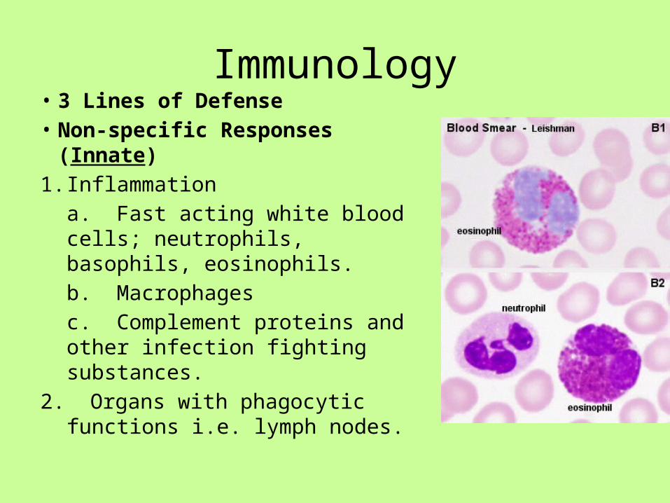
Immunology• 3 Lines of Defense• Non-specific Responses (Innate)1. Inflammation
a. Fast acting white blood cells; neutrophils, basophils, eosinophils.b. Macrophagesc. Complement proteins and other infection fighting substances.
2. Organs with phagocytic functions i.e. lymph nodes.
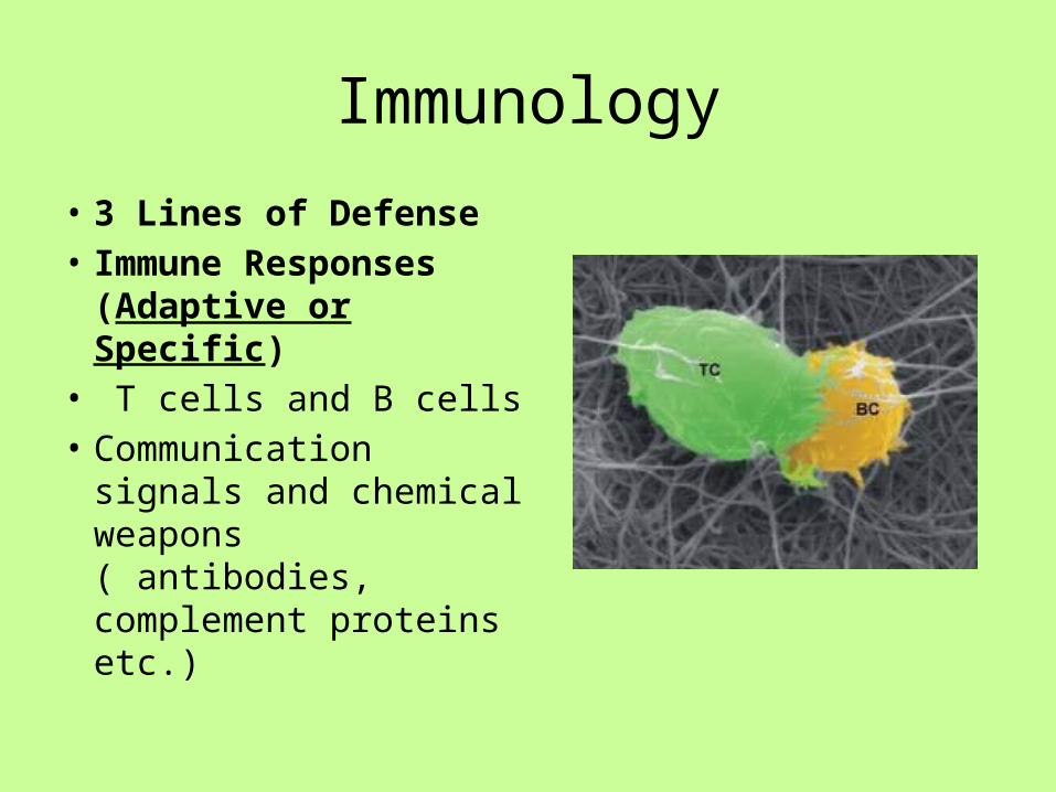
Immunology
• 3 Lines of Defense• Immune Responses
(Adaptive or Specific)• T cells and B cells• Communication
signals and chemical weapons ( antibodies, complement proteins etc.)
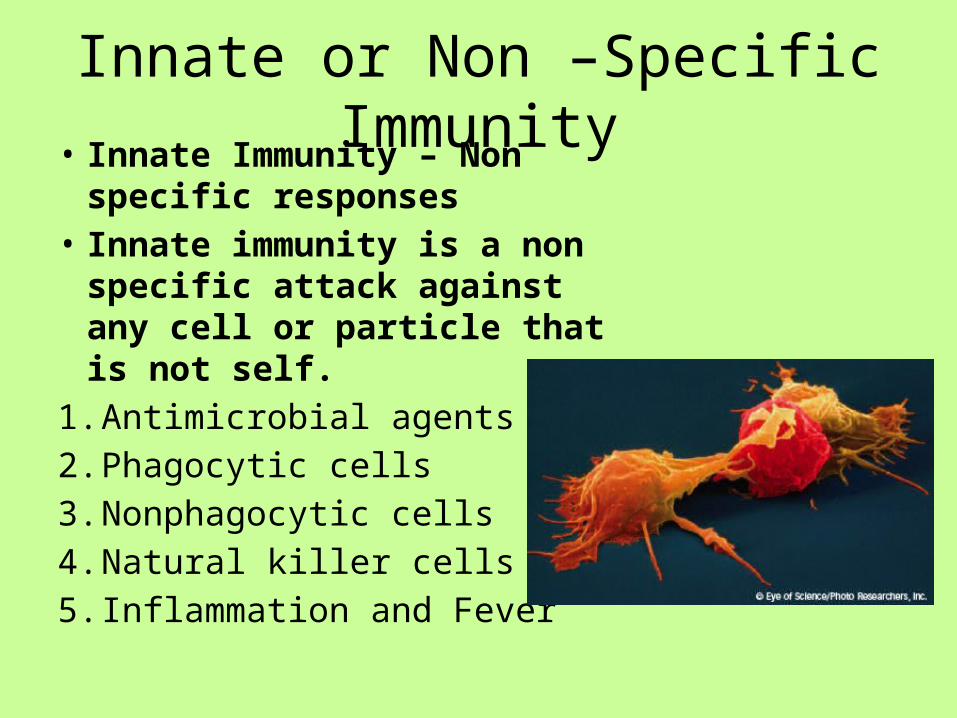
Innate or Non –Specific Immunity• Innate Immunity – Non specific
responses• Innate immunity is a non specific
attack against any cell or particle that is not self.
1. Antimicrobial agents2. Phagocytic cells3. Nonphagocytic cells4. Natural killer cells 5. Inflammation and Fever
Innate or Non-Specific Immunity
• Innate - Antimicrobial agents• Antimicrobial agents are chemicals or molecules
that act to deter or destroy microorganisms. Some of them act in conjunction with physical barriers.
There are 3 types:1. Interferon2. Interleukins3. Complement

Innate or Non-specific Immunity• Interferon• Interferons are a large group of
proteins that acts as signals both during innate and adaptive immune responses.
• Interferon is produced early in viral infections by a cell.
• The interferon will not keep the cell from viral infection but it is released and warns other cells to synthesize antiviral cell surface proteins.
• http://www.youtube.com/watch?v=3qFu6Fv4cJk&feature=related

Innate or Non-specific Immunity
• Interleukins• Interleukins are another
class of proteins which are produced by cells of the immune system.
• Tumor necrosis factor (TNF) , a type of interleukin, stimulates cells to create an inflammatory response.
• TNF cells can also kill tumor cells.

Innate or Non-specific Immumity
• Complement: a family of 20 different proteins.• Found in blood serum and protect the body
from infection.• Works together with other components of the
innate and adaptive immune systems.• Generally, complement is inactive.• In the presence of an antigen, complement
becomes activated.
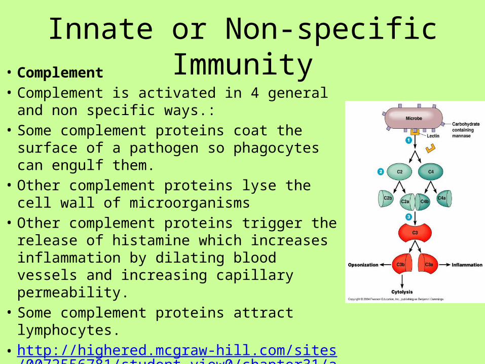
Innate or Non-specific Immunity• Complement• Complement is activated in 4 general and non
specific ways.:• Some complement proteins coat the surface
of a pathogen so phagocytes can engulf them.• Other complement proteins lyse the cell wall
of microorganisms• Other complement proteins trigger the
release of histamine which increases inflammation by dilating blood vessels and increasing capillary permeability.
• Some complement proteins attract lymphocytes.
• http://highered.mcgraw-hill.com/sites/0072556781/student_view0/chapter31/animation_quiz_1.html

Innate or Non-specific Immunity• Innate – Phagocytic Cells• Sometimes an infectious agent avoids
physical barriers and antimicrobial agents in the body.
• A 3rd line of defense is available.• Phagocytic cells engulf microorganisms
in digestive vacuoles and break down cells.
• Phagocytic cells contain many enzymes: lysozyme to breakdown peptidoglycan, proteases to break down proteins, nucleases to break down DNA and RNA, and lipases to break down lipids.

Innate or Non-specific Immunity
• Phagocytic Cells• There are 2 types of phagocytic cells:1. Stationary phagocytes – reside along blood
vessel walls and in connective tissue2. Wandering phagocytes – circulate in the
blood.• Both types are made in the bone marrow

Innate or Non-specific Immunity• Stationary phagocytes• Macrophages are large phagocytic cells.• Made in the bone marrow, they circulate
in the blood for a few days and are called monocytes at this stage.
• Monocytes are released into connective tissue and are now referred to as macrophages.
• Macrophages are scavenger cells. They engulf:
1. Microorganisms2. Dead body cells3. Cancer cells4. Cells infected with viruses.• The life span of a macrophage is from a
few months to many years.
http://www.youtube.com/watch?v=m6qJ69wcSnc
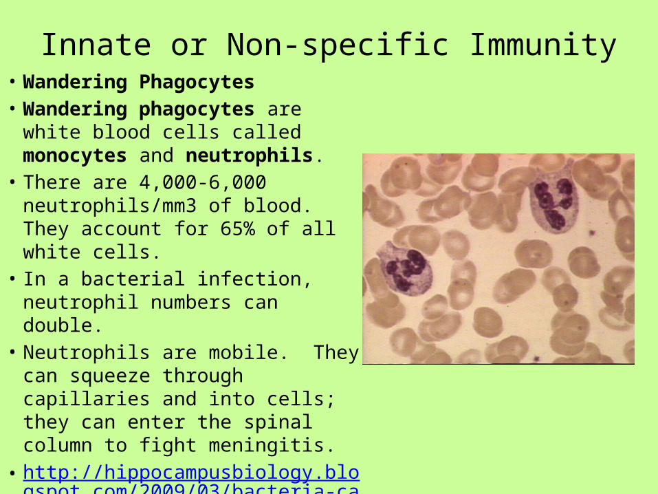
Innate or Non-specific Immunity• Wandering Phagocytes• Wandering phagocytes are white
blood cells called monocytes and neutrophils.
• There are 4,000-6,000 neutrophils/mm3 of blood. They account for 65% of all white cells.
• In a bacterial infection, neutrophil numbers can double.
• Neutrophils are mobile. They can squeeze through capillaries and into cells; they can enter the spinal column to fight meningitis.
• http://hippocampusbiology.blogspot.com/2009/03/bacteria-can-run-but-they-cant-hide.html

Innate or Non-Specific Immunity• Innate – Nonphagocytic Cells• Eosinophils – White blood cells that secrete enzymes
that attack parasitic worms; reside in blood.• Basophils - White blood cells that contain heparin
(stops blood from clotting) and histamine (vasodilator which promotes blood flow to tissues). Play a role in inflammation and allgergies. Reside in blood.
• Mast Cell - Cells residing in many tissues that contain heparin and histamine. Look like basophils but come from different cell line. Important in inflammation.

Innate or Non Specific Immunity
Mast Cell
Eosinophil Basophil

Innate or Non-specific Immunity
• Innate – Natural Killer Cells• Are not phagocytic.• Are a white blood cell called a
lymphocytes• Attach to cell surfaces and produce
enzymes that destroy antibody covered cells that have been infected with microorganisms or viruses.
• Also able to destroy cancer cells. • http://www.youtube.com/watch?v=
HNP1EAYLhOs

Innate or Non-specific Immunity• Innate – Inflammation• Inflammatory response is a major component of the
innate immune system.• In inflammation:1. When tissue is injured or microorganisms enter the
tissue, mast cells release chemicals called histamine.

Innate or Non-specific Immunity• Inflammation2. The chemicals spark the mobilization of various defenses.a. Histamine induces blood vessels to dilate and increases blood flow
to the injured tissue.b. Blood plasma leaks out of the blood vessels to the affected tissues.c. Phagocytic cells squeeze out of the blood vessels and migrate to
the tissue.d. Increased blood flow, fluid, and cells produce redness, heat, and
swelling of tissues.

Innate of Non-specific Immunity
• Inflammation3. The major results of inflammation is to
disinfect and clean injured tissue.a. Phagocytic cells engulf bacteria and
body cells killed by them.b. Many phagocytic cells die in this process
and they are engulfed and digested.c. Pus that accumulates at the injury site
consists of dead cells and fluid from leaking capillaries.
Innate or Non-specific Immunity
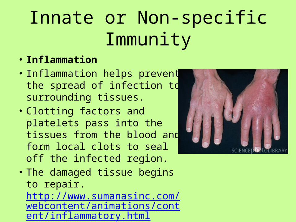
• Inflammation• Inflammation helps prevent the
spread of infection to surrounding tissues.
• Clotting factors and platelets pass into the tissues from the blood and form local clots to seal off the infected region.
• The damaged tissue begins to repair. http://www.sumanasinc.com/webcontent/animations/content/inflammatory.html

Innate or Non-specific Immunity• Innate- Fever• During some infections, fever occurs.• Fever is induced by toxins released by
microorganisms.• The increase in body temperature:1. Kills some microorganisms.2. Increases inflammation.3. Stimulates phagocytic activity.4. Stimulates adaptive (acquired)
immune response.5. Reduces iron concentration in blood
and limits amount of iron available to microorganisms.
Adaptive or Acquired Immunity
• Adaptive immunity is a complex set of interactions that is highly specific against one type of antigen.
• Whereas innate immunity reacts to a variety of pathogens, adaptive immunity must be primed by the presence of an antigen.
• Adaptive immunity either attacks a specific antigenic invader directly or it produces antibodies.
• There are two types of adaptive immunity:1. Cell mediated immunity2. Humoral immunity

Adaptive or Acquire Immunity• Cell mediated immunity
involves a type of lymphocyte called a T (thymus) cell. T cells work against infections caused by fungus and protozoans. Also, they are important in eliminating cancer cells.
• Humoral immunity involves a type of lymphocyte called a B cell. B cells protect against viruses and bacteria in body fluids by producing antibodies.
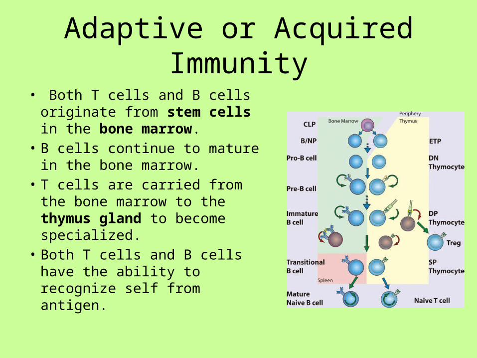
Adaptive or Acquired Immunity
• Both T cells and B cells originate from stem cells in the bone marrow.
• B cells continue to mature in the bone marrow.
• T cells are carried from the bone marrow to the thymus gland to become specialized.
• Both T cells and B cells have the ability to recognize self from antigen.

Adaptive or Acquired Immunity• Cell Mediated Immunity• T cells after maturation are
moved to the lymphatic system.• T cells are primarily involved in
attacking cells directly.• While T cells are maturing, they
develop the ability to recognize specific antigens (Non-self)
• For T cells to go into action, they need the antigen presented to them.

Adaptive or Acquire Immunity
• Cell mediated immunity• All cells have surface glycoproteins
call the Major Histocompatibilty Complex (MHC).
• In humans, the MHC is called HLA (human leukocyte antigen).
• These classes of molecules mark our cells as self.
• Invading cells have an MHC and this marks them as foreign.

Adaptive or Acquire Immunity• Cell mediated immunity• Cells like macrophages recognize
foreign (MHC) cells and ingest them.• Macrophages(or other cells) then
display antigenic fragments from the microorganism on their cell surfaces.
• Macrophages displaying antigens are called antigen presenting cells (APC).
• T cells bind to the macrophage/antigen complex and this activates the T cell.
• T cells will then proliferate and carry out their functions.
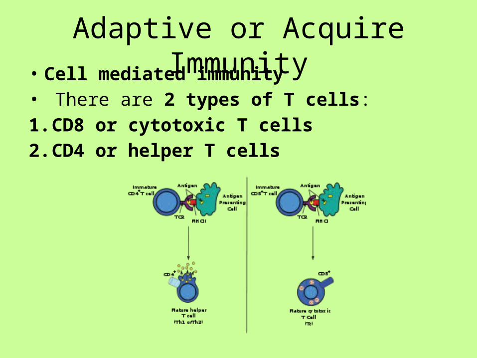
Adaptive or Acquire Immunity• Cell mediated immunity• There are 2 types of T cells:1. CD8 or cytotoxic T cells2. CD4 or helper T cells

Adaptive or Acquire Immunity• Cell mediated immunity• CD8 cells• CD8 cells respond to foreign
antigens on the cell’s surface by binding to the antigenic MHC on the invading cells and directly killing them.
• CD8 cells release perforin, a protein that creates pores on the invading cells membrane. Water and ions flow into the cells and the cell lyses.
• Cancer cells, foreign cells from a transplant or graft, pathogen infected cells, and virus infected cells are targeted by CD8 cells.
http://www.theimmunology.com/animations/Cytotoxic.T.Cell.htm

Adaptive or Acquired Immunity

Adaptive or Acquired Immunity• Cell mediated immunity• CD4 cells• CD4 cells that have been activated by an APC secrete
cytokines, a protein that stimulates other lymphocytes.• If B cells have contacted an antigen, the signal from the CD4
cell differentiates the B cell into an antibody producing cell.• CD4 cells also play a role in stimulating CD8 cells to
proliferate. • http://highered.mcgraw-hill.com/sites/0072507470/studen
t_view0/chapter22/animation__t-cell_dependent_antigens__quiz_2_.html

Adaptive or Acquired Immunity
• Humoral Immunity• B cells are involved in humoral immunity.• B cells produce antibody. The antibody
produced by a B cell is specific for a particular antigen.
• There are two types of B cells:1. Plasma Cells2. Memory Cells

Adaptive or Acquired Immunity• Humoral immunity• Plasma Cells• A plasma cell encounters an
antigen and secretes a specific antibody.
• Plasma cells are activated when they encounter an antigen and are stimulated by helper T cells.
• Plasma cells can produce more than 10 million antibodies in an hour.
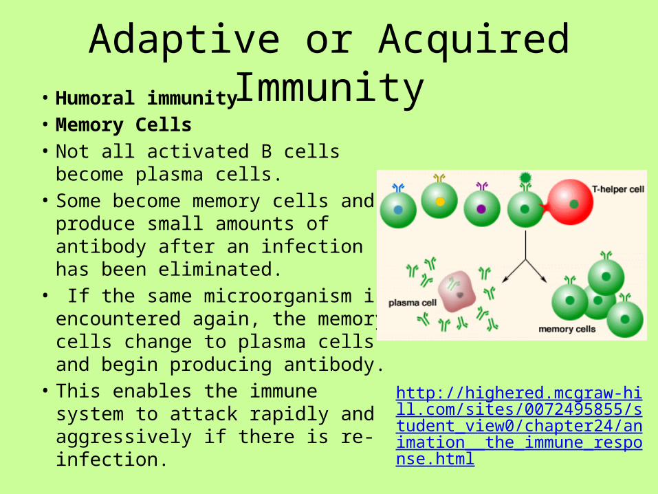
Adaptive or Acquired Immunity• Humoral immunity• Memory Cells• Not all activated B cells become
plasma cells.• Some become memory cells and
produce small amounts of antibody after an infection has been eliminated.
• If the same microorganism is encountered again, the memory cells change to plasma cells and begin producing antibody.
• This enables the immune system to attack rapidly and aggressively if there is re-infection.
http://highered.mcgraw-hill.com/sites/0072495855/student_view0/chapter24/animation__the_immune_response.html

Adaptive or Acquire Immunity
• Antibodies1. Antibody structure and function2. Disposal of antibody/antigen complex.

Adaptive or Acquired Immunity
• Antibody structure and function• Antigens that elicit an antibody
response are typically a protein or polysaccharide surface component of microbes.
• The antibody does not bind to the total antigen.
• A small accessible portion of antigen called an epitope or antigenic determinant is available for binding to the antibody.
• A single antigen has several epitopes and each epitope binds to a different antibody.
Adaptive or Acquired Immunity
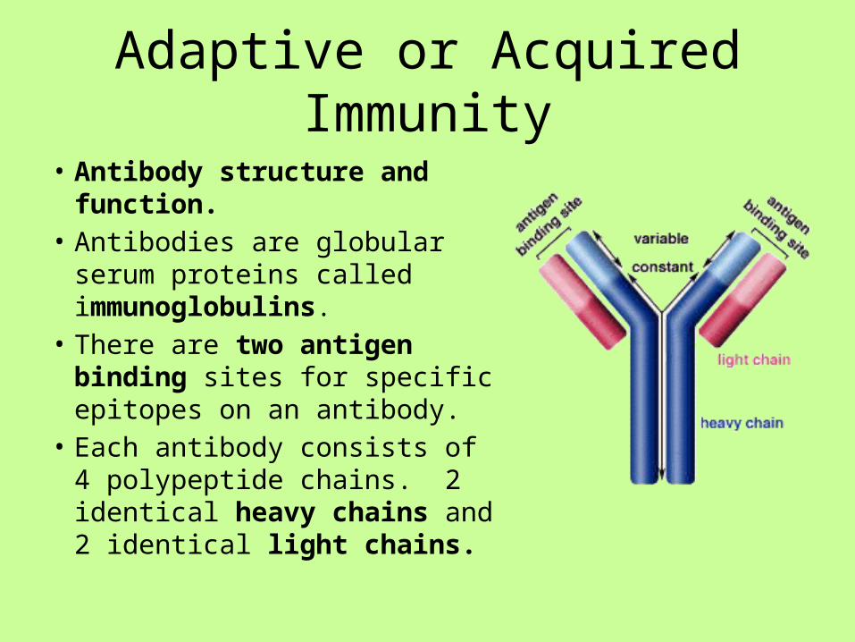
• Antibody structure and function.• Antibodies are globular serum
proteins called immunoglobulins.• There are two antigen binding
sites for specific epitopes on an antibody.
• Each antibody consists of 4 polypeptide chains. 2 identical heavy chains and 2 identical light chains.

Adaptive or Acquired Immunity• Antibody Structure and
Function• At the 2 tips of the antibody
are variable regions (on light and heavy chains).
• The amino acid sequence varies from antibody to antibody.
• The variable region gives the antibody specificity for an antigen.
• The binding of the epitope and the binding site is similar to an enzyme substrate reaction.

Adaptive or Acquired Immunity• Antibody structure and
function• The tail of the antibody is
formed by the constant region.• The constant region is
responsible for distribution of the antibody within the body and for mechanisms that mediate the disposal of the antibody/antigen complex.
• There are 5 types of heavy chain constant regions: IgA, IgG, IgM, IgD, and IgE.


Adaptive or Acquire Immunity
• Disposal antibody/antigen complex• The binding of antibody to antigen creates
complexes that must be disposed.• Three types of disposal1. Neutralization/Opsonization2. Agglutination/ Precipitation3. Complement fixation


Adaptive or Acquire Immunity
• Disposal antibody/antigen complex• Neutralization/Opsonization• In neutralization, the antibody binds to the antigen.• The microbe covered in antibodies is phagocytized
by macrophages.• Opsonization is similar to neutralization.
Compounds called opsins bind to antibody/antigen complexes and this enhances the ability of the macrophage to phagocytize the microorganism


Adaptive or Acquired Immunity
• Disposal antibody/antigen complex• Agglutination• Agglutination is possible because antibodies have 2 antigen
binding sites.• One site can attach to one bacteria and the second site can
attach to a second bacteria.• When thousands of antibodies behave in this way, a clumping
of the microorganisms occurs.• Precipitation is similar to agglutination as clumping occurs.• In precipitation, the antigens cross link and form a precipitate.• Both processes are followed by macrophage phagocytosis.


Adaptive or Acquired Immunity• Disposal antibody/antigen complex• Complement Fixation• Antibody/antigen complexes
activate a complement cascade.• Complement consists of 20 proteins
and in the cascade one type of complement triggers the production of the next type in a series of reactions.
• Completion of the complement cascade results in lysis of viruses and pathogenic cells or opsonization.
• http://highered.mcgraw-hill.com/sites/0072556781/student_view0/chapter31/animation_quiz_1.html

Lesson 6
• ELISA Lab – SARS virus• Conduct ELISA test.• Write lab report• Refer to your handouts
Lesson 6
• Vaccines• Read article: How do vaccines work?• Discussion: How is this related to discussion of immune
system• Lecture- Types of vaccines (biotechnology)• Whole class review with questions of vaccine types.• Video: Mothers who do not vaccinate their children.• Read and review article on causal relationships between
vaccines and adverse effects.• Discussion: Should childhood vaccination be mandatory?

Lesson 5
• Vaccines

Vaccines
• How do vaccines work?• http://www.healthychildren.org/English/safet
y-prevention/immunizations/pages/How-do-Vaccines-Work.aspx?nfstatus=401&nftoken=00000000-0000-0000-0000-000000000000&nfstatusdescription=ERROR%3a+No+local+token
• http://www.historyofvaccines.org/content/how-vaccines-work

Vaccines• Vaccination has proven effective
against fighting diseases caused by microorganisms.
• Infectious disease is one of the major causes of death worldwide. 60% of children worldwide under the age of 4 die from infectious disease.
• The world’s first vaccine was made by Edward Jenner in 1796. He discovered that the live cowpox virus could be used to immunize patients against smallpox.

Vaccines
• Vaccines• Vaccines are parts of a pathogen or
whole organisms that are given to humans or animals by mouth or by injection to stimulate the immune system.
• When people or animals are vaccinated, the immune system recognizes the vaccine as antigen and produces antibodies and memory B cells.

Vaccines
• Vaccines• Four major strategies
are used to make vaccines:
1. Subunit vaccines2. Attenuated vaccines3. Inactivated (killed)
vaccines4. DNA vaccines

Vaccines
• Vaccines• Subunit vaccines• Subunit vaccines are made by injecting
portions of viral or bacterial structures, usually proteins or lipids from the microbe, to which the immune system responds.
• EX: Vaccines for hepatitis B, anthrax, tetanus, and meningococcal disease.
• http://library.thinkquest.org/20994/data/task8-2.html

Vaccines
• Vaccines• Attenuated vaccines• Attenuated vaccines involve using live
bacteria or viruses that have been weakened (attenuated) by aging or alteration of growth conditions.
• Attenuation prevents replication after the vaccine is introduced into the recipient.
• EX: Vaccines for MMR, tuberculosis, cholera, Saban polio, and chickenpox.

Vaccines
• Vaccines• Inactivated vaccines• The pathogen is killed and
the dead microorganism is used for the vaccine.
• EX. Vaccines for rabies, influenza, DPT, and Salk polio.

Vaccines

Vaccines• Vaccines• DNA Vaccines• DNA vaccines have demonstrated
that injecting small pieces of DNA from a microbe create an antibody response.
• DNA vaccines are composed of bacterial plasmids. Expression plasmids used in DNA-based vaccination normally contain two units: the antigen expression unit, followed by antigen-encoding and polyadenylation sequences and the production unit that is composed of bacterial sequences necessary for plamid amplification and selection
• DNA vaccines for HIV and malaria are in clinical trials.

Vaccines
• Vaccines• http://www.pbs.org/wgbh/pages/frontline/te
ach/vaccine/• Pros and cons of vaccination• http://www.hrsa.gov/vaccinecompensation/a
dverseeffects.pdf• Causality vaccines
Related Documents