Unit 6--Microbiology Chapter 19 Bacteria & Viruses

Unit 6--Microbiology Chapter 19 Bacteria & Viruses.
Dec 18, 2015
Welcome message from author
This document is posted to help you gain knowledge. Please leave a comment to let me know what you think about it! Share it to your friends and learn new things together.
Transcript

Unit 6--Microbiology
Chapter 19
Bacteria & Viruses

Early microbiologists
• Louis Pasteur• Concluded that
microorganisms cannot spontaneously generate
• Showed world how heat kills microorganisms (pasteurization)
Microscopic organisms
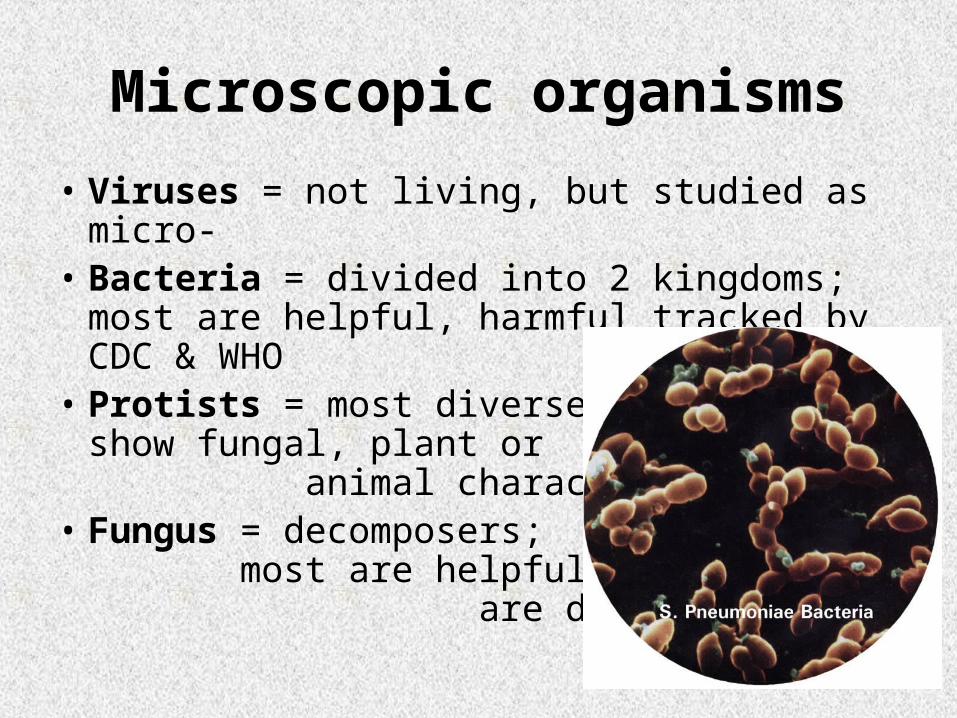
• Viruses = not living, but studied as micro-• Bacteria = divided into 2 kingdoms; most
are helpful, harmful tracked by CDC & WHO
• Protists = most diverse; show fungal, plant or animal characteristics
• Fungus = decomposers; most are helpful, some are disease-causing


(Eubacteria & Archaebacteria)

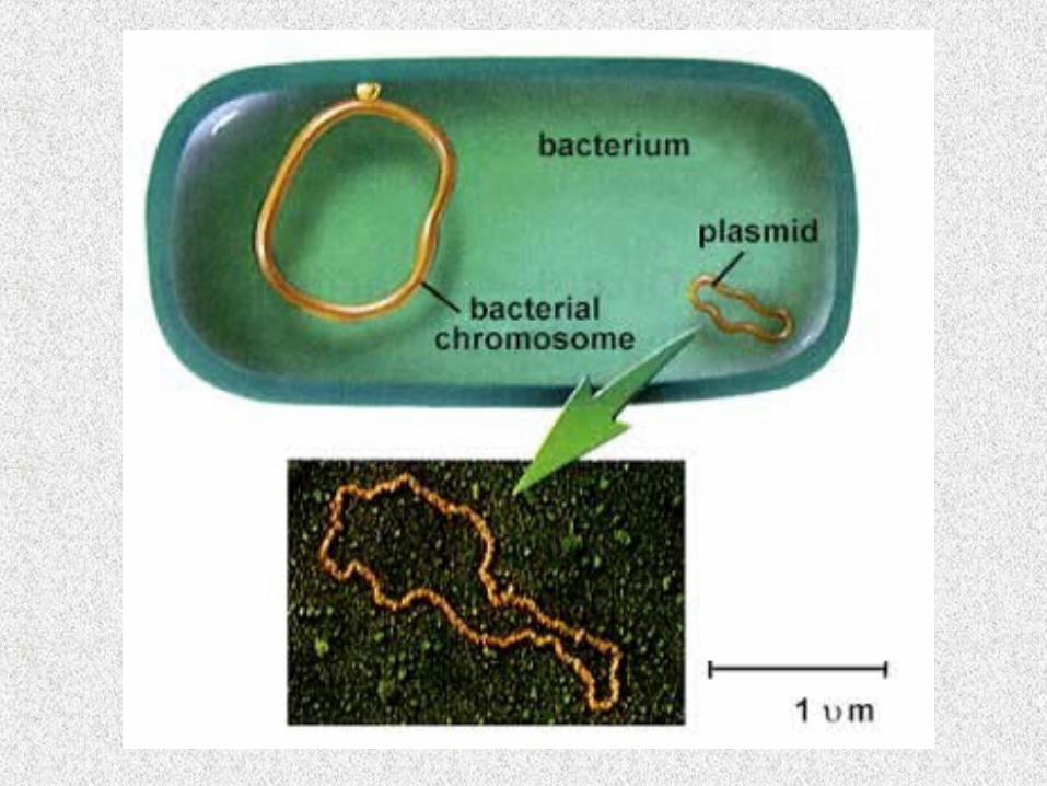
Bacterial characteristics:
• Prokaryotic (no nuclear membrane)– Ribosomes only– Pili = for attachment– Capsule = outermost layer for extra protection
• Unicellular (some colonial)• Varied metabolism & nutritional types• Often flagellated• May contain endospores…
to survive harsh conditions• Binary fission to reproduce
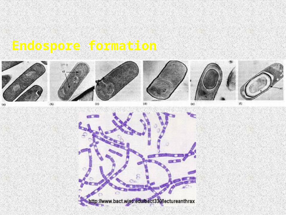
Endospore formation

See page 465


Practice

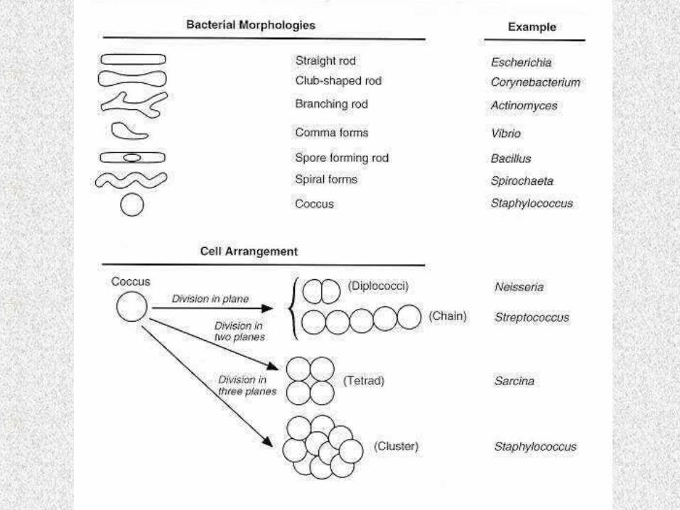
Bacterial shapes
• Bacillus = rod-shaped (ex: Lactobacillus)
• Coccus = sphere (ex: Streptococcus)
• Spirillum = coiled (ex: Spirochete)
• Strepto- (chains)
• Staphylo- (clusters)

Blue = causes Lyme disease Black = causes syphilis

Metabolic diversity• Obligate aerobes =
– Must have oxygen to grow normally
• Facultative aerobes =– Prefers oxygen, but not necessary
• Facultative anaerobes =– Prefers no oxygen, but not necessary
• Obligate anaerobes =– May not have oxygen to grow normally
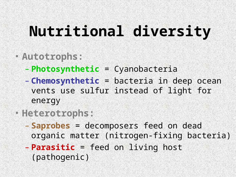
Nutritional diversity
• Autotrophs:– Photosynthetic = Cyanobacteria– Chemosynthetic = bacteria in deep ocean
vents use sulfur instead of light for energy
• Heterotrophs:– Saprobes = decomposers feed on dead
organic matter (nitrogen-fixing bacteria)– Parasitic = feed on living host (pathogenic)

Chemosynthetic bacteria in deep sea vents
Photosynthetic Cyanobacteria in fresh-water ponds or streams

Binary fission
• Cell Replication
• (cloning) for prokaryotic cell
• Much simpler than mitosis (like cytokinesis without the 4 other stages)
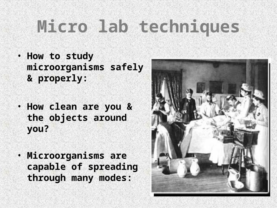
Micro lab techniques
• How to study microorganisms safely & properly:
• How clean are you & the objects around you?
• Microorganisms are capable of spreading through many modes:

1) Direct Contact:
2) Air:
3) Fomites: (inanimate objects)

Gram-staining:
• Bacteria cell walls differ from plant cell walls which are made of cellulose
• Bacteria cell walls are made of peptidoglycan
• Bacteria have either thick or thin peptidoglycan cell walls
• Bacteria are classified as Gram positive (thick) or negative (thin cell walls)

See page 463


Staining techniques:
Step 1 = smear thin layer of sample on slide.
Step 2 = flame quickly to fix (stick) to slide.
Step 3 = add Crystal Violet stain which adheres to peptidoglycan.
Step 4 = add iodine to fix Crystal Violet.
Step 5 = add alcohol to remove unfixed C.V.
Step 6 = add Safranin O (a pink stain) to stain cell membrane.
**Gram+ is purple and Gram- is pink.

negative
Related Documents