Understanding molecular recognition of promiscuity of thermophilic methionine adenosyltransferase sMAT from Sulfolobus solfataricus Fengbin Wang 1 , Shanteri Singh 2 , Jianjun Zhang 2 , Tyler D. Huber 2 , Kate E. Helmich 3 , Manjula Sunkara 4 , Katherine A. Hurley 3 , Randal D. Goff 5 , Craig A. Bingman 3 , Andrew J. Morris 4 , Jon S. Thorson 2 and George N. Phillips Jr 1,3 1 Department of Biochemistry and Cell Biology, Rice University, Houston, TX, USA 2 Center for Pharmaceutical Research and Innovation, College of Pharmacy, University of Kentucky, Lexington, KY, USA 3 Department of Biochemistry, University of Wisconsin, Madison, WI, USA 4 Division of Cardiovascular Medicine, Gill Heart Institute, University of Kentucky, Lexington, KY, USA 5 Western Wyoming Community College, Rock Springs, WY, USA Keywords enzyme engineering; methionine adenosyltransferase; natural product; S-adenosylmethionine; X-ray diffraction Correspondence G. N. Phillips Jr, George R. Brown Hall, W200Q, 6100 Main Street, Rice University, Houston, TX 77005, USA Fax: +(713) 348 5154 Tel: +(713) 348 6951 E-mail: [email protected] J. S. Thorson, University of Kentucky College of Pharmacy, 789 South Limestone Street, Lexington, KY 40536, USA Fax: +(859) 218 0140 Tel: +(859) 218 0140 E-mail: [email protected] (Received 15 January 2014, revised 6 March 2014, accepted 12 March 2014) doi:10.1111/febs.12784 Methionine adenosyltransferase (MAT) is a family of enzymes that utilizes ATP and methionine to produce S-adenosylmethionine (AdoMet), the most crucial methyl donor in the biological methylation of biomolecules and bioactive natural products. Here, we report that the MAT from Sulfol- obus solfataricus (sMAT), an enzyme from a poorly explored class of the MAT family, has the ability to produce a range of differentially alkylated AdoMet analogs in the presence of non-native methionine analogs and ATP. To investigate the molecular basis for AdoMet analog production, we have crystallized the sMAT in the AdoMet bound, S-adenosylethionine (AdoEth) bound and unbound forms. Notably, among these structures, the AdoEth bound form offers the first MAT structure containing a non-native product, and cumulatively these structures add new structural insight into the MAT family and allow for detailed active site comparison with its homologs in Escherichia coli and human. As a thermostable MAT struc- ture from archaea, the structures herein also provide a basis for future engineering to potentially broaden AdoMet analog production as reagents for methyltransferase-catalyzed ‘alkylrandomization’ and/or the study of methylation in the context of biological processes. Databases PDB IDs: 4HPV, 4L7I, 4K0B and 4L2Z. EC 2.5.1.6 Structured digital abstract sMAT and sMAT bind by x-ray crystallography ( View interaction) Abbreviations AdoEth, S-adenosylethionine; AdoMet, S-adenosylmethionine; eMAT, Escherichia coli methionine adenosyltransferase; hMAT1A, hMAT2A, human methionine adenosyltransferases; MAT, methionine adenosyltransferase; mjMAT, Methanococcus jannaschii methionine adenosyltransferase; PDB, Protein Data Bank; Pi, phosphate; PPi, diphosphate; rlMAT, rat liver methionine adenosyltransferase; sMAT, Sulfolobus solfataricus methionine adenosyltransferase. 4224 FEBS Journal 281 (2014) 4224–4239 ª 2014 FEBS

Welcome message from author
This document is posted to help you gain knowledge. Please leave a comment to let me know what you think about it! Share it to your friends and learn new things together.
Transcript
Understanding molecular recognition of promiscuity ofthermophilic methionine adenosyltransferase sMAT fromSulfolobus solfataricusFengbin Wang1, Shanteri Singh2, Jianjun Zhang2, Tyler D. Huber2, Kate E. Helmich3,Manjula Sunkara4, Katherine A. Hurley3, Randal D. Goff5, Craig A. Bingman3, Andrew J. Morris4,Jon S. Thorson2 and George N. Phillips Jr1,3
1 Department of Biochemistry and Cell Biology, Rice University, Houston, TX, USA
2 Center for Pharmaceutical Research and Innovation, College of Pharmacy, University of Kentucky, Lexington, KY, USA
3 Department of Biochemistry, University of Wisconsin, Madison, WI, USA
4 Division of Cardiovascular Medicine, Gill Heart Institute, University of Kentucky, Lexington, KY, USA
5 Western Wyoming Community College, Rock Springs, WY, USA
Keywords
enzyme engineering; methionine
adenosyltransferase; natural product;
S-adenosylmethionine; X-ray diffraction
Correspondence
G. N. Phillips Jr, George R. Brown Hall,
W200Q, 6100 Main Street, Rice University,
Houston, TX 77005, USA
Fax: +(713) 348 5154
Tel: +(713) 348 6951
E-mail: [email protected]
J. S. Thorson, University of Kentucky
College of Pharmacy, 789 South Limestone
Street, Lexington, KY 40536, USA
Fax: +(859) 218 0140
Tel: +(859) 218 0140
E-mail: [email protected]
(Received 15 January 2014, revised 6
March 2014, accepted 12 March 2014)
doi:10.1111/febs.12784
Methionine adenosyltransferase (MAT) is a family of enzymes that utilizes
ATP and methionine to produce S-adenosylmethionine (AdoMet), the
most crucial methyl donor in the biological methylation of biomolecules
and bioactive natural products. Here, we report that the MAT from Sulfol-
obus solfataricus (sMAT), an enzyme from a poorly explored class of the
MAT family, has the ability to produce a range of differentially alkylated
AdoMet analogs in the presence of non-native methionine analogs and
ATP. To investigate the molecular basis for AdoMet analog production,
we have crystallized the sMAT in the AdoMet bound, S-adenosylethionine
(AdoEth) bound and unbound forms. Notably, among these structures, the
AdoEth bound form offers the first MAT structure containing a non-native
product, and cumulatively these structures add new structural insight into
the MAT family and allow for detailed active site comparison with its
homologs in Escherichia coli and human. As a thermostable MAT struc-
ture from archaea, the structures herein also provide a basis for future
engineering to potentially broaden AdoMet analog production as reagents
for methyltransferase-catalyzed ‘alkylrandomization’ and/or the study of
methylation in the context of biological processes.
Databases
PDB IDs: 4HPV, 4L7I, 4K0B and 4L2Z.
EC 2.5.1.6
Structured digital abstract
� sMAT and sMAT bind by x-ray crystallography (View interaction)
Abbreviations
AdoEth, S-adenosylethionine; AdoMet, S-adenosylmethionine; eMAT, Escherichia coli methionine adenosyltransferase; hMAT1A, hMAT2A,
human methionine adenosyltransferases; MAT, methionine adenosyltransferase; mjMAT, Methanococcus jannaschii methionine
adenosyltransferase; PDB, Protein Data Bank; Pi, phosphate; PPi, diphosphate; rlMAT, rat liver methionine adenosyltransferase; sMAT,
Sulfolobus solfataricus methionine adenosyltransferase.
4224 FEBS Journal 281 (2014) 4224–4239 ª 2014 FEBS
Introduction
S-adenosylmethionine (AdoMet) is the most widely
used methyl donor in biological systems, and the only
known family of enzymes that synthesize AdoMet is
methionine adenosyltransferase (MAT), which utilizes
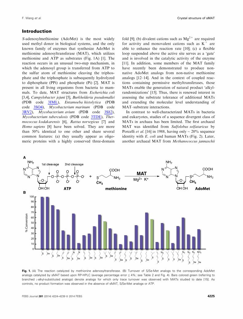
methionine and ATP as substrates (Fig. 1A) [1]. The
reaction occurs in an unusual two-step mechanism, in
which the adenosyl group is transferred from ATP to
the sulfur atom of methionine cleaving the triphos-
phate and the triphosphate is subsequently hydrolyzed
to diphosphate (PPi) and phosphate (Pi) [2]. MAT is
present in all living organisms from bacteria to mam-
mals. To date, MAT structures from Escherichia coli
[3,4], Campylobacter jejuni [5], Burkholderia pseudomallei
(PDB code 3IML), Entamoeba histolytica (PDB
code 3SO4), Mycobacterium marinum (PDB code
3RV2), Mycobacterium avium (PDB code 3S82),
Mycobacterium tuberculosis (PDB code 3TDE), Ther-
mococcus kodakarensis [6], Rattus norvegicus [7] and
Homo sapiens [8] have been solved. They are more
than 50% identical to one other and share several
common features: (a) they usually appear as oligo-
meric proteins with a highly conserved three-domain
fold [9]; (b) divalent cations such as Mg2+ are required
for activity and monovalent cations such as K+ are
able to enhance the reaction rate [10]; (c) a flexible
loop suspended above the active site serves as a ‘gate’
and is involved in the catalytic activity of the enzyme
[11]. In addition, some members of the MAT family
have recently been demonstrated to produce non-
native AdoMet analogs from non-native methionine
analogs [12–14]. And in the context of coupled reac-
tions containing permissive methyltransferases, those
MATs enable the generation of natural product ‘alkyl-
randomizations’ [15]. Thus, there is renewed interest in
assessing the substrate tolerance of additional MATs
and extending the molecular level understanding of
MAT–substrate interactions.
In contrast to well-characterized MATs in bacteria
and eukaryotes, studies of a sequence divergent class of
MATs in archaea has been limited. The first archaeal
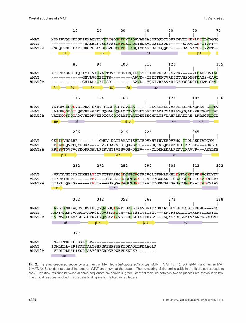
MAT was identified from Sulfolobus solfataricus by
Porcelli et al. [16] in 1988, having only ~ 20% sequence
identity with E. coli and human MATs (Fig. 2). Later,
another archaeal MAT from Methanococcus jannaschii
100
90
80
70
60
50
40
30
20
10
0
% C
onve
rsio
n
Se
NH2
Se
N3
S
N3
S
N3
SeSSS
N3
S
SSeS
SeSSSeS
SeSSeSSeSSe
N
S
N
SeSSeS
SeS
ATP methionine AdoMet
A
B
Fig. 1. (A) The reaction catalyzed by methionine adenosyltransferase. (B) Turnover of S/Se-Met analogs to the corresponding AdoMet
analogs catalyzed by sMAT based upon RP-HPLC (average percentage error ≤ 4%; see Table 2 and Fig. 4). Bars colored green (referring to
branched L-alkyl-substituted analogs) denote analogs for which only trace turnover was observed with MATs studied to date [15]. As
controls, no product formation was observed in the absence of sMAT, S/Se-Met analogs or ATP.
FEBS Journal 281 (2014) 4224–4239 ª 2014 FEBS 4225
F. Wang et al. Crystal structure of sMAT
β1 β2 β3
β4 β6 β7 β8
β9 β10
β11 β12
β13
β14
α1
α2
α3 α4 α5
α6
α7
α9
α10
β15
β5
α8
Fig. 2. The structure-based sequence alignment of MAT from Sulfolobus solfataricus (sMAT), MAT from E. coli (eMAT) and human MAT
(hMAT2A). Secondary structural features of sMAT are shown at the bottom. The numbering of the amino acids in the figure corresponds to
sMAT. Identical residues between all three sequences are shown in green; identical residues between two sequences are shown in yellow.
The critical residues involved in substrate binding are highlighted in red letters.
4226 FEBS Journal 281 (2014) 4224–4239 ª 2014 FEBS
Crystal structure of sMAT F. Wang et al.
(mjMAT) was characterized in detail in terms of
kinetics parameters, substrate specificity and folding
[12,17]. More recently, an apo crystal structure of
MAT from Thermococcus kodakarensis has been
reported, which provides new structural insights on
archaeal MAT [6]. The improved thermostability but
comparable kinetics parameters with MATs in bacte-
ria and eukaryotes show archaeal MATs to be of
great interest for enzyme engineering applications.
However, structural information of active site con-
tents and broad substrate specificity assessment for
archaeal MATs has been lacking. Here we report
Sulfolobus solfataricus MAT (sMAT) to enable the
cumulative synthesis of a broad panel of unnatural
AdoMet analogs (31 analogs detected) starting from
synthetic S/Se-alkylated Met analogs (42 analogs) or
commercial sources (three analogs). In addition, this
study highlights the crystal structures of a thermosta-
ble MAT (sMAT), in three different forms: AdoMet
bound, a non-native product S-adenosylethionine
(AdoEth) bound, and the unbound form. Interestingly,
in contrast to its low sequence similarity to other
MATs, sMAT displays the typical three-domain fold
and partly conserved active site architecture. Unlike
other known MAT structures, the activity of sMAT
cannot be stimulated by ionic potassium [16]. This
can be supported structurally by the presence of a
lysine side chain (K63) in sMAT, which probably has
a similar function to potassium ion in other MATs.
Further, the capture of the first atypical ligand bound
structure of MAT provides insights on the nature of
sMAT broad substrate specificity and a potential
template for future engineering toward expanding the
substrate scope. Cumulatively, the results in this
study provide the first atomic view of the poorly
explored class of MATs from archaea and expose
sMAT as an efficient catalyst for AdoMet analog
production that is amenable to downstream AdoMet-
utilizing processes.
Results and discussion
Overall structural organization
The crystal structures of sMAT have been determined
successfully at 2.19 �A or 2.39 �A resolution for the Ado-
Met-liganded form, 2.49 �A for the AdoEth-liganded
form and 2.21 �A for the unliganded form (Table 1).
Similar to E. coli MAT (eMAT) [18], rat liver MAT
(rlMAT) [19] and human MATs (hMAT1A and
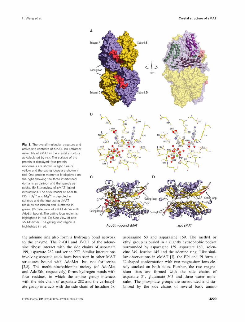
hMAT2A) [8], sMAT packs as a tetramer (Fig. 3A). All
four sMAT structures contain two subunits, A and B,
in the asymmetric unit and the tetramer is formed by a
two-fold crystallographic symmetry axis. The buried
surface interfaces between the two subunits A and B
and the two dimers AB and CD are calculated to be
2570 and 1870 �A2 respectively [20]. These areas are sim-
ilar to those from other bacterial MATs [3–5], but largerthan rlMAT [7] and slightly smaller than hMAT [8]. In
the tetramer, there are four potential ligand binding
sites: two sites sandwiched by A and B and the other
two sites between C and D. Compared with other
known MAT structures [9], the sMAT monomer adopts
a similar three-domain architecture with secondary
structure variants (Fig. 3A). Interestingly, unlike other
MATs, structural variations were observed between
subunits A and B in sMAT with an average rmsd of
0.51 �A in both the liganded and unliganded structures.
In addition, the maximum rmsd between all the A
subunits and the maximum rmsd between all the B
subunits in all sMAT structures are calculated as 0.29 �A
and 0.21 �A, respectively. A similar observation was also
reported in a recently solved archaeal MAT structure
[6]. As a result, half of the active sites within sMAT
have a more open conformation than the others.
Consistently, in all the ligand bound sMAT structures,
only half of the active sites within the sMAT tetramer
are occupied while the other half are unoccupied. In
addition, only the gating loops outside the occupied
active site become ordered (Fig. 3C,D).
The substrate specificity for sMAT based upon RP-
HPLC is illustrated in Fig. 1B wherein observed 50
methyl-thio(seleno)-50-deoxyadenosine (MSeA) produc-
tion (via RP-HPLC, Fig. 4) was interpreted as product
based upon the well-established AdoMet decay path-
ways indicating MSeA to directly derive from AdoMet
(not ATP) [21–23]. The putative substrates tested were
those recently reported to profile the substrate specific-
ity of a range of MATs and these analogs were specifi-
cally designed to interrogate both steric and electronic
contributions to turnover [15]. Of the 45 putative sub-
strates (Table 2) tested with sMAT, 11 led to apprecia-
ble (> 50%) AdoMet analog production, an additional
15 led to moderate (25%–50%) conversion, while five
offered detectable product (< 25%) under the condi-
tions described. In general, smaller alkyl substitutions
were better tolerated, suggesting steric infringement to
possibly prohibit larger substitutions. Interestingly, in
the case where direct comparisons could be made, the
degree of unsaturation correlated with a reduction in
turnover (e.g. propyl > allyl > propargyl). Importantly,
notable turnover was observed with branched analogs
(Fig. 1B, highlighted in green) that previously led to
only trace product with MATs studied to date.
Table 3 highlights a comparison of the kinetic param-
eters for L-methionine and the non-native substrate for
FEBS Journal 281 (2014) 4224–4239 ª 2014 FEBS 4227
F. Wang et al. Crystal structure of sMAT
which a ligand bound structure is available (L-ethio-
nine). The changes in kinetic parameters of the sMAT
for both the substrates are moderate from 37 °C to
65 °C. Compared with Thermococcus kodakarensis
MAT and mjMAT, sMAT appears to be a somewhat
better enzyme because sMAT has a 100 times smaller
Km for methionine, a slightly smaller Km for ATP, and a
similar kcat. More importantly, the data reflect that
L-ethionine is kinetically competent and comparable to
the native substrate L-methionine. At either tempera-
ture, the kcat values for the sMAT reaction with
L-methionine or L-ethionine are similar and the reduced
proficiency with L-ethionine compared with the native
substrate L-methionine derives from a combination of
higher Km values for both L-ethionine and ATP.
Active site contents
The MAT catalyzed AdoMet formation, as mentioned
in the Introduction, occurs via a sequential two-step
mechanism. In the first step, AdoMet is formed by a
direct SN2 reaction, in which the sulfur atom of methi-
onine attacks the C50 position of ATP and thus cleaves
the polyphosphate chain from ATP. In the second
step, the triphosphate is further hydrolyzed to PPi and
Pi [9]. Komoto et al. [3] identified two critical residues,
lysine 165 and histidine 14, in eMAT for this proposed
SN2 reaction based on their ligand bound structures.
Interestingly, even with significant sequential variations
to eMAT and other MATs, several conserved residues
were observed in sMAT, mainly located around the
active site, including the two crucial residues lysine
(K201) and histidine (H29) for the proposed SN2 reac-
tion (Table 4).
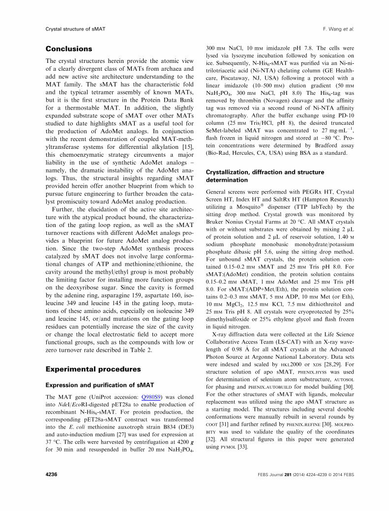
The interactions between sMAT and products in
the active site are multifaceted as illustrated in
Fig. 3B. The adenine ring of AdoMet is recognized
by a hydrogen bond with the side chain of aspartate
144 and a stacking interaction with the aromatic ring
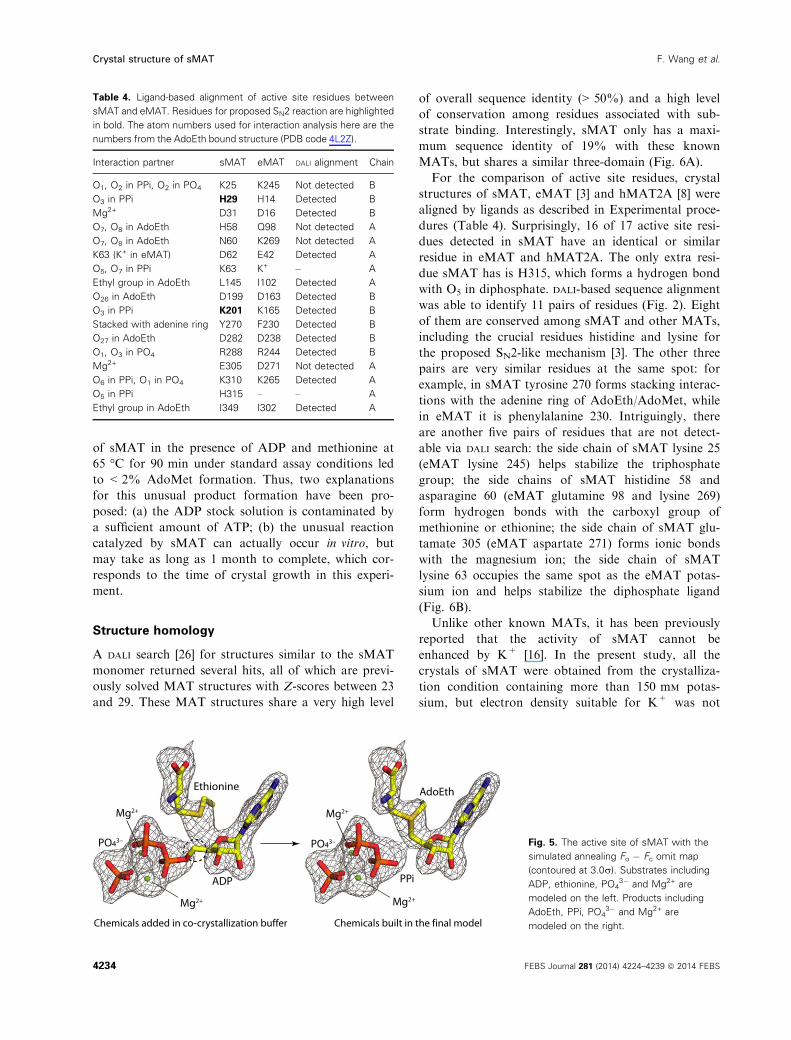
of tyrosine 270. Several water molecules surrounding
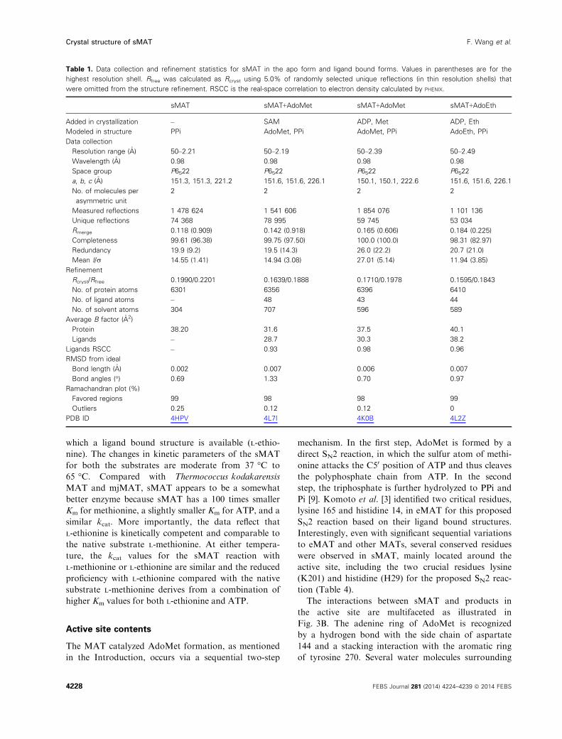
Table 1. Data collection and refinement statistics for sMAT in the apo form and ligand bound forms. Values in parentheses are for the
highest resolution shell. Rfree was calculated as Rcryst using 5.0% of randomly selected unique reflections (in thin resolution shells) that
were omitted from the structure refinement. RSCC is the real-space correlation to electron density calculated by PHENIX.
sMAT sMAT+AdoMet sMAT+AdoMet sMAT+AdoEth
Added in crystallization – SAM ADP, Met ADP, Eth
Modeled in structure PPi AdoMet, PPi AdoMet, PPi AdoEth, PPi
Data collection
Resolution range (�A) 50–2.21 50–2.19 50–2.39 50–2.49
Wavelength (�A) 0.98 0.98 0.98 0.98
Space group P6522 P6522 P6522 P6522
a, b, c (�A) 151.3, 151.3, 221.2 151.6, 151.6, 226.1 150.1, 150.1, 222.6 151.6, 151.6, 226.1
No. of molecules per
asymmetric unit
2 2 2 2
Measured reflections 1 478 624 1 541 606 1 854 076 1 101 136
Unique reflections 74 368 78 995 59 745 53 034
Rmerge 0.118 (0.909) 0.142 (0.918) 0.165 (0.606) 0.184 (0.225)
Completeness 99.61 (96.38) 99.75 (97.50) 100.0 (100.0) 98.31 (82.97)
Redundancy 19.9 (9.2) 19.5 (14.3) 26.0 (22.2) 20.7 (21.0)
Mean I/r 14.55 (1.41) 14.94 (3.08) 27.01 (5.14) 11.94 (3.85)
Refinement
Rcryst/Rfree 0.1990/0.2201 0.1639/0.1888 0.1710/0.1978 0.1595/0.1843
No. of protein atoms 6301 6356 6396 6410
No. of ligand atoms – 48 43 44
No. of solvent atoms 304 707 596 589
Average B factor (�A2)
Protein 38.20 31.6 37.5 40.1
Ligands – 28.7 30.3 38.2
Ligands RSCC – 0.93 0.98 0.96
RMSD from ideal
Bond length (�A) 0.002 0.007 0.006 0.007
Bond angles (°) 0.69 1.33 0.70 0.97
Ramachandran plot (%)
Favored regions 99 98 98 99
Outliers 0.25 0.12 0.12 0
PDB ID 4HPV 4L7I 4K0B 4L2Z
4228 FEBS Journal 281 (2014) 4224–4239 ª 2014 FEBS
Crystal structure of sMAT F. Wang et al.
the adenine ring also form a hydrogen bond network
to the enzyme. The 20-OH and 30-OH of the adeno-
sine ribose interact with the side chains of aspartate
199, aspartate 282 and serine 277. Similar interactions
involving aspartic acids have been seen in other MAT
structures bound with AdoMet, but not for serine
[3,8]. The methionine/ethionine moiety (of AdoMet
and AdoEth, respectively) forms hydrogen bonds with
four residues, in which the amino group interacts
with the side chain of aspartate 282 and the carboxyl-
ate group interacts with the side chain of histidine 58,
asparagine 60 and asparagine 159. The methyl or
ethyl group is buried in a slightly hydrophobic pocket
surrounded by asparagine 159, aspartate 160, isoleu-
cine 349, leucine 145 and the adenine ring. Like simi-
lar observations in eMAT [3], the PPi and Pi form a
U-shaped conformation with two magnesium ions clo-
sely stacked on both sides. Further, the two magne-
sium sites are formed with the side chains of
aspartate 31, glutamate 305 and three water mole-
cules. The phosphate groups are surrounded and sta-
bilized by the side chains of several basic amino
A
90o
C D
apo sMATAdoEth-bound sMAT
Gating loop
Gating loop Gating loop
Gating loop
Subunit A Subunit B
Subunit C Subunit D
Gating loop
B
Fig. 3. The overall molecular structure and
active site contents of sMAT. (A) Tetramer
assembly of sMAT in the crystal structure
as calculated by PISA. The surface of the
protein is displayed: four protein
monomers are shown in light blue or
yellow and the gating loops are shown in
red. One protein monomer is displayed on
the right showing the three intertwined
domains as cartoon and the ligands as
sticks. (B) Stereoview of sMAT–ligand
interactions. The stick model of AdoEth,
PPi, PO43� and Mg2+ is depicted in
spheres and the interacting sMAT
residues are labeled and illustrated in
green. (C) Side view of sMAT dimer with
AdoEth bound. The gating loop region is
highlighted in red. (D) Side view of apo
sMAT dimer. The gating loop region is
highlighted in red.
FEBS Journal 281 (2014) 4224–4239 ª 2014 FEBS 4229
F. Wang et al. Crystal structure of sMAT
acids: lysine 25, histidine 29, lysine 201, arginine 288
and lysine 310.
As described, the ligands are solvent inaccessible
and thus the entrance of the active site requires a
dynamic and flexible region. A flexible loop region was
previously identified as the gate for the active site in
MATs [24]. This gate loop feature has been confirmed
by crystal structures of eMAT and hMAT2A, in which
the loop becomes ordered when ligands are bound and
it becomes disordered when the active site is empty
[3,8]. A similar gating loop (residues 141–155) region
was identified in sMAT (Fig. 3), which has a similar
pose to eMAT and hMAT2A and interacts with the
adenine ring and the methionine or ethionine moiety.
When the active site is empty, residues 141–144 form a
b-sheet with residues 95–96 while the rest of the loop
is poorly seen or unidentifiable in electron density.
When ligands are bound in the active site, the loop
region becomes ordered and is anchored above the
active site with residues 141–144 shifting from the ori-
gin position and with residues 145–149 forming a small
a-helix.In various structures of eMAT [3] and hMAT2A [8],
the active site ligand occupation is correlated with an
ordered active site gating loop. These studies are consis-
tent with our observations in sMAT structures. In addi-
tion to this, ligand orientations in the eMAT and
human MAT studies are similar to sMAT’s. Different
ligand orientations with an unordered gating loop have
been reported before in rlMAT structures [7] and an
earlier set of eMAT structures [4], but have some unu-
sual features: (a) in rlMAT the temperature factors of
most of the ligand coordinates are above 70 �A2, and in
earlier eMAT structures the temperature factors of
ADP (or ADP mimic) are above 114 �A2; (b) the flexible
loop above the active site was undefined even though
the active sites were reported to be occupied [25]; (c)
even though rlMAT and eMAT have a 59% sequence
identity and all critical residues are conserved, they
show completely different ligand orientations in the
active site; (d) X-ray data for rlMAT and earlier eMAT
structures are not available online, and thus ligand real-
space correlation coefficients cannot be calculated.
These unusual features suggest that the active site con-
tents of rlMAT and earlier eMAT are questionable. In
contrast to rlMAT and the earlier eMAT structures, all
the hMAT2A, later eMAT and sMAT structures have
reasonable temperature factors and a good ligand real-
space correlation to electron density at the active site.
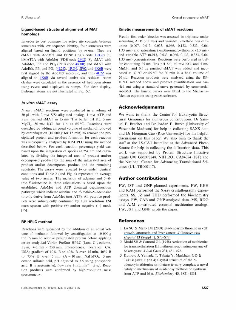
Unusual product formation during crystallization
As described in Experimental procedures, two sets of
ligand bound crystals were obtained in the presence of
Fig. 4. HPLC traces for representative
sMAT reactions illustrating the production
of AdoMet analogs and/or 50-methyl-thio
(seleno)-50-deoxyadenosine (MSeA) in the
presence of a select set of L-Met analogs.
Starting material (ATP) is designated by a
dot.
4230 FEBS Journal 281 (2014) 4224–4239 ª 2014 FEBS
Crystal structure of sMAT F. Wang et al.
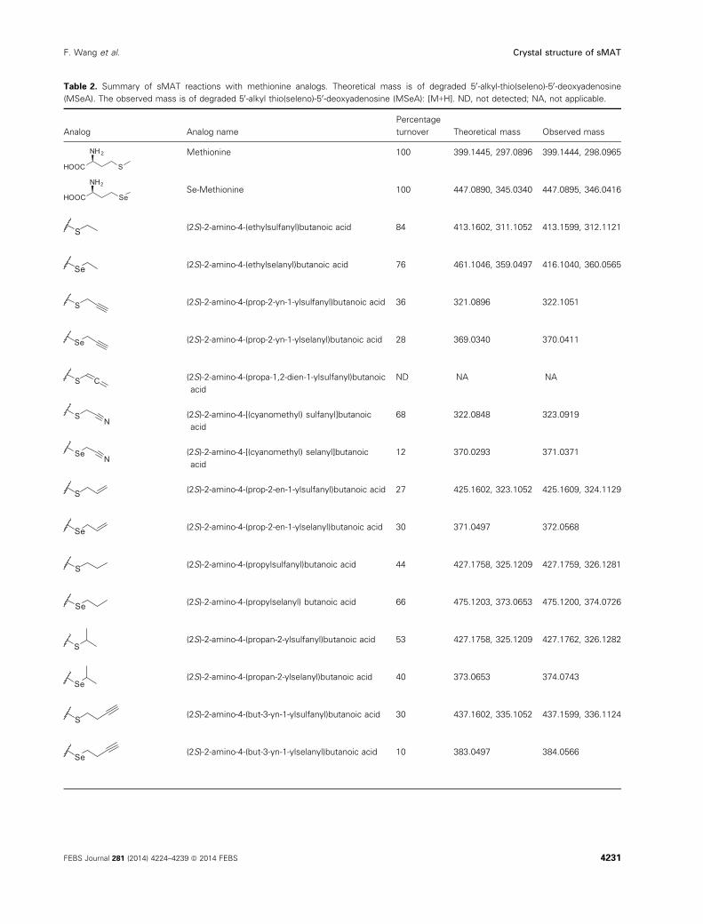
Table 2. Summary of sMAT reactions with methionine analogs. Theoretical mass is of degraded 50-alkyl-thio(seleno)-50-deoxyadenosine(MSeA). The observed mass is of degraded 50-alkyl thio(seleno)-50-deoxyadenosine (MSeA): [M+H]. ND, not detected; NA, not applicable.
Analog Analog name
Percentage
turnover Theoretical mass Observed mass
HOOC S
NH2 Methionine 100 399.1445, 297.0896 399.1444, 298.0965
HOOC Se
NH2Se-Methionine 100 447.0890, 345.0340 447.0895, 346.0416
S (2S)-2-amino-4-(ethylsulfanyl)butanoic acid 84 413.1602, 311.1052 413.1599, 312.1121
Se (2S)-2-amino-4-(ethylselanyl)butanoic acid 76 461.1046, 359.0497 416.1040, 360.0565
S (2S)-2-amino-4-(prop-2-yn-1-ylsulfanyl)butanoic acid 36 321.0896 322.1051
Se (2S)-2-amino-4-(prop-2-yn-1-ylselanyl)butanoic acid 28 369.0340 370.0411
S C (2S)-2-amino-4-(propa-1,2-dien-1-ylsulfanyl)butanoic
acid
ND NA NA
SN
(2S)-2-amino-4-[(cyanomethyl) sulfanyl]butanoic
acid
68 322.0848 323.0919
SeN
(2S)-2-amino-4-[(cyanomethyl) selanyl]butanoic
acid
12 370.0293 371.0371
S (2S)-2-amino-4-(prop-2-en-1-ylsulfanyl)butanoic acid 27 425.1602, 323.1052 425.1609, 324.1129
Se (2S)-2-amino-4-(prop-2-en-1-ylselanyl)butanoic acid 30 371.0497 372.0568
S (2S)-2-amino-4-(propylsulfanyl)butanoic acid 44 427.1758, 325.1209 427.1759, 326.1281
Se (2S)-2-amino-4-(propylselanyl) butanoic acid 66 475.1203, 373.0653 475.1200, 374.0726
S(2S)-2-amino-4-(propan-2-ylsulfanyl)butanoic acid 53 427.1758, 325.1209 427.1762, 326.1282
Se(2S)-2-amino-4-(propan-2-ylselanyl)butanoic acid 40 373.0653 374.0743
S (2S)-2-amino-4-(but-3-yn-1-ylsulfanyl)butanoic acid 30 437.1602, 335.1052 437.1599, 336.1124
Se (2S)-2-amino-4-(but-3-yn-1-ylselanyl)butanoic acid 10 383.0497 384.0566
FEBS Journal 281 (2014) 4224–4239 ª 2014 FEBS 4231
F. Wang et al. Crystal structure of sMAT
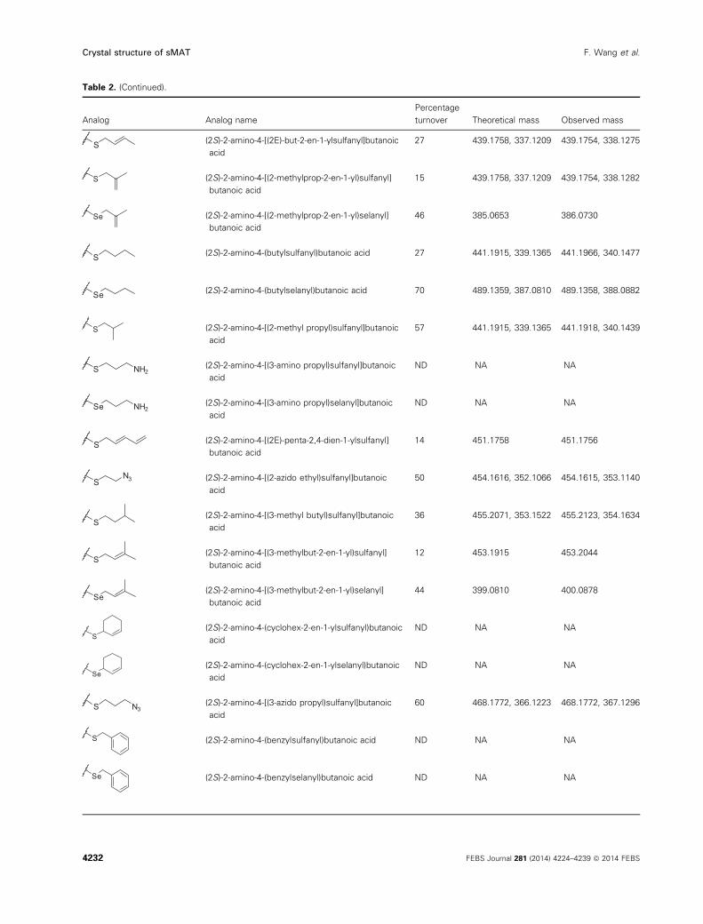
Table 2. (Continued).
Analog Analog name
Percentage
turnover Theoretical mass Observed mass
S (2S)-2-amino-4-[(2E)-but-2-en-1-ylsulfanyl]butanoic
acid
27 439.1758, 337.1209 439.1754, 338.1275
S (2S)-2-amino-4-[(2-methylprop-2-en-1-yl)sulfanyl]
butanoic acid
15 439.1758, 337.1209 439.1754, 338.1282
Se (2S)-2-amino-4-[(2-methylprop-2-en-1-yl)selanyl]
butanoic acid
46 385.0653 386.0730
S (2S)-2-amino-4-(butylsulfanyl)butanoic acid 27 441.1915, 339.1365 441.1966, 340.1477
Se (2S)-2-amino-4-(butylselanyl)butanoic acid 70 489.1359, 387.0810 489.1358, 388.0882
S (2S)-2-amino-4-[(2-methyl propyl)sulfanyl]butanoic
acid
57 441.1915, 339.1365 441.1918, 340.1439
S NH2 (2S)-2-amino-4-[(3-amino propyl)sulfanyl]butanoic
acid
ND NA NA
Se NH2 (2S)-2-amino-4-[(3-amino propyl)selanyl]butanoic
acid
ND NA NA
S (2S)-2-amino-4-[(2E)-penta-2,4-dien-1-ylsulfanyl]
butanoic acid
14 451.1758 451.1756
SN3 (2S)-2-amino-4-[(2-azido ethyl)sulfanyl]butanoic
acid
50 454.1616, 352.1066 454.1615, 353.1140
S(2S)-2-amino-4-[(3-methyl butyl)sulfanyl]butanoic
acid
36 455.2071, 353.1522 455.2123, 354.1634
S(2S)-2-amino-4-[(3-methylbut-2-en-1-yl)sulfanyl]
butanoic acid
12 453.1915 453.2044
Se(2S)-2-amino-4-[(3-methylbut-2-en-1-yl)selanyl]
butanoic acid
44 399.0810 400.0878
S(2S)-2-amino-4-(cyclohex-2-en-1-ylsulfanyl)butanoic
acid
ND NA NA
Se(2S)-2-amino-4-(cyclohex-2-en-1-ylselanyl)butanoic
acid
ND NA NA
S N3(2S)-2-amino-4-[(3-azido propyl)sulfanyl]butanoic
acid
60 468.1772, 366.1223 468.1772, 367.1296
S (2S)-2-amino-4-(benzylsulfanyl)butanoic acid ND NA NA
Se (2S)-2-amino-4-(benzylselanyl)butanoic acid ND NA NA
4232 FEBS Journal 281 (2014) 4224–4239 ª 2014 FEBS
Crystal structure of sMAT F. Wang et al.
5 mM ADP, 10 mM ethionine (or methionine), 10 mM
Mg2+ ion and 1.4 M NaHPO4/K2HPO4. Thus based
on the simulated annealing Fo � Fc omit map of the
active site, one ADP, one PO4, two Mg2+ and one
ethionine (or methionine) molecule were initially built
in (Fig. 5, left). However, this model does not fit the
electron density perfectly, because the Fo � Fc omit
map does not agree with the placement of the crucial
carbon atom circled in Fig. 5. Thus, it is very clear
that the product has already formed and a model
including PPi and AdoEth (or AdoMet) is more
appropriate. The new model (Fig. 5, right) has a lower
temperature factor and a better real-space correlation
to electron density in the active site. MAT-catalyzed
AdoMet/AdoEth formation via ADP and Met/Eth has
not been previously observed. In addition, incubation
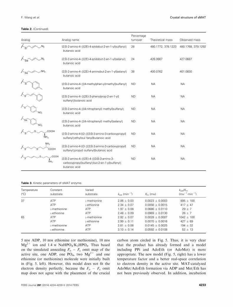
Table 2. (Continued).
Analog Analog name
Percentage
turnover Theoretical mass Observed mass
SN3 (2S)-2-amino-4-{[(2E)-4-azidobut-2-en-1-yl]sulfanyl}
butanoic acid
28 480.1772, 378.1223 480.1768, 379.1292
SeN3 (2S)-2-amino-4-{[(2E)-4-azidobut-2-en-1-yl]selanyl}
butanoic acid
24 426.0667 427.0687
SeNH2 (2S)-2-amino-4-{[(2E)-4-aminobut-2-en-1-yl]selanyl}
butanoic acid
38 400.0762 401.0830
S (2S)-2-amino-4-{[(4-methylphen-yl)methyl]sulfanyl}
butanoic acid
ND NA NA
S (2S)-2-amino-4-{[(2E)-3-phenylprop-2-en-1-yl]
sulfanyl}butanoic acid
ND NA NA
S
NO2
(2S)-2-amino-4-{[(4-nitrophenyl) methyl]sulfanyl}
butanoic acid
ND NA NA
Se
NO2
(2S)-2-amino-4-{[(4-nitrophenyl) methyl]selanyl}
butanoic acid
ND NA NA
SS COOH
NH2(2S)-2-amino-4-[(2-{[(3S)-3-amino-3-carboxypropyl]
sulfanyl}ethyl)sul fanyl]butanoic acid
ND NA NA
S S COOH
NH2(2S)-2-amino-4-[(3-{[(3S)-3-amino-3-carboxypropyl]
sulfanyl}propyl) sulfanyl]butanoic acid
ND NA NA
SS COOH
NH2(2S)-2-amino-4-{[(2E)-4-{[(3S)-3-amino-3-
carboxypropyl]sulfanyl}but-2-en-1-yl]sulfanyl}
butanoic acid
ND NA NA
Table 3. Kinetic parameters of sMAT enzyme.
Temperature
(°C)
Constant
substrate
Varied
substrate kcat (min�1) Km (mM)
kcat/Km
(mM�1�min�1)
37 ATP L-methionine 2.06 � 0.03 0.0023 � 0.0003 895 � 100
ATP L-ethionine 2.34 � 0.07 0.0056 � 0.0015 417 � 47
L-methionine ATP 1.97 � 0.08 0.0686 � 0.0110 29 � 7
L-ethionine ATP 2.48 � 0.09 0.0969 � 0.0130 26 � 7
65 ATP L-methionine 2.92 � 0.07 0.0028 � 0.0007 1042 � 100
ATP L-ethionine 2.99 � 0.11 0.0070 � 0.0016 427 � 69
L-methionine ATP 2.81 � 0.08 0.0145 � 0.0025 194 � 32
L-ethionine ATP 3.10 � 0.14 0.0592 � 0.0108 52 � 13
FEBS Journal 281 (2014) 4224–4239 ª 2014 FEBS 4233
F. Wang et al. Crystal structure of sMAT
of sMAT in the presence of ADP and methionine at
65 °C for 90 min under standard assay conditions led
to < 2% AdoMet formation. Thus, two explanations
for this unusual product formation have been pro-
posed: (a) the ADP stock solution is contaminated by
a sufficient amount of ATP; (b) the unusual reaction
catalyzed by sMAT can actually occur in vitro, but
may take as long as 1 month to complete, which cor-
responds to the time of crystal growth in this experi-
ment.
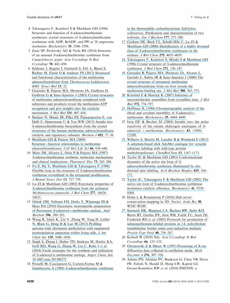
Structure homology
A DALI search [26] for structures similar to the sMAT
monomer returned several hits, all of which are previ-
ously solved MAT structures with Z-scores between 23
and 29. These MAT structures share a very high level
of overall sequence identity (> 50%) and a high level
of conservation among residues associated with sub-
strate binding. Interestingly, sMAT only has a maxi-
mum sequence identity of 19% with these known
MATs, but shares a similar three-domain (Fig. 6A).
For the comparison of active site residues, crystal
structures of sMAT, eMAT [3] and hMAT2A [8] were
aligned by ligands as described in Experimental proce-
dures (Table 4). Surprisingly, 16 of 17 active site resi-
dues detected in sMAT have an identical or similar
residue in eMAT and hMAT2A. The only extra resi-
due sMAT has is H315, which forms a hydrogen bond
with O5 in diphosphate. DALI-based sequence alignment
was able to identify 11 pairs of residues (Fig. 2). Eight
of them are conserved among sMAT and other MATs,
including the crucial residues histidine and lysine for
the proposed SN2-like mechanism [3]. The other three
pairs are very similar residues at the same spot: for
example, in sMAT tyrosine 270 forms stacking interac-
tions with the adenine ring of AdoEth/AdoMet, while
in eMAT it is phenylalanine 230. Intriguingly, there
are another five pairs of residues that are not detect-
able via DALI search: the side chain of sMAT lysine 25
(eMAT lysine 245) helps stabilize the triphosphate
group; the side chains of sMAT histidine 58 and
asparagine 60 (eMAT glutamine 98 and lysine 269)
form hydrogen bonds with the carboxyl group of
methionine or ethionine; the side chain of sMAT glu-
tamate 305 (eMAT aspartate 271) forms ionic bonds
with the magnesium ion; the side chain of sMAT
lysine 63 occupies the same spot as the eMAT potas-
sium ion and helps stabilize the diphosphate ligand
(Fig. 6B).
Unlike other known MATs, it has been previously
reported that the activity of sMAT cannot be
enhanced by K+ [16]. In the present study, all the
crystals of sMAT were obtained from the crystalliza-
tion condition containing more than 150 mM potas-
sium, but electron density suitable for K+ was not
Table 4. Ligand-based alignment of active site residues between
sMAT and eMAT. Residues for proposed SN2 reaction are highlighted
in bold. The atom numbers used for interaction analysis here are the
numbers from the AdoEth bound structure (PDB code 4L2Z).
Interaction partner sMAT eMAT DALI alignment Chain
O1, O2 in PPi, O2 in PO4 K25 K245 Not detected B
O3 in PPi H29 H14 Detected B
Mg2+ D31 D16 Detected B
O7, O8 in AdoEth H58 Q98 Not detected A
O7, O8 in AdoEth N60 K269 Not detected A
K63 (K+ in eMAT) D62 E42 Detected A
O5, O7 in PPi K63 K+ – A
Ethyl group in AdoEth L145 I102 Detected A
O26 in AdoEth D199 D163 Detected B
O3 in PPi K201 K165 Detected B
Stacked with adenine ring Y270 F230 Detected B
O27 in AdoEth D282 D238 Detected B
O1, O3 in PO4 R288 R244 Detected B
Mg2+ E305 D271 Not detected A
O6 in PPi, O1 in PO4 K310 K265 Detected A
O5 in PPi H315 – – A
Ethyl group in AdoEth I349 I302 Detected A
AdoEth
ADP
Chemicals added in co-crystallization buffer Chemicals built in the final model
Ethionine
PPi
PO43–PO43–
Mg2+
Mg2+
Mg2+
Mg2+
Fig. 5. The active site of sMAT with the
simulated annealing Fo � Fc omit map
(contoured at 3.0r). Substrates including
ADP, ethionine, PO43� and Mg2+ are
modeled on the left. Products including
AdoEth, PPi, PO43� and Mg2+ are
modeled on the right.
4234 FEBS Journal 281 (2014) 4224–4239 ª 2014 FEBS
Crystal structure of sMAT F. Wang et al.
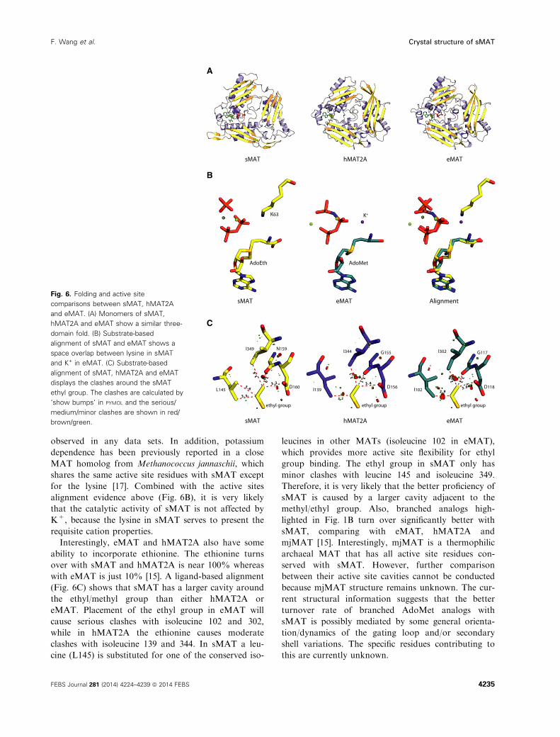
observed in any data sets. In addition, potassium
dependence has been previously reported in a close
MAT homolog from Methanococcus jannaschii, which
shares the same active site residues with sMAT except
for the lysine [17]. Combined with the active sites
alignment evidence above (Fig. 6B), it is very likely
that the catalytic activity of sMAT is not affected by
K+, because the lysine in sMAT serves to present the
requisite cation properties.
Interestingly, eMAT and hMAT2A also have some
ability to incorporate ethionine. The ethionine turns
over with sMAT and hMAT2A is near 100% whereas
with eMAT is just 10% [15]. A ligand-based alignment
(Fig. 6C) shows that sMAT has a larger cavity around
the ethyl/methyl group than either hMAT2A or
eMAT. Placement of the ethyl group in eMAT will
cause serious clashes with isoleucine 102 and 302,
while in hMAT2A the ethionine causes moderate
clashes with isoleucine 139 and 344. In sMAT a leu-
cine (L145) is substituted for one of the conserved iso-
leucines in other MATs (isoleucine 102 in eMAT),
which provides more active site flexibility for ethyl
group binding. The ethyl group in sMAT only has
minor clashes with leucine 145 and isoleucine 349.
Therefore, it is very likely that the better proficiency of
sMAT is caused by a larger cavity adjacent to the
methyl/ethyl group. Also, branched analogs high-
lighted in Fig. 1B turn over significantly better with
sMAT, comparing with eMAT, hMAT2A and
mjMAT [15]. Interestingly, mjMAT is a thermophilic
archaeal MAT that has all active site residues con-
served with sMAT. However, further comparison
between their active site cavities cannot be conducted
because mjMAT structure remains unknown. The cur-
rent structural information suggests that the better
turnover rate of branched AdoMet analogs with
sMAT is possibly mediated by some general orienta-
tion/dynamics of the gating loop and/or secondary
shell variations. The specific residues contributing to
this are currently unknown.
B
C
eMATsMAT Alignment
K63 K+
AdoEth
L145
AdoMet
sMAT eMAThMAT2A
I349 N159
D160 D156 D118
G155 G117I344
I139 I102
I302
3.5
3.6
3.8
2.9
3 .2
3 .2
3.1 3.1
3 .4
2.9
2.9 3
.2
3 .0
ethyl group ethyl group ethyl group
A
sMAT eMAThMAT2A
Fig. 6. Folding and active site
comparisons between sMAT, hMAT2A
and eMAT. (A) Monomers of sMAT,
hMAT2A and eMAT show a similar three-
domain fold. (B) Substrate-based
alignment of sMAT and eMAT shows a
space overlap between lysine in sMAT
and K+ in eMAT. (C) Substrate-based
alignment of sMAT, hMAT2A and eMAT
displays the clashes around the sMAT
ethyl group. The clashes are calculated by
‘show bumps’ in PYMOL and the serious/
medium/minor clashes are shown in red/
brown/green.
FEBS Journal 281 (2014) 4224–4239 ª 2014 FEBS 4235
F. Wang et al. Crystal structure of sMAT
Conclusions
The crystal structures herein provide the atomic view
of a clearly divergent class of MATs from archaea and
add new active site architecture understanding to the
MAT family. The sMAT has the characteristic fold
and the typical tetramer assembly of known MATs,
but it is the first structure in the Protein Data Bank
for a thermostable MAT. In addition, the slightly
expanded substrate scope of sMAT over other MATs
studied to date highlights sMAT as a useful tool for
the production of AdoMet analogs. In conjunction
with the recent demonstration of coupled MAT-meth-
yltransferase systems for differential alkylation [15],
this chemoenzymatic strategy circumvents a major
liability in the use of synthetic AdoMet analogs –namely, the dramatic instability of the AdoMet ana-
logs. Thus, the structural insights regarding sMAT
provided herein offer another blueprint from which to
pursue future engineering to further broaden the cata-
lyst promiscuity toward AdoMet analog production.
Further, the elucidation of the active site architec-
ture with the atypical product bound, the characteriza-
tion of the gating loop region, as well as the sMAT
turnover reactions with different AdoMet analogs pro-
vides a blueprint for future AdoMet analog produc-
tion. Since the two-step AdoMet synthesis process
catalyzed by sMAT does not involve large conforma-
tional changes of ATP and methionine/ethionine, the
cavity around the methyl/ethyl group is most probably
the limiting factor for installing more function groups
on the deoxyribose sugar. Since the cavity is formed
by the adenine ring, asparagine 159, aspartate 160, iso-
leucine 349 and leucine 145 in the gating loop, muta-
tions of these amino acids, especially on isoleucine 349
and leucine 145, or/and mutations on the gating loop
residues can potentially increase the size of the cavity
or change the local electrostatic field to accept more
functional groups, such as the compounds with low or
zero turnover rate described in Table 2.
Experimental procedures
Expression and purification of sMAT
The MAT gene (UniProt accession: Q980S9) was cloned
into NdeI/EcoRI-digested pET28a to enable production of
recombinant N-His6-sMAT. For protein production, the
corresponding pET28a-sMAT construct was transformed
into the E. coli methionine auxotroph strain B834 (DE3)
and auto-induction medium [27] was used for expression at
37 °C. The cells were harvested by centrifugation at 4200 g
for 30 min and resuspended in buffer 20 mM NaH2PO4,
300 mM NaCl, 10 mM imidazole pH 7.8. The cells were
lysed via lysozyme incubation followed by sonication on
ice. Subsequently, N-His6-sMAT was purified via an Ni-ni-
trilotriacetic acid (Ni-NTA) chelating column (GE Health-
care, Piscataway, NJ, USA) following a protocol with a
linear imidazole (10–500 mM) elution gradient (50 mM
NaH2PO4, 300 mM NaCl, pH 8.0) The His6-tag was
removed by thrombin (Novagen) cleavage and the affinity
tag was removed via a second round of Ni-NTA affinity
chromatography. After the buffer exchange using PD-10
column (25 mM Tris/HCl, pH 8), the desired truncated
SeMet-labeled sMAT was concentrated to 27 mg�mL�1,
flash frozen in liquid nitrogen and stored at �80 °C. Pro-tein concentrations were determined by Bradford assay
(Bio-Rad, Hercules, CA, USA) using BSA as a standard.
Crystallization, diffraction and structure
determination
General screens were performed with PEGRx HT, Crystal
Screen HT, Index HT and SaltRx HT (Hampton Research)
utilizing a Mosquito� dispenser (TTP labTech) by the
sitting drop method. Crystal growth was monitored by
Bruker Nonius Crystal Farms at 20 °C. All sMAT crystals
with or without substrates were obtained by mixing 2 lLof protein solution and 2 lL of reservoir solution, 1.40 M
sodium phosphate monobasic monohydrate/potassium
phosphate dibasic pH 5.6, using the sitting drop method.
For unbound sMAT crystals, the protein solution con-
tained 0.15–0.2 mM sMAT and 25 mM Tris pH 8.0. For
sMAT:(AdoMet) condition, the protein solution contains
0.15–0.2 mM sMAT, 1 mM AdoMet and 25 mM Tris pH
8.0. For sMAT:(ADP+Met/Eth), the protein solution con-
tains 0.2–0.3 mM sMAT, 5 mM ADP, 10 mM Met (or Eth),
10 mM MgCl2, 12.5 mM KCl, 7.5 mM dithiothreitol and
25 mM Tris pH 8. All crystals were cryoprotected by 25%
dimethylsulfoxide or 25% ethylene glycol and flash frozen
in liquid nitrogen.
X-ray diffraction data were collected at the Life Science
Collaborative Access Team (LS-CAT) with an X-ray wave-
length of 0.98 �A for all sMAT crystals at the Advanced
Photon Source at Argonne National Laboratory. Data sets
were indexed and scaled by HKL2000 or XDS [28,29]. For
structure solution of apo sMAT, PHENIX.HYSS was used
for determination of selenium atom substructure, AUTOSOL
for phasing and PHENIX.AUTOBUILD for model building [30].
For the other structures of sMAT with ligands, molecular
replacement was utilized using the apo sMAT structure as
a starting model. The structures including several double
conformations were manually rebuilt in several rounds by
COOT [31] and further refined by PHENIX.REFINE [30]. MOLPRO-
BITY was used to validate the quality of the coordinates
[32]. All structural figures in this paper were generated
using PYMOL [33].
4236 FEBS Journal 281 (2014) 4224–4239 ª 2014 FEBS
Crystal structure of sMAT F. Wang et al.
Ligand-based structural alignment of MAT
homologs
In order to best compare the active site contents between
structures with low sequence identity, four structures were
aligned based on ligand positions by PYMOL. They are
eMAT with AdoMet and PPNP (PDB code 1RG9) [3],
hMAT2A with AdoMet (PDB code 2P02) [8], sMAT with
AdoMet, PPi and PO4 (PDB code 4K0B) and sMAT with
AdoEth, PPi and PO4 (4L2Z). 1RG9, 2P02 and 4K0B were
first aligned by the AdoMet molecule, and thus 4L2Z was
aligned to 4K0B via several active site residues. Atom
clashes were calculated in the presence of hydrogen atoms
using PYMOL and displayed as bumps. For clear display,
hydrogen atoms are not illustrated in Fig. 6C.
In vitro sMAT assay
In vitro sMAT reactions were conducted in a volume of
50 lL with 2 mM S/Se-alkylated analog, 1 mM ATP and
5 lM purified sMAT in 25 mM Tris buffer pH 8.0, 5 mM
MgCl2, 50 mM KCl for 4 h at 65 °C. Reactions were
quenched by adding an equal volume of methanol followed
by centrifugation (10 000 g for 15 min) to remove the pre-
cipitated protein and product formation for each reaction
was subsequently analyzed by RP-HPLC using the method
described below. For each reaction, percentage yield was
based upon the integration of species at 254 nm and calcu-
lated by dividing the integrated area of product and/or
decomposed product by the sum of the integrated area of
product and/or decomposed product and the remaining
substrate. The assays were repeated twice under identical
conditions and Table 2 (and Fig. 4) represents an average
value of two assays. The inclusion of adenine and 50-R-thio-50-adenosine in these calculations is based upon the
established AdoMet and ATP chemical decomposition
pathways which indicate adenine and 50-R-thio-50-adenosineto only derive from AdoMet (not ATP). All putative prod-
ucts were subsequently confirmed by high resolution ESI
mass spectra with positive (+) and/or negative (�) mode
[15].
RP-HPLC method
Reactions were quenched by the addition of an equal vol-
ume of methanol followed by centrifugation at 10 000 g
for 15 min to remove precipitated protein before applying
on an analytical Varian ProStar HPLC [Luna C18 column,
5 lm, 4.6 mm 9 250 mm; Phenomenex, Torrance, CA,
USA; gradient of 10% B to 40% B over 15 min, 40% B
to 75% B over 5 min (A = 10 mM NaH2PO4, 5 mM
octane sulfonic acid, pH adjusted to 3.5 using phosphoric
acid; B is acetonitrile); flow rate 1 mL�min�1; A254]. Reac-
tion products were confirmed by high-resolution mass
spectrometry.
Kinetic measurements of sMAT reactions
Pseudo first-order kinetics was assessed in triplicate under
saturating ATP (2.5 mM) and variable L-methionine/L-ethi-
onine (0.007, 0.013, 0.033, 0.066, 0.133, 0.333, 0.66,
1.33 mM) and saturating L-methionine/L-ethionine (2.5 mM)
and variable ATP (0.013, 0.033, 0.066, 0.133, 0.333, 0.66,
1.33 mM) concentrations. Reactions were performed in buf-
fer containing 25 mM Tris pH 8.0, 40 mM KCl and 5 mM
MgCl2, and 0.5 lg purified sMAT was added and incu-
bated at 37 °C or 65 °C for 30 min in a final volume of
20 lL. Reaction products were analyzed using the RP-
HPLC method above and product quantification was car-
ried out using a standard curve generated by commercial
AdoMet. The kinetic curves were fitted to the Michaelis–
Menten equation using PRISM software.
Acknowledgements
We want to thank the Center for Eukaryotic Struc-
tural Genomics for numerous contributions, Dr Sam-
uel E. Butcher and Dr Jordan E. Burke (University of
Wisconsin Madison) for help in collecting SAXS data
and Dr Hongnan Cao (Rice University) for his helpful
discussions on this paper. We also wish to thank the
staff at the LS-CAT beamline at the Advanced Photo
Source for help in collecting the diffraction data. This
work was supported by Protein Structure Initiative
grants U01 GM098248, NIH RO1 CA84374 (JST) and
the National Center for Advancing Translational Sci-
ences (UL1TR000117).
Author contributions
FW, JST and GNP planned experiments. FW, KEH
and KAH performed the X-ray crystallography experi-
ments. SS, JZ and THD performed the biochemistry
assays. FW, CAB and GNP analyzed data. MS, RDG
and AJM contributed essential methionine analogs.
FW, JST and GNP wrote the paper.
References
1 Lu SC & Mato JM (2008) S-adenosylmethionine in cell
growth, apoptosis and liver cancer. J Gastroenterol
Hepatol 23 (Suppl 1), S73–S77.
2 Mudd SH & Cantoni GL (1958) Activation of methionine
for transmethylation III methionine-activating enzyme of
bakers yeast. J Biol Chem 231, 481–492.
3 Komoto J, Yamada T, Takata Y, Markham GD &
Takusagawa F (2004) Crystal structure of the S-
adenosylmethionine synthetase ternary complex: a novel
catalytic mechanism of S-adenosylmethionine synthesis
from ATP and Met. Biochemistry 43, 1821–1831.
FEBS Journal 281 (2014) 4224–4239 ª 2014 FEBS 4237
F. Wang et al. Crystal structure of sMAT
4 Takusagawa F, Kamitori S & Markham GD (1996)
Structure and function of S-adenosylmethionine
synthetase: crystal structures of S-adenosylmethionine
synthetase with ADP, BrADP, and PPi at 28 angstroms
resolution. Biochemistry 35, 2586–2596.
5 Zano SP, Pavlovsky AG & Viola RE (2014) Structure
of an unusual S-adenosylmethionine synthetase from
Campylobacter jejuni. Acta Crystallogr D Biol
Crystallogr 70, 442–450.
6 Schlesier J, Siegrist J, Gerhardt S, Erb A, Blaesi S,
Richter M, Einsle O & Andexer JN (2013) Structural
and functional characterisation of the methionine
adenosyltransferase from Thermococcus kodakarensis.
BMC Struct Biol 13, 22.
7 Gonzalez B, Pajares MA, Hermoso JA, Guillerm D,
Guillerm G & Sanz-Aparicio J (2003) Crystal structures
of methionine adenosyltransferase complexed with
substrates and products reveal the methionine-ATP
recognition and give insights into the catalytic
mechanism. J Mol Biol 331, 407–416.
8 Shafqat N, Muniz JR, Pilka ES, Papagrigoriou E, von
Delft F, Oppermann U & Yue WW (2013) Insight into
S-adenosylmethionine biosynthesis from the crystal
structures of the human methionine adenosyltransferase
catalytic and regulatory subunits. Biochem J 452, 27–36.
9 Markham GD & Pajares MA (2009)
Structure�function relationships in methionine
adenosyltransferases. Cell Mol Life Sci 66, 636–648.
10 Mato JM, Alvarez L, Ortiz P & Pajares MA (1997)
S-adenosylmethionine synthesis: molecular mechanisms
and clinical implications. Pharmacol Ther 73, 265–280.
11 Fu Z, Hu Y, Markham GD & Takusagawa F (1996)
Flexible loop in the structure of S-adenosylmethionine
synthetase crystallized in the tetragonal modification.
J Biomol Struct Dyn 13, 727–739.
12 Lu ZJ & Markham GD (2002) Enzymatic properties of
S-adenosylmethionine synthetase from the archaeon
Methanococcus jannaschii. J Biol Chem 277, 16624–
16631.
13 Ottink OM, Nelissen FH, Derks Y, Wijmenga SS &
Heus HA (2010) Enzymatic stereospecific preparation
of fluorescent S-adenosyl-L-methionine analogs. Anal
Biochem 396, 280–283.
14 Wang R, Islam K, Liu Y, Zheng W, Tang H, Lailler
N, Blum G, Deng H & Luo M (2013) Profiling
genome-wide chromatin methylation with engineered
posttranslation apparatus within living cells. J Am
Chem Soc 135, 1048–1056.
15 Singh S, Zhang J, Huber TD, Sunkara M, Hurley KA,
Goff RD, Wang G, Zhang W, Liu C, Rohr J et al.
(2014) Facile strategies for the synthesis and utilization
of S-adenosyl-L-methionine analogs. Angew Chem, doi:
10.1002/anie.201308272
16 Porcelli M, Cacciapuoti G, Carteni-Farina M &
Gambacorta A (1988) S-adenosylmethionine synthetase
in the thermophilic archaebacterium Sulfolobus
solfataricus. Purification and characterization of two
isoforms. Eur J Biochem 177, 273–280.
17 Graham DE, Bock CL, Schalk-Hihi C, Lu ZJ &
Markham GD (2000) Identification of a highly diverged
class of S-adenosylmethionine synthetases in the
archaea. J Biol Chem 275, 4055–4059.
18 Takusagawa F, Kamitori S, Misaki S & Markham GD
(1996) Crystal structure of S-adenosylmethionine
synthetase. J Biol Chem 271, 136–147.
19 Gonzalez B, Pajares MA, Hermoso JA, Alvarez L,
Garrido F, Sufrin JR & Sanz-Aparicio J (2000) The
crystal structure of tetrameric methionine
adenosyltransferase from rat liver reveals the
methionine-binding site. J Mol Biol 300, 363–375.
20 Krissinel E & Henrick K (2007) Inference of
macromolecular assemblies from crystalline state. J Mol
Biol 372, 774–797.
21 Hoffman JL (1986) Chromatographic analysis of the
chiral and covalent instability of S-adenosyl-L-
methionine. Biochemistry 25, 4444–4449.
22 Iwig DF & Booker SJ (2004) Insight into the polar
reactivity of the onium chalcogen analogues of S-
adenosyl- L-methionine. Biochemistry 43, 13496–
13509.
23 Willnow S, Martin M, Luscher B & Weinhold E (2012)
A selenium-based click AdoMet analogue for versatile
substrate labeling with wild-type protein
methyltransferases. ChemBioChem 13, 1167–1173.
24 Taylor JC & Markham GD (2003) Conformational
dynamics of the active site loop of S-
adenosylmethionine synthetase illuminated by site-
directed spin labeling. Arch Biochem Biophys 415, 164–
171.
25 Taylor JC, Takusagawa F & Markham GD (2002) The
active site loop of S-adenosylmethionine synthetase
modulates catalytic efficiency. Biochemistry 41, 9358–
9369.
26 Holm L & Rosenstrom P (2010) Dali server:
conservation mapping in 3D. Nucleic Acids Res 38,
W545–W549.
27 Sreenath HK, Bingman CA, Buchan BW, Seder KD,
Burns BT, Geetha HV, Jeon WB, Vojtik FC, Aceti DJ,
Frederick RO et al. (2005) Protocols for production of
selenomethionine-labeled proteins in 2-L polyethylene
terephthalate bottles using auto-induction medium.
Protein Expr Purif 40, 256–267.
28 Kabsch W (2010) Xds. Acta Crystallogr D Biol
Crystallogr 66, 125–132.
29 Otwinowski Z & Minor W (1997) Processing of X-ray
diffraction data collected in oscillation mode. Meth
Enzymol A 276, 307–326.
30 Adams PD, Afonine PV, Bunkoczi G, Chen VB, Davis
IW, Echols N, Headd JJ, Hung LW, Kapral GJ,
Grosse-Kunstleve RW et al. (2010) PHENIX: a
4238 FEBS Journal 281 (2014) 4224–4239 ª 2014 FEBS
Crystal structure of sMAT F. Wang et al.

comprehensive Python-based system for
macromolecular structure solution. Acta Crystallogr D
Biol Crystallogr 66, 213–221.
31 Emsley P, Lohkamp B, Scott WG & Cowtan K (2010)
Features and development of Coot. Acta Crystallogr D
Biol Crystallogr 66, 486–501.
32 Chen VB, Arendall WB 3rd, Headd JJ, Keedy DA,
Immormino RM, Kapral GJ, Murray LW, Richardson
JS & Richardson DC (2010) MolProbity: all-atom
structure validation for macromolecular
crystallography. Acta Crystallogr D Biol Crystallogr 66,
12–21.
33 Schr€odinger LLC (2010) The PyMOL Molecular
Graphics System, Version 1.5.0.4
FEBS Journal 281 (2014) 4224–4239 ª 2014 FEBS 4239
F. Wang et al. Crystal structure of sMAT
Related Documents