Understanding Bacterial Cell-Cell Communication with Computational Modeling Andrew B. Goryachev* Centre for Systems Biology, School of Biological Sciences, University of Edinburgh, Mayfield Road, Edinburgh EH9 3JR, United Kingdom Received August 31, 2010 Contents 1. Introduction 238 2. Quorum Sensing Gene Networks: From Design to Function 239 2.1. Are Quorum Sensing Networks “Switches” or “Rheostats”? 239 2.2. Quorum Sensing in the Presence of Molecular Noise 243 2.3. Integration of QSNs within Bacterial Decision-Making Circuitry 244 3. Why Do Bacteria Communicate? 244 4. Quorum Sensing in Bacterial Biofilms 245 5. Controlling Quorum Sensing to Reduce Bacterial Pathogenicity 246 6. Cell-Cell Communication in Engineered Synthetic Systems 246 7. Cell-Cell Communication beyond Quorum Sensing 247 8. Conclusions 248 9. References 248 1. Introduction In the past decades, bacterial cell-cell communication has captivated interest of a broad scientific community drawn from a wide spectrum of disciplines including biology, physics, chemistry, mathematics, and engineering. Extensive exchange of experimental techniques and theoretical para- digms resulted in burgeoning development of the field as well as inevitable mixing of research cultures. As is often the case when multiple disciplines address a complex scientific problem, mathematical equations can provide a unifying platform which synergizes the efforts. Indeed, integration of many disparate experimental results in the form of models that span multiple scales from molecules to populations has already greatly benefited the field. In the present contribution, I will briefly survey the key develop- ments in the rapidly growing field of modeling approaches toward understanding bacterial cell-cell communication on a systemic level. Complex prokaryotic metabolism generates a diverse array of chemicals that enter the extracellular environment and can potentially function as signaling molecules. The list of bacteria-produced substances known to function as cell-cell communication signals grows constantly 1,2 and is likely to continue expansion in the foreseeable future. Once outside the bacterial cell, these molecules find themselves in diverse, often hostile, environments where they freely diffuse until adsorbed to surfaces, chemically degraded, assimilated by other organisms, or perceived by potential signal recipients. Even in the absence of degradation, the intensity of this undirected, diffusion-propagated chemical signal rapidly falls with the distance from the signal source. Thus, success of any cell-cell communication mediated by freely diffusing molecules, defined as the ratio of received to the total number of secreted molecules, strongly depends on the characteristic cell-to-cell distance. Apart from some exceptional situations in which bacteria might find themselves enclosed in tiny diffusion-impermeable compartments (see section 3), this implies that cell-cell communication becomes a significant factor only when the local cell density reaches certain threshold level. Not surprisingly, most of the known cell-cell signaling and communication in bacteria is cell density dependent. In the majority of examined systems (a few notable exceptions have also been characterized, see e.g., ref 3 and discussion in section 7), the mode of bacterial cell-cell communication is autocrine, i.e., cells capable of producing the signal are also the cells that can receive the signal. The received signal is directly translated into a change in transcription regulation, the decision-making level of a prokaryotic cell at which cell-cell communication is inte- grated with other sensory inputs. The ability of bacteria to regulate gene expression programs in response to autocrine diffusible signals is typically referred to as quorum sensing, * To whom correspondence should be addressed. E-mail: Andrew.Goryachev@ ed.ac.uk. Phone: 44-131-650-7807. Andrew Goryachev is a computational cell biologist with a multifaceted background and broad interests in the dynamics of cellular regulatory networks. Trained as a biophysicist at the Moscow Institute of Physics and Technology, he received his Ph.D. in theoretical computational Chemistry at the University of Toronto. Presently, he is a Lecturer at the Centre for Systems Biology, University of Edinburgh, UK. He has a long- standing interest in bacterial quorum sensing to understanding of which he contributed by developing computational models on intracellular as well as population-wide scales. Chem. Rev. 2011, 111, 238–250 238 10.1021/cr100286z 2011 American Chemical Society Published on Web 12/22/2010

Welcome message from author
This document is posted to help you gain knowledge. Please leave a comment to let me know what you think about it! Share it to your friends and learn new things together.
Transcript

Understanding Bacterial Cell-Cell Communication with ComputationalModeling
Andrew B. Goryachev*
Centre for Systems Biology, School of Biological Sciences, University of Edinburgh, Mayfield Road, Edinburgh EH9 3JR, United Kingdom
Received August 31, 2010
Contents
1. Introduction 2382. Quorum Sensing Gene Networks: From Design to
Function239
2.1. Are Quorum Sensing Networks “Switches” or“Rheostats”?
239
2.2. Quorum Sensing in the Presence of MolecularNoise
243
2.3. Integration of QSNs within BacterialDecision-Making Circuitry
244
3. Why Do Bacteria Communicate? 2444. Quorum Sensing in Bacterial Biofilms 2455. Controlling Quorum Sensing to Reduce Bacterial
Pathogenicity246
6. Cell-Cell Communication in Engineered SyntheticSystems
246
7. Cell-Cell Communication beyond QuorumSensing
247
8. Conclusions 2489. References 248
1. IntroductionIn the past decades, bacterial cell-cell communication has
captivated interest of a broad scientific community drawnfrom a wide spectrum of disciplines including biology,physics, chemistry, mathematics, and engineering. Extensiveexchange of experimental techniques and theoretical para-digms resulted in burgeoning development of the field aswell as inevitable mixing of research cultures. As is oftenthe case when multiple disciplines address a complexscientific problem, mathematical equations can provide aunifying platform which synergizes the efforts. Indeed,integration of many disparate experimental results in the formof models that span multiple scales from molecules topopulations has already greatly benefited the field. In thepresent contribution, I will briefly survey the key develop-ments in the rapidly growing field of modeling approachestoward understanding bacterial cell-cell communication ona systemic level.
Complex prokaryotic metabolism generates a diverse arrayof chemicals that enter the extracellular environment and canpotentially function as signaling molecules. The list ofbacteria-produced substances known to function as cell-cellcommunication signals grows constantly1,2 and is likely tocontinue expansion in the foreseeable future. Once outsidethe bacterial cell, these molecules find themselves in diverse,
often hostile, environments where they freely diffuse untiladsorbed to surfaces, chemically degraded, assimilated byother organisms, or perceived by potential signal recipients.Even in the absence of degradation, the intensity of thisundirected, diffusion-propagated chemical signal rapidly fallswith the distance from the signal source. Thus, success ofany cell-cell communication mediated by freely diffusingmolecules, defined as the ratio of received to the total numberof secreted molecules, strongly depends on the characteristiccell-to-cell distance. Apart from some exceptional situationsin which bacteria might find themselves enclosed in tinydiffusion-impermeable compartments (see section 3), thisimplies that cell-cell communication becomes a significantfactor only when the local cell density reaches certainthreshold level. Not surprisingly, most of the known cell-cellsignaling and communication in bacteria is cell densitydependent. In the majority of examined systems (a fewnotable exceptions have also been characterized, see e.g.,ref 3 and discussion in section 7), the mode of bacterialcell-cell communication is autocrine, i.e., cells capable ofproducing the signal are also the cells that can receive thesignal. The received signal is directly translated into a changein transcription regulation, the decision-making level of aprokaryotic cell at which cell-cell communication is inte-grated with other sensory inputs. The ability of bacteria toregulate gene expression programs in response to autocrinediffusible signals is typically referred to as quorum sensing,
* To whom correspondence should be addressed. E-mail: [email protected]. Phone: 44-131-650-7807.
Andrew Goryachev is a computational cell biologist with a multifacetedbackground and broad interests in the dynamics of cellular regulatorynetworks. Trained as a biophysicist at the Moscow Institute of Physicsand Technology, he received his Ph.D. in theoretical computationalChemistry at the University of Toronto. Presently, he is a Lecturer at theCentre for Systems Biology, University of Edinburgh, UK. He has a long-standing interest in bacterial quorum sensing to understanding of whichhe contributed by developing computational models on intracellular aswell as population-wide scales.
Chem. Rev. 2011, 111, 238–250238
10.1021/cr100286z 2011 American Chemical SocietyPublished on Web 12/22/2010
which is by far the most explored mode of bacterial cell-cellcommunication and, thus, is the major focus of this review.
Since its first observation in marine luminescent bacteriumVibrio fischeri in the early 1970s,4 quorum sensing (QS) hasbeen attracting ever increasing attention.2,5-12 With theincreased availability of sequencing technologies, it becameclear that homologues of key V. fischeri QS genes are presentin the genomes of many Gram negative bacteria.6 Indepen-dently, QS has been described in several Gram positivespecies, further highlighting the panprokaryotic nature of thisphenomenon. Ubiquity, cooperative nature as well as theecological and medical significance of QS ignited the interestof researchers in physical and mathematical disciplines.Being simple unicellular organisms, bacteria have a shortpath from extracellular signals to gene regulation and fromswitching gene expression programs within individual cellsto changing behavior of the whole populations. This opensan unprecedented opportunity for the systems and syntheticbiology studies in which technically feasible genetic alter-ations can be predicted in silico to produce specific behav-ioral changes, and these theoretical predictions can then bereadily tested in vivo (see ref 13 for review and the discussionin section 6). As a result, realistic quantitative models canbe built in a timely fashion through several convergingiterations of experimental and theoretical work. Success ofsuch integrated efforts has been greatly facilitated by thedesign of genetically encoded fluorescent reporters andadvancement of fluorescent microscopy that nowadays permitmeasurement of gene expression within individual cells.14
Being tractable experimentally, bacteria are also attractivetargets for theoretical analyses. Thus, bacteria have relativelywell-understood modes of gene regulation and short signal-transduction cascades frequently consisting of only one ortwo levels. Small sizes of bacterial cells often permit ignoringof spatial heterogeneity and thus reduce the complexity ofmodeling the intracellular environment to ordinary dif-ferential equations and space-independent stochastic methods.Beyond these features, QS has an additional appeal totheoreticians. Cell-cell communication by means of math-ematically well-defined diffusion-mediated transport allowsfor creation of spatially explicit models of whole populationsin diverse habitats. This has been achieved by using eithercontinuous description, such as partial differential equations,or discrete agent-based methods. Combining mathematicaldescription of intracellular molecular networks with that ofpopulation-wide cell-cell communication provides for in-sightful multiscale dynamical models capable of predictingoutcomes of complex experiments in which both intracellularand ecological factors are varied.
The following content is subdivided as follows. In section2, I focus on the structure-function relationship in theorganization of quorum sensing gene networks whosemolecular details have been studied in some detail. Section3 changes the perspective from the mechanisms of imple-mentation to the ecological and evolutionary function ofquorum sensing. The role of quorum sensing within bacterialbiofilms is the subject of section 4, while biomedicalapplications related to suppression of cell-cell communica-tion in pathogenic bacteria are considered in section 5. Anovel field of research, prokaryotic synthetic biology thatuses controlled cell-cell communication to achieve desiredpatterns of population-wide behavior, is introduced in section6. To complement the main theme of this review, in section7, I discuss a few known examples of cell-cell communica-
tion that do not fall in the wide category of quorum sensingphenomena. Finally, some unsolved problems and potentialfuture directions are highlighted in section 8.
2. Quorum Sensing Gene Networks: From Designto Function
Experimental and theoretical accessibility of QS made ita subject of choice for studies on environmentally regulatedgene networks. One common theme that motivated much ofthe work in the field is how the architecture of the quorumsensing gene network (QSN) translates into its function as asensor and an element of bacterial decision-making machin-ery. A host of associated questions has been raised andvigorously discussed in the literature. Is QSN a true switchwith distinct “on” and “off” states? Which elements of theQSN are responsible for the switch-like behavior? Whatdefines the critical level of extracellular signal that turns QSNon? How does this critical level of signal translate into thebacterial density and, indeed, how many cells constitute aquorum? How do QSNs operate in the conditions of extrememolecular noise that stems from the microscopic size of abacterial cell? These and other questions focusing onbiochemical and biophysical properties of QS necessitatedthe use of quantitative modeling and, in fact, have been foundparticularly amenable to the combined experimental-theoreti-cal analyses.
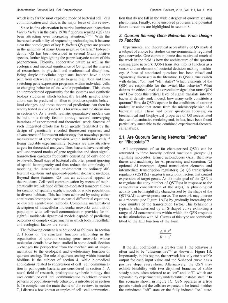
2.1. Are Quorum Sensing Networks “Switches”or “Rheostats”?
All components of so far characterized QSNs can beattributed to three broadly defined functional groups: (1)signaling molecules, termed autoinducers (AIs), their syn-thases and machinery for AI processing and secretion; (2)optional AI receptors, signal transduction elements andintermediate transcription regulators; (3) QS transcriptionregulators (QSTRs) - master transcription factors that controlexpression of target genes. As the main goal of the QSN isto regulate the copy number of QSTR(s) in response to theextracellular concentration of the AI(s), its physiologicalactivity can be insightfully characterized by the shape of theQSTR(AI) dose-response curve. The network may operateas a rheostat (see Figure 1A,B) by gradually increasing thecopy number of the transcription factor. This behavior istypically characterized by an S-shaped curve exhibiting arange of AI concentrations within which the QSN respondsto the stimulation with AI. Curves of this type are commonlyfitted to the Hill function of the form
If the Hill coefficient n is greater than 1, the behavior isoften said to be “ultrasensitive”15 as shown in Figure 1B.Importantly, in this regime, the network has only one possibleoutput for each input value and the S-shaped curve has apositive slope everywhere. Alternatively, the QSN mayexhibit bistability with two disjoined branches of stablesteady states, often referred to as “on” and “off”, which areseparated by experimentally undetectable unstable state.16 Inthis scenario shown in Figure 1C, QSN operates as a truegenetic switch and the cells are expected to be found in eitherthe uninduced “off” state or the fully induced “on” state.
A × xn
B + xn
Understanding Bacterial Cell-Cell Communication Chemical Reviews, 2011, Vol. 111, No. 1 239
This sets it apart from the first case in which the majority ofthe population is likely to exhibit intermediate expressionlevels.
Gram negative QSNs discovered early in the history ofthe QS research, such as the paradigmatic luxR/I networkcharacterized first in V. fischeri, seemed to present aparticularly simple design. Indeed, QSTRs of the LuxR type,as many other bacterial transcription factors, are alsosimultaneously receptors for their cognate signaling mol-ecules, N-acylhomoserine lactones (AHLs).17 AHLs aresynthesized from commodity cellular metabolites by severalenzyme classes, of which LuxI is the best characterized.18,19
Despite intracellular localization of their receptors, cell-cellcommunication with AHLs is possible because most AHLscan freely diffuse in and out of the cell through bothmembranes and the cell wall. However, additional QSNelements, such as efflux pumps, might be required if AHL’spassive diffusion transport is hindered by their long acylchains (>10).20
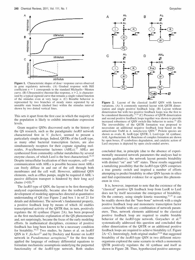
The luxR/I type of QSN, the layout to be first thoroughlyanalyzed experimentally, became also the testbed for thedevelopment of modeling approaches directed at quantitativeunderstanding of QS (see Figure 2 and box 1 in ref 21 fordetails and definitions). The network’s fundamental property,a positive feedback loop by means of which AI enablestranscriptional activity of the QSTR that, in turn, positivelyregulates the AI synthase (Figure 2A), has been proposedas the first mechanistic explanation of the QS phenomenon5
and, not surprisingly, became the focus of the early modelingefforts. In mathematical disciplines, existence of positivefeedback has long been known to be a necessary conditionfor bistability.16,22 Two studies, by James et al. on luxR/IQSN in V. fischeri23 and by Dockery and Keener on lasR/Inetwork in Pseudomonas aeruginosa,24 for the first timeapplied the language of ordinary differential equations toformulate mechanistic assumptions underlying the purportedfunctionality of the luxR/I-type network. Both studies
concluded that, in principle (due to the absence of experi-mentally measured network parameters the analyses had toremain qualitative), the network layout permits bistabilitywith distinct “on” and “off” states. These results suggesteda tantalizing possibility that the luxR/I-type QSN comprisesa true genetic switch and inspired a number of effortsattempting to predict bistability in other QSN layouts in silicoand find experimental evidence for or against this phenom-enon in vivo.
It is, however, important to note that the existence of the“classical” positive QS feedback loop from LuxR to LuxIdoes not by itself necessitate the emergence of bistability.On the contrary, using simple kinetic reasoning,21,25 it canbe readily shown that the “bare-bone” network with a singlepositive feedback loop and monomeric transcription factorcannot be bistable with any combination of network param-eters. Thus, network elements additional to the classicalpositive feedback loop are required to enable bistablebehavior of the luxR/I-type network. Goryachev et al.25
systematically addressed this question and concluded thateither dimerization of the QSTR or an additional positivefeedback loops are required to achieve bistability (cf. Figure2A-C). Interestingly, both original studies23,24 that suggestedbistability of the luxR/I-type QSN in two different micro-organisms explored the same scenario in which a monomericQSTR positively regulates the AI synthase and itself asshown in Figure 2B. Thus, hypothesized positive autoregu-
Figure 1. Characteristic shapes of dose-response curves observedin gene regulatory networks. (A) Gradual response with Hillcoefficient n ) 1 corresponds to the standard Michaelis-Mentencurve. (B) Ultrasensitive rheostat-like response, n > 2, is character-ized by a typical sigmoid curve that remains a single-valued functionof the stimulus even at very large n. (C) Bistable behavior isrepresented by two branches of steady states separated by anunstable state branch (dashed line) within the stimulus intervalshown by two dotted vertical lines.
Figure 2. Layout of the classical luxR/I QSN with knownvariations. (A) A commonly reported layout with QSTR dimer-ization and single positive feedback loop. (B) Layout withoutdimerization but with two positive feedback loops was the first tobe considered theoretically.23,24 (C) Presence of QSTR dimerizationand second positive feedback loops together was shown to provideincreased robustness of QSN switch-like behavior to noise.25 (D)The irreversibility of the QSTR formation was proposed tonecessitate an additional negative feedback loop based on theantiactivator TraM in A. tumefaciens QSN.37 Protein species areshown as ovals; R, luxR-type QSTR; I, LuxI-type AI synthase;AAI, Agrobacterium AI. Reactions of complex formation are shownby open boxes, L symbolizes degradation, and catalytic action ofLuxI enzymes is depicted by open circle-ended arrows.
240 Chemical Reviews, 2011, Vol. 111, No. 1 Goryachev

lation of the QSTR provided second, additional positivefeedback loop that brought about bistability.
Providing some support to the bistability hypothesis, ampleevidence exists that many LuxR-type QSTRs operate asdimers. Moreover, activator-type LuxR homologues havebeen shown to become dimerization-competent only uponbinding their cognate AHLs.26,27 Dimerization, in turn,enables these QSTRs to bind upstream cis-regulatory ele-ments of the target genes and activate their transcription.Even P. aeruginosa LasR, assumed monomeric in earlymodeling studies,24,28 more recently was shown to be dimericin vitro29 and is likely active as a dimer or higher-orderoligomer in vivo.30 Evidence for the existence of additionalpositive feedback loops has also been presented in somesystems. Thus positive autoregulation has been reported forQSTRs carR in Erwinia caratoVora,31 traR in Agrobacteriumtumefaciens,32 and lasR in P. aeruginosa.33,34 Originallydescribed in ref 35, positive autoregulation of V. fischeri luxRhas been revisited recently by Williams et al.36 In an elegantstudy, they combined theory and experiment to provideadditional evidence in support of luxR positive autoregulationand the existence of bistability in the classical luxR/I network.
Fundamental limitation for the luxR/I-type network layoutsis the requirement for the relative instability of the QSTRdimer and reversibility of the QSTR-AI interaction.37 Indeed,due to the constitutive intracellular production of AI andtranslation of the QSTR, strong interaction between themonomeric QSTR and AI followed by largely irreversibleQSTR dimerization could result in undesirable network“short-circuit”saccumulation of active transcription factorand eventual transition to the induced state even in theabsence of cell-cell communication. One possible evolu-tionary solution avoiding this “runaway” activation is to keepthe interactions between the QSTR and AI and between thetwo AI-bound QSTR monomers weak. This assumption,implicitly made in the majority of luxR/I-type networkmodels, has been experimentally confirmed in some species38
but clearly disproven in others. Perhaps the best characterizedcounterexample is provided by the quorum sensing systemof Ti plasmids in A. tumefaciens32 in which QSTR TraRforms essentially irreversible complex with its cognate AIduring translation.26 Moreover, if translated in the absenceof AI, TraR is poorly folded and insoluble. Experiments inA. tumefaciens identified a protein, TraM, whose deletionresulted in constitutively induced QSN state.39 Paradoxically,TraM was found to be positively regulated by the veryTraR.40 Modeling analysis of the traR/I network by Gory-achev et al.37 resolved this seeming contradiction anddemonstrated that TraR-TraM negative feedback loopshown in Figure 2D is indeed the network element thatenables sensing extracellular AI despite the irreversibleinteraction of QSTR TraR with its AI. This study alsopredicted bistable, switch-like mode of operation for thetraR/I QSN. Interestingly, P. aeruginosa LasR was foundto bind its cognate AI 3OC12-HSL with picomolar-rangedissociation constant while LasR, translated in the absenceof AI, was largely insoluble.29 These features, reminiscentof TraR and its interaction with the cognate AI, suggest thatmechanisms similar to those characterized in A. tumefaciensmight be at play in the highly complex QSN of P.aeruginosa.
Although V. fischeri-type luxR/I network remains probablythe most popular example of QSN, many more networklayouts have been discovered and characterized to various
degrees of detail (see refs 21 and 41 for recent reviews onthe QSN architecture and design principles). Analyses ofluxR/I homologues in some γ-proteobacteria demonstratedthat despite sequence similarity with V. fischeri genes, theirfunctional mode of operation is fundamentally different,warranting separation of these QSNs into a distinct functionalclass21,42 that is also characterized by different genomicorganization as shown in Figure 3. QSTRs of this networktype (class B) are generally dimeric repressors that negativelyregulate transcription of their target genes in the absence oftheir cognate AIs. Binding to the AIs induces conformationalchanges that reduce affinity of the QSTRs to DNA andrelieves repression of the target genes in the state of quorum.These QSTRs frequently repress themselves, but in the mostreported cases, they do not regulate the respective AIsynthases.42 Therefore, mechanisms based on the “classical”positive QS feedback loop and positive autoregulation ofQSTRs characterized in V. fischeri-type (class A) networksare not applicable to this class of QSNs. Although complexregulatory patterns have been suggested by some studies43
(see Figure 3), more experimental and theoretical work isrequired before their mode of operation can be fullyunderstood.
Of other functionally distinct QSN layouts in Gramnegative bacteria, perhaps the best characterized are networksof Vibrio species. QSTRs LuxR in V. harVeyi (not relatedto V. fischeri LuxR) and HapR in V. cholera belong to alarge family of TetR transcription factors. They do not bindAIs but instead are regulated by sRNA species downstreamof the cell-surface AI receptors.44,45 Intricate architecture andfunctional principles governing Vibrio QSNs have beenunraveled recently in a series of insightful papers by Basslerand colleagues (see refs 46-49 and the discussion in section2.3).
Cell-cell communication in Gram positive bacteria mostlyrelies on genetically encoded autoinducer peptides (AIPs).50,51
Translated as precursors, these peptides are proteolyticallyprocessed and extruded into the extracellular space byenzymes positioned in the cell membrane. Reception isachieved by either transmembrane receptor histidine-kinasesor through transporter-facilitated import into the cell wherepeptides are recognized by intracellular receptors, such asBacillus subtilis phosphatase Rap.52 Prototypical example ofa peptide-based Gram positive QSN is provided by the agrsystem in Staphylococcus aureus,53 which is schematicallyshown in Figure 4A. The agr operon encodes the messenger
Figure 3. luxR/I QSNs of Gram negative bacteria belong to twodistinct classes with different genomic layout and mode ofregulation (see text). A complex regulatory pattern described inYersinia pseudotuberculosis has been attributed to the interactionbetween its two sequentially connected class B QSNs.43 Arrowheadsrepresent transcriptional activation, and hammerheads representrepression. Elements reported only in some organisms are shownby dashed line. Open arrows indicate QSTR genes, and gray arrowsdepict AI synthase genes.
Understanding Bacterial Cell-Cell Communication Chemical Reviews, 2011, Vol. 111, No. 1 241
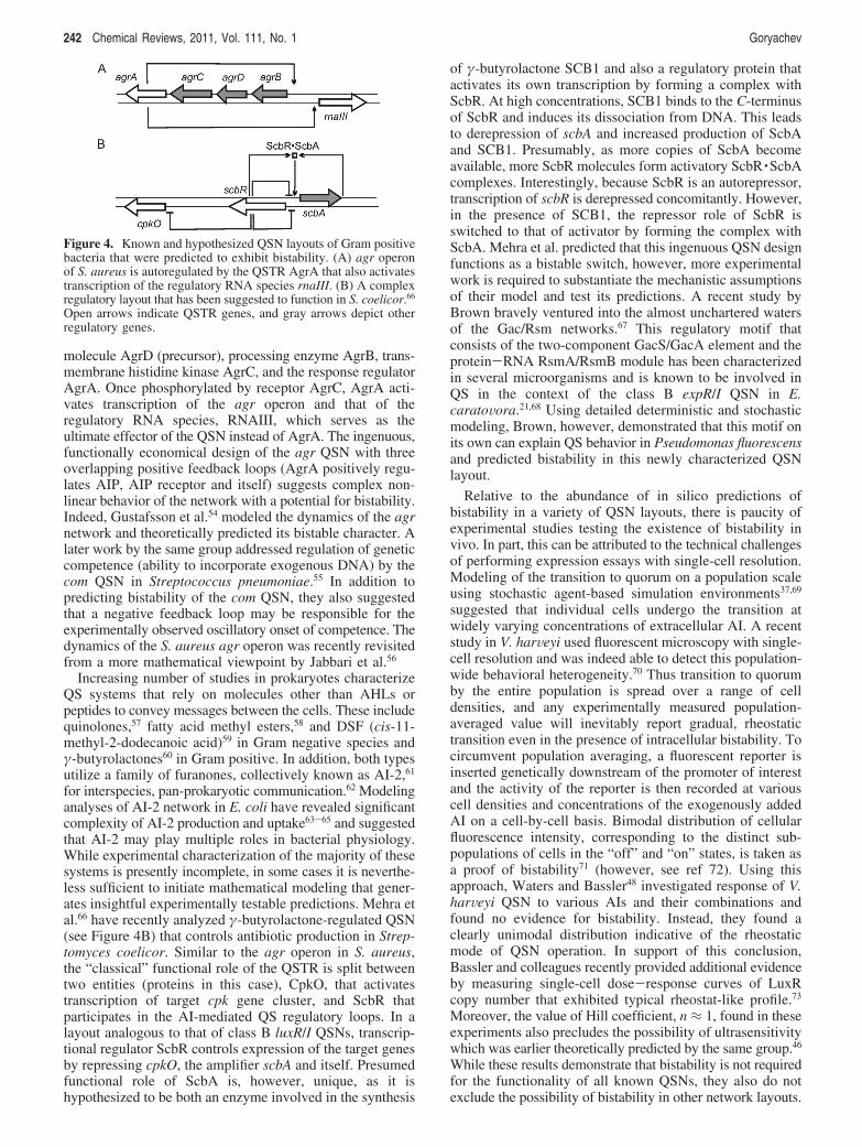
molecule AgrD (precursor), processing enzyme AgrB, trans-membrane histidine kinase AgrC, and the response regulatorAgrA. Once phosphorylated by receptor AgrC, AgrA acti-vates transcription of the agr operon and that of theregulatory RNA species, RNAIII, which serves as theultimate effector of the QSN instead of AgrA. The ingenuous,functionally economical design of the agr QSN with threeoverlapping positive feedback loops (AgrA positively regu-lates AIP, AIP receptor and itself) suggests complex non-linear behavior of the network with a potential for bistability.Indeed, Gustafsson et al.54 modeled the dynamics of the agrnetwork and theoretically predicted its bistable character. Alater work by the same group addressed regulation of geneticcompetence (ability to incorporate exogenous DNA) by thecom QSN in Streptococcus pneumoniae.55 In addition topredicting bistability of the com QSN, they also suggestedthat a negative feedback loop may be responsible for theexperimentally observed oscillatory onset of competence. Thedynamics of the S. aureus agr operon was recently revisitedfrom a more mathematical viewpoint by Jabbari et al.56
Increasing number of studies in prokaryotes characterizeQS systems that rely on molecules other than AHLs orpeptides to convey messages between the cells. These includequinolones,57 fatty acid methyl esters,58 and DSF (cis-11-methyl-2-dodecanoic acid)59 in Gram negative species andγ-butyrolactones60 in Gram positive. In addition, both typesutilize a family of furanones, collectively known as AI-2,61
for interspecies, pan-prokaryotic communication.62 Modelinganalyses of AI-2 network in E. coli have revealed significantcomplexity of AI-2 production and uptake63-65 and suggestedthat AI-2 may play multiple roles in bacterial physiology.While experimental characterization of the majority of thesesystems is presently incomplete, in some cases it is neverthe-less sufficient to initiate mathematical modeling that gener-ates insightful experimentally testable predictions. Mehra etal.66 have recently analyzed γ-butyrolactone-regulated QSN(see Figure 4B) that controls antibiotic production in Strep-tomyces coelicor. Similar to the agr operon in S. aureus,the “classical” functional role of the QSTR is split betweentwo entities (proteins in this case), CpkO, that activatestranscription of target cpk gene cluster, and ScbR thatparticipates in the AI-mediated QS regulatory loops. In alayout analogous to that of class B luxR/I QSNs, transcrip-tional regulator ScbR controls expression of the target genesby repressing cpkO, the amplifier scbA and itself. Presumedfunctional role of ScbA is, however, unique, as it ishypothesized to be both an enzyme involved in the synthesis
of γ-butyrolactone SCB1 and also a regulatory protein thatactivates its own transcription by forming a complex withScbR. At high concentrations, SCB1 binds to the C-terminusof ScbR and induces its dissociation from DNA. This leadsto derepression of scbA and increased production of ScbAand SCB1. Presumably, as more copies of ScbA becomeavailable, more ScbR molecules form activatory ScbR ·ScbAcomplexes. Interestingly, because ScbR is an autorepressor,transcription of scbR is derepressed concomitantly. However,in the presence of SCB1, the repressor role of ScbR isswitched to that of activator by forming the complex withScbA. Mehra et al. predicted that this ingenuous QSN designfunctions as a bistable switch, however, more experimentalwork is required to substantiate the mechanistic assumptionsof their model and test its predictions. A recent study byBrown bravely ventured into the almost unchartered watersof the Gac/Rsm networks.67 This regulatory motif thatconsists of the two-component GacS/GacA element and theprotein-RNA RsmA/RsmB module has been characterizedin several microorganisms and is known to be involved inQS in the context of the class B expR/I QSN in E.caratoVora.21,68 Using detailed deterministic and stochasticmodeling, Brown, however, demonstrated that this motif onits own can explain QS behavior in Pseudomonas fluorescensand predicted bistability in this newly characterized QSNlayout.
Relative to the abundance of in silico predictions ofbistability in a variety of QSN layouts, there is paucity ofexperimental studies testing the existence of bistability invivo. In part, this can be attributed to the technical challengesof performing expression essays with single-cell resolution.Modeling of the transition to quorum on a population scaleusing stochastic agent-based simulation environments37,69
suggested that individual cells undergo the transition atwidely varying concentrations of extracellular AI. A recentstudy in V. harVeyi used fluorescent microscopy with single-cell resolution and was indeed able to detect this population-wide behavioral heterogeneity.70 Thus transition to quorumby the entire population is spread over a range of celldensities, and any experimentally measured population-averaged value will inevitably report gradual, rheostatictransition even in the presence of intracellular bistability. Tocircumvent population averaging, a fluorescent reporter isinserted genetically downstream of the promoter of interestand the activity of the reporter is then recorded at variouscell densities and concentrations of the exogenously addedAI on a cell-by-cell basis. Bimodal distribution of cellularfluorescence intensity, corresponding to the distinct sub-populations of cells in the “off” and “on” states, is taken asa proof of bistability71 (however, see ref 72). Using thisapproach, Waters and Bassler48 investigated response of V.harVeyi QSN to various AIs and their combinations andfound no evidence for bistability. Instead, they found aclearly unimodal distribution indicative of the rheostaticmode of QSN operation. In support of this conclusion,Bassler and colleagues recently provided additional evidenceby measuring single-cell dose-response curves of LuxRcopy number that exhibited typical rheostat-like profile.73
Moreover, the value of Hill coefficient, n ≈ 1, found in theseexperiments also precludes the possibility of ultrasensitivitywhich was earlier theoretically predicted by the same group.46
While these results demonstrate that bistability is not requiredfor the functionality of all known QSNs, they also do notexclude the possibility of bistability in other network layouts.
Figure 4. Known and hypothesized QSN layouts of Gram positivebacteria that were predicted to exhibit bistability. (A) agr operonof S. aureus is autoregulated by the QSTR AgrA that also activatestranscription of the regulatory RNA species rnaIII. (B) A complexregulatory layout that has been suggested to function in S. coelicor.66
Open arrows indicate QSTR genes, and gray arrows depict otherregulatory genes.
242 Chemical Reviews, 2011, Vol. 111, No. 1 Goryachev
Indeed, although integrative model has not yet been con-structed for V. harVeyi-like QSN, interactions within thisnetwork discovered and characterized so far do not suggestbistability also from the theoretical point of view. Analyseswith single-cell resolution, similar to that in ref 48, aredesirable in the systems where theoretical prediction ofbistability had been provided. To date, the study byLevchenko, Stevens, and co-workers36 remains the solepositive experimental verification of bistability in a bacterialquorum-sensing network.
2.2. Quorum Sensing in the Presence ofMolecular Noise
A typical Escherichia coli cell with length 2 µm anddiameter 0.5 µm has a cell volume of only ∼0.72 µm3 (or7.2 × 10-16 L). Therefore, for a signaling molecule freelydiffusing in and out of the cell to be present within the cellat only a single copy, its concentration in the extracellularenvironment should be g2.3 nM. So-called slim rods, e.g.,A. tumefaciens, and Gram positive cocci are even smaller(V ≈ 0.18 µm3). For them, the minimal detectable concentra-tion of an extracellular molecule is on the order of 10 nM.Accordingly, reception of a freely diffusing cell-cell com-munication signal is subjected to intense extrinsic molecularnoise which arises solely due to the small size of the“detector”, the bacterial cell itself, and not because offluctuations in the extracellular concentration per se. Anotherimportant and well-known factor that affects bacterial generegulation is the intrinsic molecular noise, which in part arisesdue to fluctuation of the copy numbers of the key participat-ing molecules, in particular transcription factors.74 Experi-mental observation of these fluctuations became possiblechiefly due to the introduction of genetically encodedfluorescent reporters and the associated detection methods(see discussion in section 2.1). These technological develop-ments enabled the giant leap forward from the inevitablypopulation-averaged picture provided by the classical bio-chemical methods to the full appreciation of highly stochasticnature of bacterial intracellular environment.
In the framework of QS, both the extrinsic noise (detectedAI) and the intrinsic noise are highest in the low populationdensity state. With only a handful of potentially detectableAI molecules and their intracellular receptors per cell (e.g.,in the case of luxR/I-type networks), stochastic fluctuationsare greater than the average values that are normallyinterpreted as deterministic “concentrations”. This makes abacterial cell, particularly in the low population density state,a fundamentally stochastic system. As the dynamics of suchsystems generally has been shown to depart from thatpredicted by purely deterministic methods,75,76 stochasticmodeling techniques are required to confirm, refine, or refutethe predictions of deterministic models. A number ofstudies25,37,77 used the stochastic simulation algorithm (SSA),also known as the Gillespie algorithm,78 to simulate intra-cellular dynamics of QSNs numerically. To account formolecular noise, Li et al.63 adopted stochastic Petri nets(SPN) for modeling AI-2 network in E. coli. Interestingly,taking noise into consideration demonstrated that somepredictions from purely deterministic models may need arevision. Goryachev et al. used SSA to study a number ofluxR/I-type network layouts with increasing complexity.25
They found that, with biologically realistic network param-eters, some of the tested layouts, while predicted bistableby respective ODE-based models, failed to exhibit bistability
in the presence of molecular noise which essentially obliter-ated the difference between the “on” and “off” states. Theyconcluded that seemingly redundant network elements, e.g.,multiple positive feedback loops, often found in the real-life QSNs, might be there to provide sufficient quantitativedifference between the distinct states of the network to enableits robust, noise-resistant operation. Not only can noise negatesome predicted QSN properties, it may potentially also bringabout novel behavior, unaccounted for deterministically.Using a synthetic biology strategy, To and Maheshri haverecently demonstrated bimodal population distribution of atranscription factor copy number in a yeast-based systemwhere deterministic model did not warrant bistability.72 In atheory-experiment systems study in E. coli, Tozaki et al.tested the applicability of ergodicity principle, an assumptionthat the percentage of cells in a given state is identical tothe probability to find a cell in this state.79 They found thatergodicity, typically taken for granted, may be readily brokenif the bacterial growth rate is dependent on the network state,which is a reasonable assumption in the case of QS. Thus toavoid erroneous prediction and interpretation of experiments,stochastic effects arising from the molecular noise andrandom switching of QSN states require careful consideration.
To extend stochastic methodology to whole populations,Goryachev and colleagues37 designed a parallel agent-basedsimulation environment in which individual cell-agents,whose intracellular QSN dynamics was simulated using SSA,were able to randomly move, divide, and exchange signalingmolecules with the environment. Cell-cell communicationwas enabled by the common extracellular space in whichdiffusion of signaling molecules was treated in a continuousdeterministic approximation. This unique tool permittedstochastic simulation of the transition of a whole populationof A. tumefaciens to the induced, high population densitystate. Results of these simulations revealed astonishingdiversity of individual cell behavior even in a spatiallyhomogeneous extracellular environment, the phenomenonwhich was recently observed also experimentally.70 As thecomputing power becomes more readily available, suchcomputationally intensive methods may become viable andvaluable tools for the analysis of cell-cell communicationin complex habitats such as biofilms.
Are there any mechanisms embedded into the design ofQSNs that allow them to harness molecular noise or at leastreduce its harmful influence? Levchenko and colleaguesnoted that positive autoregulation of a luxR-type QSTR mayplay an additional noise reduction role because the intensityof the molecular noise in AI-LuxR interaction scalesinversely with the square root of the copy number of LuxRin the cell.36 Tanouchi et al.80 highlighted a different noisereduction feature of QSNs, fast degradation of the QSTR inthe absence of AI, which has been reported for a number ofluxR-type QSTRs, such as A. tumefaciens TraR.81 Zhou etal. theoretically analyzed a hypothetical cell-cell com-munication network inspired by QS but radically differentfrom the natural QSNs by the inhibitory action of theQSTR-AI complex on the transcription.82 This designexhibited spontaneous oscillations which could be synchro-nized between individual communicating cells in the presenceof extracellular noise. Curiously, these synchronized oscil-lations were found to be the direct consequence of theextracellular noise and provided a potentially interestingexample of a fundamentally noise-induced behavior whosebiological relevance is yet to be demonstrated.
Understanding Bacterial Cell-Cell Communication Chemical Reviews, 2011, Vol. 111, No. 1 243
2.3. Integration of QSNs within BacterialDecision-Making Circuitry
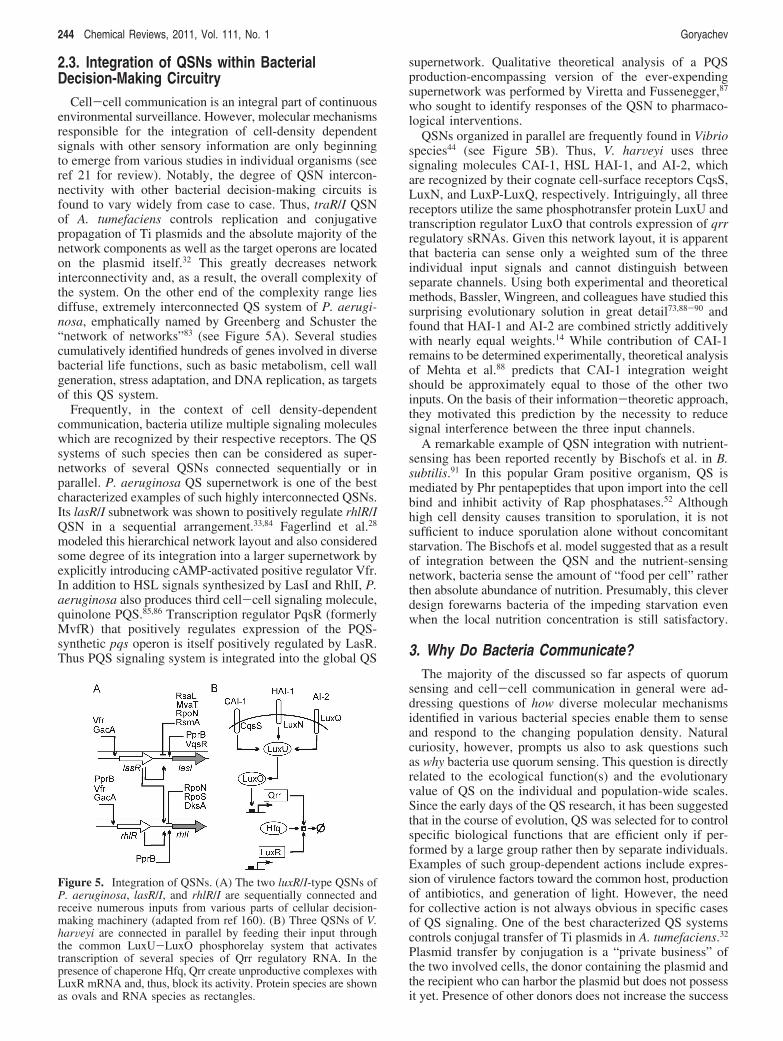
Cell-cell communication is an integral part of continuousenvironmental surveillance. However, molecular mechanismsresponsible for the integration of cell-density dependentsignals with other sensory information are only beginningto emerge from various studies in individual organisms (seeref 21 for review). Notably, the degree of QSN intercon-nectivity with other bacterial decision-making circuits isfound to vary widely from case to case. Thus, traR/I QSNof A. tumefaciens controls replication and conjugativepropagation of Ti plasmids and the absolute majority of thenetwork components as well as the target operons are locatedon the plasmid itself.32 This greatly decreases networkinterconnectivity and, as a result, the overall complexity ofthe system. On the other end of the complexity range liesdiffuse, extremely interconnected QS system of P. aerugi-nosa, emphatically named by Greenberg and Schuster the“network of networks”83 (see Figure 5A). Several studiescumulatively identified hundreds of genes involved in diversebacterial life functions, such as basic metabolism, cell wallgeneration, stress adaptation, and DNA replication, as targetsof this QS system.
Frequently, in the context of cell density-dependentcommunication, bacteria utilize multiple signaling moleculeswhich are recognized by their respective receptors. The QSsystems of such species then can be considered as super-networks of several QSNs connected sequentially or inparallel. P. aeruginosa QS supernetwork is one of the bestcharacterized examples of such highly interconnected QSNs.Its lasR/I subnetwork was shown to positively regulate rhlR/IQSN in a sequential arrangement.33,84 Fagerlind et al.28
modeled this hierarchical network layout and also consideredsome degree of its integration into a larger supernetwork byexplicitly introducing cAMP-activated positive regulator Vfr.In addition to HSL signals synthesized by LasI and RhlI, P.aeruginosa also produces third cell-cell signaling molecule,quinolone PQS.85,86 Transcription regulator PqsR (formerlyMvfR) that positively regulates expression of the PQS-synthetic pqs operon is itself positively regulated by LasR.Thus PQS signaling system is integrated into the global QS
supernetwork. Qualitative theoretical analysis of a PQSproduction-encompassing version of the ever-expendingsupernetwork was performed by Viretta and Fussenegger,87
who sought to identify responses of the QSN to pharmaco-logical interventions.
QSNs organized in parallel are frequently found in Vibriospecies44 (see Figure 5B). Thus, V. harVeyi uses threesignaling molecules CAI-1, HSL HAI-1, and AI-2, whichare recognized by their cognate cell-surface receptors CqsS,LuxN, and LuxP-LuxQ, respectively. Intriguingly, all threereceptors utilize the same phosphotransfer protein LuxU andtranscription regulator LuxO that controls expression of qrrregulatory sRNAs. Given this network layout, it is apparentthat bacteria can sense only a weighted sum of the threeindividual input signals and cannot distinguish betweenseparate channels. Using both experimental and theoreticalmethods, Bassler, Wingreen, and colleagues have studied thissurprising evolutionary solution in great detail73,88-90 andfound that HAI-1 and AI-2 are combined strictly additivelywith nearly equal weights.14 While contribution of CAI-1remains to be determined experimentally, theoretical analysisof Mehta et al.88 predicts that CAI-1 integration weightshould be approximately equal to those of the other twoinputs. On the basis of their information-theoretic approach,they motivated this prediction by the necessity to reducesignal interference between the three input channels.
A remarkable example of QSN integration with nutrient-sensing has been reported recently by Bischofs et al. in B.subtilis.91 In this popular Gram positive organism, QS ismediated by Phr pentapeptides that upon import into the cellbind and inhibit activity of Rap phosphatases.52 Althoughhigh cell density causes transition to sporulation, it is notsufficient to induce sporulation alone without concomitantstarvation. The Bischofs et al. model suggested that as a resultof integration between the QSN and the nutrient-sensingnetwork, bacteria sense the amount of “food per cell” ratherthen absolute abundance of nutrition. Presumably, this cleverdesign forewarns bacteria of the impeding starvation evenwhen the local nutrition concentration is still satisfactory.
3. Why Do Bacteria Communicate?The majority of the discussed so far aspects of quorum
sensing and cell-cell communication in general were ad-dressing questions of how diverse molecular mechanismsidentified in various bacterial species enable them to senseand respond to the changing population density. Naturalcuriosity, however, prompts us also to ask questions suchas why bacteria use quorum sensing. This question is directlyrelated to the ecological function(s) and the evolutionaryvalue of QS on the individual and population-wide scales.Since the early days of the QS research, it has been suggestedthat in the course of evolution, QS was selected for to controlspecific biological functions that are efficient only if per-formed by a large group rather then by separate individuals.Examples of such group-dependent actions include expres-sion of virulence factors toward the common host, productionof antibiotics, and generation of light. However, the needfor collective action is not always obvious in specific casesof QS signaling. One of the best characterized QS systemscontrols conjugal transfer of Ti plasmids in A. tumefaciens.32
Plasmid transfer by conjugation is a “private business” ofthe two involved cells, the donor containing the plasmid andthe recipient who can harbor the plasmid but does not possessit yet. Presence of other donors does not increase the success
Figure 5. Integration of QSNs. (A) The two luxR/I-type QSNs ofP. aeruginosa, lasR/I, and rhlR/I are sequentially connected andreceive numerous inputs from various parts of cellular decision-making machinery (adapted from ref 160). (B) Three QSNs of V.harVeyi are connected in parallel by feeding their input throughthe common LuxU-LuxO phosphorelay system that activatestranscription of several species of Qrr regulatory RNA. In thepresence of chaperone Hfq, Qrr create unproductive complexes withLuxR mRNA and, thus, block its activity. Protein species are shownas ovals and RNA species as rectangles.
244 Chemical Reviews, 2011, Vol. 111, No. 1 Goryachev

rate of any chosen donor cell per se. On the contrary, abilityto sense the local density of potential recipients could havecome handy by reducing the cost of unsuccessful transfers.This, however, is not possible because the QSN is locatedon the plasmid itself and thus all potential recipients are “QSsilent”sthey neither produce nor respond to the signal.Goryachev et al. addressed this paradoxical situation byexplicitly simulating transition to quorum in distinct envi-ronmental conditions.37 Specifically, they contrasted celldensities required to reach quorum in the batch culture wherecells are distributed uniformly in suspension and in a moreecologically realistic assumption of growth as a biofilm. Theyfound that while transition to quorum in the batch culturerequires unrealistically high cell densities, transition in thebiofilm can take place at the cell densities readily found inexperiment. The reasons for this difference were clearly themore compact spatial distribution of cells and the loss of AIonly into a half of the space in the case of biofilm. Inconclusion, it was suggested that while Ti plasmids cannot“learn” about the density of potential recipients by using QS,they nevertheless can detect certain advanced stage of biofilmmaturation. From that they can “determine” that their hostcells are firmly attached within a dense bacterial mass whichwill inevitably also contain some potential recipients.
Analyses of cases, such as the above example, promptedthe opinion that the concept of population “density-sensing”should be entirely replaced by “diffusion-sensing”.92 Aspecific example of a situation in which “density-sensing”is clearly irrelevant is the situation when a small number ofbacterial cells is enclosed in a diffusion-impermeable com-partment. S. aureus, a primarily extracellular pathogen, hasbeen occasionally found within endosomes of nonprofes-sional phagocytes in the quantity of 1-2 cells per endosome.Internalized bacteria apparently manage to escape from theendosomes, proliferate in the cytoplasm, and eventually causeapoptosis of the infected cell.93 Intriguingly, QS agr operonof S. aureus (see section 2.1 for details) is activated prior tothe endosomal escape while the agr-defective mutant wasfound unable to proliferate inside the cells.94 Koerber et al.modeled this peculiar instance of QS and computed analyti-cally and numerically the bacterial escape time.95 “Solitaryconfinement” of S. aureus cells was recently revisitedexperimentally by Carnes et al.,96 who confirmed inductionof agr operon during confinement of bacterial cells in theengineered nanostructured matrix and demonstrated enhancedsurvival of QS-competent cells. To unify “density-sensing”and “diffusion-sensing”, a concept of “efficiency-sensing”was proposed recently.97 This hypothesis, that essentiallyreiterated the conclusions of ref 37, postulated that cellscompute overall efficiency of secreting extracellular mol-ecules by factoring in cell density, spatial distribution of cells,and diffusivity of the medium. Although more biophysicallybalanced, this hypothesis also does not cover all possiblefunctions of QS because secretion of diffusible extracellulareffectors is not the universal “goal” of all known QS-regulated gene expression systems (conjugation of Ti plas-mids is just one counterexample). Regardless of potentiallysubjective interpretations, QS allows bacteria to survey theirenvironment by releasing and receiving diffusible signalingmolecules.98 Accumulating knowledge from numerous QSsystems demonstrates that different species use this surveil-lance information in different ways, sometimes makingentirely opposite decisions. Thus while some bacteria, e.g.,Pantoea stewartii, build biofilm at high cell density11
(collective action), others, like V. cholera, instead disassociatefrom the biofilm and become motile when “overcrowded”.Particular decision will optimally suit specific needs andecological strategy of a given microorganism. Given thediversity of ecological niches, the exact function of QS needsto be determined on a species-specific basis.
4. Quorum Sensing in Bacterial BiofilmsBiofilms, compact bacterial structures cemented together
by extracellular matrix that form naturally on various surfacesand interfaces, have recently gained broad recognition as themajor native prokaryotic habitat.99-103 Pseudomonas aerugi-nosa is notoriously hard to eradicate with standard treatmentprotocols when it grows as a biofilm in medical cathetersand in the lungs of cystic fibrosis patients. In 1998,Greenberg and colleagues suggested that QS plays a key rolein the morphogenesis of P. aeruginosa biofilms.104 Theyfound that a mutant with genetically disrupted QS wasshowing abnormal biofilm morphology with concomitantincrease in susceptibility to antimicrobials. This short reportin Science magazine sparked a great amount of interesttoward the role of QS in biofilm formation across severaldisciplines. Therefore, already the first theoretical studiesconcerned with QS modeling also attempted to address QSin the context of biofilms.24,105,106 Nilsson et al. for the firsttime asked the question how many bacterial cells constitutea “quorum” and demonstrated that compact organization ofcells in the biofilm has a potential to result in the inductionof the activated state even at relatively low cell density.106
Koerber et al. constructed a reaction-diffusion model describ-ing QS and formation of P. aeruginosa biofilm in burnwounds.105 Explicitly introducing velocity of biofilm propa-gation due to the growth of its biomass, the same group laterformulated a different mathematical approach to QS ingrowing biofilms by representing it as an advection-diffusionproblem.107 Extending established bioengineering approachesto modeling bacterial biofilms, Chopp et al. developed a 1Dmodel of QS in P. aeruginosa biofilm.108 Biological realismof their model was improved by taking into the considerationcell death and decay as well as oxygen consumption, factorsthat naturally limit the biomass growth. They concluded thatbacteria, dwelling in anaerobic conditions close to thesubstratum and often considered metabolically dormant,should actively produce AI and reach the induced state first.In the follow-up study,109 Chopp et al. extended andconfirmed this theoretical prediction by imaging fluorescentQS reporter at various depths within the biofilm. They foundlocalized clusters of induced bacteria in the immediateproximity to the underlying glass slide.
In realistic conditions, such as porous caverns in the soilor the lumen of a medical catheter, bacterial biofilms growin the presence of a hydrodynamic flow which is likely toaffect both the distribution of nutrients and the chemicalcommunication among the bacteria. To address this importantyet often ignored factor, Parsek, Chopp, and colleaguesexplicitly introduced advective flow into their models. In ref110, they used the Stokes flow model to represent the flowof fluid above the biofilm and compared model predictionswith the results of their experiments at different flow rates.As expected, introduction of even moderate flow significantlyincreased the cell density requirement to achieve quorum.Thus at the flow rate 0.04 mL/min (velocity 0.15 mm/s),the required density was 4 × 10⁶ CFU/mm2, while at 4.0mL/min (15 mm/s), it was found to be 6 × 10⁷ CFU/mm2.
Understanding Bacterial Cell-Cell Communication Chemical Reviews, 2011, Vol. 111, No. 1 245
Further technological advance in modeling hydrodynamicsof biofilms was reported by Duddu et al.,111 where introduc-tion of sophisticated numerical methods allowed the authorsto consider complex profiles of cell density as well asdetachment of biofilm fragments induced by the sheer stress.Armed with these tools, Chopp and colleagues recentlyrevisited induction of QS in biofilms subjected to flow.112
They found that, to make the situation even more complex,the influence of flow strongly depends on the morphologyof the biofilm. Biofilms with rough, mushroom-shapedsurface that are frequently found in experiments exhibit morecomplex dependence of the critical biomass on the flow rateand, generally require less biomass to reach quorum thantheir flat counterparts.
While continuous methods based on partial differentialequations permit modeling large system sizes, they fail tocapture discrete nature of biofilms composed of individualcells. Given the highly stochastic nature of the bacterialintracellular environment (see section 2.2), discrete methods,based on representation of cells as individual entities, mayprovide valuable complementary results. Proposed first inlate 1990s113,114 as simple cellular automata representing cellsas nodes on a regular 2D lattice, discrete models haveevolved into advanced tools with a potential to realisticallysimulate cell-cell communication in complex conditions ofa biofilm. Thus, individual-based models developed by vanLoosdrecht and colleagues115-117 utilize spherical particlescharacterized by continuous location in 3D space and areable to independently grow, move, and divide. Rather thancell-cell communication, metabolic processes in biofilms,such as production of methane and nitrification, had beenthe primary focus of application of these methods. Agent-based methods, however, provide encapsulated sets ofintracellular variables that can be used to simulate thedynamics of molecular networks, such as QSN, withinindividual cells. This approach was taken by Goryachev andcolleagues in the development of their stochastic simulationenvironment in which cell agents could freely move anddivide and had individual intracellular environments chemi-cally connected to the outside medium.37 Representation ofcells as zero-dimensional points that can occupy the sameor unrealistically close locations in the continuous 3D space,however, prevented this tool from being able to describebiofilm structure at a fine level of spatial resolution. Morerecently, agent-based methods were used by Xavier, Foster,and colleagues to address evolutionary aspects of cell-cellcommunication in biofilms.118,119 In ref 119, they assumedthat bacteria produce AI at a constant rate, and once thecritical concentration is exceeded locally, some strains(termed QS+) terminate production of extracellular matrix(EPS) and instead invest all their resources into growth, whileEPS+ and EPS- strains either produce EPS constitutivelyor do not produce it at all. They concluded that such quorum-sensing strategy will be evolutionary advantageous if biofilmdispersal is favored (the case of V. cholera) and detrimentalin chronic, long-term biofilms (the case of P. aeruginosa).A platform for simulation of QS on a population scale butwith a single cell resolution has also been reported by Melkeet al., who combined intracellular gene-regulation dynamicsand mechanical cell-cell and cell-wall interaction to modeltransition to quorum in various spatial layouts on the exampleof V. fischeri.69
5. Controlling Quorum Sensing to ReduceBacterial Pathogenicity
Closely related to the field of quorum sensing in biofilmsis the area of quorum sensing control in medical applications.Since the seminal work of Greenberg and colleagues on P.aeruginosa,104 bacterial cell-cell communication has beenprogressively growing in importance as a potential target foranti-infective interventions.120 Suppression of QS is advisiblein two mutually nonexclusive situations, namely if: (1) inthe quorum-induced state pathogenic bacteria produce biofilmthat protects them from antibiotics and the immune system,(2) the state of quorum is required to mount aggressivevirulence program. In the first scenario, reduction or completedestruction of autoinducer molecules can augment other, e.g.,antibiotic, treatments, while in the second it may decreasepathogenicity without killing the microorganism, leaving thisjob to the immune system now relieved from the virulentattack. Thus a number of strategies, some of them based onthe use of native autoinducer-degrading bacterial enzymes,have been proposed to fight pathogens of both animal121,122
and plant123,124 hosts.Complementing these mostly experimental efforts, theo-
retical studies have also been conducted to identify optimalintervention targets and predict the therapy outcomes.64,87,125-128
Thus, Fagerlind et al. suggested utilizing the property ofmany native QSNs to rapidly degrade the QSTR in theabsence of the cognate AI by supplying cells with AHLantagonists.129 Presumably, chemical compounds that stronglybind QSTR and block AHL-binding pocket without inducingactive conformation of the transcription factor would suitthe purpose. King, Ward, and colleagues utilized theirpopulation-based approach to QS130 to study anti-AI strate-gies in both liquid culture and biofilms, predominantly onthe example of P. aeruginosa105,107,131-134 but more recentlyalso in S. aureus.135
6. Cell-Cell Communication in EngineeredSynthetic Systems
Synthetic biology, a novel branch of bioengineering, strivesto develop standardized library of well-characterized regula-tory elements to design modified microorganisms withdesired properties.13 Naturally, a module that can providesynchronization of gene expression across the whole popula-tion is a high-priority item on the list of synthetic elements.Because of their simple, well-characterized layout (seesection 2.1), luxR/I type QSNs are promising candidates forsuch a module. Design and “debugging” of synthetic biologyprojects would hardly be possible without mathematicalmodeling that guides and supports the experimental effortthroughout.
Among applications of QS in synthetic biology, popula-tion-wide synchronization of intracellular oscillators hascaptured perhaps the most attention. Collins and colleaguespresented one of the first models predicting synchronizationof synthetic intracellular oscillators by a luxR/I-type QSN.136
Garcia-Ojalvo et al. constructed a model of an E. colipopulation modified by a “repressilator”-type oscillatorynetwork whose individual intracellular dynamics was coupledacross the population by a QS system.137 In their model, theyobserved that individual cells were able to robustly synchro-nize into a collective rhythm despite intracellular noise. Anumber of theoretical studies further explored various aspectsof oscillation synchronization by means of QS.82,138-140
246 Chemical Reviews, 2011, Vol. 111, No. 1 Goryachev
Despite much invested effort, experimental realization of theproposed designs, however, has not been achieved untilrecently. At last, in early 2010, Hasty and colleagues reportedthe successful implementation of an engineered gene networkthat is capable of generating global oscillations in a growingpopulation of cells.141 In addition to the commonly used V.fischeri luxI and luxR, they utilized aiiA gene from Bacillusthuringiensis that encodes an AHL-degrading enzyme, lac-tonase. This gene was put under control of the luxI promoter,thus establishing a negative feedback loop that is necessaryfor oscillations. By using a microfluidic device, theymanipulated the cell density and monitored the activity of afluorescent reporter. Period and amplitude of observedsynchronized oscillations were found to be dependent on thevelocity of flow in the main channel of the microfluidicdevice. Interestingly, at low flow rates, they also detectedstriking waves of spatiotemporal activity propagating throughthe 100 µm chamber at velocities ∼8-35 µm per min.
Beyond synchronization of oscillations, QS-based couplinghas been modeled and experimentally implemented in anumber of engineered designs. Thus Kobayashi et al.implemented an E. coli strain that could activate transcriptionof any gene in a density-dependent manner by engineeringa plasmid-born QS system based on the V. fischeri luxR andluxI genes.13 Interestingly, by using a fluorescent reporter,they clearly observed bistability throughout the transition oftheir engineered QSN to the induced state. By furtherincreasing complexity of the engineered behavior, Weiss andcolleagues generated sender and receiver strains of E. coliso that the receiver cells responded to AI produced by thesenders in a pulse-like manner.142 Because the rate, withwhich the input signal increased, determined in this designthe response amplitude, a number of interesting spatiotem-poral patterns of induction has been observed. Specifically,the receivers were able to respond to rapidly increasing signalfrom the nearby senders and ignored slowly increasingsignals from the farther located cells. In a subsequentpaper,143 Chen and Weiss demonstrated that, to establish aQS system, no bacterial components are absolutely necessary.Instead, they used budding yeast S. cereVisiae as a host thatcarried a plant (Arabidopsis thaliana) signaling system basedon secretion and reception of a cytokinin, isopentenyladenine.This elegant and meticulous genetic-engineering task, whichresulted in creation of a eukaryotic QSN entirely de novo,required the state-of-the-art experimental techniques andmechanistic knowledge accumulated by yeast genetics as wellas careful planning involving substantial use of mathematicalmodeling.
If quorum-sensing allows bacteria to “measure” populationdensity, can this information be fed back to, in fact, controlthe population size? Arnold and colleagues asked thisquestion in a series of elegant synthetic biology studies andobtained positive answer. In ref 144, they inserted a toxin-encoding gene ccdB under the control of the V. fischeri QSNso that it was activated only at a sufficiently high cell densityand observed that the engineered network indeed maintainedcell density of the E. coli host at a controllable steady level.Assuming the existence of some time delay in this negativefeedback system, it should be possible to observe at leasttransient oscillations. This indeed was reported by Balagaddeet al., who cultivated the engineered strain in the micro-chemostat.145 Although in nature QS is based on the autocrinesignaling, theoretically nothing prevents a synergistic density-dependent action to be undertaken by two (or more)
genetically distinct strains or even species. Brenner et al.146
explored this possibility by engineering strains of E. coliharboring two distinct circuits so that circuit A expressed P.aeruginosa lasI and rhlR, while circuit B encoded rhlI andlasR. Thus, AI produced by strain A activated response instrain B and vice versa. As a result, mixed populationconsisting of both stains, named by the authors a microbialconsensus consortium, exhibited transition to the inducedstate in a synergistic, mutually dependent manner. Finally,by crossing the QS-inducible toxin-mediated populationcontrol with the idea of cross-talking populations, Balagaddeet al. generated a bacterial equivalent of the predator-preyecosystem.147 In this intricate design, the predator-producedAI (LasI generated 3oC12HSL) induced killing of the prey,while the prey-produced AI (LuxI generated 3oC6HSL)rescued the predator from expression of the toxin. Theresulting synthetic ecosystem exhibited a variety of regimesincluding extinction of the prey, coexistence, and cell densityoscillations. Another synthetic system with emergent oscil-lations was recently reported by Marguet et al. in E. colistrain engineered with a suicide circuit containing QScomponents and a lysis gene.148
7. Cell-Cell Communication beyond QuorumSensing
While the majority of known bacterial cell-cell com-munication can be characterized as quorum sensing or, moregenerally, autoinduction,98 a number of notable exceptionshas also been described in the literature. In the Introduction,I defined QS as an (i) autocrine signaling system that (ii)regulates gene expression and relies on (iii) diffusivemessengers. Violation of any or several of the above criteriarenders the cell-cell communication system distinct fromthe classical “quorum sensing”. Indeed, bacterial com-munication need not be always autocrine. The possibility ofparacrine signaling emerges in bacterial species capable ofdifferentiation into several distinct cell types, so that onecell type is a signal sender while another is a receiver. Indeed,Kolter and colleagues have recently described an exampleof such a communication in B. subtilis.3 Formation of biofilmin B. subtilis is under control of two major signals: ComXand surfactin. ComX activates production of surfactin, whilesurfactin, in turn, induces production of extracellular matrixin a certain cell type. Cells that produce surfactin do notrespond to it themselves. Moreover, it turns out that surfactinrespondents become insensitive to ComX, thus precludingtheir differentiation into the surfactin producers.
In addition to, or instead of, altering the gene expressionpattern, a signaling molecule can affect bacterial behaviorthrough chemotaxis. Austin, Stock, and colleagues describeda self-attractive mode of E. coli behavior149,150 in whichbacteria followed a chemotactic molecule, likely an aminoacid, secreted by bacteria themselves. As a result, bacteriawere found to accumulate in small confined spaces withinmicromanufactured chambers and mazes. Modeling of thisbehavior with Keller-Segel type reaction-diffusion equa-tions fully reproduced their experimental observations.149
Extending these results, Levchenko and colleagues con-structed a microfluidic device to study formation of E. colimicrocolonies in small confined spaces represented in theirexperiments by microfluidic chambers.151 They demonstratedthat the long-term growth in such environments can resultin self-organized states with highly correlated organizationof cells. With the help of in silico modeling, this effect was
Understanding Bacterial Cell-Cell Communication Chemical Reviews, 2011, Vol. 111, No. 1 247
attributed to the mechanical interaction between the cells andthe chamber walls.
Another example of signaling with secreted amino acidshas been reported by Salman and Libchaber, who found thatdirection of thermotaxis in E. coli is population-density-dependent.152 Signaling molecule in this case is glycine thatis both secreted and sensed by the cells, thus allowingbacteria to monitor the population density. Curiously, at lowpopulation density, bacteria swim toward higher temperature,but upon reaching a “quorum,” they reverse the directiontoward lower temperature. In both examples, signalingmolecules affect chemotactic behavior rather then geneexpression and the classical QS positive feedback loop isnot closed because the signal is perceived but its productionis not amplified. An interesting variation on the theme couldbe possibly observed if the chemotactic molecule was alsoa true quorum-sensing signal. Computational modelingsuggests that in this case a spatially homogeneous bacterialpopulation can spontaneously evolve into a stable patternwith multiple isolated maxima of cell density (Goryachevet al., unpublished results). While such a behavior as yethas not been reported in a wild-type bacterial population,potentially it could be achieved in a suitably engineeredsynthetic strain.
Finally, just like the cells of multicellular eukaryotes,bacteria can relay signals by direct physical contact ofneighbors.153 Perhaps the most studied example of thiscommunication mode is the C-signaling in Myxococcusxanthus.154 C-Signaling is mediated by a product of pro-teolytic cleavage of CsgA protein that is exposed on the outermembrane of M. xanthus and conveys information betweentwo polarized cells aligned end-to-end. While the detailedbiology of C-signaling and the associated multicellulardevelopment are beyond the scope of this review, it isnoteworthy that mathematical modeling has been and is likelyto continue playing a crucial role in understanding complexintra- and multicellular behavior of this extraordinary socialmicrobe.155-158
8. ConclusionsCell-cell communication among prokaryotes is an excit-
ing multifaceted phenomenon that justly has attractedlimelight in the past few decades. The field in itself has beena living testament to the systems biology cause by showinghow experiment and theory can mutually enrich each other.Although the enormous progress in our understanding of theunderlying molecular mechanisms and dynamic principleshas been already achieved, much yet remains to be learned.Until recently, most of attention has been focused on thecharacterization of class A luxR/I quorum-sensing networks.Ample modeling literature proposed bistable, switch-likebehavior of these QSNs in various Gram negative organisms.Yet, apart from a few notable examples described above,experimental validation of these predictions has been scarce.Outside of the class A QSNs, mechanistic knowledge is yetmostly fragmentary and our understanding of the governingprinciples is still far from complete. In particular, elusiveoperation principles of the class B luxR/I QSNs remainlargely enigmatic. More work will be necessary also to betterunderstand the integration of cell-cell communication intothe overall bacterial decision-making machinery. Theoreticalanalysis and modeling have already proven their worth, yettheir potential is far from being exhausted or even fullyutilized. As follows from a number of recent successes, the
best result is achieved when experimentation and modelingwork closely together by iteratively informing and guidingeach other. To achieve this, experimentalists and theoreticiansshould consciously reach for each other willing to break awayfrom certain confounding dogmas and instead embrace theculture of their respective counterpart.159
9. References(1) Barnard, A. M.; Salmond, G. Complexus 2005, 2, 87.(2) Williams, P. Microbiology 2007, 153, 3923.(3) Lopez, D.; Vlamakis, H.; Losick, R.; Kolter, R. Genes DeV. 2009,
23, 1631.(4) Nealson, K. H.; Platt, T.; Hastings, J. W. J. Bacteriol. 1970, 104,
313.(5) Fuqua, W. C.; Winans, S. C.; Greenberg, E. P. J. Bacteriol. 1994,
176, 269.(6) Fuqua, C.; Winans, S. C.; Greenberg, E. P. Annu. ReV. Microbiol.
1996, 50, 727.(7) Swift, S.; Throup, J. P.; Williams, P.; Salmond, G. P.; Stewart, G. S.
Trends Biochem. Sci. 1996, 21, 214.(8) Surette, M. G.; Miller, M. B.; Bassler, B. L. Proc. Natl. Acad. Sci.
U.S.A 1999, 96, 1639.(9) Parsek, M. R.; Val, D. L.; Hanzelka, B. L.; Cronan, J. E., Jr.;
Greenberg, E. P. Proc. Natl. Acad. Sci. U.S.A 1999, 96, 4360.(10) Miller, M. B.; Bassler, B. L. Annu. ReV. Microbiol. 2001, 55, 165.(11) von Bodman, S. B.; Bauer, W. D.; Coplin, D. L. Annu. ReV.
Phytopathol. 2003, 41, 455.(12) Waters, C. M.; Bassler, B. L. Annu. ReV. Cell DeV. Biol. 2005, 21,
319.(13) Kobayashi, H.; Kaern, M.; Araki, M.; Chung, K.; Gardner, T. S.;
Cantor, C. R.; Collins, J. J. Proc. Natl. Acad. Sci. U.S.A. 2004, 101,8414.
(14) Long, T.; Tu, K. C.; Wang, Y.; Mehta, P.; Ong, N. P.; Bassler, B. L.;Wingreen, N. S. PLoS Biol. 2009, 7, e68.
(15) Ferrell, J. E., Jr. Trends Biochem. Sci. 1996, 21, 460.(16) Tyson, J. J.; Novak, B. Annu. ReV. Phys. Chem. 2010, 61, 219.(17) Whitehead, N. A.; Barnard, A. M.; Slater, H.; Simpson, N. J.;
Salmond, G. P. FEMS Microbiol. ReV. 2001, 25, 365.(18) Fuqua, C.; Parsek, M. R.; Greenberg, E. P. Annu. ReV. Genet. 2001,
35, 439.(19) Gray, K. M.; Garey, J. R. Microbiology 2001, 147, 2379.(20) Pearson, J. P.; van Delden, C.; Iglewski, B. H. J. Bacteriol. 1999,
181, 1203.(21) Goryachev, A. B. Wiley Interdiscip. ReV. Syst. Biol. Med. 2009, 1,
45.(22) Mitrophanov, A. Y.; Groisman, E. A. Bioessays 2008, 30, 542.(23) James, S.; Nilsson, P.; James, G.; Kjelleberg, S.; Fagerstrom, T. J.
Mol. Biol. 2000, 296, 1127.(24) Dockery, J. D.; Keener, J. P. Bull. Math. Biol. 2001, 63, 95.(25) Goryachev, A. B.; Toh, D. J.; Lee, T. Biosystems 2006, 83, 178.(26) Zhu, J.; Winans, S. C. Proc. Natl. Acad. Sci. U.S.A. 2001, 98, 1507.(27) Nasser, W.; Reverchon, S. Anal. Bioanal. Chem. 2007, 387, 381.(28) Fagerlind, M. G.; Rice, S. A.; Nilsson, P.; Harlen, M.; James, S.;
Charlton, T.; Kjelleberg, S. J. Mol. Microbiol. Biotechnol. 2003, 6,88.
(29) Schuster, M.; Urbanowski, M. L.; Greenberg, E. P. Proc. Natl. Acad.Sci. U.S.A. 2004, 101, 15833.
(30) Kiratisin, P.; Tucker, K. D.; Passador, L. J. Bacteriol. 2002, 184,4912.
(31) McGowan, S. J.; Barnard, A. M.; Bosgelmez, G.; Sebaihia, M.;Simpson, N. J.; Thomson, N. R.; Todd, D. E.; Welch, M.; Whitehead,N. A.; Salmond, G. P. Mol. Microbiol. 2005, 55, 526.
(32) Zhu, J.; Oger, P. M.; Schrammeijer, B.; Hooykaas, P. J.; Farrand,S. K.; Winans, S. C. J. Bacteriol. 2000, 182, 3885.
(33) Pesci, E. C.; Iglewski, B. H. Trends Microbiol. 1997, 5, 132.(34) Pesci, E. C.; Pearson, J. P.; Seed, P. C.; Iglewski, B. H. J. Bacteriol.
1997, 179, 3127.(35) Shadel, G. S.; Baldwin, T. O. J. Biol. Chem. 1992, 267, 7696.(36) Williams, J. W.; Cui, X.; Levchenko, A.; Stevens, A. M. Mol. Syst.
Biol. 2008, 4, 234.(37) Goryachev, A. B.; Toh, D. J.; Wee, K. B.; Lee, T.; Zhang, H. B.;
Zhang, L. H. PLoS Comput. Biol. 2005, 1, e37.(38) Urbanowski, M. L.; Lostroh, C. P.; Greenberg, E. P. J. Bacteriol.
2004, 186, 631.(39) Hwang, I.; Smyth, A. J.; Luo, Z. Q.; Farrand, S. K. Mol. Microbiol.
1999, 34, 282.(40) Hwang, I.; Cook, D. M.; Farrand, S. K. J. Bacteriol. 1995, 177, 449.(41) Ng, W. L.; Bassler, B. L. Annu. ReV. Genet. 2009, 43, 197.(42) Tsai, C. S.; Winans, S. C. Mol. Microbiol. 2010, 77, 1072.
248 Chemical Reviews, 2011, Vol. 111, No. 1 Goryachev
(43) Atkinson, S.; Chang, C. Y.; Patrick, H. L.; Buckley, C. M.; Wang,Y.; Sockett, R. E.; Camara, M.; Williams, P. Mol. Microbiol. 2008,69, 137.
(44) Milton, D. L. Int. J. Med. Microbiol. 2006, 296, 61.(45) Bejerano-Sagie, M.; Xavier, K. B. Curr. Opin. Microbiol. 2007, 10,
189.(46) Lenz, D. H.; Mok, K. C.; Lilley, B. N.; Kulkarni, R. V.; Wingreen,
N. S.; Bassler, B. L. Cell 2004, 118, 69.(47) Lenz, D. H.; Miller, M. B.; Zhu, J.; Kulkarni, R. V.; Bassler, B. L.
Mol. Microbiol. 2005, 58, 1186.(48) Waters, C. M.; Bassler, B. L. Genes DeV. 2006, 20, 2754.(49) Tu, K. C.; Bassler, B. L. Genes DeV. 2007, 21, 221.(50) Dunny, G. M.; Leonard, B. A. Annu. ReV. Microbiol. 1997, 51, 527.(51) Lazazzera, B. A.; Grossman, A. D. Trends Microbiol. 1998, 6, 288.(52) Pottathil, M.; Lazazzera, B. A. Front. Biosci. 2003, 8, d32.(53) Lyon, G. J.; Novick, R. P. Peptides 2004, 25, 1389.(54) Gustafsson, E.; Nilsson, P.; Karlsson, S.; Arvidson, S. J. Mol.
Microbiol. Biotechnol. 2004, 8, 232.(55) Karlsson, D.; Karlsson, S.; Gustafsson, E.; Normark, B. H.; Nilsson,
P. Biosystems 2007, 90, 211.(56) Jabbari, S.; King, J. R.; Koerber, A. J.; Williams, P. J. Math. Biol.
2010, 61, 17.(57) Diggle, S. P.; Cornelis, P.; Williams, P.; Camara, M. Int. J. Med.
Microbiol. 2006, 296, 83.(58) Van Houdt, R.; Givskov, M.; Michiels, C. W. FEMS Microbiol. ReV.
2007, 31, 407.(59) Fouhy, Y.; Lucey, J. F.; Ryan, R. P.; Dow, J. M. Res. Microbiol.
2006, 157, 899.(60) Takano, E. Curr. Opin. Microbiol. 2006, 9, 287.(61) Federle, M. J. Contrib. Microbiol. 2009, 16, 18.(62) Xavier, K. B.; Bassler, B. L. Curr. Opin. Microbiol. 2003, 6, 191.(63) Li, J.; Wang, L.; Hashimoto, Y.; Tsao, C. Y.; Wood, T. K.; Valdes,
J. J.; Zafiriou, E.; Bentley, W. E. Mol. Syst. Biol. 2006, 2, 67.(64) Barrios, A. F.; Covo, V.; Medina, L. M.; Vives-Florez, M.; Achenie,
L. Bioprocess. Biosyst. Eng 2009, 32, 545.(65) Gonzalez Barrios, A. F.; Achenie, L. E. Biosystems 2010, 99, 17.(66) Mehra, S.; Charaniya, S.; Takano, E.; Hu, W. S. PLoS One 2008, 3,
e2724.(67) Brown, D. Biosystems 2010, 101, 200.(68) Cui, Y.; Chatterjee, A.; Hasegawa, H.; Chatterjee, A. K. J. Bacteriol.
2006, 188, 4715.(69) Melke, P.; Sahlin, P.; Levchenko, A.; Jonsson, H. PLoS. Comput.
Biol 2010, 6, e1000819.(70) Anetzberger, C.; Pirch, T.; Jung, K. Mol. Microbiol. 2009, 73, 267.(71) Ozbudak, E. M.; Thattai, M.; Lim, H. N.; Shraiman, B. I.; Van
Oudenaarden, A. Nature 2004, 427, 737.(72) To, T. L.; Maheshri, N. Science 2010, 327, 1142.(73) Tu, K. C.; Long, T.; Svenningsen, S. L.; Wingreen, N. S.; Bassler,
B. L. Mol. Cell 2010, 37, 567.(74) Elowitz, M. B.; Levine, A. J.; Siggia, E. D.; Swain, P. S. Science
2002, 297, 1183.(75) Shahrezaei, V.; Ollivier, J. F.; Swain, P. S. Mol. Syst. Biol. 2008, 4,
196.(76) Samoilov, M. S.; Arkin, A. P. Nature Biotechnol. 2006, 24, 1235.(77) Cox, C. D.; Peterson, G. D.; Allen, M. S.; Lancaster, J. M.;
McCollum, J. M.; Austin, D.; Yan, L.; Sayler, G. S.; Simpson, M. L.OMICS 2003, 7, 317.
(78) Gillespie, D. T. J. Phys. Chem. 1977, 81, 2340.(79) Tozaki, H.; Kobayashi, T. J.; Okano, H.; Yamamoto, R.; Aihara,
K.; Kimura, H. FEBS Lett. 2008, 582, 1067.(80) Tanouchi, Y.; Tu, D.; Kim, J.; You, L. PLoS Comput. Biol. 2008, 4,
e1000167.(81) Zhu, J.; Winans, S. C. Proc. Natl. Acad. Sci. U.S.A. 1999, 96, 4832.(82) Zhou, T.; Zhang, J.; Yuan, Z.; Chen, L. Chaos 2008, 18, 037126.(83) Schuster, M.; Greenberg, E. P. Int. J. Med. Microbiol. 2006, 296,
73.(84) Latifi, A.; Foglino, M.; Tanaka, K.; Williams, P.; Lazdunski, A. Mol.
Microbiol. 1996, 21, 1137.(85) Pesci, E. C.; Milbank, J. B.; Pearson, J. P.; McKnight, S.; Kende,
A. S.; Greenberg, E. P.; Iglewski, B. H. Proc. Natl. Acad. Sci. U.S.A.1999, 96, 11229.
(86) Dubern, J. F.; Diggle, S. P. Mol Biosyst. 2008, 4, 882.(87) Viretta, A. U.; Fussenegger, M. Biotechnol. Prog. 2004, 20, 670.(88) Mehta, P.; Goyal, S.; Long, T.; Bassler, B. L.; Wingreen, N. S. Mol.
Syst. Biol. 2009, 5, 325.(89) Swem, L. R.; Swem, D. L.; Wingreen, N. S.; Bassler, B. L. Cell
2008, 134, 461.(90) Teng, S. W.; Wang, Y.; Tu, K. C.; Long, T.; Mehta, P.; Wingreen,
N. S.; Bassler, B. L.; Ong, N. P. Biophys. J. 2010, 98, 2024.(91) Bischofs, I. B.; Hug, J. A.; Liu, A. W.; Wolf, D. M.; Arkin, A. P.
Proc. Natl. Acad. Sci. U.S.A 2009, 106, 6459.(92) Redfield, R. J. Trends Microbiol. 2002, 10, 365.
(93) Bayles, K. W.; Wesson, C. A.; Liou, L. E.; Fox, L. K.; Bohach,G. A.; Trumble, W. R. Infect. Immun. 1998, 66, 336.
(94) Wesson, C. A.; Liou, L. E.; Todd, K. M.; Bohach, G. A.; Trumble,W. R.; Bayles, K. W. Infect. Immun. 1998, 66, 5238.
(95) Koerber, A. J.; King, J. R.; Williams, P. J. Math. Biol. 2005, 50,440.
(96) Carnes, E. C.; Lopez, D. M.; Donegan, N. P.; Cheung, A.; Gresham,H.; Timmins, G. S.; Brinker, C. J. Nature Chem. Biol. 2010, 6, 41.
(97) Hense, B. A.; Kuttler, C.; Muller, J.; Rothballer, M.; Hartmann, A.;Kreft, J. U. Nature ReV. Microbiol. 2007, 5, 230.
(98) Platt, T. G.; Fuqua, C. Trends Microbiol. 2010, 18, 383.(99) Branda, S. S.; Vik, S.; Friedman, L.; Kolter, R. Trends Microbiol.
2005, 13, 20.(100) Hall-Stoodley, L.; Costerton, J. W.; Stoodley, P. Nature ReV.
Microbiol. 2004, 2, 95.(101) Parsek, M. R.; Singh, P. K. Annu. ReV. Microbiol. 2003, 57, 677.(102) Webb, J. S.; Givskov, M.; Kjelleberg, S. Curr. Opin. Microbiol. 2003,
6, 578.(103) Danhorn, T.; Fuqua, C. Annu. ReV. Microbiol. 2007, 61, 401.(104) Davies, D. G.; Parsek, M. R.; Pearson, J. P.; Iglewski, B. H.;
Costerton, J. W.; Greenberg, E. P. Science 1998, 280, 295.(105) Koerber, A. J.; King, J. R.; Ward, J. P.; Williams, P.; Croft, J. M.;
Sockett, R. E. Bull. Math. Biol. 2002, 64, 239.(106) Nilsson, P.; Olofsson, A.; Fagerlind, M.; Fagerstrom, T.; Rice, S.;
Kjelleberg, S.; Steinberg, P. J. Mol. Biol. 2001, 309, 631.(107) Anguige, K.; King, J. R.; Ward, J. P. Math. Biosci. 2006, 203, 240.(108) Chopp, D. L.; Kirisits, M. J.; Moran, B.; Parsek, M. R. J. Ind.
Microbiol. Biotechnol. 2002, 29, 339.(109) Chopp, D. L.; Kirisits, M. J.; Moran, B.; Parsek, M. R. Bull. Math.
Biol. 2003, 65, 1053.(110) Kirisits, M. J.; Margolis, J. J.; Purevdorj-Gage, B. L.; Vaughan, B.;
Chopp, D. L.; Stoodley, P.; Parsek, M. R. J. Bacteriol. 2007, 189,8357.
(111) Duddu, R.; Chopp, D. L.; Moran, B. Biotechnol. Bioeng. 2009, 103,92.
(112) Vaughan, B. L., Jr.; Smith, B. G.; Chopp, D. L. Bull. Math. Biol.2010, 72, 1143.
(113) Hermanowicz, S. W. Water Sci. Technol. 1998, 37, 219.(114) Picioreanu, C.; van Loosdrecht, M. C.; Heijnen, J. J. Biotechnol.
Bioeng. 1998, 58, 101.(115) Kreft, J. U.; Picioreanu, C.; Wimpenny, J. W.; van Loosdrecht, M. C.
Microbiology 2001, 147, 2897.(116) Picioreanu, C.; Kreft, J. U.; Van Loosdrecht, M. C. Appl. EnViron.
Microbiol. 2004, 70, 3024.(117) Xavier, J. B.; Picioreanu, C.; van Loosdrecht, M. C. EnViron.
Microbiol. 2005, 7, 1085.(118) Nadell, C. D.; Foster, K. R.; Xavier, J. B. PLoS Comput. Biol. 2010,
6, e1000716.(119) Nadell, C. D.; Xavier, J. B.; Levin, S. A.; Foster, K. R. PLoS Biol.
2008, 6, e14.(120) Hentzer, M.; Eberl, L.; Nielsen, J.; Givskov, M. BioDrugs 2003, 17,
241.(121) Hentzer, M.; Wu, H.; Andersen, J. B.; Riedel, K.; Rasmussen, T. B.;
Bagge, N.; Kumar, N.; Schembri, M. A.; Song, Z.; Kristoffersen,P.; Manefield, M.; Costerton, J. W.; Molin, S.; Eberl, L.; Steinberg,P.; Kjelleberg, S.; Hoiby, N.; Givskov, M. EMBO J. 2003, 22, 3803.
(122) Hoffmann, N.; Lee, B.; Hentzer, M.; Rasmussen, T. B.; Song, Z.;Johansen, H. K.; Givskov, M.; Hoiby, N. Antimicrob. AgentsChemother. 2007, 51, 3677.
(123) Dong, Y. H.; Wang, L. H.; Xu, J. L.; Zhang, H. B.; Zhang, X. F.;Zhang, L. H. Nature 2001, 411, 813.
(124) Dong, Y. H.; Xu, J. L.; Li, X. Z.; Zhang, L. H. Proc. Natl. Acad.Sci. U.S.A. 2000, 97, 3526.
(125) Chen, F.; Chen, C. C.; Riadi, L.; Ju, L. K. Biotechnol. Prog. 2004,20, 1325.
(126) Chen, C. C.; Riadi, L.; Suh, S. J.; Ohman, D. E.; Ju, L. K.J. Biotechnol. 2005, 117, 1.
(127) Cogan, N. G.; Cortez, R.; Fauci, L. Bull. Math. Biol. 2005, 67, 831.(128) Sepulchre, J. A.; Reverchon, S.; Nasser, W. J. Theor. Biol. 2007,
244, 239.(129) Fagerlind, M. G.; Nilsson, P.; Harlen, M.; Karlsson, S.; Rice, S. A.;
Kjelleberg, S. Biosystems 2005, 80, 201.(130) Ward, J. P.; King, J. R.; Koerber, A. J.; Williams, P.; Croft, J. M.;
Sockett, R. E. IMA J. Math. Appl. Med. Biol. 2001, 18, 263.(131) Anguige, K.; King, J. R.; Ward, J. P. J Math. Biol. 2005, 51, 557.(132) Anguige, K.; King, J. R.; Ward, J. P.; Williams, P. Math. Biosci.
2004, 192, 39.(133) Ward, J. P.; King, J. R.; Koerber, A. J.; Croft, J. M.; Sockett, R. E.;
Williams, P. J. Math. Biol. 2003, 47, 23.(134) Ward, J. P.; King, J. R.; Koerber, A. J.; Croft, J. M.; Sockett, R. E.;
Williams, P. Math. Med. Biol. 2004, 21, 169.(135) Jabbari, S.; King, J. R.; Williams, P. Math. Biosci. 2010, 225, 115.
Understanding Bacterial Cell-Cell Communication Chemical Reviews, 2011, Vol. 111, No. 1 249

(136) McMillen, D.; Kopell, N.; Hasty, J.; Collins, J. J. Proc. Natl. Acad.Sci. U.S.A. 2002, 99, 679.
(137) Garcia-Ojalvo, J.; Elowitz, M. B.; Strogatz, S. H. Proc. Natl. Acad.Sci. U.S.A. 2004, 101, 10955.
(138) Kuznetsov, A.; Kaern, M.; Kopell, N. SIAM J. Appl. Math. 2004,65, 392.
(139) Koseska, A.; Volkov, E.; Zaikin, A.; Kurths, J. Phys. ReV. E: Stat.,Nonlinear, Soft Matter Phys. 2007, 76, 020901.
(140) Ullner, E.; Zaikin, A.; Volkov, E. I.; Garcia-Ojalvo, J. Phys. ReV.Lett. 2007, 99, 148103.
(141) Danino, T.; Mondragon-Palomino, O.; Tsimring, L.; Hasty, J. Nature2010, 463, 326.
(142) Basu, S.; Mehreja, R.; Thiberge, S.; Chen, M. T.; Weiss, R. Proc.Natl. Acad. Sci. U.S.A 2004, 101, 6355.
(143) Chen, M. T.; Weiss, R. Nature Biotechnol. 2005, 23, 1551.(144) You, L.; Cox, R. S., III; Weiss, R.; Arnold, F. H. Nature 2004, 428,
868.(145) Balagadde, F. K.; You, L.; Hansen, C. L.; Arnold, F. H.; Quake,
S. R. Science 2005, 309, 137.(146) Brenner, K.; Karig, D. K.; Weiss, R.; Arnold, F. H. Proc. Natl. Acad.
Sci. U.S.A 2007, 104, 17300.(147) Balagadde, F. K.; Song, H.; Ozaki, J.; Collins, C. H.; Barnet, M.;
Arnold, F. H.; Quake, S. R.; You, L. Mol. Syst. Biol. 2008, 4, 187.(148) Marguet, P.; Tanouchi, Y.; Spitz, E.; Smith, C.; You, L. PLoS One
2010, 5, e11909.
(149) Park, S.; Wolanin, P. M.; Yuzbashyan, E. A.; Lin, H.; Darnton, N. C.;Stock, J. B.; Silberzan, P.; Austin, R. Proc. Natl. Acad. Sci. U.S.A.2003, 100, 13910.
(150) Park, S.; Wolanin, P. M.; Yuzbashyan, E. A.; Silberzan, P.; Stock,J. B.; Austin, R. H. Science 2003, 301, 188.
(151) Cho, H.; Jonsson, H.; Campbell, K.; Melke, P.; Williams, J. W.;Jedynak, B.; Stevens, A. M.; Groisman, A.; Levchenko, A. PLoSBiol. 2007, 5, e302.
(152) Salman, H.; Libchaber, A. Nature Cell Biol. 2007, 9, 1098.(153) Blango, M. G.; Mulvey, M. A. Curr. Opin. Microbiol. 2009, 12, 177.(154) Kaiser, D. Annu. ReV. Microbiol. 2004, 58, 75.(155) Holmes, A. B.; Kalvala, S.; Whitworth, D. E. PLoS Comput. Biol.
2010, 6, e1000686.(156) Igoshin, O. A.; Goldbeter, A.; Kaiser, D.; Oster, G. Proc. Natl. Acad.
Sci. U.S.A. 2004, 101, 15760.(157) Igoshin, O. A.; Welch, R.; Kaiser, D.; Oster, G. Proc. Natl. Acad.
Sci. U.S.A 2004, 101, 4256.(158) Wu, Y.; Jiang, Y.; Kaiser, D.; Alber, M. PLoS Comput. Biol. 2007,
3, e253.(159) Drubin, D. G.; Oster, G. Mol. Biol. Cell 2010, 21, 2099.(160) Venturi, V. FEMS Microbiol. ReV. 2006, 30, 274.
CR100286Z
250 Chemical Reviews, 2011, Vol. 111, No. 1 Goryachev
Related Documents


