
Under the Skeletal System Function Classification Histology Formation Gross Anatomy.
Dec 27, 2015
Welcome message from author
This document is posted to help you gain knowledge. Please leave a comment to let me know what you think about it! Share it to your friends and learn new things together.
Transcript
Under the Skeletal System
• Function
• Classification
• Histology
• Formation
• Gross Anatomy
• Support- framework that supports body and cradles its soft organs
• Protection- for delicate organs, heart, lungs, brain
• Movement- bones act as levers for muscles
• Mineral storage- calcium & phosphate
• Blood cell formation- hematopoiesis
Functions:1. Support
• The bones of the legs, pelvic girdle, and vertebral column support the weight of the erect body.
• The mandible (jawbone) supports the teeth.
• Other bones support various organs and tissues.
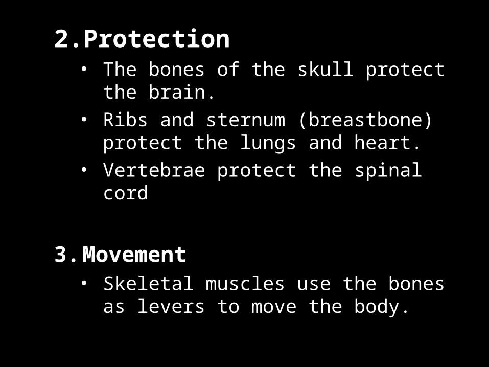
2.Protection • The bones of the skull protect the brain.• Ribs and sternum (breastbone) protect
the lungs and heart.• Vertebrae protect the spinal cord
3. Movement • Skeletal muscles use the bones as levers
to move the body.

3. Reservoir for minerals and adipose tissue• 99% of the body’s calcium is stored in
bone.• 85% of the body’s phosphorous is stored
in bone.• Adipose tissue is found in the marrow of
certain bones
4. Hematopoiesis • Blood cell formation.• All blood cells are made in the marrow of
certain bones
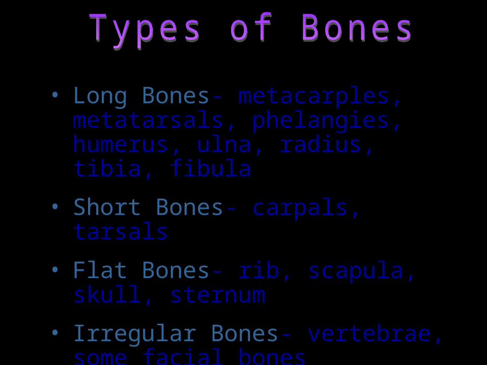
• Long Bones- metacarples, metatarsals, phelangies, humerus, ulna, radius, tibia, fibula
• Short Bones- carpals, tarsals
• Flat Bones- rib, scapula, skull, sternum
• Irregular Bones- vertebrae, some facial bones
• Sesamoid- patella
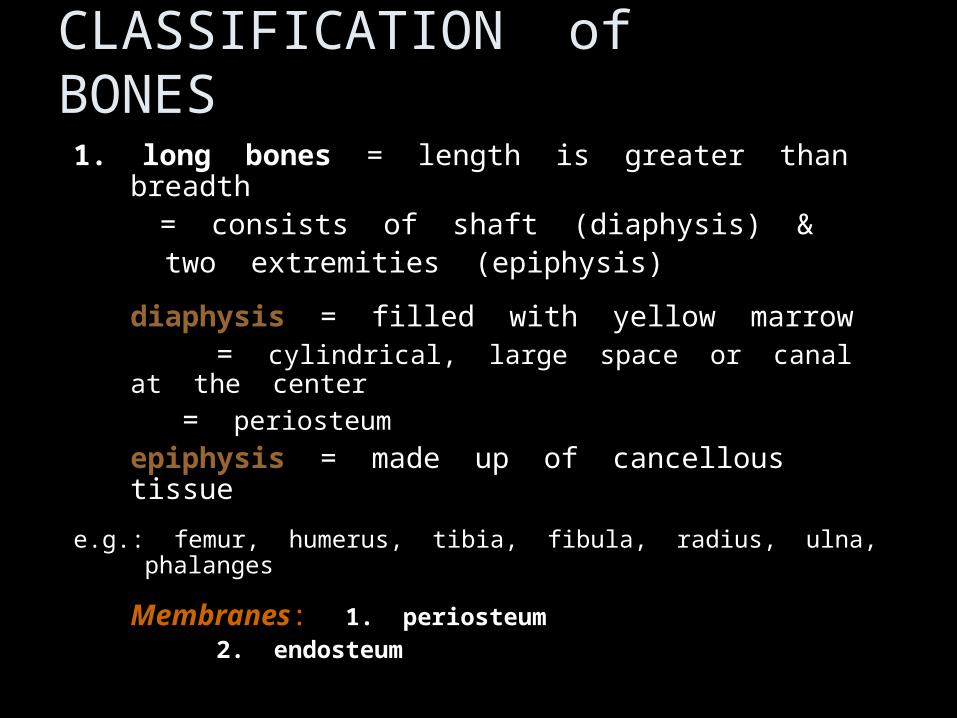
CLASSIFICATION of BONES
1.1. long boneslong bones = length is greater than breadth = length is greater than breadth = consists of shaft (diaphysis) & = consists of shaft (diaphysis) &
two extremities (epiphysis)two extremities (epiphysis)
diaphysis diaphysis = filled with yellow marrow = filled with yellow marrow= = cylindrical, large space or canal at the cylindrical, large space or canal at the
centercenter
= = periosteumperiosteum
epiphysis epiphysis = made up of cancellous tissue = made up of cancellous tissue
e.g.: femur, humerus, tibia, fibula, radius, ulna, phalangese.g.: femur, humerus, tibia, fibula, radius, ulna, phalanges
MembranesMembranes: : 1. periosteum1. periosteum
2. endosteum2. endosteum

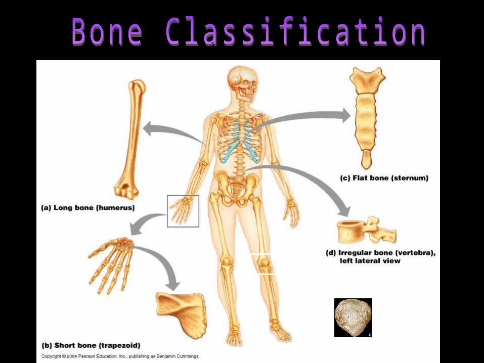
Bone Classification
1. Long Bones
• Much longer than they are wide.• All bones of the limbs except for the
patella (kneecap),
and the bones of the wrist and ankle.• Consists of a shaft plus 2 expanded ends.• Your finger bones are long bones even
though they’re very short
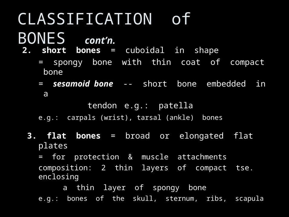
CLASSIFICATION of BONES cont’n.2. 2. short bonesshort bones = cuboidal in shape = cuboidal in shape
= spongy bone with thin coat of compact bone= spongy bone with thin coat of compact bone
= = sesamoid bonesesamoid bone -- short bone embedded in a -- short bone embedded in a
tendontendone.g.: patellae.g.: patella
e.g.: carpals (wrist), tarsal (ankle) bonese.g.: carpals (wrist), tarsal (ankle) bones
3. flat bones3. flat bones = broad or elongated flat plates = broad or elongated flat plates= for protection & muscle attachments= for protection & muscle attachments
composition: 2 thin layers of compact tse. enclosingcomposition: 2 thin layers of compact tse. enclosing
a thin layer of spongy bonea thin layer of spongy bone
e.g.: bones of the skull, sternum, ribs, scapulae.g.: bones of the skull, sternum, ribs, scapula
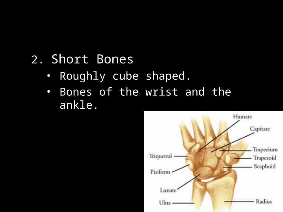
2. Short Bones• Roughly cube shaped.• Bones of the wrist and the ankle.

3. Flat Bones• Thin, flattened, and usually a bit curved.• Scapulae, sternum, (shoulder blades),
ribs and most bones of the skull.
Sternum
CLASSIFICATION of BONES cont’n.
4. Irregular bones4. Irregular bones = all other bones not assigned = all other bones not assigned to the previous groupsto the previous groups
e.g.: e.g.: vertebraevertebrae
pelvic bones pelvic bones
bones of the base of the skullbones of the base of the skull
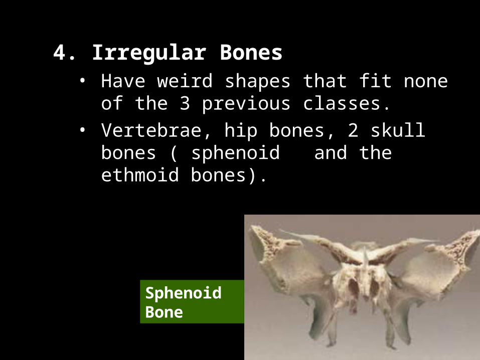
4. Irregular Bones• Have weird shapes that fit none of the 3
previous classes.• Vertebrae, hip bones, 2 skull bones
( sphenoid and the ethmoid bones).
Sphenoid Bone
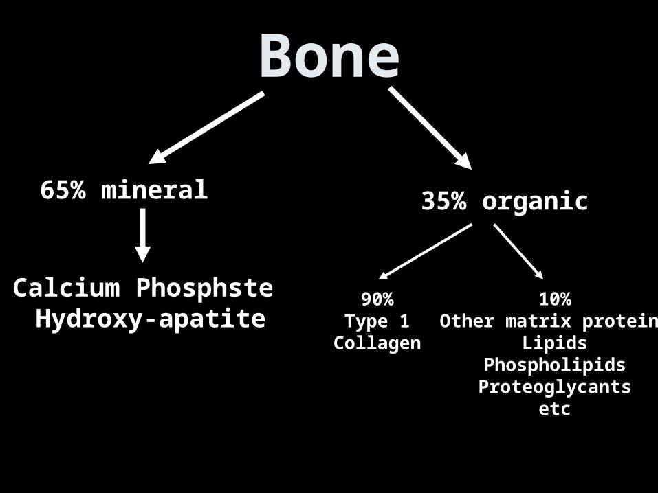
Bone
65% mineral 35% organic
Calcium Phosphste Hydroxy-apatite
90%Type 1
Collagen
10%Other matrix proteins
LipidsPhospholipidsProteoglycants
etc
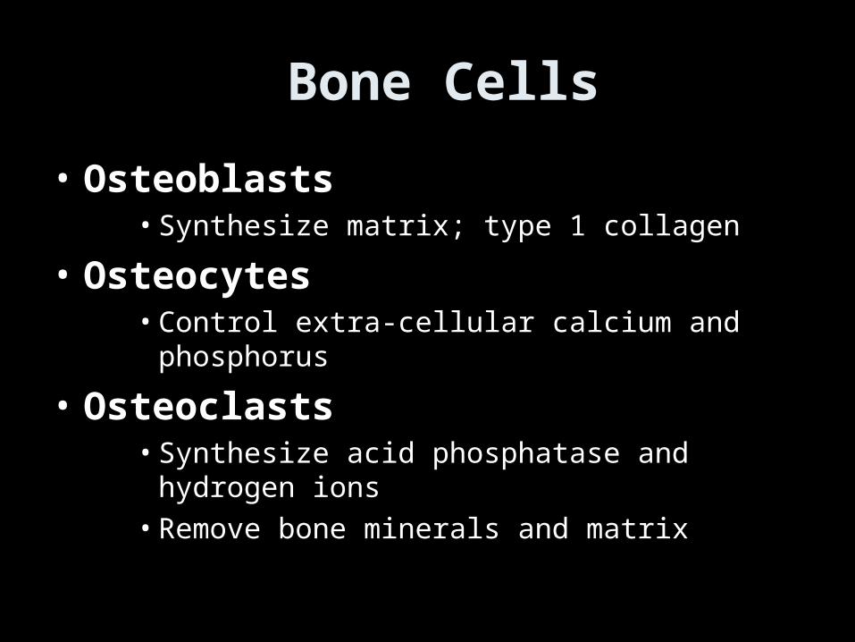
Bone Cells
• Osteoblasts• Synthesize matrix; type 1 collagen
• Osteocytes• Control extra-cellular calcium and phosphorus
• Osteoclasts• Synthesize acid phosphatase and hydrogen ions• Remove bone minerals and matrix
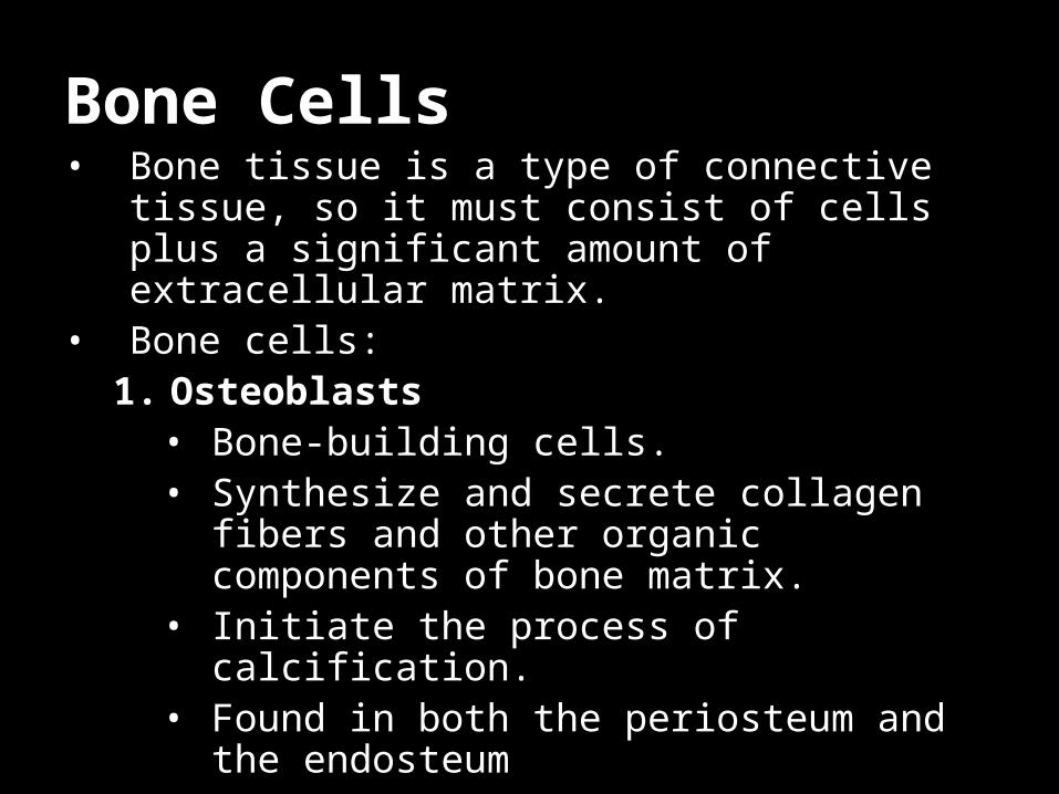
Bone Cells• Bone tissue is a type of connective tissue, so it
must consist of cells plus a significant amount of extracellular matrix.
• Bone cells:1. Osteoblasts
• Bone-building cells.• Synthesize and secrete collagen fibers
and other organic components of bone matrix.
• Initiate the process of calcification.• Found in both the periosteum and the
endosteum
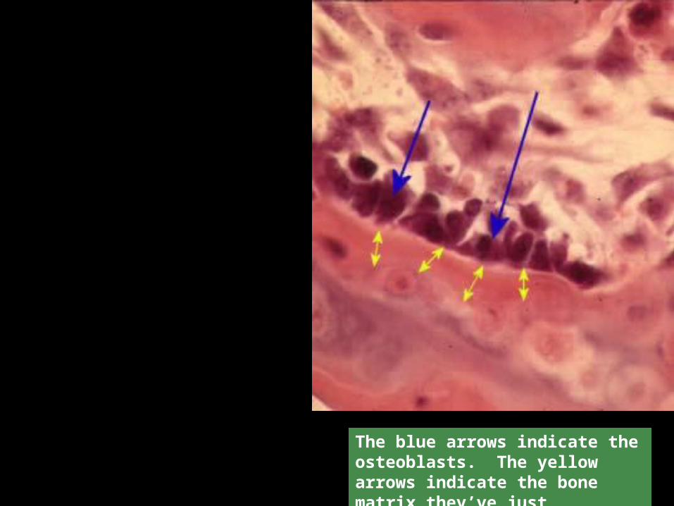
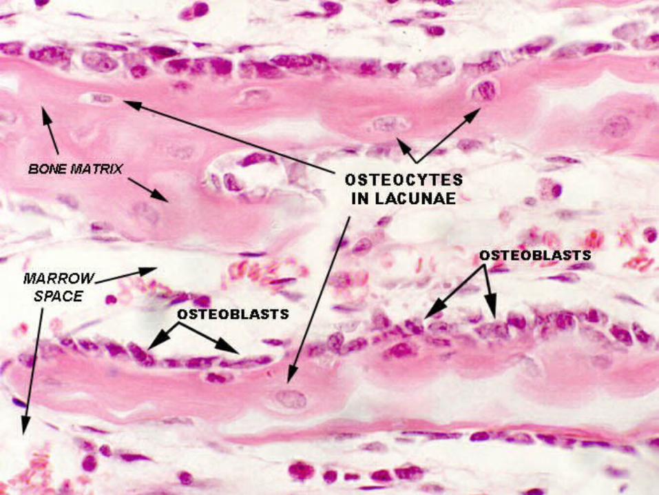
The blue arrows indicate the osteoblasts. The yellow arrows indicate the bone matrix they’ve just secreted.
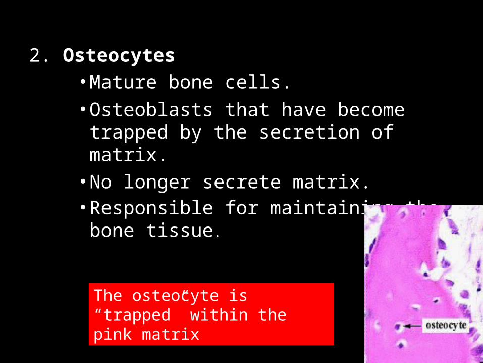
2. Osteocytes• Mature bone cells.• Osteoblasts that have become trapped by
the secretion of matrix.• No longer secrete matrix.• Responsible for maintaining the bone
tissue.
The osteocyte is “trapped” within the pink matrix
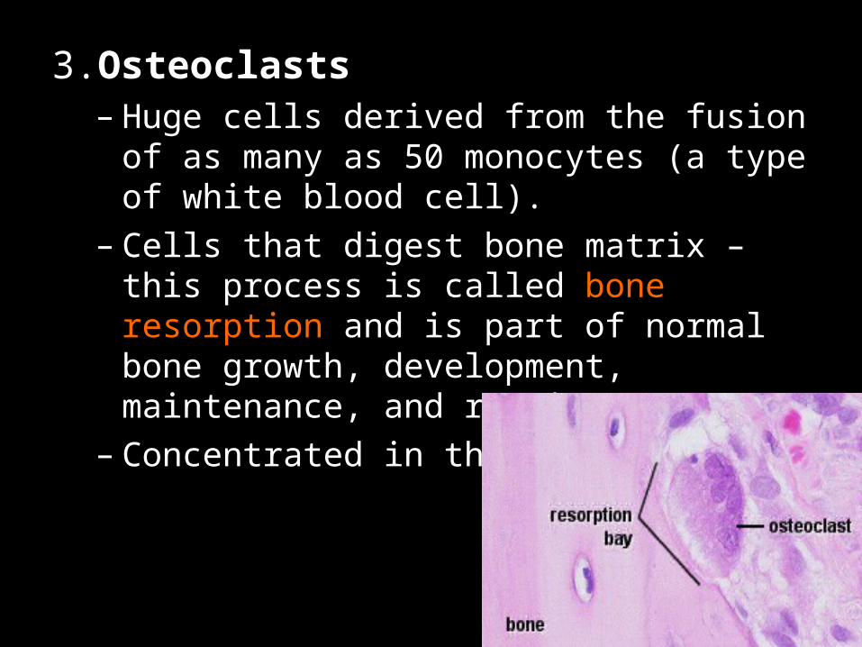
3.Osteoclasts– Huge cells derived from the fusion of as many
as 50 monocytes (a type of white blood cell).– Cells that digest bone matrix – this process is
called bone resorption and is part of normal bone growth, development, maintenance, and repair.
– Concentrated in the endosteum.
Bone Matrix:– Consists of organic and inorganic
components.• Organic component consists of several
materials that are secreted by the osteoblasts:
–Collagen fibers and other organic materials
»These (particularly the collagen) provide the bone with resilience and the ability to resist stretching and twisting
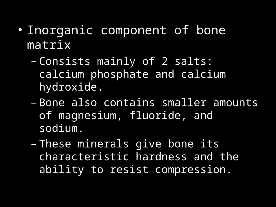
• Inorganic component of bone matrix– Consists mainly of 2 salts: calcium phosphate
and calcium hydroxide.– Bone also contains smaller amounts of
magnesium, fluoride, and sodium.– These minerals give bone its characteristic
hardness and the ability to resist compression.

Distal
epiphysis
Proximal
epiphysis
diaphysis
yellow marrow
epiphyseal line
periosteum
compact bone
spongy bone
Endosteum
hyaline cartilage
Sharpey’s fibers

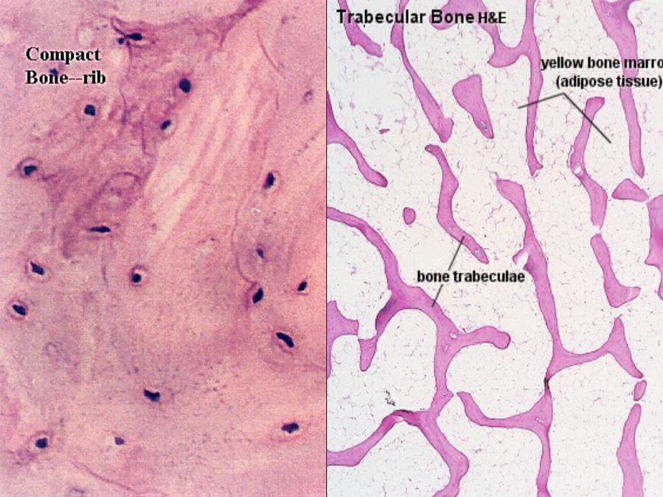
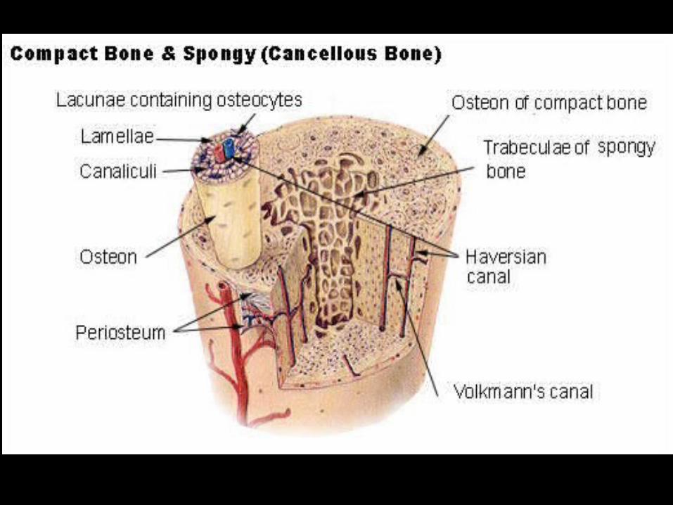
2 Types of Bone
Compact bone
Spongy bone
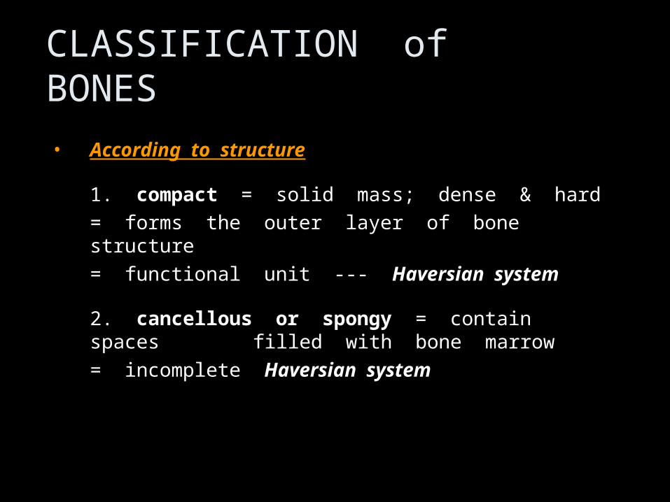
CLASSIFICATION of BONES
• According to structureAccording to structure
1. compact = solid mass; dense & hard
= forms the outer layer of bone structure
= functional unit --- Haversian system
2. cancellous or spongy = contain spaces filled with bone marrow
= incomplete Haversian system

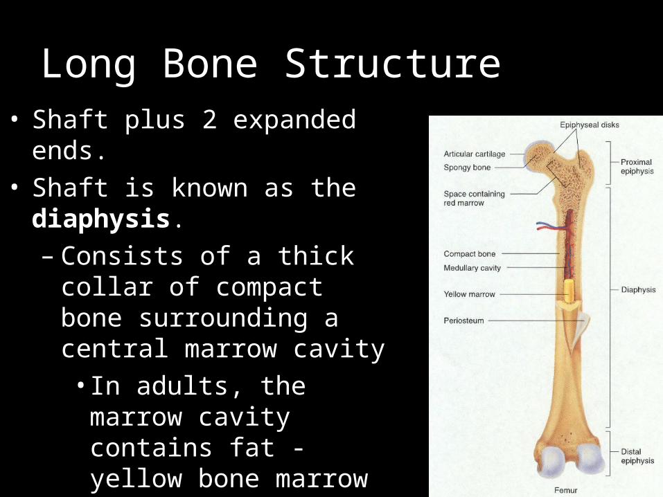
Long Bone Structure• Shaft plus 2 expanded ends.• Shaft is known as the
diaphysis.– Consists of a thick collar of
compact bone surrounding a central marrow cavity• In adults, the marrow
cavity contains fat - yellow bone marrow
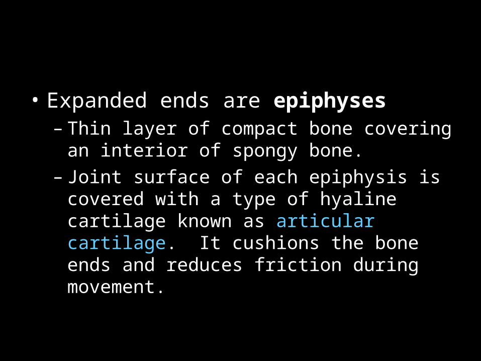
• Expanded ends are epiphyses– Thin layer of compact bone covering an
interior of spongy bone.– Joint surface of each epiphysis is covered
with a type of hyaline cartilage known as articular cartilage. It cushions the bone ends and reduces friction during movement.
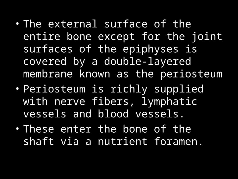
• The external surface of the entire bone except for the joint surfaces of the epiphyses is covered by a double-layered membrane known as the periosteum
• Periosteum is richly supplied with nerve fibers, lymphatic vessels and blood vessels.
• These enter the bone of the shaft via a nutrient foramen.
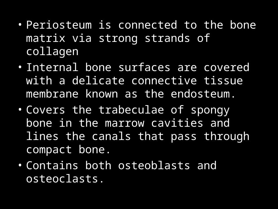
• Periosteum is connected to the bone matrix via strong strands of collagen
• Internal bone surfaces are covered with a delicate connective tissue membrane known as the endosteum.
• Covers the trabeculae of spongy bone in the marrow cavities and lines the canals that pass through compact bone.
• Contains both osteoblasts and osteoclasts.
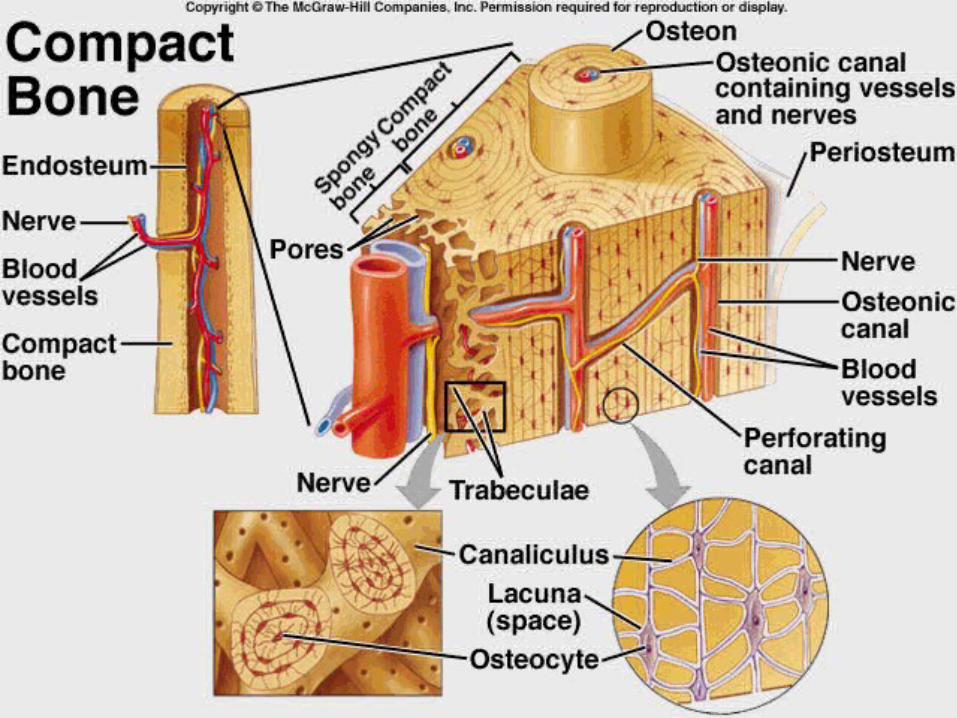
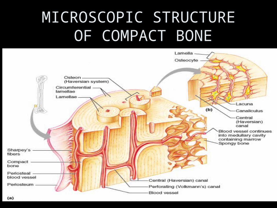
MICROSCOPIC STRUCTURE OF COMPACT BONE
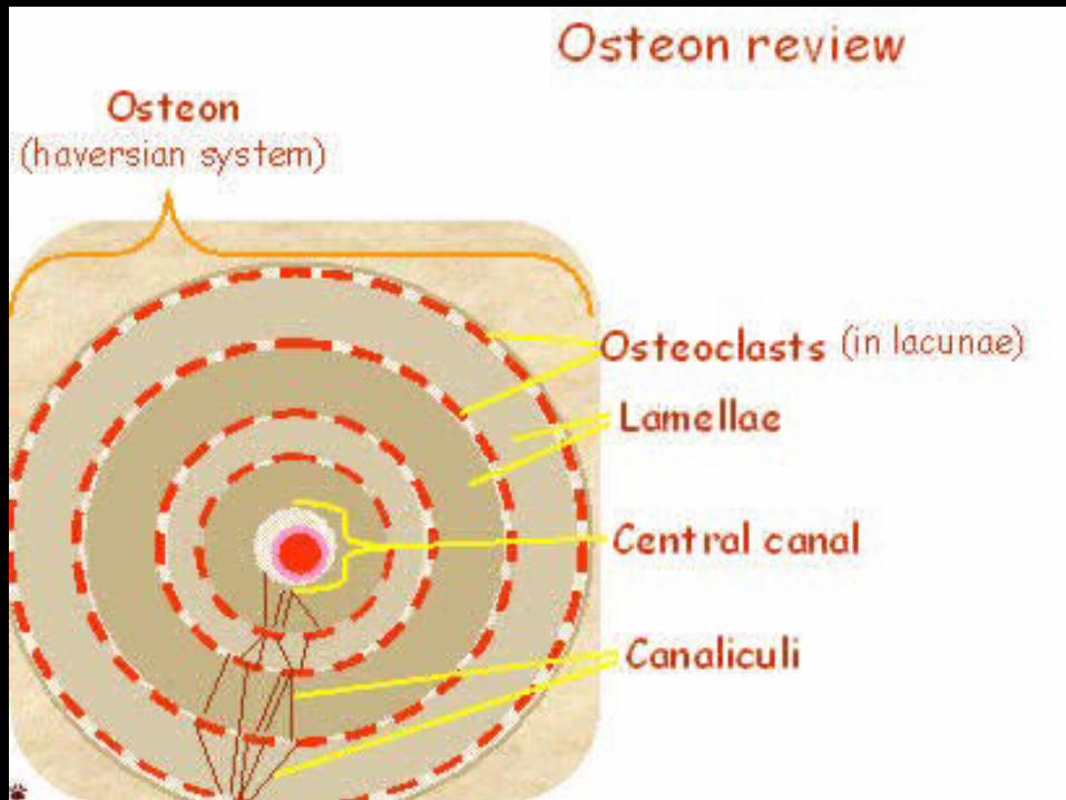
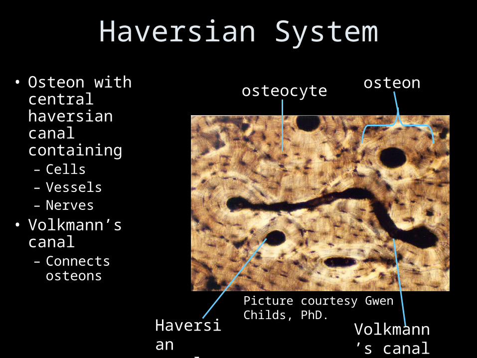
Haversian System
• Osteon with central haversian canal containing– Cells – Vessels– Nerves
• Volkmann’s canal – Connects
osteons
Picture courtesy Gwen Childs, PhD.
osteon
Haversian canal
osteocyte
Volkmann’s canal
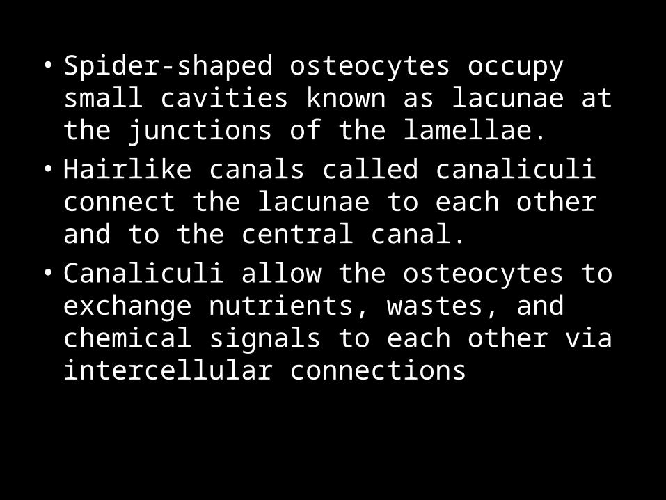
• Spider-shaped osteocytes occupy small cavities known as lacunae at the junctions of the lamellae.
• Hairlike canals called canaliculi connect the lacunae to each other and to the central canal.
• Canaliculi allow the osteocytes to exchange nutrients, wastes, and chemical signals to each other via intercellular connections

Bones - Animation
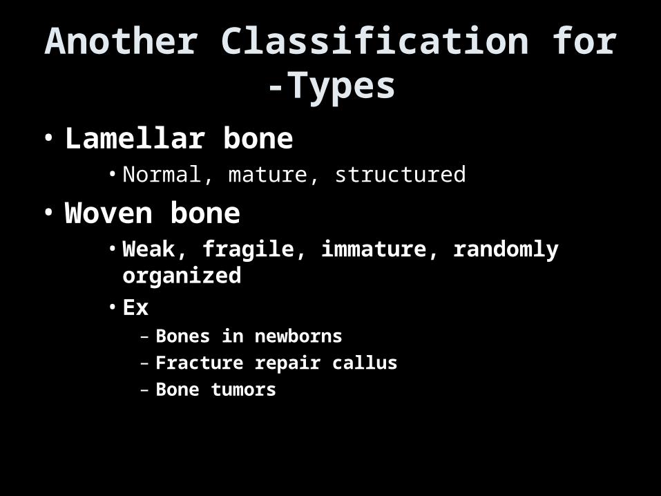
Another Classification for -Types
• Lamellar bone• Normal, mature, structured
• Woven bone• Weak, fragile, immature, randomly organized• Ex
– Bones in newborns– Fracture repair callus– Bone tumors
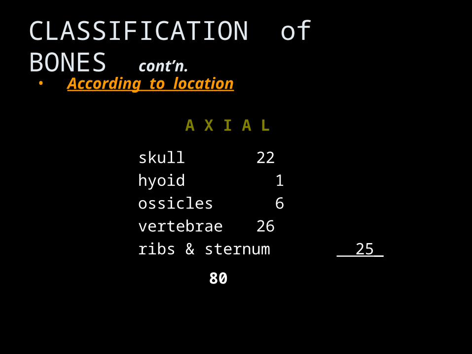
CLASSIFICATION of BONES cont’n.• According to locationAccording to location
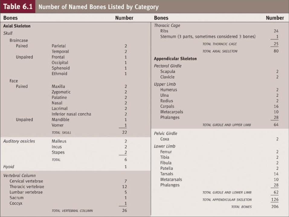
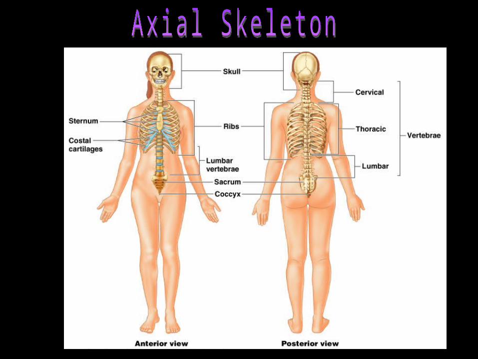
A X I A LA X I A L
skullskull 2222
hyoidhyoid 1 1
ossiclesossicles 6 6
vertebraevertebrae 2626
ribs & sternum ribs & sternum 25_ 25_
8080
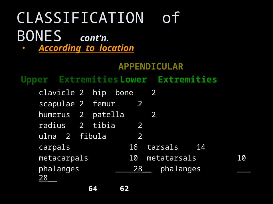
CLASSIFICATION of BONES cont’n.• According to locationAccording to location
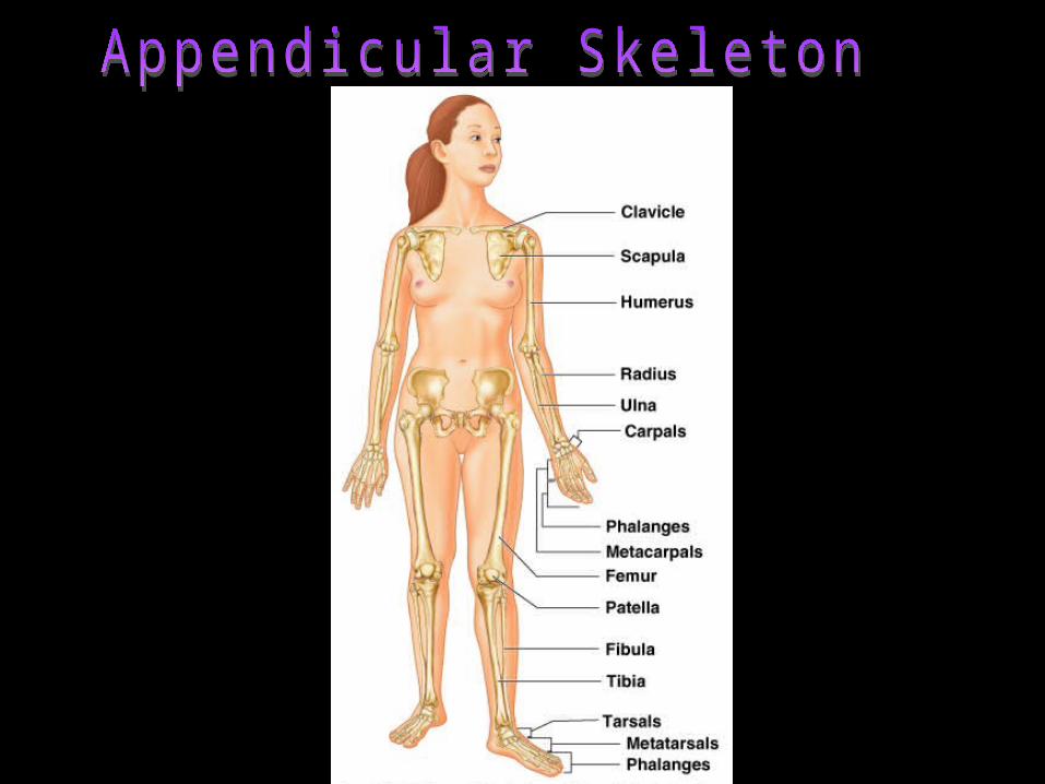
APPENDICULARAPPENDICULAR
Upper ExtremitiesUpper Extremities Lower ExtremitiesLower Extremities
clavicleclavicle 22 hip bonehip bone 2 2
scapulaescapulae 22 femurfemur 2 2
humerushumerus 22 patellapatella 2 2
radiusradius 22 tibiatibia 2 2
ulnaulna 22 fibulafibula 2 2
carpalscarpals 16 16 tarsalstarsals 14 14
metacarpals 10metacarpals 10 metatarsals metatarsals 10 10
phalanges phalanges 28__ 28__ phalanges phalanges 28__ 28__
6464 62 62

Table. 6.2

GROSS ANATOMY OF A LONG BONE
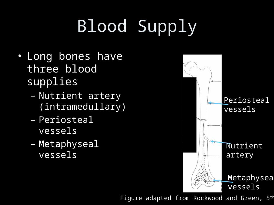
Blood Supply
• Long bones have three blood supplies– Nutrient artery
(intramedullary)– Periosteal vessels– Metaphyseal vessels
Nutrient artery
Metaphysealvessels
Periosteal vessels
Figure adapted from Rockwood and Green, 5th Ed
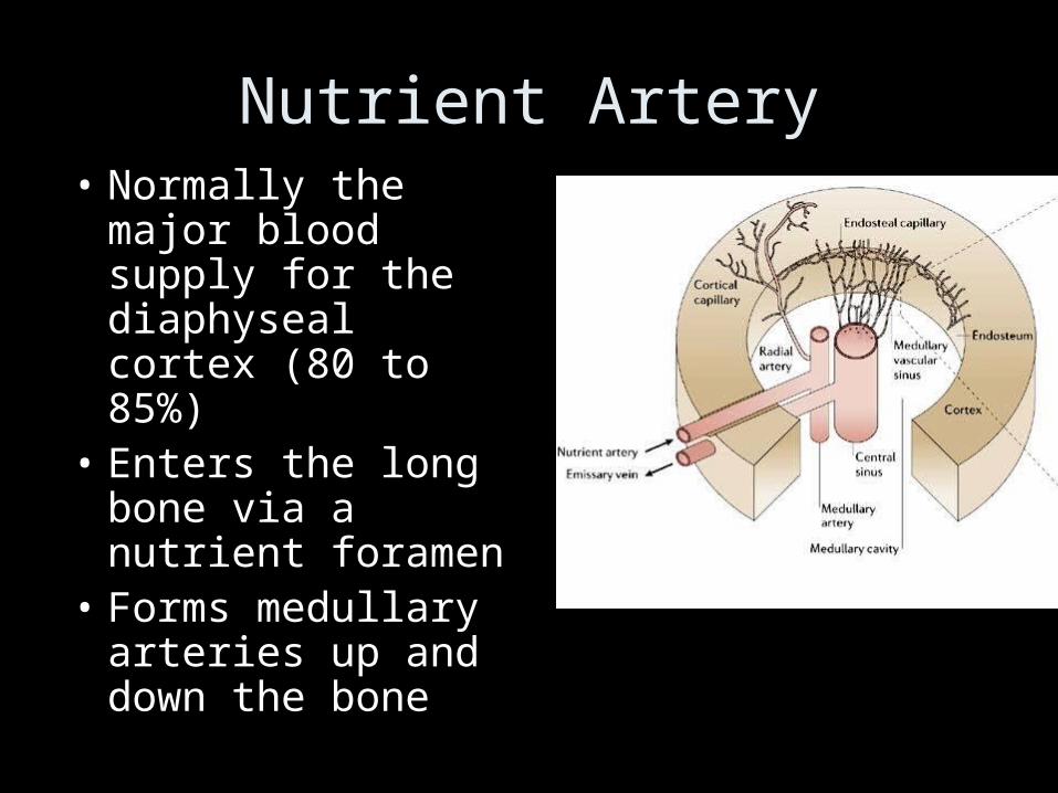
Nutrient Artery• Normally the major
blood supply for the diaphyseal cortex (80 to 85%)
• Enters the long bone via a nutrient foramen
• Forms medullary arteries up and down the bone
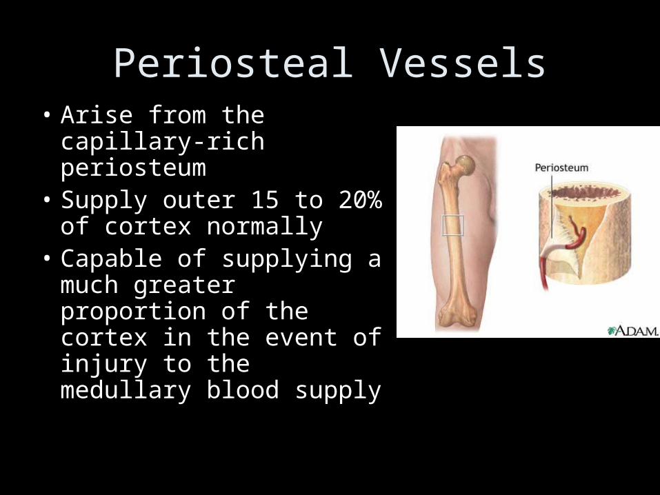
Periosteal Vessels• Arise from the capillary-
rich periosteum• Supply outer 15 to 20%
of cortex normally• Capable of supplying a
much greater proportion of the cortex in the event of injury to the medullary blood supply
Metaphyseal Vessels
• Arise from periarticular vessels
• Penetrate the thin cortex in the metaphyseal region and anastomose with the medullary blood supply
Bone Marrow
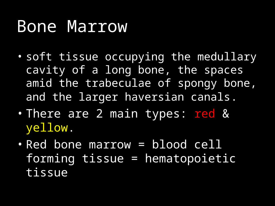
• soft tissue occupying the medullary cavity of a long bone, the spaces amid the trabeculae of spongy bone, and the larger haversian canals.
• There are 2 main types: red & yellow.• Red bone marrow = blood cell forming
tissue = hematopoietic tissue
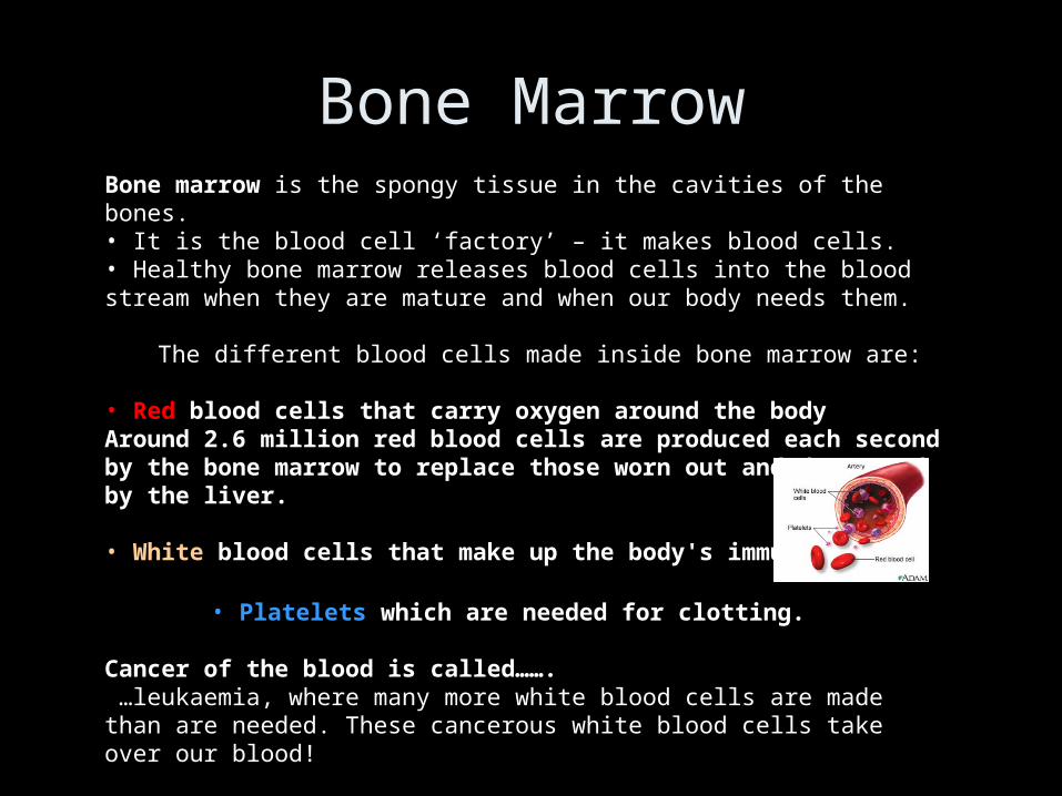
Bone marrow is the spongy tissue in the cavities of the bones. • It is the blood cell ‘factory’ – it makes blood cells. • Healthy bone marrow releases blood cells into the blood stream when they are mature and when our body needs them.
The different blood cells made inside bone marrow are:
• Red blood cells that carry oxygen around the body Around 2.6 million red blood cells are produced each second by the bone marrow to replace those worn out and destroyed by the liver.
• White blood cells that make up the body's immune system
• Platelets which are needed for clotting.
Cancer of the blood is called……. …leukaemia, where many more white blood cells are made than are needed. These cancerous white blood cells take over our blood!
Bone Marrow
Bone Remodeling - Steps
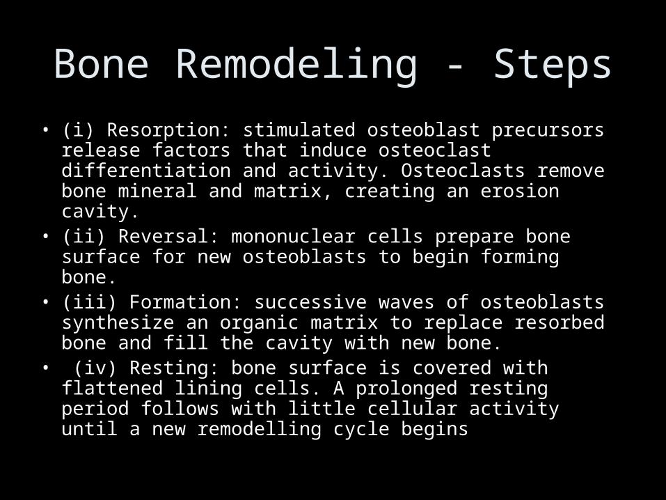
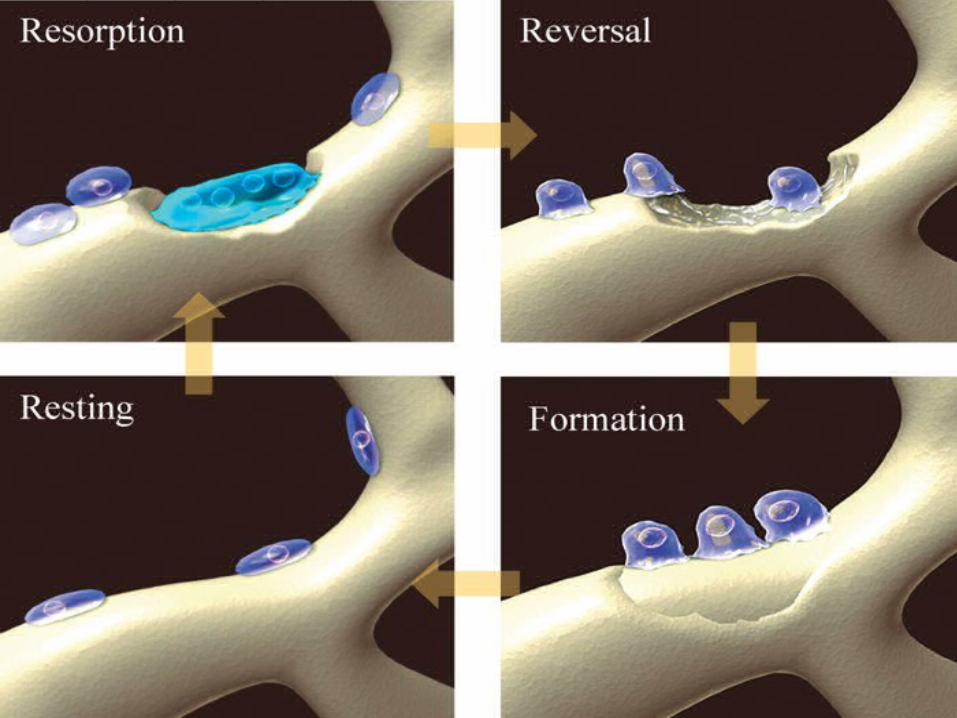
• (i) Resorption: stimulated osteoblast precursors release factors that induce osteoclast differentiation and activity. Osteoclasts remove bone mineral and matrix, creating an erosion cavity.
• (ii) Reversal: mononuclear cells prepare bone surface for new osteoblasts to begin forming bone.
• (iii) Formation: successive waves of osteoblasts synthesize an organic matrix to replace resorbed bone and fill the cavity with new bone.
• (iv) Resting: bone surface is covered with flattened lining cells. A prolonged resting period follows with little cellular activity until a new remodelling cycle begins

Bone Remodelling
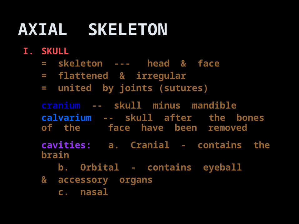
AXIAL SKELETONI.I. SKULLSKULL
= skeleton --- head & face= skeleton --- head & face= flattened & irregular= flattened & irregular= united by joints (sutures)= united by joints (sutures)
cranium cranium -- skull minus mandible-- skull minus mandiblecalvarium calvarium -- skull after the bones of the -- skull after the bones of the
face have been removed face have been removed
cavities:cavities: a. Cranial - contains the brain a. Cranial - contains the brain b. Orbital - contains eyeball b. Orbital - contains eyeball
& accessory organs& accessory organs c. nasalc. nasal
The skull
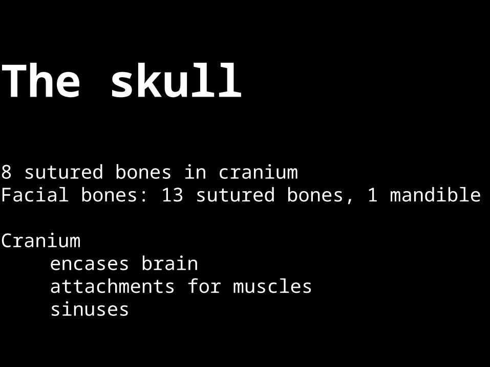
8 sutured bones in craniumFacial bones: 13 sutured bones, 1 mandible
Craniumencases brainattachments for musclessinuses

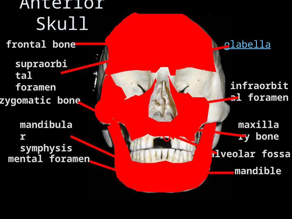
Anterior Skull
frontal bonefrontal bone
supraorbital supraorbital foramenforamen
zygomatic bonezygomatic bone
maxillary maxillary bonebone
alveolar fossaalveolar fossa
infraorbital infraorbital foramenforamen
glabella
mental foramenmental foramenmandiblemandible
mandibular mandibular symphysissymphysis
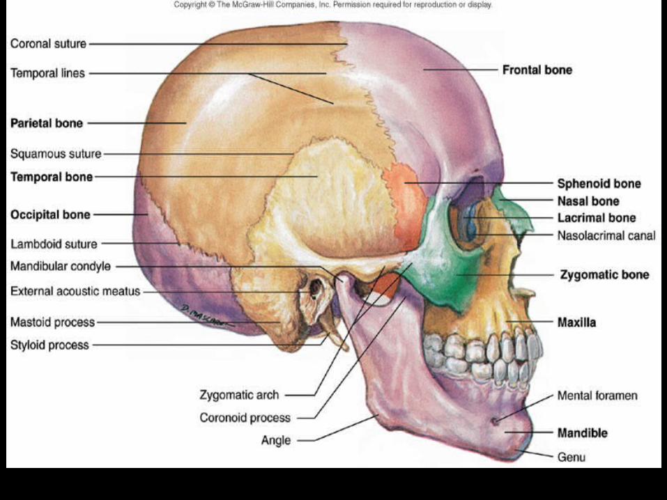
Fig. 6.11
Sutures
• Sutures are the tight connections between skull bones
• In the adult skull , the skull bones are unable to move or separate due to this tight connections
• In infant skull this sutures are not well formed and the skull bones are mobile which facilitates the delivery of the baby
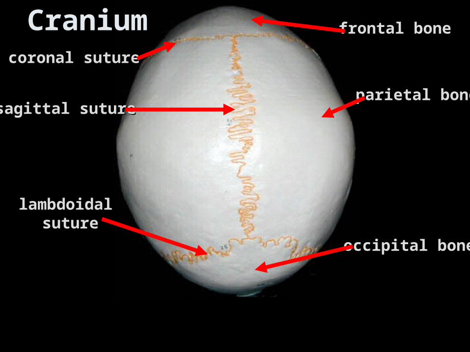
Cranium frontal bonefrontal bone
parietal boneparietal bone
occipital boneoccipital bone
lambdoidallambdoidal suturesuture
sagittal suturesagittal suture
coronal suturecoronal suture
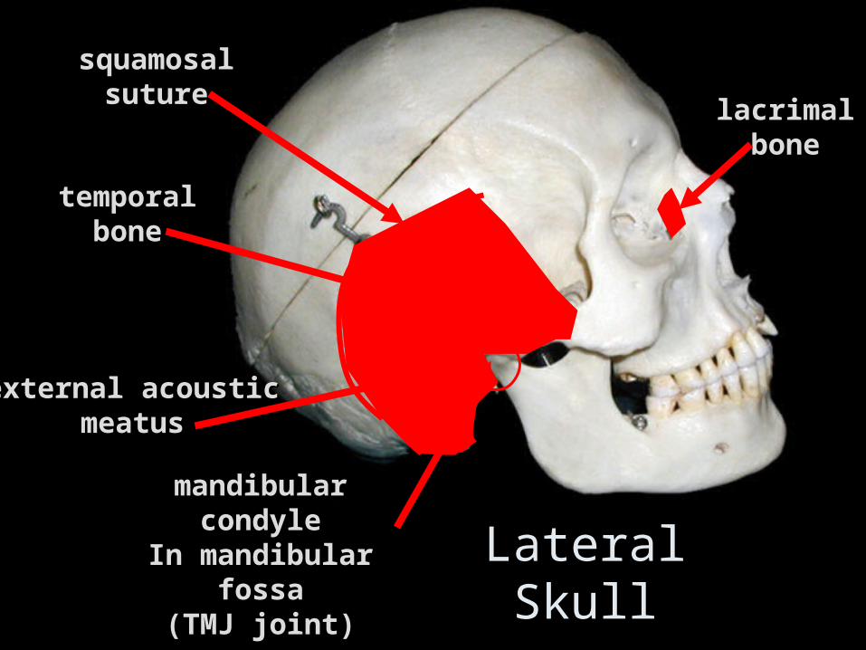
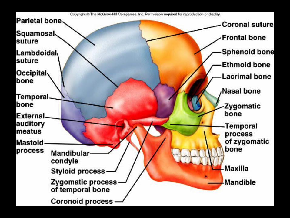
Lateral Skull
lacrimallacrimalbonebone
temporaltemporalbonebone
squamosalsquamosalsuturesuture
mandibular condylemandibular condyleIn mandibular fossaIn mandibular fossa
(TMJ joint)(TMJ joint)
external acousticexternal acousticmeatusmeatus
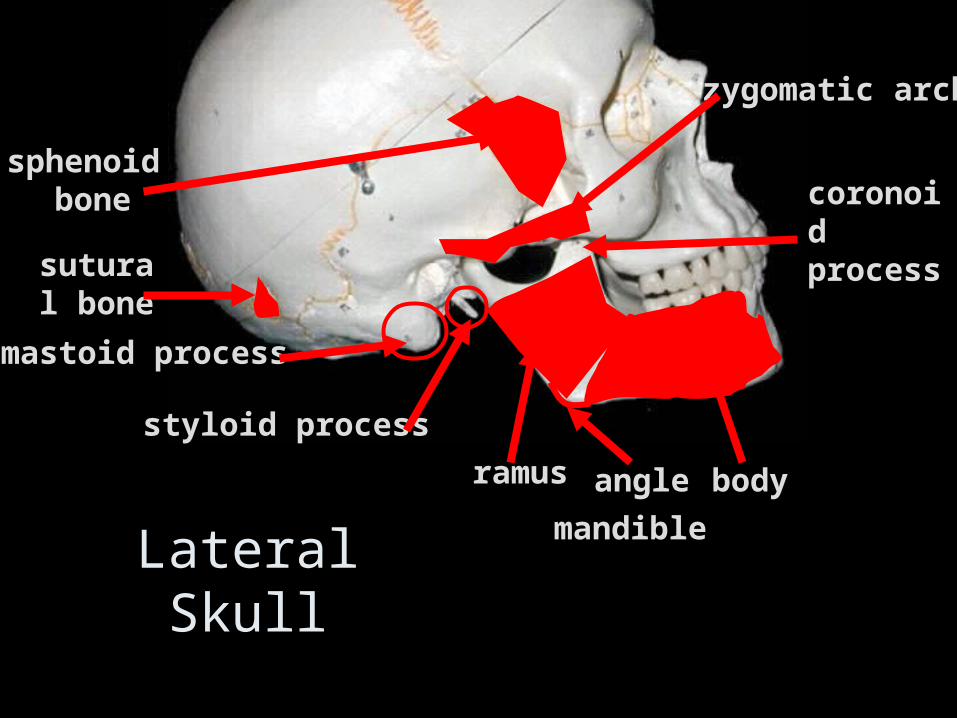
angleangle
coronoid coronoid processprocess
zygomatic archzygomatic arch
mastoid processmastoid process
styloid processstyloid process
sphenoid sphenoid bonebone
bodybodyramusramus
mandiblemandibleLateral Skull
sutural sutural bonebone
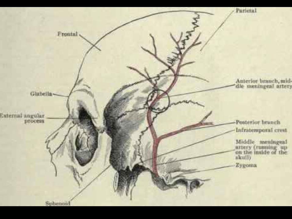
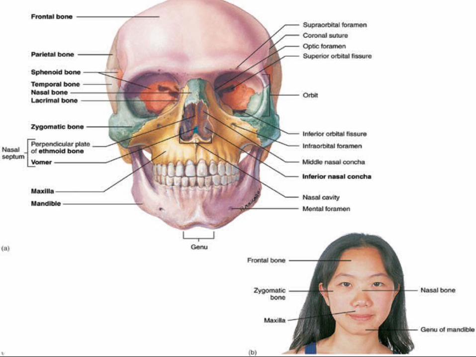
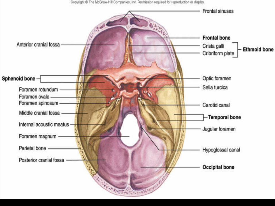
Fig. 6.14
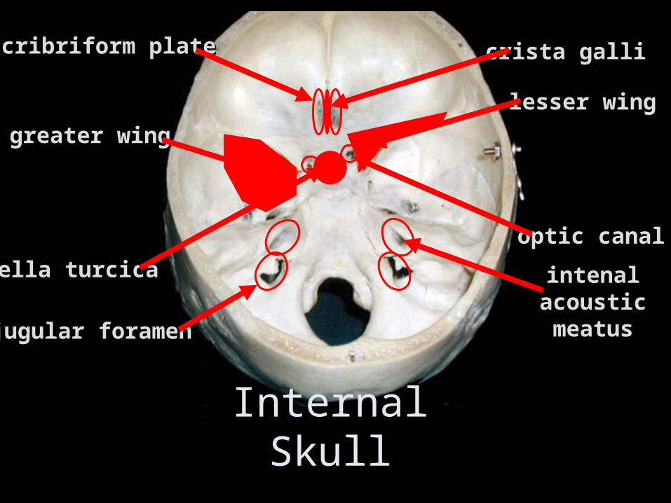
crista gallicrista gallicribriform platecribriform plate
intenal intenal acoustic acoustic meatusmeatus
greater winggreater wing
lesser winglesser wing
optic canaloptic canal
sella turcicasella turcica
jugular foramenjugular foramen
Internal Skull
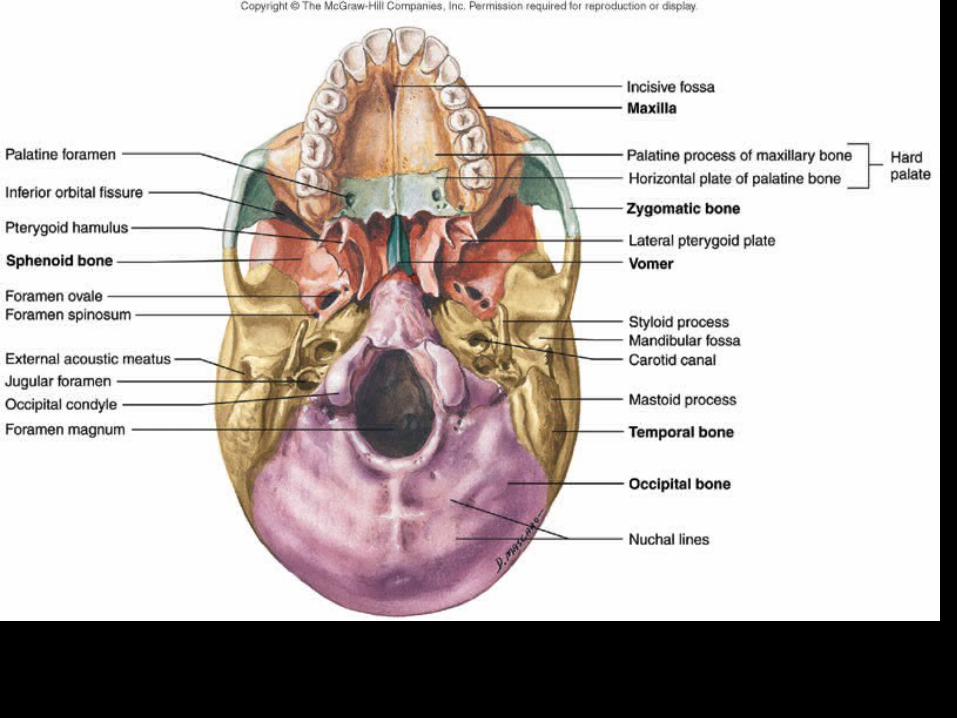
Fig. 6.15

Ventral Skull
palatine processpalatine process
palatine bonepalatine bone
vomer bonevomer bone
mastoid processmastoid process
styloid processstyloid process
external occipitalexternal occipitalprotuberanceprotuberance
sphenoid bonesphenoid bone
temporal bonetemporal bone
occipital boneoccipital bone
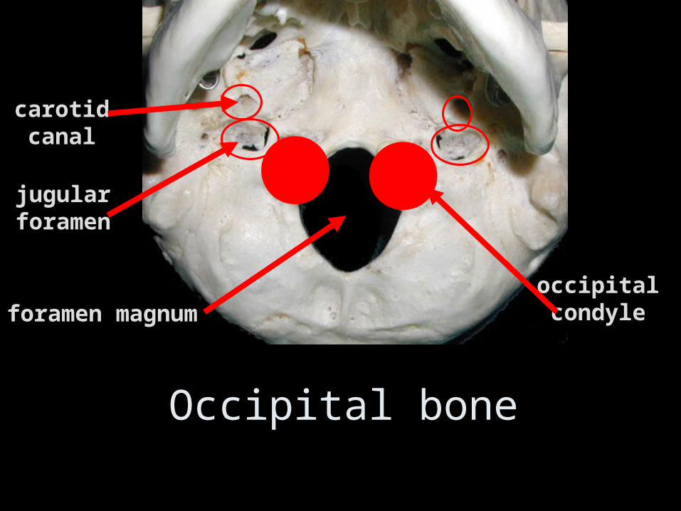
Occipital bone
occipitaloccipitalcondylecondyle
jugularjugularforamenforamen
carotidcarotidcanalcanal
foramen magnumforamen magnum
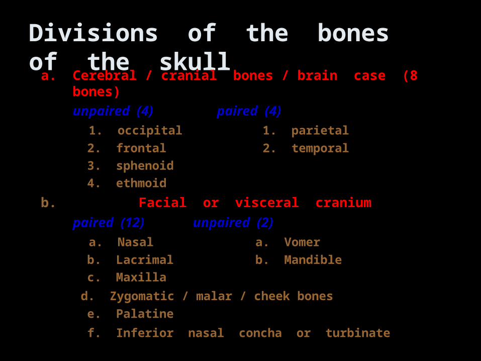
Divisions of the bones of the skull
a.a. Cerebral / cranial bones / brain case (8 bones)Cerebral / cranial bones / brain case (8 bones)
unpaired (4)unpaired (4) paired (4)paired (4)
1. occipital1. occipital 1. parietal 1. parietal
2. frontal2. frontal 2. temporal 2. temporal
3. sphenoid3. sphenoid
4. ethmoid4. ethmoid
b.b. Facial or visceral craniumFacial or visceral cranium
paired (12)paired (12) unpaired (2)unpaired (2)
a. Nasala. Nasal a. Vomer a. Vomer
b. Lacrimalb. Lacrimal b. Mandible b. Mandible
c. Maxillac. Maxilla
d. Zygomatic / malar / cheek bonesd. Zygomatic / malar / cheek bones
e. Palatinee. Palatine
f. Inferior nasal concha or turbinatef. Inferior nasal concha or turbinate
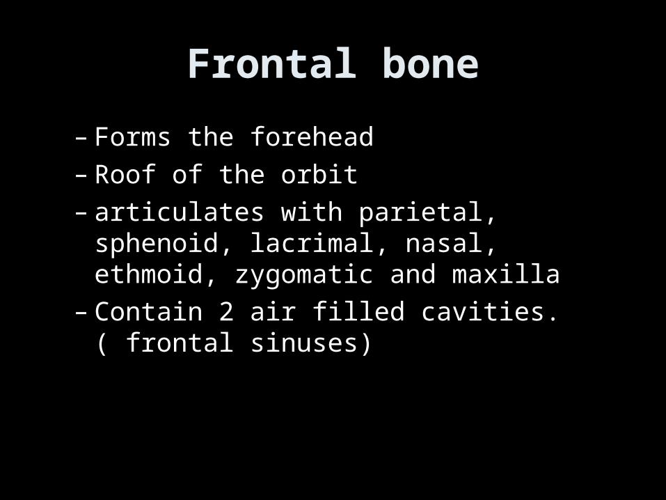
Frontal bone
– Forms the forehead– Roof of the orbit– articulates with parietal, sphenoid, lacrimal,
nasal, ethmoid, zygomatic and maxilla– Contain 2 air filled cavities. ( frontal sinuses)
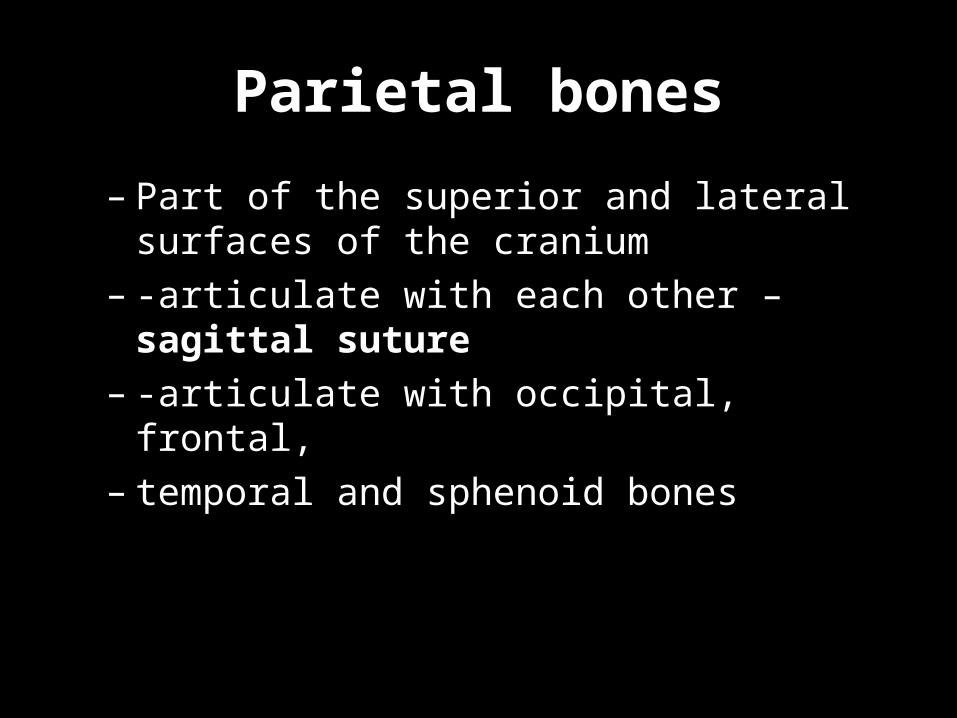
Parietal bones
– Part of the superior and lateral surfaces of the cranium
– -articulate with each other – sagittal suture– -articulate with occipital, frontal,– temporal and sphenoid bones
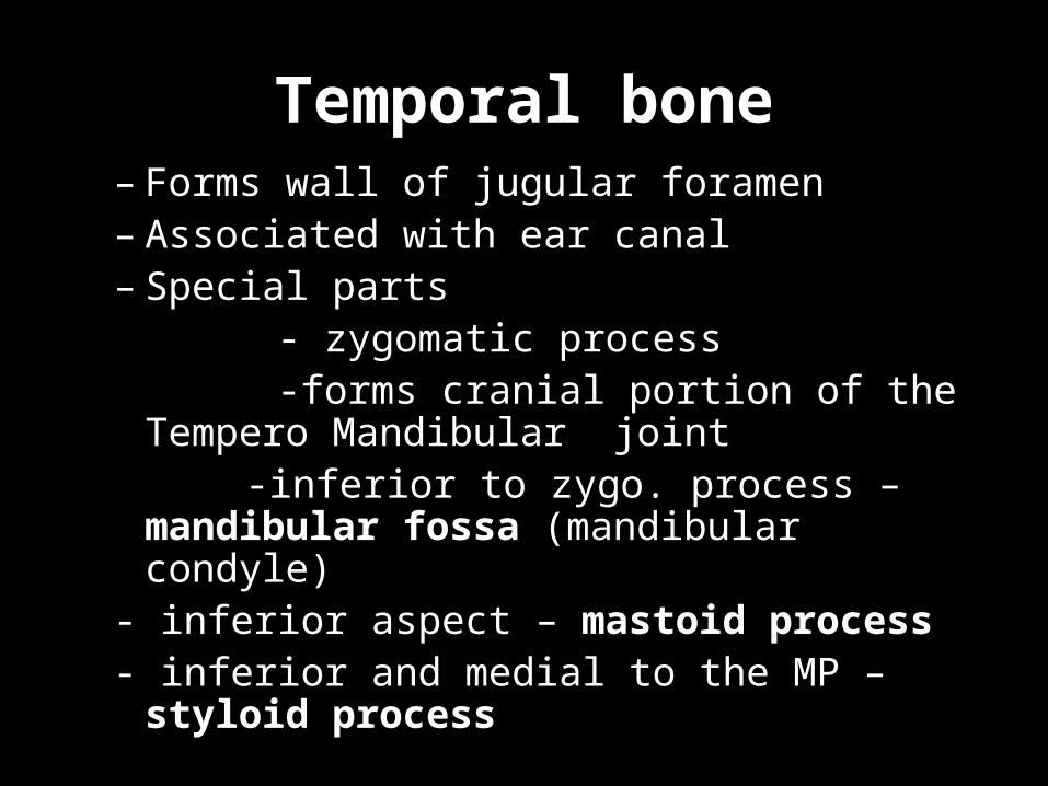
Temporal bone– Forms wall of jugular foramen– Associated with ear canal– Special parts - zygomatic process -forms cranial portion of the Tempero
Mandibular joint -inferior to zygo. process – mandibular
fossa (mandibular condyle)- inferior aspect – mastoid process- inferior and medial to the MP – styloid
process
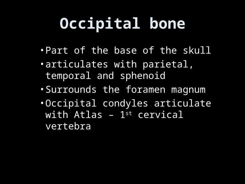
Occipital bone
• Part of the base of the skull• articulates with parietal, temporal and
sphenoid• Surrounds the foramen magnum• Occipital condyles articulate with Atlas – 1st
cervical vertebra

The uppermost portion of the human respiratory system, the nose is a hollow air passage that functions in breathing and in the sense of smell. The nasal cavity moistens and warms incoming air, while small hairs and mucus filter out harmful particles and micro-organisms. This illustration depicts the interior of the human nose.
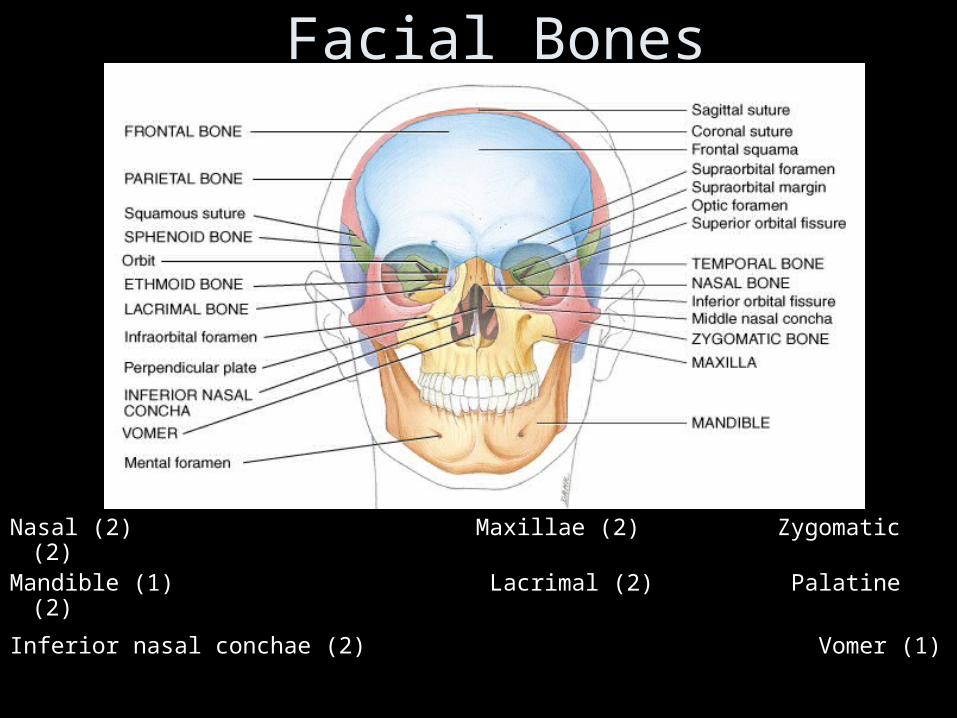
Facial Bones
Nasal (2) Maxillae (2) Zygomatic (2)Mandible (1) Lacrimal (2) Palatine (2)
Inferior nasal conchae (2) Vomer (1)
Anterior Skull
frontal bonefrontal bone
supraorbital supraorbital foramenforamen
zygomatic bonezygomatic bone
maxillary maxillary bonebone
alveolar fossaalveolar fossa
infraorbital infraorbital foramenforamen
glabella
mental foramenmental foramenmandiblemandible
mandibular mandibular symphysissymphysis
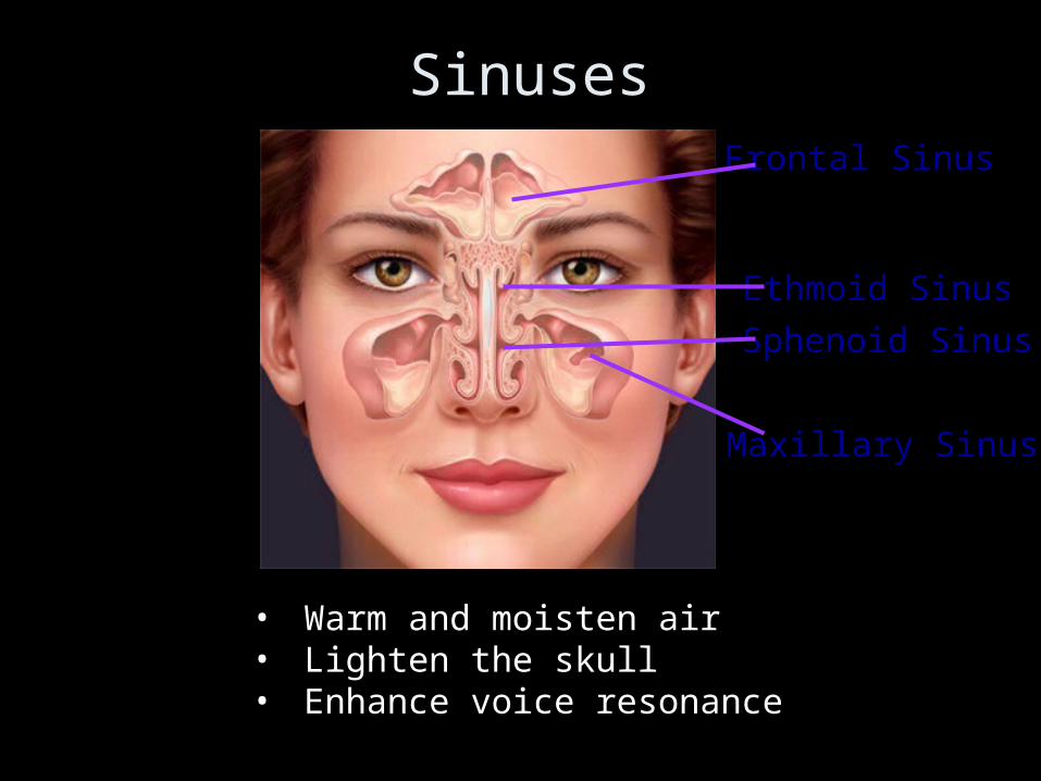
• Warm and moisten air• Lighten the skull• Enhance voice resonance
Frontal Sinus
Ethmoid Sinus
Sphenoid Sinus
Maxillary Sinus
Sinuses
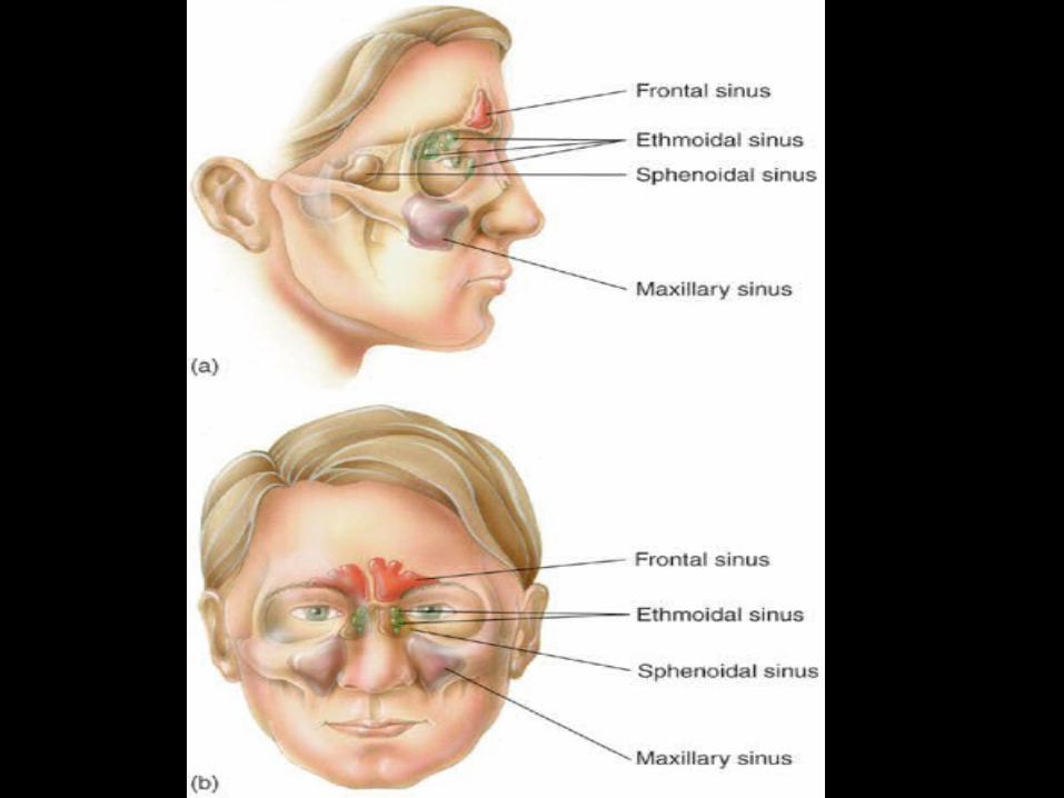
Fig. 6.13
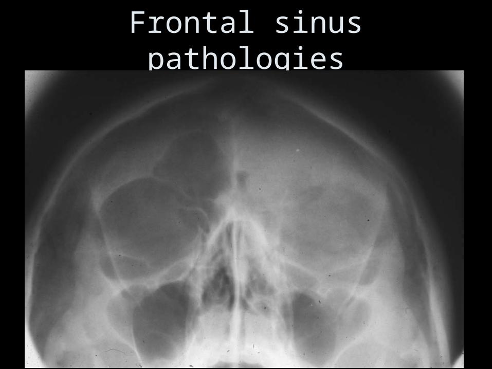
Frontal sinus pathologies
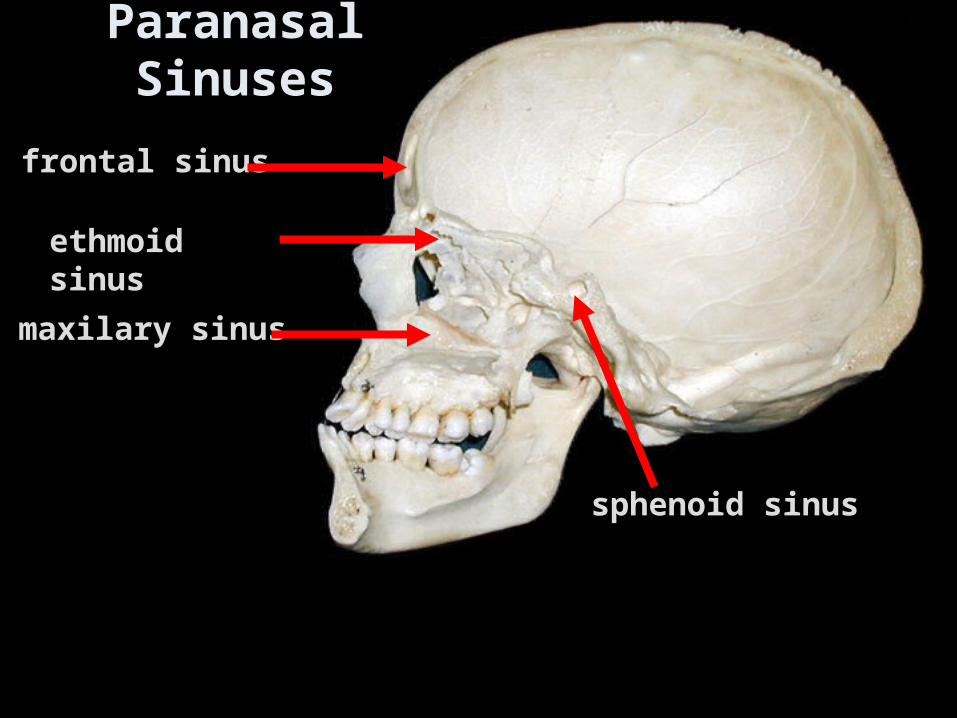
Paranasal Sinuses
frontal sinusfrontal sinus
ethmoid sinusethmoid sinus
maxilary sinusmaxilary sinus
sphenoid sinussphenoid sinus

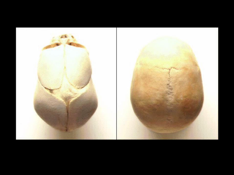
Fontanelles
• These are seen in new borns
• At birth the junction between skull bones covered with a membrane
• This allows the expansion and growth of the brain
• Later they become ossified ( bone formation)

Fontanelle = membrane filled spaces found in the skull of= membrane filled spaces found in the skull of
newborn infantsnewborn infants
e.g.:e.g.: 1. 1. anterior anterior = largest= largest
2. 2. posterior posterior 3. anterolateral (sphenoidal)3. anterolateral (sphenoidal)
4. posterolateral (mastoid)4. posterolateral (mastoid)
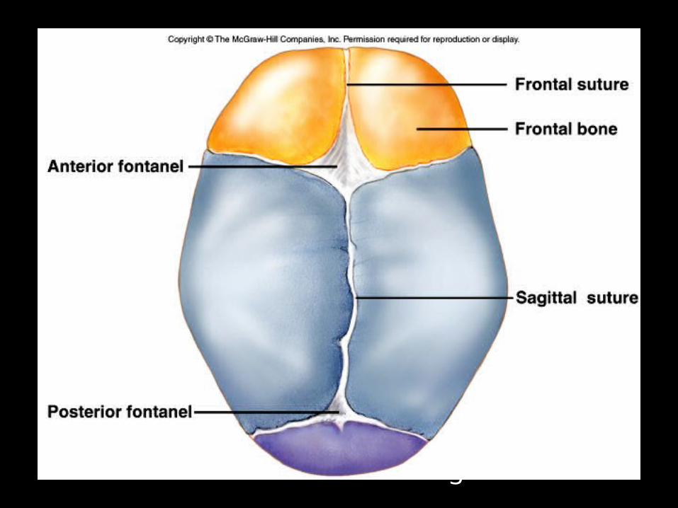
Allows forgrowth

Hydrocephalus
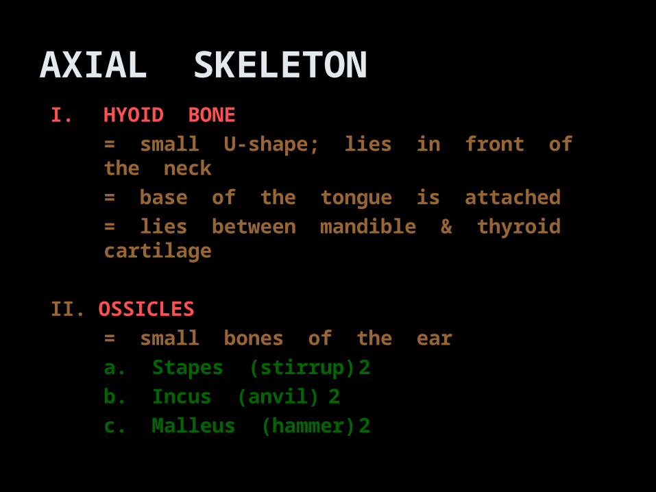
AXIAL SKELETONI.I. HYOID BONE HYOID BONE
= small U-shape; lies in front of the neck= small U-shape; lies in front of the neck
= base of the tongue is attached= base of the tongue is attached
= lies between mandible & thyroid cartilage= lies between mandible & thyroid cartilage
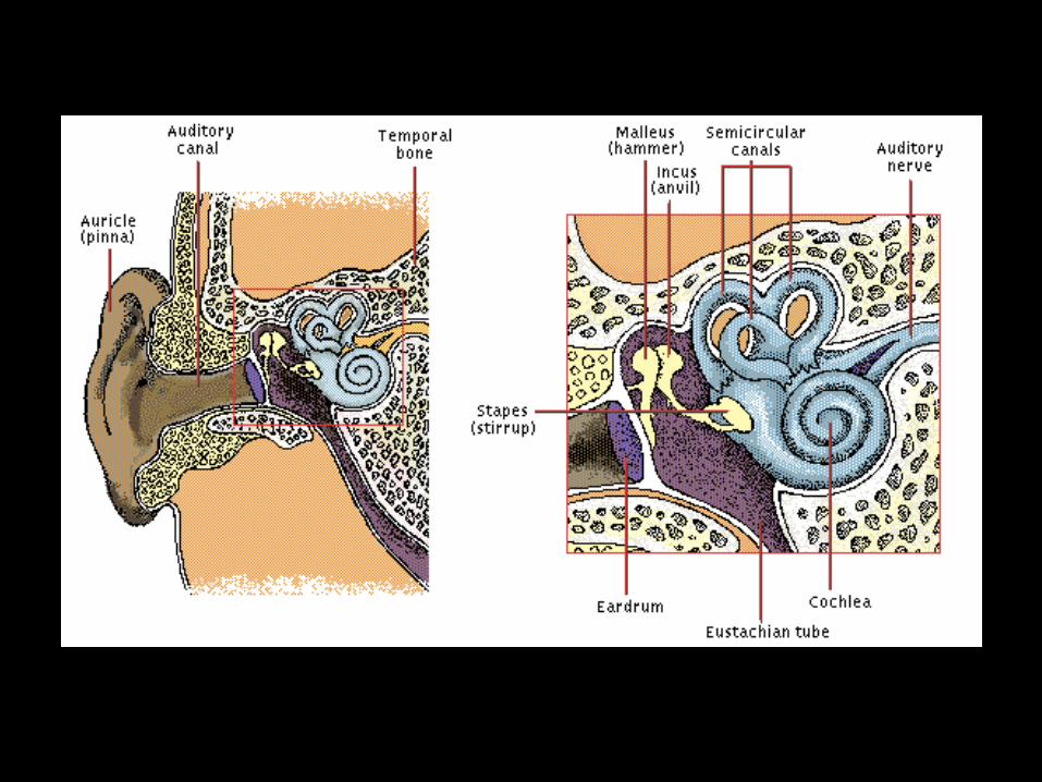
II. II. OSSICLESOSSICLES
= small bones of the ear= small bones of the ear
a. Stapes (stirrup)a. Stapes (stirrup) 22
b. Incus (anvil)b. Incus (anvil) 22
c. Malleus (hammer)c. Malleus (hammer) 22
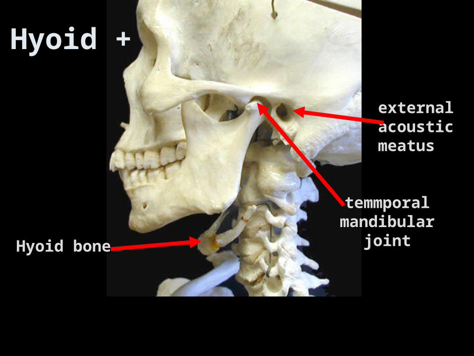
Hyoid boneHyoid bone
temmporaltemmporalmandibularmandibular
jointjoint
external external acousticacousticmeatusmeatus
Hyoid +
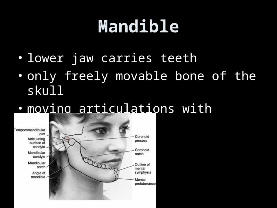
Mandible
• lower jaw carries teeth
• only freely movable bone of the skull
• moving articulations with temporal bone
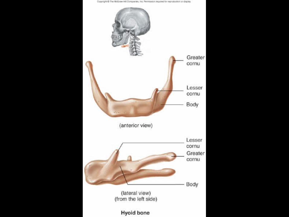
Fig. 6.16
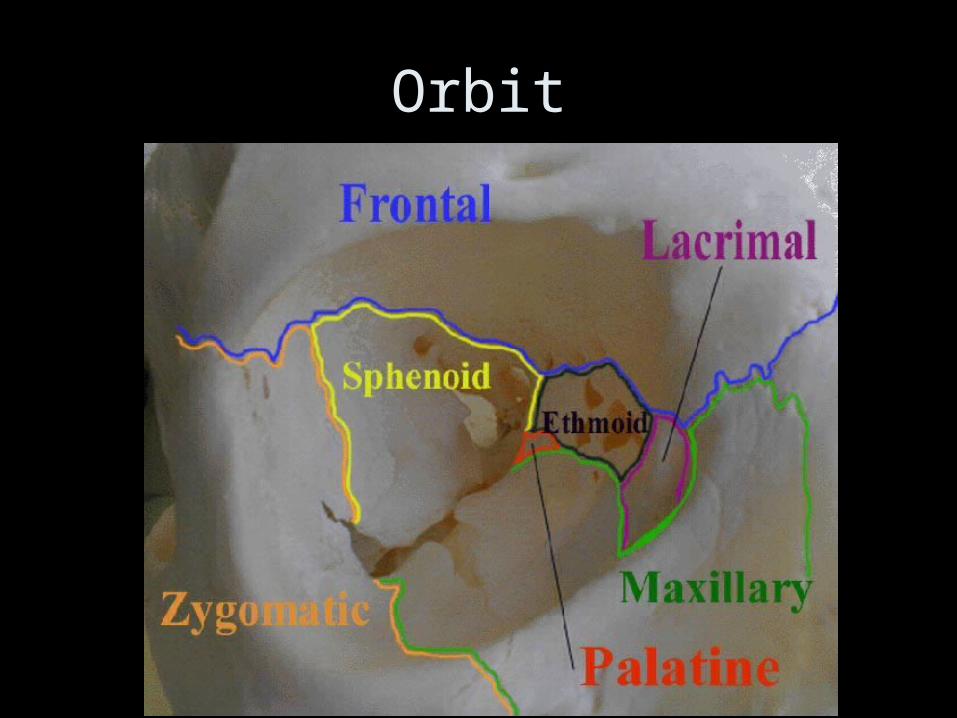
Orbit
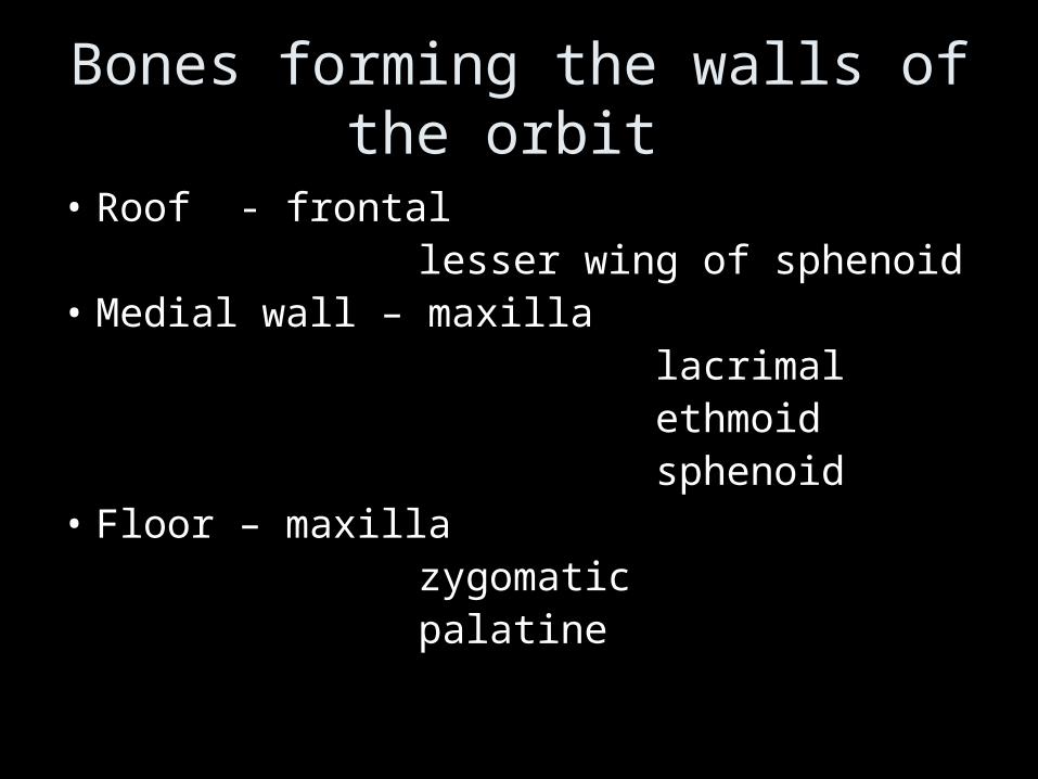
Bones forming the walls of the orbit
• Roof - frontal lesser wing of sphenoid• Medial wall – maxilla lacrimal ethmoid sphenoid• Floor – maxilla zygomatic palatine


Related Documents


