This article appeared in a journal published by Elsevier. The attached copy is furnished to the author for internal non-commercial research and education use, including for instruction at the authors institution and sharing with colleagues. Other uses, including reproduction and distribution, or selling or licensing copies, or posting to personal, institutional or third party websites are prohibited. In most cases authors are permitted to post their version of the article (e.g. in Word or Tex form) to their personal website or institutional repository. Authors requiring further information regarding Elsevier’s archiving and manuscript policies are encouraged to visit: http://www.elsevier.com/authorsrights

Welcome message from author
This document is posted to help you gain knowledge. Please leave a comment to let me know what you think about it! Share it to your friends and learn new things together.
Transcript

This article appeared in a journal published by Elsevier. The attachedcopy is furnished to the author for internal non-commercial researchand education use, including for instruction at the authors institution
and sharing with colleagues.
Other uses, including reproduction and distribution, or selling orlicensing copies, or posting to personal, institutional or third party
websites are prohibited.
In most cases authors are permitted to post their version of thearticle (e.g. in Word or Tex form) to their personal website orinstitutional repository. Authors requiring further information
regarding Elsevier’s archiving and manuscript policies areencouraged to visit:
http://www.elsevier.com/authorsrights
Author's personal copy
Journal of Microscopy and Ultrastructure 3 (2015) 38–43
Contents lists available at ScienceDirect
Journal of Microscopy and Ultrastructure
jou rn al hom ep age : www.elsev ier .com/ locate / jmau
Original Article
Ultrastructural effect on mastitis pathogens by extract ofendophytic fungi associated with ethnoveterinary plant,Hibiscus sabdariffa L.
Archana Nath, S.R. Joshi ∗
Microbiology Laboratory, Department of Biotechnology & Bioinformatics, North-Eastern Hill University, Shillong 793 022, Meghalaya,India
a r t i c l e i n f o
Article history:Received 19 June 2014Received in revised form 4 September 2014Accepted 2 October 2014Available online 22 October 2014
Keywords:Endophytic fungiScanning electron microscopyUltrastructureEscherichia coliKlebsiella pneumoniaeMastitis
a b s t r a c t
Three endophytic fungi isolated from different parts of Hibiscus sabdariffa L. were identifiedusing morphological and molecular approaches. Ethanolic extract of endophytic fungi aswell as plant extracts were evaluated for in vitro antibacterial activity by using well diffu-sion method and their minimum inhibitory concentration estimated. The culture extract ofone endophytic fungus Glomerella acutata EF15 was found to be potent antibacterial agentagainst pathogenic coliform bacteria Klebsiella pneumoniae and Escherichia coli responsiblefor causing clinical mastitis. Scanning electron microscopy (SEM) revealed the ultrastruc-tural alteration in the cells of K. pneumoniae and E. coli when treated with crude ethanolicextract of G. acutata. The ethanolic extract of the endophytic fungus revealed potential tobe bioprospected as antibacterial agent against pathogens causing coliform mastitis, theexisting havoc of dairy industries.
© 2014 Saudi Society of Microscopes. Published by Elsevier Ltd. All rights reserved.
1. Introduction
Medicinal plants constitute a source of both traditionaland modern medicines [1]. About 80% of rural popula-tion depends on herbal medicine as their first line curativemedicine. There are reports on the antimicrobial activity ofthe ethnoveterinary plants [2,3] but no reports are avail-able on the antibacterial activity of the fungal endophytesisolated from these ethnoveterinary plants. Rosselle (Hibis-cus sabdariffa L.; Family Malvaceae) also known as Rangatengamara in Assamese is mostly used as an ethnoveteri-nary plant by the local people of Northeast India to treatvarious ailments of the livestock [4]. The leaf juice is mostlyfed to the animal in empty stomach for the treatment of
∗ Corresponding author. Tel.: +91 9436102171; fax: +91 3642550076.E-mail address: [email protected] (S.R. Joshi).
dysentery. Several reports on the antimicrobial activity ofthe plant H. sabdariffa have also been reported [5,6].
Escherichia coli and Klebsiella pneumoniae are character-ized under Enterobacteriaceae family that normally inhabitthe digestive tract of human and animals but can be trans-mitted through contaminated food and water and become acause for enterobacterial infections [7]. The coliform bacte-ria, E. coli and K. pneumoniae are also the most commongram-negative causes of clinical mastitis [8–10]. K. pneu-moniae is the most common Klebsiella species found tocause mastitis. Klebsiella mastitis causes more losses in live-stock industry in comparison to the losses made by E. colimastitis in terms of milk production and survival [11,12].E. coli infections respond well toward the treatment andtake a shorter time to recover as compared to Klebsiellainfections [8,13].
Endophytic fungi are fungi that colonize living, internaltissues of various parts of the plants without harm-ing them [14]. Antimicrobial activity of the natural
http://dx.doi.org/10.1016/j.jmau.2014.10.0012213-879X/© 2014 Saudi Society of Microscopes. Published by Elsevier Ltd. All rights reserved.
Author's personal copy
A. Nath, S.R. Joshi / Journal of Microscopy and Ultrastructure 3 (2015) 38–43 39
products from plants have been extensively studied againstnewly emerged antibiotic resistant pathogens in a viewthat these new inhibitory agents from natural productsare effective, have low toxicity and have a low environ-mental impact [15]. Recently endophytes are viewed asoutstanding source of secondary metabolites and bioac-tive antimicrobial natural products [16]. The aim of thepresent study was to assess the antibacterial effect of endo-phytic fungi isolated from the H. sabdariffa plant. This studyreports the isolation and antimicrobial potential, againstmastitis pathogens, of endophytic fungi associated withethnoveterinary plant H. sabdariffa used in treatment ofcows, buffaloes and goats. The antimicrobial effect on theultrastructure of the pathogens was evaluated using scan-ning electron micrographs.
2. Materials and methods
2.1. Plant collection and isolation of endophytic fungi
Fresh plants (stem, root and leaves) of H. sabdariffa L.were collected from Assam, India under sterilized conditionin polythene bags. The plant parts were separated and cutinto small pieces and washed thoroughly with tap water.They were surface sterilized in 70% ethanol for 3 min, 4%sodium hypochlorite for 1 min and again in 70% ethanolfor 3 min and rinsed twice in sterile distilled water. Sam-ples were blotted dry with sterile paper towels, then driedin laminar air flow and placed on potato dextrose agar(PDA) containing 100 �g/ml of streptomycin sulphate tosuppress bacterial growth. Plates were incubated at 25 ◦Cuntil the outgrowth of endophytic fungi from the explantswas observed. The outgrowths were subcultured to pro-duce pure culture on PDA plates [17]. All isolates weremaintained in PDA slants and kept at 4 ◦C.
2.2. Morphological features
The endophytic fungal isolates were identified initiallyby their morphological characteristics such as hyphal fea-ture, arrangement of spores and reproductive structures.
2.3. Molecular characterization of the endophytic fungi
Fungal mycelia grown in potato dextrose broth (PDB)for more than one week were taken and fungal genomicDNA was extracted using the HiPurA fungal DNA isolationkit (Himedia) following manufacturer’s instruction. FungalrDNA-ITS region was amplified from the extracted genomicDNA by using fungal domain specific primer ITS1F andITS4R [18]. PCR was performed in a GeneAmp 9700 ThermalCycler (Applied Biosystems, USA) under the following con-ditions, initial denaturation step at 94 ◦C for 5 min, followedby 30 cycles of 94 ◦C for 1 min, 52 ◦C for 30 s, and 72 ◦Cfor 1 min, with a final extension step of 72 ◦C for 10 min.The PCR amplicons were excised and purified using a QIAQuick Gel Extraction Kit (Qiagen, Germany). The ampliconswere sequenced in Applied Biosystems 3700 Genetic Anal-yser (Applied Biosystems, USA) with Big Dye Terminatorver. 3.1. The sequences were then compared with the Gen-Bank database by the BLASTN program. Sequences obtained
were then searched for similarity with other depositedsequences in GenBank. Alignments and phylogenetic anal-ysis were performed using MEGA 4.0 software [19].
2.4. Preparation of plant extract
The collected plants were first washed properly withdistilled water and then dried in an oven at 50 ◦C for72 h with forced air, after which the dried plant wereground into a fine powder using a clean pestle and mortar.Dried sample (approximately 100 g) was soaked in ethanol(300 mL) for 4 days at room temperature (25 ± 2 ◦C) withan intermittent stirring to allow the powder to fully dis-solve in the ethanol. The mixture was then filtered throughWhatman No. 1 paper and then the filtrate was concen-trated under vacuum using a rotary evaporator. The drycrude extracts were then finally dissolved in dimethylsulf-oxide (DMSO) to make a final concentration of 1 mg/ml andthen sterilized by filtration using a 0.22 �m membrane forantimicrobial assay [20]. The resultant extract was kept at4 ◦C for further analysis.
2.5. Fungal broth fermentation and preparation ofculture extract
Endophytic fungi were cultured in potato dextrosebroth (PDB) at 25 ◦C for 4 weeks. The mycelia of fungiwere separated from the fermentation broth by filtration.The mycelia filtrates were extracted thrice with an equalvolume of 70% ethanol. Ethanol was then evaporated todryness under reduced pressure at 45 ◦C using a rotaryevaporator to obtain the crude extract [21] of the mycelialproduct. The dry crude extracts were then finally dissolvedin dimethylsulfoxide (DMSO) to make a final concentra-tion of 1 mg/ml of stock solution which was further usedfor antimicrobial assay.
2.6. Antimicrobial assay using well diffusion method
The crude ethanolic fungal extracts were screened forantimicrobial activity against two pathogenic coliformbacteria K. pneumoniae and E. coli isolated from bovinemastitis samples which were collected from College of Vet-erinary Science, Assam. The test organisms were grownovernight at 37 ◦C in brain heart infusion broth. The antimi-crobial assay using the crude extract was carried out onMueller Hinton agar medium plates. One milliliter of inocu-lum was swapped on molten agar plates. Using the sterilecork borer 7 mm wells were made in the plates in which50 �L of fungal extract was loaded. The control well wasloaded with DMSO. The plates were incubated for 24 h at37 ◦C and then were observed for zone of inhibition [22].
2.7. Determination of minimal inhibitory concentration
Minimum inhibitory concentration of all the fungalextract and plant extract were done by using microtiterbroth dilution technique of Keskin et al. [23] with minormodification. A sterile 96 round-bottom well plate wasused and was labeled properly. Crude extract of vol-ume 100 �L was added into the first row of the plate.
Author's personal copy
40 A. Nath, S.R. Joshi / Journal of Microscopy and Ultrastructure 3 (2015) 38–43
To rest of the wells, 50 �L of potato dextrose broth wasadded and then serial dilutions were performed using amicropipette (A1–A10). Then, 50 �L of broth containingbacterial suspension (106 cfu/mL) was added to each well.Single antimicrobial extract in progressive dilutions wasadded in each column of wells. Each plate had a set of bothgrowth (A11) and sterility control (A12). Plates were sealedwith parafilm and placed in an incubator at 37 ◦C for 24 h.After incubation, 10 �L of 0.2% 2,3,5-triphenyl tetrazoliumchloride (TTC) solution was added to each well. The plateshaving TTC were then incubated at 37 ◦C for 1 h. A visi-ble color changes from purple to pink, indicated growthof microorganism and were recorded as negative. The MICvalue of the extract was taken as the lowest concentrationthat showed no microbial growth.
2.8. Scanning electron microscopic analysis
Mastitis pathogens (K. pneumoniae and E. coli) havingsusceptibility to growth in presence of fungal extract wereanalyzed under scanning electron microscope (JSM-6360,Jeol) for their ultrastructural deformities implicated on thecells. Test organisms were treated with 4× concentrationof the MIC of the fungal extracts and kept under overnightincubation at 37 ◦C. Then the cells were pelleted after cen-trifugation at 8000 rpm for 15 min. The pellets were thenwashed with PBS pH 7.3 several times to remove the debrisand fixed in 2.5% glutaraldehyde in 0.1 M cacodylate buffer(pH 7.2) for 4 h. Glutaraldehyde was drained carefully andplaced in three consecutive 1 h washes of 0.1 M cacodylatebuffer. The samples were dehydrated with acetone series(50%, 70%, 80%, 90%, 95% and 100%) and the drying wasdone with TMS (trimethyl silane). Cells were immersed inTMS for 10 min in two changes at 4 ◦C and are brought toroom temperature for drying. Finally, samples were sput-ter coated with a thin layer of gold–palladium and scannedunder SEM.
3. Results
3.1. Isolation and identification
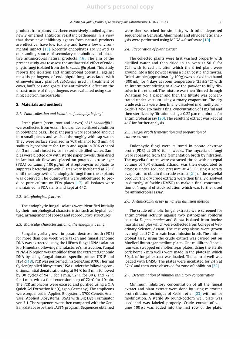
A total of eight isolates were isolated from differentparts of the plant H. sabdariffa L. most being isolated fromthe leaf segments. The colony morphological trait of theisolate such as hyphal features and arrangement of sporeswere used for the tentative identification. Molecular char-acterization was considered for confirmatory identificationof the isolate in which rDNA-ITS region was amplified, thesequence data was aligned using BLAST and the endophyticfungi were found to be closest homolog of Aspergillus niger(EF13), Corynespora cassicola (EF14) and Glomerella acutata(EF15). The ITS sequences for the fungi were deposited toNCBI and accessions obtained as KF928291, KF928292 andKF928293 respectively (Fig. 1).
3.2. Antimicrobial screening
The crude ethanolic extract of different endophyticfungi along with the plant extract was evaluated for theantimicrobial efficacy against two clinically significant
Table 1Antimicrobial activity of endophytic fungal plant and the host plantextract by well diffusion method measured as zone of inhibition (mm).
Fungal isolates and the host plant Test organisms with growthinhibition zone
Klebsiellapneumoniae
Escherichia coli
Aspergillus niger EF 13 ++ +Corynespora cassicola EF14 + ++Glomerella acutata EF15 +++ +++Hibiscus subdarifa L. ++ ++
+ indicates inhibition zone between 12 and 14 mm; ++ indicates inhibitionzone between 14 and 18 mm; +++ indicates inhibition zone between 18and 22 mm.
Table 2MIC values of the crude ethanolic extract of endophytic fungi and the shotplant.
Fungal isolates and the host plant MIC (mg/ml)
Klebsiellapneumoniae
Escherichia coli
Aspergillus niger EF 13 0.083 ± 0.021 0.33 ± 0.08Corynespora cassicola EF14 0.25 ± 0.00 0.20 ± 0.041Glomerella acutata EF15 0.021 ± 0.005 0.016 ± 0.00Hibiscus subdarifa L. 0.032 ± 0.00 0.16 ± 0.041
animal pathogens. Among the eight isolates, only three iso-lates showed potent antimicrobial activity. Crude ethanolicplant extract also showed the antimicrobial activity. Thefungal isolate EF15 showed the highest antimicrobial activ-ity against both the tested animal pathogen followedby plant extract and the fungal isolates EF13 and EF14(Table 1). The control well with DMSO did not show anyzone of inhibition.
3.3. Minimum inhibitory concentration (MIC)
All active extracts showing potent antimicrobial activ-ity were further determined for their MIC by a microtiterbroth dilution technique. The ethanolic extract of the fun-gal isolate EF15 showed MIC of 0.021 ± 0.005 mg/ml and0.016 ± 0.00 mg/ml for K. pneumoniae and E. coli respec-tively (Table 2), which showed its efficacy as a potentantimicrobial. The findings of the MIC corroborated withthe results obtained for well diffusion assay during primaryscreening.
3.4. Scanning electron micrographs
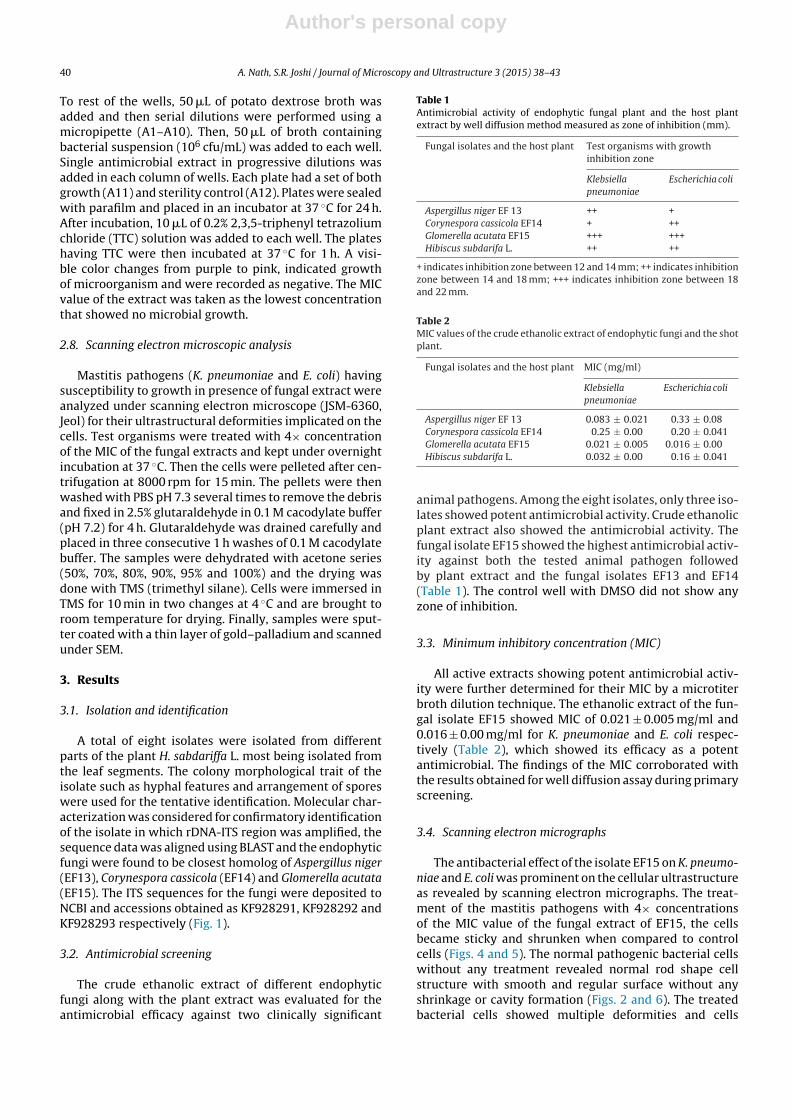
The antibacterial effect of the isolate EF15 on K. pneumo-niae and E. coli was prominent on the cellular ultrastructureas revealed by scanning electron micrographs. The treat-ment of the mastitis pathogens with 4× concentrationsof the MIC value of the fungal extract of EF15, the cellsbecame sticky and shrunken when compared to controlcells (Figs. 4 and 5). The normal pathogenic bacterial cellswithout any treatment revealed normal rod shape cellstructure with smooth and regular surface without anyshrinkage or cavity formation (Figs. 2 and 6). The treatedbacterial cells showed multiple deformities and cells
Author's personal copy
A. Nath, S.R. Joshi / Journal of Microscopy and Ultrastructure 3 (2015) 38–43 41
Fig. 1. Evolutionary positions of the three endophytic fungal isolates with other related fungal species based on rRNA-ITS sequence similarity.
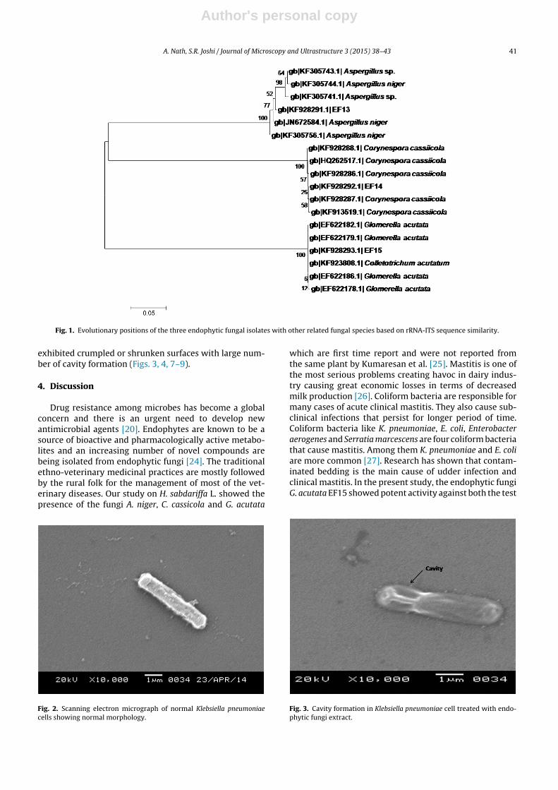
exhibited crumpled or shrunken surfaces with large num-ber of cavity formation (Figs. 3, 4, 7–9).
4. Discussion
Drug resistance among microbes has become a globalconcern and there is an urgent need to develop newantimicrobial agents [20]. Endophytes are known to be asource of bioactive and pharmacologically active metabo-lites and an increasing number of novel compounds arebeing isolated from endophytic fungi [24]. The traditionalethno-veterinary medicinal practices are mostly followedby the rural folk for the management of most of the vet-erinary diseases. Our study on H. sabdariffa L. showed thepresence of the fungi A. niger, C. cassicola and G. acutata
Fig. 2. Scanning electron micrograph of normal Klebsiella pneumoniaecells showing normal morphology.
which are first time report and were not reported fromthe same plant by Kumaresan et al. [25]. Mastitis is one ofthe most serious problems creating havoc in dairy indus-try causing great economic losses in terms of decreasedmilk production [26]. Coliform bacteria are responsible formany cases of acute clinical mastitis. They also cause sub-clinical infections that persist for longer period of time.Coliform bacteria like K. pneumoniae, E. coli, Enterobacteraerogenes and Serratia marcescens are four coliform bacteriathat cause mastitis. Among them K. pneumoniae and E. coliare more common [27]. Research has shown that contam-inated bedding is the main cause of udder infection andclinical mastitis. In the present study, the endophytic fungiG. acutata EF15 showed potent activity against both the test
Fig. 3. Cavity formation in Klebsiella pneumoniae cell treated with endo-phytic fungi extract.

Author's personal copy
42 A. Nath, S.R. Joshi / Journal of Microscopy and Ultrastructure 3 (2015) 38–43
Fig. 4. Micrograph showing shrunken and wrinkled Klebsiella pneumoniaebacterial cell.
Fig. 5. Collapsed and disintegrated Klebsiella pneumoniae bacterial cellslosing their normal morphology.
Fig. 6. Scanning electron micrograph of Escherichia coli having normal rodshaped cells.
Fig. 7. Micrographs showing rupturing of the cell membrane ofEscherichia coli.
Fig. 8. Cavity formation in the bacterial cells of Escherichia coli treatedwith endophytic fungi extract.
Fig. 9. Deformed Escherichia coli bacterial cells losing their normal cellmorphology after treatment.

Author's personal copy
A. Nath, S.R. Joshi / Journal of Microscopy and Ultrastructure 3 (2015) 38–43 43
pathogens K. pneumoniae and E. coli which is in agreementwith earlier reports [28]. The study indicated the ethanolicextract of endophytic fungi showing better antimicrobialactivity when compared to the ethanolic plant extract ofthe host plant. One of the mechanisms involved in bacte-rial cell destruction is the action of antimicrobial agentson the cell membrane of the bacteria [29,30] leading tocomplete damage of the cells. The scanning electron micro-graphs clearly indicated that the bacterial cells crumpled,shrunken and cavities formation were prominent on thecell membrane of the bacteria which lead to complete dam-age of the cells corroborating earlier findings on bacterialcell damage [30]. This finding confirms the potential forinvestigating endophytic fungi from ethnoveterinary plant,H. sabdariffa L. as a potent antimicrobial agent against col-iform bacteria causing mastitis.
Conflict of interest
None declared.
References
[1] Karimi E, Jaafar HZE. HPLC and GC–MS determination of bioactivecompounds in microwave obtained extracts of three varieties ofLabisia pumila Benth. Molecules 2011;16:6791–805.
[2] Kalayou S, Haileselassie M, Gebre-egziabher G, Tiku’e T, Sahle S,Taddele H, et al. In-vitro antimicrobial activity screening of someethnoveterinary medicinal plants traditionally used against masti-tis, wound and gastrointestinal tract complication in Tigray Region,Ethiopia. Asian Pacific J Trop Biomed 2012;2:516–22.
[3] Adamu M, Naidoo V, Eloff JN. The antibacterial activity, antioxidantactivity and selectivity index of leaf extracts of thirteen South Africantree species used in ethnoveterinary medicine to treat helminthinfections. BMC Vet Res 2014;10:52.
[4] Saikia B, Borthakur SK. Use of medicinal plants in animal healthcare –a case study from Gohpur, Assam. Indian J Trad Knowl 2010;9:49–51.
[5] Alaa G. Antioxidant and antibacterial activities of Hibiscus sabdariffaL. extracts. Afr J Food Sci 2012;21:506–11.
[6] El-kamali H, Moneer M. Antibacterial activity of Hibiscus sabdariffaand Acacia seyal against upper respiratory tract pathogens. SudanMed J 2006;2:121–6.
[7] Guentzel MN. Escherichia, Klebsiella, Enterobacter, Serratia, Citrobac-ter, and Proteus. In: Baron S, editor. Medical microbiology. 4th ed.Galveston, TX: University of Texas Medical Branch at Galveston; 1996[Chapter 26].
[8] Smith KL, Todhunter DA, Schoenberger PS. Environmental mastitis:cause, prevalence, prevention. J Dairy Sci 1985;68:1531–53.
[9] Erskine RJ, Tyler JW, Riddell Jr MG, Wilson RC. Theory, use, and real-ities of efficacy and food safety of antimicrobial treatment of acutecoliform mastitis. J Am Vet Med Assoc 1991;198:980–4.
[10] Todhunter DA, Smith KL, Hogan JS, Schoenberger PS. Gram-negativebacterial infections of the mammary gland in cows. Am J Vet Res1991;52:184–8.
[11] Erskine RJ, Barlett PC, VanLente JL, Phipps CR. Efficacy of systemicceftiofur as a therapy for severe clinical mastitis in dairy cattle. JDairy Sci 2002;85:2571–5.
[12] Grohn YT, Wilson DJ, Gonzalez RN, Hertl JA, Schulte H, Bennett G,et al. Effect of pathogen-specific clinical mastitis on milk yield indairy cows. J Dairy Sci 2004;87:3358–74.
[13] Roberson JR, Warnick LD, Moore G. Mild to moderate clinical mas-titis: efficacy of intramammary amoxicillin, frequent milk-out, acombined intramammary amoxicillin, and frequent milk-out treat-ment versus no treatment. J Dairy Sci 2004;87:583–92.
[14] Bacon CW, Stone JK, White JF. An overview of endophytic. MicrobEndophytes 2000;1:3–29.
[15] Supaphon P, Phongpaichit S, Rukachaisirikul V, Sakayaroj J.Antimicrobial potential of endophytic fungi derived from three sea-grass species: Cymodocea serrulata, Halophila ovalis and Thalassiahemprichii. PLOS ONE 2013;8:e72520.
[16] Jalgaonwala RE, Mohite BV, Mahajan RT. Evaluation of endophytesfor their antimicrobial activity from indigenous medicinal plantsbelonging to North Maharashtra region, India. Int J Pharm Biol Res2010;1:136–41.
[17] Nath A, Chattopadhyay A, Joshi SR. Biological activity of endophyticfungi of Rouwolfia serpentina Benth, an ethnomedicinal plant used infolk medicines in Northeast India. Proc Natl Acad Sci India B: Biol Sci2013, http://dx.doi.org/10.1007/s40011-013-0184-8.
[18] Bhagobaty RK, Joshi SR. Multi-loci molecular characterisation ofendophytic fungi isolated from five medicinal plants of Meghalaya,India. Mycobiology 2011;39:71–8.
[19] Tamura K, Peterson D, Peterson N, Stecher G, Nei M, Kumar S. MEGA5:molecular evolutionary genetics analysis using maximum likelihood,evolutionary distance, and maximum parsimony methods. Mol BiolEvol 2011;28:2732–9.
[20] Vijayarathna S, Zakaria Z, Chen Y, Latha LY, Kanwar JR, SasidharanS. The antimicrobial efficacy of Elaeis guineensis: characterization,in vitro and in vivo studies. Molecules 2012;17:4860–77.
[21] Nath A, Joshi SR. Bioactivity assessment of endophytic fungi asso-ciated with the ethnomedicinal plant Potentilla fulgens. World JPharmaceut Res 2013;6:2596–607.
[22] Nath A, Raghunatha P, Joshi SR. Diversity and biological activities ofendophytic fungi of Emblica officinalis, an ethnomedicinal plant ofIndia. Mycobiology 2012;40:18–30.
[23] Keskin D, Oskay D, Oskay M. Antimicrobial activity of selected plantspices marketed in the west Anatolia. Int J Agric Biol 2010;12:916–20.
[24] Strobel G, Daisy B. Bioprospecting for microbial endophytesand their natural products. Microbiol Mol Biol Rev 2003;67:491–502.
[25] Kumaresan V, Veeramohan R, Shrivastava P, Deepika R, Suganya M,Vennila S, et al. Fungal endophytes of some green leafy vegetables.World J Agric Sci 2013;9:415–20.
[26] Mubarack HM, Doss A, Dhanabalan R, Venkataswamy R. In-vitroantimicrobial effects of some selected plants against bovine mastitispathogens. Hygeia J Drugs Med 2011;3:71–5.
[27] Munoz MA, Ahlstrom C, Rauch BJ, Zadoks RN. Fecal sheddingof Klebsiella pneumoniae by dairy cows. J Dairy Sci 2006;89:3425–30.
[28] Vieira ML, Hughes AF, Gil VB, Vaz AB, Alves TM, Zani CL, et al.Diversity and antimicrobial activities of the fungal endophyte com-munity associated with the traditional Brazilian medicinal plantSolanum cernuum Vell. (Solanaceae). Can J Microbiol 2012;58:54–66.
[29] Hyldgaard M, Mygind T, Meyer RL. Essential oils in food preserva-tion: mode of action, synergies, and interactions with food matrixcomponents. Front Microbiol 2012;3:12.
[30] Darah I, Lim SH, Nithianantham K. Effects of methanol extract ofWedelia chinensis Osbeck (asteraceae) leaves against pathogenicbacteria with emphasise on Bacillus cereus. Indian J Pharm Sci2013;75:533–9.
Related Documents