Ultrastructural comparison of dissolution and apatite precipitation on hydroxyapatite and silicon-substituted hydroxyapatite in vitro and in vivo Alexandra E. Porter, 1 Claudia M. Botelho, 2,3 Maria A. Lopes, 2,3 Jose ´ D. Santos, 2,3 Serena M. Best, 1 William Bonfield 1 1 Department of Materials Science and Metallurgy, University of Cambridge, Pembroke Street, Cambridge, CB2 3QZ, United Kingdom 2 Instituto de Engenharia Biome ´dica, Laboratorio de Biomaterials, Rua do Campo Alegre, 823, 4150-180 Porto, Portugal 3 Faculdade de Engenharia da Universidade do Porto, DEMM, Rua Dr. Roberto Frias, 4200-465 Porto, Portugal Received 9 October 2003; revised 21 January 2004; accepted 9 February 2004 Published online 26 April 2004 in Wiley InterScience (www.interscience.wiley.com). DOI: 10.1002/jbm.a.30035 Abstract: Recent histological studies have demonstrated that the substitution of silicate ions into hydroxyapatite (HA) significantly increases the rate of bone apposition to HA implants. The enhanced bioactivity of silicon-substi- tuted HA (Si-HA) over pure HA has been attributed to the effect of silicate ions in accelerating dissolution. In the present study, high-resolution transmission electron micros- copy (HR-TEM) was employed to compare dissolution of HA and Si-HA in an acellular simulated body fluid (SBF) to dissolution in an in vivo model. HR-TEM observations con- firmed a difference in morphology of apatite precipitates in vivo and in SBF: apatite deposits were platelike in vivo and nodular in SBF. Compositional mapping suggested that preferential dissolution of silicon from the implant promotes the nucleation of carbonate apatite around the implant. The in vivo findings illustrated an absence of dissolution at the bone–HA or Si–HA interface, whereas dissolution was ex- tensive from within the implant. The amount of dissolution in acellular SBF was similar to dissolution from within the implant, although the site at which the dissolution nucleates was different: dissolution predominates at the crystallite surfaces in SBF, whereas grain boundary dissolution pre- dominates in vivo. These findings suggest that proteins in the in vivo milieu modify the processes of dissolution from the implant. © 2004 Wiley Periodicals, Inc. J Biomed Mater Res 69A: 670 – 679, 2004 Key words: dissolution; silicon; simulated body fluid; ultra- structure; precipitation INTRODUCTION Hydroxyapatite (HA) possesses a hexagonal Bra- vais lattice and a P6 3 /m space group with cell di- mensions of a 9.1404 and c 6.8747 Å, and it is known to be bioactive. 1,2 The substitution of silicate ions (SiO 4 4 ) for phosphate ion tetrahedra (PO 4 3 ) in the HA unit cell has been shown to have the potential to increase the rate and amount of bone apposition to HA bioceramic implants. 2 However, uncertainty remains about the mechanism by which silicate ion incorporation increases the in vivo bio- activity. In previous studies, defect structures in silicon-substituted HA (Si-HA) were observed and characterized using high-resolution transmission electron microscopy (HR-TEM). 1 Phase pure, sin- tered granules of HA and Si-HA were implanted for 6 and 12 weeks in an ovine model. Samples contain- ing the bone–implant interface were prepared for TEM via ultramicrotomy using an anhydrous sam- ple preparation procedure. HR-TEM was used to compare the in vivo reactivity of sintered granules of HA and Si-HA at both 6 and 12 weeks. 3 Recent HR-TEM results revealed the presence of dislocations and grain boundaries in pure HA and Si-HA with a significant increase in density of triple junctions per unit area in Si-HA over pure HA. 1 Dis- solution was found to be particularly prevalent at grain boundaries and triple junctions in vivo and fol- lowed the order 1.5 wt % Si-HA 0.8 wt % Si-HA pure HA. 3 These findings suggested that increased numbers of defects in Si-HA play a significant role in Correspondence to: A. E. Porter; e-mail: [email protected] Contract grant sponsor: EPSRC Contract grant sponsor: Apatech Ltd. Contract grant sponsor: Fundac ¸a ˜o para a Cie ˆncia e Tecno- logia (to C.M.B.); contract grant numbers: SFRH/BD/6173, POCTI/CTM/49238/2002 © 2004 Wiley Periodicals, Inc.

Welcome message from author
This document is posted to help you gain knowledge. Please leave a comment to let me know what you think about it! Share it to your friends and learn new things together.
Transcript
Ultrastructural comparison of dissolution and apatiteprecipitation on hydroxyapatite and silicon-substitutedhydroxyapatite in vitro and in vivo
Alexandra E. Porter,1 Claudia M. Botelho,2,3 Maria A. Lopes,2,3 Jose D. Santos,2,3 Serena M. Best,1
William Bonfield1
1Department of Materials Science and Metallurgy, University of Cambridge, Pembroke Street, Cambridge,CB2 3QZ, United Kingdom2Instituto de Engenharia Biomedica, Laboratorio de Biomaterials, Rua do Campo Alegre, 823, 4150-180 Porto, Portugal3Faculdade de Engenharia da Universidade do Porto, DEMM, Rua Dr. Roberto Frias, 4200-465 Porto, Portugal
Received 9 October 2003; revised 21 January 2004; accepted 9 February 2004Published online 26 April 2004 in Wiley InterScience (www.interscience.wiley.com). DOI: 10.1002/jbm.a.30035
Abstract: Recent histological studies have demonstratedthat the substitution of silicate ions into hydroxyapatite(HA) significantly increases the rate of bone apposition toHA implants. The enhanced bioactivity of silicon-substi-tuted HA (Si-HA) over pure HA has been attributed to theeffect of silicate ions in accelerating dissolution. In thepresent study, high-resolution transmission electron micros-copy (HR-TEM) was employed to compare dissolution ofHA and Si-HA in an acellular simulated body fluid (SBF) todissolution in an in vivo model. HR-TEM observations con-firmed a difference in morphology of apatite precipitates invivo and in SBF: apatite deposits were platelike in vivo andnodular in SBF. Compositional mapping suggested thatpreferential dissolution of silicon from the implant promotesthe nucleation of carbonate apatite around the implant. The
in vivo findings illustrated an absence of dissolution at thebone–HA or Si–HA interface, whereas dissolution was ex-tensive from within the implant. The amount of dissolutionin acellular SBF was similar to dissolution from within theimplant, although the site at which the dissolution nucleateswas different: dissolution predominates at the crystallitesurfaces in SBF, whereas grain boundary dissolution pre-dominates in vivo. These findings suggest that proteins in thein vivo milieu modify the processes of dissolution from theimplant. © 2004 Wiley Periodicals, Inc. J Biomed Mater Res69A: 670–679, 2004
Key words: dissolution; silicon; simulated body fluid; ultra-structure; precipitation
INTRODUCTION
Hydroxyapatite (HA) possesses a hexagonal Bra-vais lattice and a P63/m space group with cell di-mensions of a � 9.1404 and c � 6.8747 Å, and it isknown to be bioactive.1,2 The substitution of silicateions (SiO4
4�) for phosphate ion tetrahedra (PO43�)
in the HA unit cell has been shown to have thepotential to increase the rate and amount of boneapposition to HA bioceramic implants.2 However,uncertainty remains about the mechanism by whichsilicate ion incorporation increases the in vivo bio-
activity. In previous studies, defect structures insilicon-substituted HA (Si-HA) were observed andcharacterized using high-resolution transmissionelectron microscopy (HR-TEM).1 Phase pure, sin-tered granules of HA and Si-HA were implanted for6 and 12 weeks in an ovine model. Samples contain-ing the bone–implant interface were prepared forTEM via ultramicrotomy using an anhydrous sam-ple preparation procedure. HR-TEM was used tocompare the in vivo reactivity of sintered granules ofHA and Si-HA at both 6 and 12 weeks.3
Recent HR-TEM results revealed the presence ofdislocations and grain boundaries in pure HA andSi-HA with a significant increase in density of triplejunctions per unit area in Si-HA over pure HA.1 Dis-solution was found to be particularly prevalent atgrain boundaries and triple junctions in vivo and fol-lowed the order 1.5 wt % Si-HA � 0.8 wt % Si-HA �pure HA.3 These findings suggested that increasednumbers of defects in Si-HA play a significant role in
Correspondence to: A. E. Porter; e-mail: [email protected] grant sponsor: EPSRCContract grant sponsor: Apatech Ltd.Contract grant sponsor: Fundacao para a Ciencia e Tecno-
logia (to C.M.B.); contract grant numbers: SFRH/BD/6173,POCTI/CTM/49238/2002
© 2004 Wiley Periodicals, Inc.
increasing the rates of dissolution of calcium, phos-phate, and silicate ions from Si-HA.
As an alternative approach to using in vivo studiesfor evaluating the bioactivity at the bone–biomaterialinterface, in vitro immersions in simulated body fluid(SBF) or cell-containing media may be employed. Thecommonly proposed mechanism underlying the phe-nomenon of the bioactivity of HA involves the disso-lution of calcium and phosphate ions from HA.4 Thisprocess may occur through the surrounding environ-ment (extracellular fluid) and may also be activelymediated by osteoclast cells.5 Dissolution generatesincreased concentrations of calcium and inorganicphosphate in the spaces between the existing bone andthe implant.4 Precipitation of HA into this space willensure incorporation of the implant into the existingbone.6 It is commonly believed that dissolution andreprecipitation of apatite crystallites promote incorpo-ration of biological molecules.7
The evaluation of bioactivity using in vitro immer-sion techniques has been criticized because they donot fully replicate the complicated series of dynamicreactions that occur at the bone–biomaterial interfacein vivo. In particular, the biological response of CaPbiomaterials implanted in vivo might be different fromthat evaluated with standard in vitro tests because ofthe interaction of calcium phosphate compounds withthe complex array of constituents in the body, in par-ticular macromolecules. Nevertheless, these systemsaid the evaluation of specific cellular and proteina-cious responses at the bone–implant interface.8–11 Invitro studies also minimize the need for animal exper-iments and offer the potential to study the dissolu-tion–reprecipitation mechanism at very early timepoints.
A recent study demonstrated that many interfacialreactions are parallel in vivo and in vitro.12 A study byFujibayashi et al. comparing in vivo bone ingrowthand in vitro apatite formation on Na2O-CaO-SiO2glasses illustrated that the depth of bone ingrowthamong glass particles increased in proportion to theirapatite forming ability in vitro.12 The in vitro results ofan investigation of the apatite forming ability of Si-HAwith several compositions corroborate our histologicalin vivo studies: the time required to form a surfaceapatite layer on Si-HA in SBF decreased with increas-ing silicate ion substitution.11 Furthermore, silicate ionsubstitution simulates osteoblast-like cell activity invitro when compared to stoichiometric HA. Despitethese similarities on the microstructural scale, ques-tions remain as to whether the mechanisms of apatitedissolution and precipitation at the ultrastructurallevel are similar in both in vivo and in vitro modelsystems.
The objective of the current study was to use HR-TEM to compare in vitro dissolution of ultrathin (�90nm) sections of HA and Si-HA in acellular SBF at 6 h,
1 day, and 3 days with the results of a previous studyinvestigating dissolution and bone remodeling aroundpure HA and Si-HA (0.8 and 1.5 wt %) in vivo.13 Weproposed that the combination of in vivo and in vitrostudies may lead to an improved understanding of thesurface reactions of bioactive ceramics in the body andtheir effects on bone formation and cell function. Onlya few studies have investigated the ultrastructure ofapatite deposits in SBF solutions.14 The lack of infor-mation that describes ultrastructural characteristicsarises from the difficulty of preparing samples forTEM observation. This study develops a simple, alter-native methodology for preparing samples for in vitroTEM observation.
MATERIALS AND METHODS
Chemical synthesis
Phase pure HA with a Ca/P molar ratio of 1.67 wasprepared by a precipitation reaction between calcium hy-droxide [Ca(OH)2] and orthophosphoric acid [H3PO4] solu-tion according to methods described elsewhere.15 Two typesof Si-HA (0.8 and 1.5 wt %) were also prepared by acid–baseneutralization reactions according to the methods of Gibsonet al.16 In addition to calcium hydroxide and orthophospho-ric acid, silicon acetate [Si(CH3COOH)4] was incorporatedinto the reaction mixture as a source of silicate ions. Thequantities of reactants were calculated by assuming thatsilicate ions would substitute for phosphate ions in the HAlattice. Therefore, the number of moles of H3PO4 in phasepure HA was the same as the number of moles of H3PO4 �Si(CH3COOH)4 for the Si-HA samples, with the number ofmoles of Ca(OH)2 being kept constant. Precipitation reac-tions were carried out at room temperature and the pH wasmaintained at 10.5 by the addition of ammonium hydroxidesolution. After complete mixing of the reactants, the suspen-sions were aged overnight. The resulting precipitates werefiltered, washed, and dried at 80°C overnight.
Sample preparation
HA and Si-HA were prepared as powders to compare thedissolution in SBF and as granules to compare the dissolu-tion in vivo.
Aged, filtered, and dried precipitates of pure HA and 1.5wt % Si-HA were ground into a fine powder. The powderswere subsequently calcined at 1200°C for 120 min using aramp rate of 2.5°C/min. An X-ray fluorescence analysis wasperformed according to previous work3 showed the Ca/Pmolar ratio of the pure HA to be 1.67, which is equivalent tostoichiometric HA. The Ca/(P � Si) molar ratios of 0.8 wt %Si-HA and 1.5 wt % Si-HA were both 1.68. The X-ray dif-fraction (XRD) measurements for HA powders indicatedthat the sintered HA and 0.8 and 1.5 wt % Si-HA were phasepure, with no additional phases present. The effect of the
DISSOLUTION AND APATITE PRECIPITATION ON HA AND Si-HA 671
silicate ion substitution on the crystal structure of HA wasdetermined by Rietveld structure refinement of the XRDdata. Silicate ion substitution resulted in a decrease in the aaxis and an increase in the c axis of the unit cell of HA anda slight decrease in the unit cell volume. These results sug-gest that the larger silicate ion (SiO4
4�) substitutes for thephosphate (PO4
3�) group in the HA lattice.1
Embedding of HA and Si-HA powders
Powders of HA and Si-HA were treated with threechanges of 100% ethanol and three of propylene oxide over1 h and then infiltrated with Spurr’s resin (Agar Scientific,Essex, U.K.) over several days: Spurr’s resin was preparedwith 10 g of the epoxy monomer vinyl cyclohexene dioxide(ERL), 4.5 g of diglycidyl ether of polypropylene glycol(DER-736), 26 g of nonenyl succinic anhydride, and 0.7 g ofbenzyldimethylamine. The samples were agitated at roomtemperature in 1:1 solutions of propylene oxide and Spurr’sresin for 2 days, in 1:3 solutions of propylene oxide/Spurr’sresin for 1 day, and then in 100% Spurr’s resin for 2 weeksunder a vacuum. The Spurr’s resin was changed every 24 h.Samples were then cured in fresh Spurr’s resin for 23 h at60°C. Precasting a �200 �m layer of Spurr’s resin into thetruncated beam capsules before adding the HA powdersfacilitated sectioning of the blocks.
Dispersion of powders in SBF and TEMpreparation
Sections (90 nm) of HA, 0.8 wt % Si-HA, and 1.5 wt %Si-HA powders were cut onto a water bath using a diamondknife (Leica Ultracut). Sections were collected on lacey car-bon support, gold TEM grids and most of these were dis-persed in SBF solutions9 at a pH of 7.4 at 37°C for 6 h or 1,3, or 5 days. It was assumed that convection would besufficient to stir the SBF solutions. The fluid was prepared bydissolving reagent-grade NaCl, KCl, K2HPO4 � 3H2O,MgCl2 � H2O, CaCl2, and NaSO4 into distilled water andbuffering to pH 7.4 with tris[hydroxymethyl]aminomethane[(CH2OH3)3CNH3] and hydrochloric acid.
TEM
TEM and selected area electron diffraction (SAED) wereperformed in a Philips CM30 operating at 300 kV. Diffrac-tion contrast was employed for bright-field imaging.
Environmental scanning electron microscopy(ESEM)
ESEM was performed on a Phillips ESEM-FEG XL30 inlow vacuum mode, using an off-axis gaseous secondaryelectron detector and a 10-kV operating voltage.
RESULTS
TEM observations of sintered powders
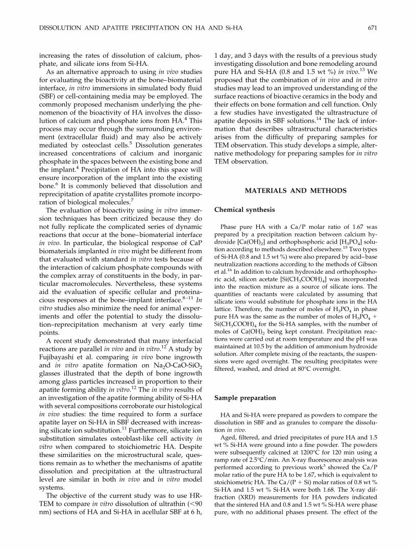
The TEM of the pure HA and 0.8 and 1.5 wt %Si-HA powders in ethanol revealed smooth, discreteedges with no loss of mineral at the subgrain bound-aries or triple junctions. An example of a representa-tive crystallite of 0.8 wt % Si-HA is illustrated inFigure 1.
Dispersion in SBF
Pure HA
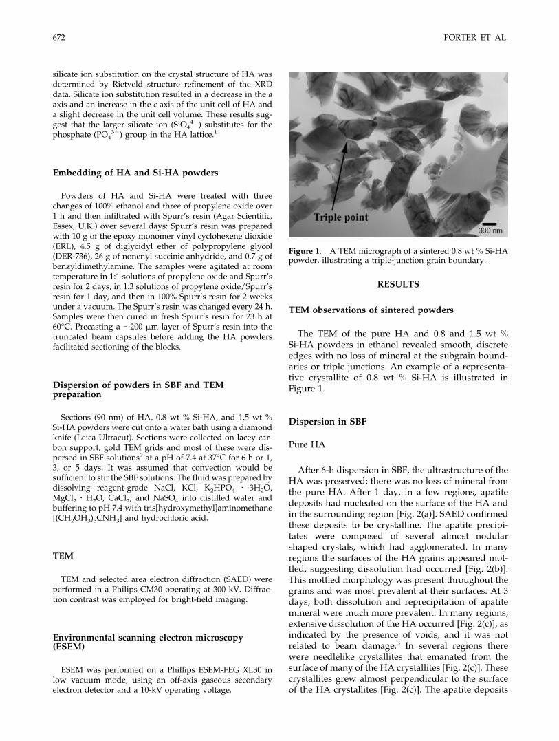
After 6-h dispersion in SBF, the ultrastructure of theHA was preserved; there was no loss of mineral fromthe pure HA. After 1 day, in a few regions, apatitedeposits had nucleated on the surface of the HA andin the surrounding region [Fig. 2(a)]. SAED confirmedthese deposits to be crystalline. The apatite precipi-tates were composed of several almost nodularshaped crystals, which had agglomerated. In manyregions the surfaces of the HA grains appeared mot-tled, suggesting dissolution had occurred [Fig. 2(b)].This mottled morphology was present throughout thegrains and was most prevalent at their surfaces. At 3days, both dissolution and reprecipitation of apatitemineral were much more prevalent. In many regions,extensive dissolution of the HA occurred [Fig. 2(c)], asindicated by the presence of voids, and it was notrelated to beam damage.3 In several regions therewere needlelike crystallites that emanated from thesurface of many of the HA crystallites [Fig. 2(c)]. Thesecrystallites grew almost perpendicular to the surfaceof the HA crystallites [Fig. 2(c)]. The apatite deposits
Figure 1. A TEM micrograph of a sintered 0.8 wt % Si-HApowder, illustrating a triple-junction grain boundary.
672 PORTER ET AL.
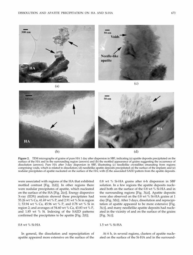
were associated with regions of the HA that exhibitedmottled contrast [Fig. 2(d)]. In other regions therewere nodular precipitates of apatite, which nucleatedon the surface of the HA [Fig. 2(e)]. Energy dispersiveX-ray (EDX) analysis showed these precipitates had55.26 wt % Ca, 41.69 wt % P, and 2.91 wt % Si in region1; 53.94 wt % Ca, 45.96 wt % P, and 0.78 wt % Si inregion 2; and averages of 54.60 wt % Ca, 43.83 wt % P,and 1.85 wt % Si. Indexing of the SAED patternsconfirmed the precipitates to be apatite [Fig. 2(f)].
0.8 wt % Si-HA
In general, the dissolution and reprecipitation ofapatite appeared more extensive on the surface of the
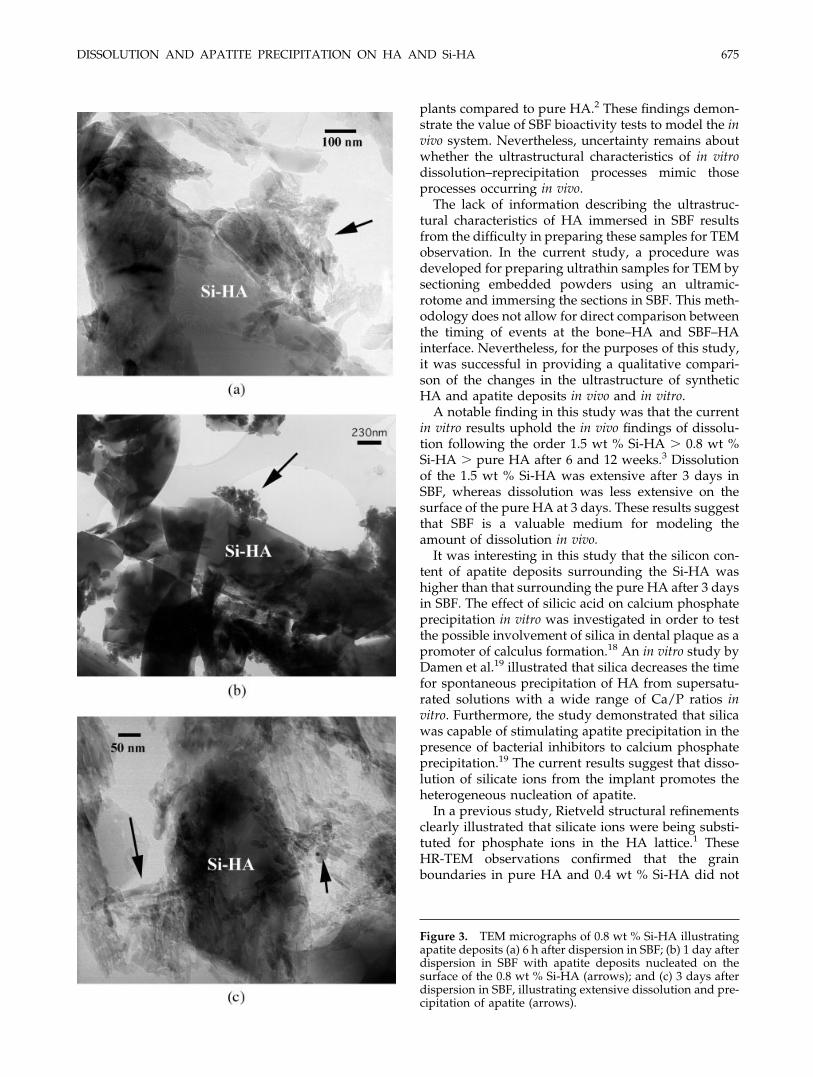
0.8 wt % Si-HA grains after 6-h dispersion in SBFsolution. In a few regions the apatite deposits nucle-ated both on the surface of the 0.8 wt % Si-HA and inthe surrounding regions [Fig. 3(a)]. Apatite depositswere also observed on the 0.8 wt % Si-HA grains at 1day [Fig. 3(b)]. After 3 days, dissolution and reprecipi-tation of apatite appeared to be more extensive [Fig.3(c)], and many needlelike apatite deposits had nucle-ated in the vicinity of and on the surface of the grains[Fig. 3(c)].
1.5 wt % Si-HA
At 6 h, in several regions, clusters of apatite nucle-ated on the surface of the Si-HA and in the surround-
Figure 2. TEM micrographs of grains of pure HA 1 day after dispersion in SBF, indicating (a) apatite deposits precipitated on thesurface of the HA and in the surrounding region (arrows) and (b) the mottled appearance of grains suggesting the occurrence ofdissolution (arrows). Pure HA after 3-day dispersion in SBF, illustrating (c) needlelike crystallites emanating from regionscomprising voids, which is related to dissolution; (d) needlelike apatite deposits precipitated on the surface of the implant; and (e)nodular precipitates of apatite nucleated on the surface of the HA; with (f) the associated SAED pattern from the apatite deposits.
DISSOLUTION AND APATITE PRECIPITATION ON HA AND Si-HA 673
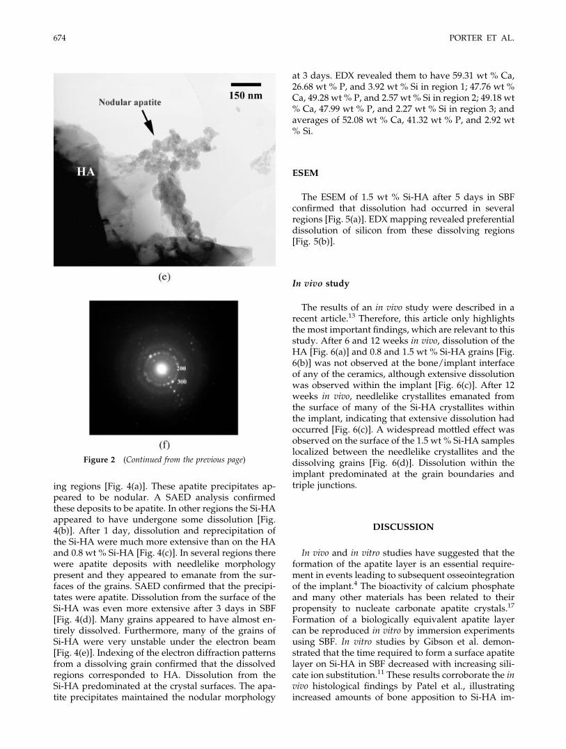
ing regions [Fig. 4(a)]. These apatite precipitates ap-peared to be nodular. A SAED analysis confirmedthese deposits to be apatite. In other regions the Si-HAappeared to have undergone some dissolution [Fig.4(b)]. After 1 day, dissolution and reprecipitation ofthe Si-HA were much more extensive than on the HAand 0.8 wt % Si-HA [Fig. 4(c)]. In several regions therewere apatite deposits with needlelike morphologypresent and they appeared to emanate from the sur-faces of the grains. SAED confirmed that the precipi-tates were apatite. Dissolution from the surface of theSi-HA was even more extensive after 3 days in SBF[Fig. 4(d)]. Many grains appeared to have almost en-tirely dissolved. Furthermore, many of the grains ofSi-HA were very unstable under the electron beam[Fig. 4(e)]. Indexing of the electron diffraction patternsfrom a dissolving grain confirmed that the dissolvedregions corresponded to HA. Dissolution from theSi-HA predominated at the crystal surfaces. The apa-tite precipitates maintained the nodular morphology
at 3 days. EDX revealed them to have 59.31 wt % Ca,26.68 wt % P, and 3.92 wt % Si in region 1; 47.76 wt %Ca, 49.28 wt % P, and 2.57 wt % Si in region 2; 49.18 wt% Ca, 47.99 wt % P, and 2.27 wt % Si in region 3; andaverages of 52.08 wt % Ca, 41.32 wt % P, and 2.92 wt% Si.
ESEM
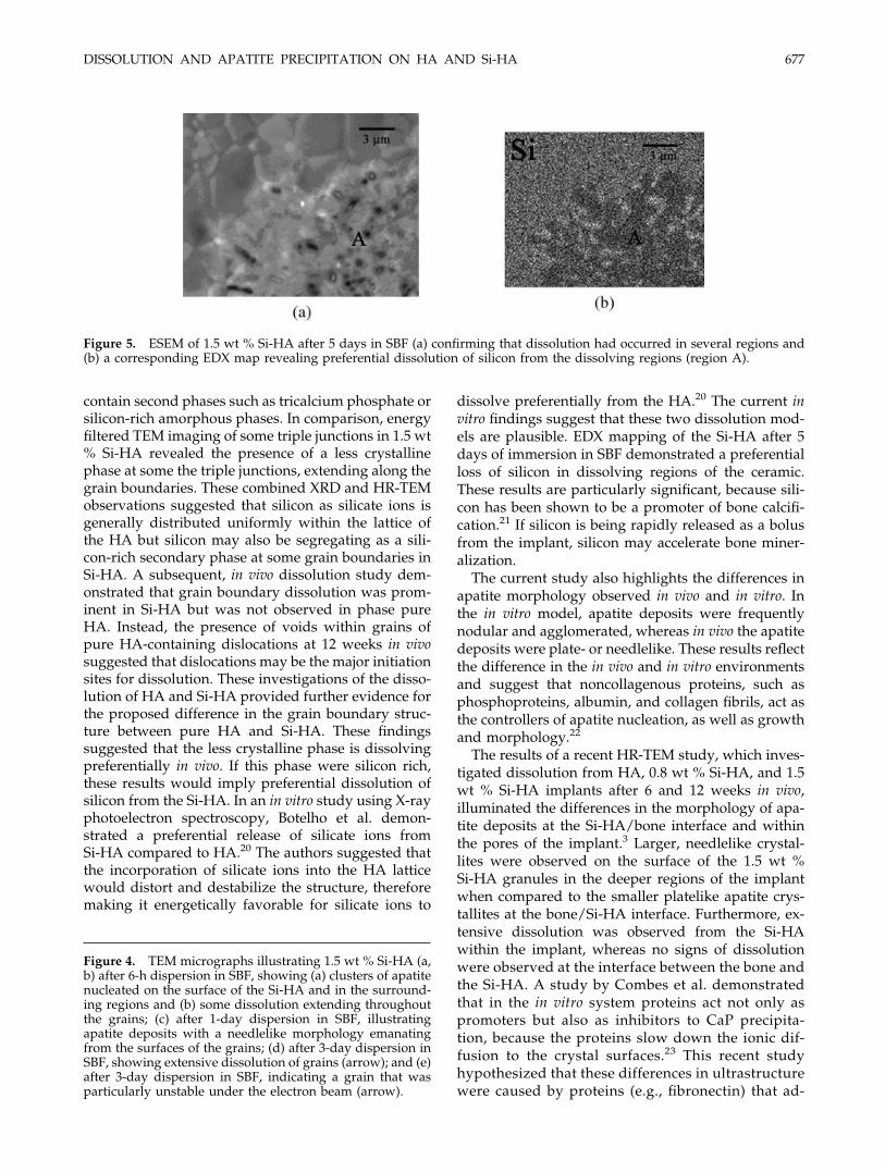
The ESEM of 1.5 wt % Si-HA after 5 days in SBFconfirmed that dissolution had occurred in severalregions [Fig. 5(a)]. EDX mapping revealed preferentialdissolution of silicon from these dissolving regions[Fig. 5(b)].
In vivo study
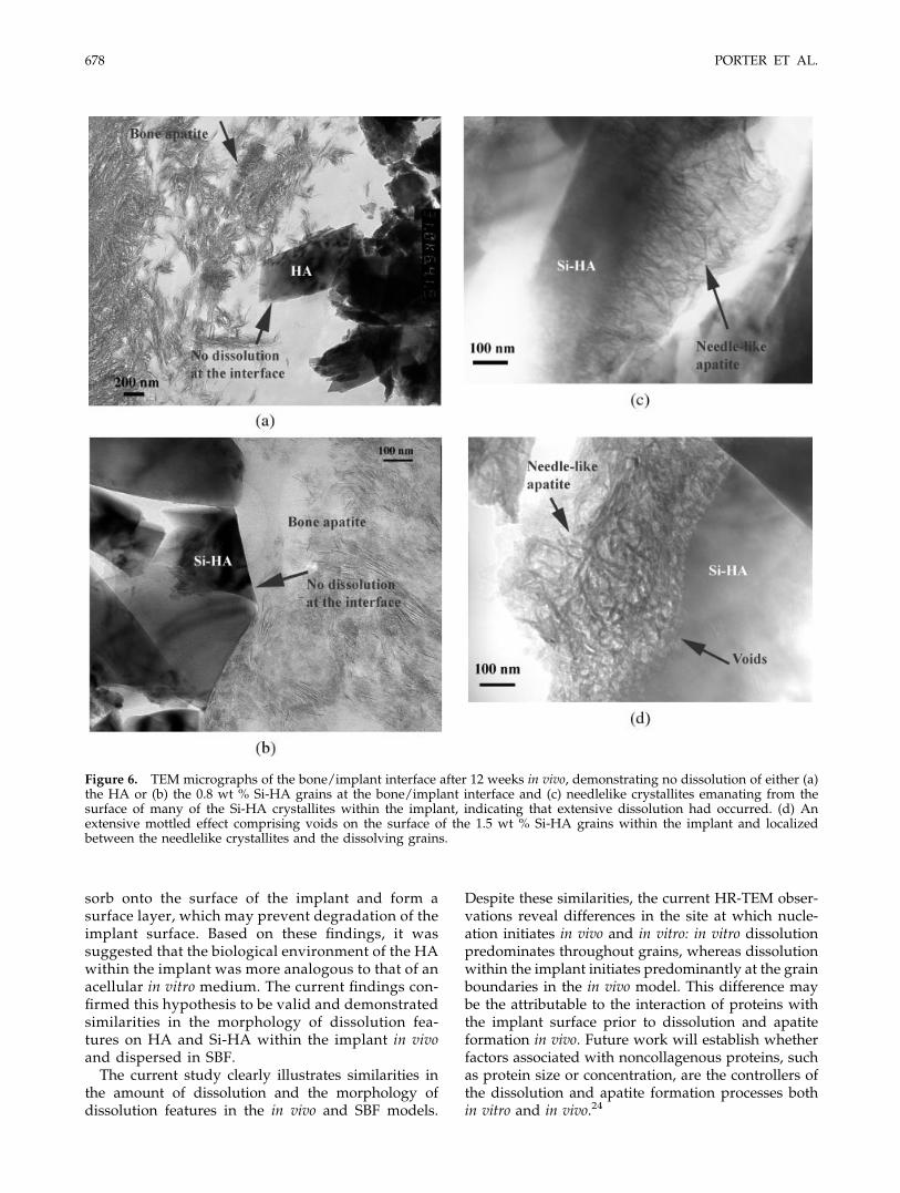
The results of an in vivo study were described in arecent article.13 Therefore, this article only highlightsthe most important findings, which are relevant to thisstudy. After 6 and 12 weeks in vivo, dissolution of theHA [Fig. 6(a)] and 0.8 and 1.5 wt % Si-HA grains [Fig.6(b)] was not observed at the bone/implant interfaceof any of the ceramics, although extensive dissolutionwas observed within the implant [Fig. 6(c)]. After 12weeks in vivo, needlelike crystallites emanated fromthe surface of many of the Si-HA crystallites withinthe implant, indicating that extensive dissolution hadoccurred [Fig. 6(c)]. A widespread mottled effect wasobserved on the surface of the 1.5 wt % Si-HA sampleslocalized between the needlelike crystallites and thedissolving grains [Fig. 6(d)]. Dissolution within theimplant predominated at the grain boundaries andtriple junctions.
DISCUSSION
In vivo and in vitro studies have suggested that theformation of the apatite layer is an essential require-ment in events leading to subsequent osseointegrationof the implant.4 The bioactivity of calcium phosphateand many other materials has been related to theirpropensity to nucleate carbonate apatite crystals.17
Formation of a biologically equivalent apatite layercan be reproduced in vitro by immersion experimentsusing SBF. In vitro studies by Gibson et al. demon-strated that the time required to form a surface apatitelayer on Si-HA in SBF decreased with increasing sili-cate ion substitution.11 These results corroborate the invivo histological findings by Patel et al., illustratingincreased amounts of bone apposition to Si-HA im-
Figure 2 (Continued from the previous page)
674 PORTER ET AL.
plants compared to pure HA.2 These findings demon-strate the value of SBF bioactivity tests to model the invivo system. Nevertheless, uncertainty remains aboutwhether the ultrastructural characteristics of in vitrodissolution–reprecipitation processes mimic thoseprocesses occurring in vivo.
The lack of information describing the ultrastruc-tural characteristics of HA immersed in SBF resultsfrom the difficulty in preparing these samples for TEMobservation. In the current study, a procedure wasdeveloped for preparing ultrathin samples for TEM bysectioning embedded powders using an ultramic-rotome and immersing the sections in SBF. This meth-odology does not allow for direct comparison betweenthe timing of events at the bone–HA and SBF–HAinterface. Nevertheless, for the purposes of this study,it was successful in providing a qualitative compari-son of the changes in the ultrastructure of syntheticHA and apatite deposits in vivo and in vitro.
A notable finding in this study was that the currentin vitro results uphold the in vivo findings of dissolu-tion following the order 1.5 wt % Si-HA � 0.8 wt %Si-HA � pure HA after 6 and 12 weeks.3 Dissolutionof the 1.5 wt % Si-HA was extensive after 3 days inSBF, whereas dissolution was less extensive on thesurface of the pure HA at 3 days. These results suggestthat SBF is a valuable medium for modeling theamount of dissolution in vivo.
It was interesting in this study that the silicon con-tent of apatite deposits surrounding the Si-HA washigher than that surrounding the pure HA after 3 daysin SBF. The effect of silicic acid on calcium phosphateprecipitation in vitro was investigated in order to testthe possible involvement of silica in dental plaque as apromoter of calculus formation.18 An in vitro study byDamen et al.19 illustrated that silica decreases the timefor spontaneous precipitation of HA from supersatu-rated solutions with a wide range of Ca/P ratios invitro. Furthermore, the study demonstrated that silicawas capable of stimulating apatite precipitation in thepresence of bacterial inhibitors to calcium phosphateprecipitation.19 The current results suggest that disso-lution of silicate ions from the implant promotes theheterogeneous nucleation of apatite.
In a previous study, Rietveld structural refinementsclearly illustrated that silicate ions were being substi-tuted for phosphate ions in the HA lattice.1 TheseHR-TEM observations confirmed that the grainboundaries in pure HA and 0.4 wt % Si-HA did not
Figure 3. TEM micrographs of 0.8 wt % Si-HA illustratingapatite deposits (a) 6 h after dispersion in SBF; (b) 1 day afterdispersion in SBF with apatite deposits nucleated on thesurface of the 0.8 wt % Si-HA (arrows); and (c) 3 days afterdispersion in SBF, illustrating extensive dissolution and pre-cipitation of apatite (arrows).
DISSOLUTION AND APATITE PRECIPITATION ON HA AND Si-HA 675
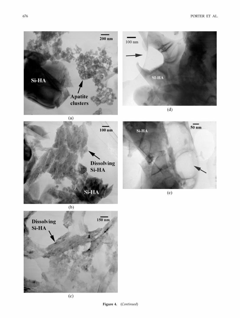
Figure 4. (Continued)
676 PORTER ET AL.
contain second phases such as tricalcium phosphate orsilicon-rich amorphous phases. In comparison, energyfiltered TEM imaging of some triple junctions in 1.5 wt% Si-HA revealed the presence of a less crystallinephase at some the triple junctions, extending along thegrain boundaries. These combined XRD and HR-TEMobservations suggested that silicon as silicate ions isgenerally distributed uniformly within the lattice ofthe HA but silicon may also be segregating as a sili-con-rich secondary phase at some grain boundaries inSi-HA. A subsequent, in vivo dissolution study dem-onstrated that grain boundary dissolution was prom-inent in Si-HA but was not observed in phase pureHA. Instead, the presence of voids within grains ofpure HA-containing dislocations at 12 weeks in vivosuggested that dislocations may be the major initiationsites for dissolution. These investigations of the disso-lution of HA and Si-HA provided further evidence forthe proposed difference in the grain boundary struc-ture between pure HA and Si-HA. These findingssuggested that the less crystalline phase is dissolvingpreferentially in vivo. If this phase were silicon rich,these results would imply preferential dissolution ofsilicon from the Si-HA. In an in vitro study using X-rayphotoelectron spectroscopy, Botelho et al. demon-strated a preferential release of silicate ions fromSi-HA compared to HA.20 The authors suggested thatthe incorporation of silicate ions into the HA latticewould distort and destabilize the structure, thereforemaking it energetically favorable for silicate ions to
dissolve preferentially from the HA.20 The current invitro findings suggest that these two dissolution mod-els are plausible. EDX mapping of the Si-HA after 5days of immersion in SBF demonstrated a preferentialloss of silicon in dissolving regions of the ceramic.These results are particularly significant, because sili-con has been shown to be a promoter of bone calcifi-cation.21 If silicon is being rapidly released as a bolusfrom the implant, silicon may accelerate bone miner-alization.
The current study also highlights the differences inapatite morphology observed in vivo and in vitro. Inthe in vitro model, apatite deposits were frequentlynodular and agglomerated, whereas in vivo the apatitedeposits were plate- or needlelike. These results reflectthe difference in the in vivo and in vitro environmentsand suggest that noncollagenous proteins, such asphosphoproteins, albumin, and collagen fibrils, act asthe controllers of apatite nucleation, as well as growthand morphology.22
The results of a recent HR-TEM study, which inves-tigated dissolution from HA, 0.8 wt % Si-HA, and 1.5wt % Si-HA implants after 6 and 12 weeks in vivo,illuminated the differences in the morphology of apa-tite deposits at the Si-HA/bone interface and withinthe pores of the implant.3 Larger, needlelike crystal-lites were observed on the surface of the 1.5 wt %Si-HA granules in the deeper regions of the implantwhen compared to the smaller platelike apatite crys-tallites at the bone/Si-HA interface. Furthermore, ex-tensive dissolution was observed from the Si-HAwithin the implant, whereas no signs of dissolutionwere observed at the interface between the bone andthe Si-HA. A study by Combes et al. demonstratedthat in the in vitro system proteins act not only aspromoters but also as inhibitors to CaP precipita-tion, because the proteins slow down the ionic dif-fusion to the crystal surfaces.23 This recent studyhypothesized that these differences in ultrastructurewere caused by proteins (e.g., fibronectin) that ad-
Figure 4. TEM micrographs illustrating 1.5 wt % Si-HA (a,b) after 6-h dispersion in SBF, showing (a) clusters of apatitenucleated on the surface of the Si-HA and in the surround-ing regions and (b) some dissolution extending throughoutthe grains; (c) after 1-day dispersion in SBF, illustratingapatite deposits with a needlelike morphology emanatingfrom the surfaces of the grains; (d) after 3-day dispersion inSBF, showing extensive dissolution of grains (arrow); and (e)after 3-day dispersion in SBF, indicating a grain that wasparticularly unstable under the electron beam (arrow).
Figure 5. ESEM of 1.5 wt % Si-HA after 5 days in SBF (a) confirming that dissolution had occurred in several regions and(b) a corresponding EDX map revealing preferential dissolution of silicon from the dissolving regions (region A).
DISSOLUTION AND APATITE PRECIPITATION ON HA AND Si-HA 677
sorb onto the surface of the implant and form asurface layer, which may prevent degradation of theimplant surface. Based on these findings, it wassuggested that the biological environment of the HAwithin the implant was more analogous to that of anacellular in vitro medium. The current findings con-firmed this hypothesis to be valid and demonstratedsimilarities in the morphology of dissolution fea-tures on HA and Si-HA within the implant in vivoand dispersed in SBF.
The current study clearly illustrates similarities inthe amount of dissolution and the morphology ofdissolution features in the in vivo and SBF models.
Despite these similarities, the current HR-TEM obser-vations reveal differences in the site at which nucle-ation initiates in vivo and in vitro: in vitro dissolutionpredominates throughout grains, whereas dissolutionwithin the implant initiates predominantly at the grainboundaries in the in vivo model. This difference maybe the attributable to the interaction of proteins withthe implant surface prior to dissolution and apatiteformation in vivo. Future work will establish whetherfactors associated with noncollagenous proteins, suchas protein size or concentration, are the controllers ofthe dissolution and apatite formation processes bothin vitro and in vivo.24
Figure 6. TEM micrographs of the bone/implant interface after 12 weeks in vivo, demonstrating no dissolution of either (a)the HA or (b) the 0.8 wt % Si-HA grains at the bone/implant interface and (c) needlelike crystallites emanating from thesurface of many of the Si-HA crystallites within the implant, indicating that extensive dissolution had occurred. (d) Anextensive mottled effect comprising voids on the surface of the 1.5 wt % Si-HA grains within the implant and localizedbetween the needlelike crystallites and the dissolving grains.
678 PORTER ET AL.
CONCLUSIONS
This study confirmed a difference in the morphol-ogy of apatite precipitates in vivo and in SBF: apatitedeposits were platelike in vivo and nodular in SBF. Thesite at which dissolution nucleates was also different:dissolution predominates at crystal surfaces in SBF,whereas grain boundary dissolution predominates invivo. Compositional mapping in this study suggestedthat silicon dissolves preferentially from the Si-HAand encourages nucleation of carbonate apatitearound the implant. A previous study illustrated thatno dissolution whatsoever occurred at the bone–HAor Si–HA interface in vivo. In comparison, dissolutionwithin the implant was similar to dissolution observedin SBF. These results suggest that the proteins andcells prevent dissolution from the surface of the im-plant. In summary, the present study suggests that,despite being unable to accurately model the ultra-structure of dissolution and reprecipitation featuresfound in vivo, SBF is a valuable medium for modelingthe amount of dissolution and precipitation.
The authors acknowledge Nelesh Patel and Jeremy Skep-per for their kind assistance with sample preparation; theHREM group at the University of Cambridge for the use oftheir facilities; and the financial support of EPSRC, ApatechLtd., and Fundacao para a Ciencia e Tecnologia for thesecond author’s (C.M.B.) grant (SFRH/BD/6173) and projectfunding (POCTI/CTM/49238/2002).
References
1. Porter AE, Best SM, Bonfield W. Ultrastructural comparison ofhydroxyapatite and silicon-substituted hydroxyapatite for bio-medical applications. J Biomed Mater Res 2003;68A:133–141.
2. Patel N, Gibson IR, Hing KA, Best SM, Revell PA, Bonfield W.A comparative study on the in vivo behaviour of hydroxyapa-tite and silicon substituted hydroxyapatite granules. J MaterSci Mater Med 2002;13:1199–1206.
3. Porter AE, Patel N, Skepper JNS, Best SM, Bonfield W. Compar-ison of in vivo dissolution processes in hydroxyapatite and silicon-hydroxyapatite bioceramics. Biomaterials 2003;24:4609–4620.
4. Weng J, Liu Q, Wolke JGC, Zhang X, de Groot K. Formationand characteristics of the apatite layer on plasma-sprayed hy-droxyapatite coatings in simulated body fluid. Biomaterials1995;18:1027–1035.
5. Doi Y, Shibutani T, Moriwake Y, Kajimoto T, Iwayama Y.Sintered carbonate apatites as bioresorbable bone substitutes.J Biomed Mater Res 1997;39:603–610.
6. Schepers E, Declercq M, Ducheyne P, Kempeneers R. Bioactiveglass particulate material as a filler for bone lesions. J OralRehab 1991;18:439–452.
7. Ducheyne P, Qiu Q. Bioactive ceramics: the effect of surfacereactivity on bone formation and bone cell function. Biomate-rials 1999;20:2287–2303.
8. Ducheyne P, Radin S, King L. The effect of calcium-phosphateceramic composition and structure on in vitro behaviour. I.Dissolution. J Biomed Mater Res 1993;27:25–34.
9. Kukubo T, Kushitani H, Sakka S. Solutions able to reproduce invivo surface-structure changes in bioactive glass-ceramic A-W.J Biomed Mater Res 1990;24:721–734.
10. Radin SR, Ducheyne P. The effect of calcium phosphate ce-ramic composition and structure on in vitro behavior. II. Pre-cipitation. J Biomed Mater Res 1993:27:35–45.
11. Gibson IR, Hing KA, Best SM, Bonfield W. Enhanced in vitrocell activity and surface apatite layer formation on novel sili-con-substituted hydroxyapatites. In: Ohgushi H, Hastings GW,Yoshikawa T, editors. 12th International Symposium on Ce-ramics in Medicine, Nara, Japan, 1999. World Scientific. p191–194.
12. Fujibayashi S, Neo M, Kim H-M, Kokubo T, Nakamura T. Acomparative study between in vivo bone ingrowth and in vitroapatite formation on Na2-CaO-SiO2 glasses. Biomaterials 2003;24:1349–1356.
13. Porter AE, Patel N, Skepper JN, Best SM, Bonfield W. Effect ofsintered silicate-substituted hydroxyapatite on remodellingprocesses at the bone–implant interface. Biomaterials 2003:25;3303–3314.
14. Leng Y, Chen J, Qu S. TEM study of calcium phosphate pre-cipitation on HA/TCP ceramics. Biomaterials 2003;24:2125–2131.
15. Akao M, Aoki H, Kato K. Mechanical properties of sinteredhydroxyapatite for prosthetic applications. J Mater Sci 1981;28:809.
16. Gibson IR, Best SM, Bonfield W. Chemical characterization ofsilicon-substituted hydroxyapatite. J Biomed Mater Res 1999;44:422–428.
17. Rey C. Calcium phosphates for medical applications. In: Am-jad Z, editor. Calcium phosphates in biological and industrialsystems. Kluwer Academic: Boston; 1998. p 217–251.
18. Tanizawa Y, Suzuki T. Effects of silica ions on the formationand transformation of calcium phosphates in neutral aqueoussolutions. J Chem Soc Faraday Trans 1995;91:3499–3503.
19. Damen JJM, Ten Cate JM. Silica-induced precipitation of cal-cium phosphate in the presence of inhibitors of hydroxyapatiteformation. J Dent Res 1992;71:453–457.
20. Botelho CM, Lopes MA, Gibson IR, Best SM, Santos JD. Struc-tural analysis of Si-substituted hydroxyapatite: zeta potentialand X-ray photoelectron spectroscopy (XPS). J Mater Sci MaterMed 2002;13:1123–1127.
21. Carlisle EM. Silicon. Nutr Rev 1975;33:257–260.22. Termine JD, Eanes ED, Conn KM. Phosphoprotein modulation
of apatite crystallization. Calcif Tissue Int 1980;31:247–251.23. Combes C, Rey C. Adsorption of proteins and calcium phos-
phate materials bioactivity. Biomaterials 2002;23:2817–2823.24. Horbett TA. Proteins at interfaces—an overview. ACS Symp
Ser 1995;602–623.
DISSOLUTION AND APATITE PRECIPITATION ON HA AND Si-HA 679
Related Documents