Ultrasound-Mediated Stimulation of Microbubbles after Acute Myocardial Infarction and Reperfusion Ameliorates Left-Ventricular Remodelling in Mice via Improvement of Borderzone Vascularization Jonas Do ¨ rner 1 , Rafael Struck 1 , Sebastian Zimmer 1 , Christine Peigney 2 , Georg Daniel Duerr 2 , Oliver Dewald 2 , Se-Chan Kim 3 , Daniela Malan 4 , Thierry Bettinger 5 , Georg Nickenig 1 , Alexander Ghanem 1 * 1 Department of Medicine/Cardiology, University of Bonn, Bonn, Germany, 2 Department of Cardiac Surgery, University Clinical Center Bonn, Bonn, Germany, 3 Department of Anaesthesiology and Intensive Care Medicine, University of Bonn, Bonn, Germany, 4 Institute of Physiology I, University of Bonn, Bonn, Germany, 5 Bracco Suisse SA, Geneva Research Center, Plan-les Ouates, Switzerland Abstract Aims: Post-infarction remodelling (PIR) determines left-ventricular (LV) function and prognosis after myocardial infarction. The aim of this study was to evaluate transthoracic ultrasound-mediated microbubble stimulation (UMS) as a novel gene- and cell-free therapeutic option after acute myocardial infarction and reperfusion (AMI/R) in mice. Methods and Results: For myocardial delivery of UMS, a novel therapeutic ultrasound-system (TIPS, Philips Medical) and commercially available microbubbles (BR1, Bracco Suisse SA) were utilized in a closed-chest mouse model. UMS was performed as myocardial post-conditioning (PC) on day four after 30 minutes of coronary occlusion and reperfusion. LV- morphology, as well as global and regional function were measured repeatedly with reconstructive 3-dimensional echocardiography applying an additional low-dose dobutamine protocol after two weeks. Scar size was quantified by means of histomorphometry. A total of 41 mice were investigated; 17 received PC with UMS. Mean ejection fraction (EF) prior UMS was similar in both groups 53%610 (w/o UMS) and 53%614 (UMS, p = 0.5), reflecting comparable myocardial mass at risk 17%68 (w/o UMS), 16%613 (UMS, p = 0.5). Two weeks after AMI/R, mice undergoing UMS demonstrated significantly better global LV-function (EF = 53%67) as compared to the group without PC (EF = 39%611, p,0.01). The fraction of akinetic myocardial mass was significantly lower among mice undergoing UMS after AMI/R [27%610 (w/o UMS), 13%68 (UMS), p,0.001)]. Our experiments showed a fast onset of transient, UMS-induced upregulation of vascular- endothelial and insulin-like growth factor (VEGF-a, IGF-1), as well as caveolin-3 (Cav-3). The mice undergoing PC with UMS after AMI/R showed a significantly lower scar size. In addition, the microvascular density was significantly higher in the borderzone of UMS-treated animals. Conclusion: UMS following AMI/R ameliorates PIR in mice via up-regulation of VEGF-a, IGF-1 and Cav-3, and consecutive improvement of myocardial borderzone vascularization. Citation: Do ¨ rner J, Struck R, Zimmer S, Peigney C, Duerr GD, et al. (2013) Ultrasound-Mediated Stimulation of Microbubbles after Acute Myocardial Infarction and Reperfusion Ameliorates Left-Ventricular Remodelling in Mice via Improvement of Borderzone Vascularization. PLoS ONE 8(2): e56841. doi:10.1371/ journal.pone.0056841 Editor: Rakesh Kukreja, Virginia Commonwealth University Medical Center, United States of America Received October 11, 2012; Accepted January 14, 2013; Published February 20, 2013 Copyright: ß 2013 Do ¨ rner et al. This is an open-access article distributed under the terms of the Creative Commons Attribution License, which permits unrestricted use, distribution, and reproduction in any medium, provided the original author and source are credited. Funding: J.D. and A.G. were supported by BONFOR (Intramural research foundation of the University of Bonn). A.G. was supported by the German Heart Foundation. The funders had no role in study design, data collection and analysis, decision to publish, or preparation of the manuscript. Competing Interests: The authors have the following interest. Thierry Bettinger is employed by Bracco Suisse SA and provided the commercially available micro bubbles (BR1) for this study. There are no further patents, products in development or marketed products to declare. This does not alter the authors’ adherence to all the PLOS ONE policies on sharing data and materials, as detailed online in the guide for authors. * E-mail: [email protected] Introduction Acute myocardial infarction (AMI) and its sequelae are some of the most common causes of death in the western world, even worldwide. The cicatrization of the infarcted left-ventricular (LV) myocardium leads to morphological and functional changes of the contractile tissue, also referred to as post-infarction remodelling (PIR) [1]. This process is progressive and comprises: a) LV- dilatation, b) deterioration of global and regional LV-function, c) progression of scar size and d) loss of viable myocardial tissue [1,2]. Each component is associated with increased mortality [3]. Hence, current therapeutic approaches for AMI aim at attenuating PIR [2]. The main objective of our study is to test the functional impact of a novel, non-gene, non-cell based intervention to ameliorate both, morphological and functional changes after AMI and reperfusion in mice. Clinically, myocardial revascularization is the method of choice in treatment of AMI and the minimization of PIR. Experimental PLOS ONE | www.plosone.org 1 February 2013 | Volume 8 | Issue 2 | e56841

Welcome message from author
This document is posted to help you gain knowledge. Please leave a comment to let me know what you think about it! Share it to your friends and learn new things together.
Transcript

Ultrasound-Mediated Stimulation of Microbubbles afterAcute Myocardial Infarction and ReperfusionAmeliorates Left-Ventricular Remodelling in Mice viaImprovement of Borderzone VascularizationJonas Dorner1, Rafael Struck1, Sebastian Zimmer1, Christine Peigney2, Georg Daniel Duerr2,
Oliver Dewald2, Se-Chan Kim3, Daniela Malan4, Thierry Bettinger5, Georg Nickenig1,
Alexander Ghanem1*
1Department of Medicine/Cardiology, University of Bonn, Bonn, Germany, 2Department of Cardiac Surgery, University Clinical Center Bonn, Bonn, Germany,
3Department of Anaesthesiology and Intensive Care Medicine, University of Bonn, Bonn, Germany, 4 Institute of Physiology I, University of Bonn, Bonn, Germany,
5 Bracco Suisse SA, Geneva Research Center, Plan-les Ouates, Switzerland
Abstract
Aims: Post-infarction remodelling (PIR) determines left-ventricular (LV) function and prognosis after myocardial infarction.The aim of this study was to evaluate transthoracic ultrasound-mediated microbubble stimulation (UMS) as a novel gene-and cell-free therapeutic option after acute myocardial infarction and reperfusion (AMI/R) in mice.
Methods and Results: For myocardial delivery of UMS, a novel therapeutic ultrasound-system (TIPS, Philips Medical) andcommercially available microbubbles (BR1, Bracco Suisse SA) were utilized in a closed-chest mouse model. UMS wasperformed as myocardial post-conditioning (PC) on day four after 30 minutes of coronary occlusion and reperfusion. LV-morphology, as well as global and regional function were measured repeatedly with reconstructive 3-dimensionalechocardiography applying an additional low-dose dobutamine protocol after two weeks. Scar size was quantified bymeans of histomorphometry. A total of 41 mice were investigated; 17 received PC with UMS. Mean ejection fraction (EF)prior UMS was similar in both groups 53%610 (w/o UMS) and 53%614 (UMS, p = 0.5), reflecting comparable myocardialmass at risk 17%68 (w/o UMS), 16%613 (UMS, p = 0.5). Two weeks after AMI/R, mice undergoing UMS demonstratedsignificantly better global LV-function (EF = 53%67) as compared to the group without PC (EF = 39%611, p,0.01). Thefraction of akinetic myocardial mass was significantly lower among mice undergoing UMS after AMI/R [27%610 (w/o UMS),13%68 (UMS), p,0.001)]. Our experiments showed a fast onset of transient, UMS-induced upregulation of vascular-endothelial and insulin-like growth factor (VEGF-a, IGF-1), as well as caveolin-3 (Cav-3). The mice undergoing PC with UMSafter AMI/R showed a significantly lower scar size. In addition, the microvascular density was significantly higher in theborderzone of UMS-treated animals.
Conclusion: UMS following AMI/R ameliorates PIR in mice via up-regulation of VEGF-a, IGF-1 and Cav-3, and consecutiveimprovement of myocardial borderzone vascularization.
Citation: Dorner J, Struck R, Zimmer S, Peigney C, Duerr GD, et al. (2013) Ultrasound-Mediated Stimulation of Microbubbles after Acute Myocardial Infarction andReperfusion Ameliorates Left-Ventricular Remodelling in Mice via Improvement of Borderzone Vascularization. PLoS ONE 8(2): e56841. doi:10.1371/journal.pone.0056841
Editor: Rakesh Kukreja, Virginia Commonwealth University Medical Center, United States of America
Received October 11, 2012; Accepted January 14, 2013; Published February 20, 2013
Copyright: � 2013 Dorner et al. This is an open-access article distributed under the terms of the Creative Commons Attribution License, which permitsunrestricted use, distribution, and reproduction in any medium, provided the original author and source are credited.
Funding: J.D. and A.G. were supported by BONFOR (Intramural research foundation of the University of Bonn). A.G. was supported by the German HeartFoundation. The funders had no role in study design, data collection and analysis, decision to publish, or preparation of the manuscript.
Competing Interests: The authors have the following interest. Thierry Bettinger is employed by Bracco Suisse SA and provided the commercially availablemicro bubbles (BR1) for this study. There are no further patents, products in development or marketed products to declare. This does not alter the authors’adherence to all the PLOS ONE policies on sharing data and materials, as detailed online in the guide for authors.
* E-mail: [email protected]
Introduction
Acute myocardial infarction (AMI) and its sequelae are some of
the most common causes of death in the western world, even
worldwide. The cicatrization of the infarcted left-ventricular (LV)
myocardium leads to morphological and functional changes of the
contractile tissue, also referred to as post-infarction remodelling
(PIR) [1]. This process is progressive and comprises: a) LV-
dilatation, b) deterioration of global and regional LV-function, c)
progression of scar size and d) loss of viable myocardial tissue [1,2].
Each component is associated with increased mortality [3]. Hence,
current therapeutic approaches for AMI aim at attenuating PIR
[2]. The main objective of our study is to test the functional impact
of a novel, non-gene, non-cell based intervention to ameliorate
both, morphological and functional changes after AMI and
reperfusion in mice.
Clinically, myocardial revascularization is the method of choice
in treatment of AMI and the minimization of PIR. Experimental
PLOS ONE | www.plosone.org 1 February 2013 | Volume 8 | Issue 2 | e56841

treatment options encompass gene- and cell-based interventions to
preserve the contractile performance after AMI. Firstly, myocar-
dial perfusion can be improved by growth factor induced neo-
vascularisation (e.g. vascular-endothelial growth factor (VEGF-a)
or insulin-like growth factor 1 (IGF-1)) [4]. Secondly, myocardial
overexpression of distinct structural proteins (e.g. Caveolin-3, Cav-
3) can affect the survival rate of cardiomyocytes after AMI [5].
Previous approaches utilizing gene- and cell-based methods to
deliver IGF-1 after myocardial infarction through permanent
occlusion of the left coronary artery demonstrated an amelioration
of PIR [6]. However, gene- and cell-free methods to stimulate
intrinsic overexpression of the mentioned mechanisms are under
current investigation [7].
Ultrasound-mediated stimulation of microbubbles (UMS) has
been shown to modulate myocardial expression patterns and to
improve myocardial transplantation of bone marrow derived cells
in rats [7]. However, the additive value of this therapeutic option
to reperfusion after AMI in mice has not been elucidated yet.
Materials and Methods
All experiments have been approved by the animal care
committee at the University of Bonn and the local government
authorities. Also, they conform to the guidelines of the American
Heart Association for the use of animals in research and
corresponds to the Guide for the Care and Use of Laboratory
Animals published by the US National Institutes of Health (NIH
Publication No. 85–23, revised 1985). All animals were housed at
a constant room temperature of 24uC and 12 h light–dark cycle
and maintained on an ad libitum diet.
Mouse Model of Acute Myocardial Infarction andReperfusion
Coronary instrumentation. In order to minimize inflam-
matory interaction of surgical trauma and AMI, an established
closed-chest model of AMI and reperfusion (AMI/R) was utilized
(Figure 1) [8]. 56 mice (8–12 weeks old, female, C57BL/6; Charles
River, Sulzfeld, Germany) underwent the procedure as recently
described [9]. 41 mice were included for functional longitudinal
studies and 15 for RNA and protein analysis. Briefly, general
anaesthesia was induced with 4% isoflurane (Abbott, Germany) in
1 L per minute O2-flow. Mice were intubated with a 22 gauge
cannula (Braun, Melsungen, Germany) and connected to a venti-
lation system (Type Small Animal Ventilator KTR4, Harvard
Apparatus GmbH, March-Hugstetten, Germany). Correct in-
tubation and ventilation was confirmed by observing thorax
excursions. The animals were placed in a supine position on
a feedback heat pad. Their body temperature was measured with
a rectal probe and maintained at 36.5uC. The anaesthesia was
maintained with 1.2–1.5% isoflurane in 100% O2, the animals
heart rates were monitored and kept within physiological ranges to
minimize cardio-depressant effects. The respiratory rate was set at
115/min and 10 mL tidal volume per gram body weight. Sufficient
analgesia was determined by testing the rear foot reflex.
A left lateral thoracotomy was performed in the forth
intercostal-space. The pericardium was opened and gently re-
moved to identify the left anterior descending coronary artery
(LAD). The LAD was cautiously under-stitched with an 8–
0 prolene suture (Mopylen, Resorba Wundversorgung GmbH &
Co. KG, Germany) with a U-shaped tapered needle 1 mm distal
of the left auricle. The suture was cut at the needle side and both
ends were threaded through 1.0 mm section of a PE 10 tube to
form a loose snare around the LAD. To check the correct position
of the LAD ligature both ends were transiently tightened. If the
position of the ligature was correct paleness of the distal antero-
lateral myocardial segments could be observed. Both ends were
exteriorized one through the 3rd, the other through the 5th
intercostal space and stored subcutaneously. The thorax was
closed with a prolene suture (6–0, Mopylen, Resorba Wundver-
sorgung GmbH & Co. KG, Germany).
After closing the skin, the mice were weaned from the ventilator
and kept in a warm cage. Post-operative care comprised fentanyl
0.1 mg/g bodyweight bid s.c. for analgesia and 7.5 mg/kg
bodyweight of the antibiotic enrofloxacine (BaytrilTM, Bayer
Healthcare, Leverkusen, Germany) s.c. for 5 days.
Figure 1. Experimental protocol. The instrumentive surgery of the left anterior descending coronary artery (LAD-Instrumentation) was performedseven days prior to the acute myocardial infarction and reperfusion (AMI/R) to avoid a pro-inflammatory influence of trauma on post-infarctionremodelling (PIR). The treatment group received UMS four days after AMI/R. On day +4 and +14, a reconstructive 3-dimensional echocardiography(r3DE) was performed to quantify global and regional left-ventricular function. Additionally, r3DE was carried out with low-dose dobutamine on day+14. Ultimately, hearts were harvested for histological workup.doi:10.1371/journal.pone.0056841.g001
Amelioration of Myocardial Remodelling with UMS
PLOS ONE | www.plosone.org 2 February 2013 | Volume 8 | Issue 2 | e56841

Myocardial infarction and reperfusion. For our AMI/R
experiments myocardial infarction took place seven days after
coronary instrumentation (Figure 1). The Mice were anesthetized
with ketamine (65 mg/kg bodyweight), xylazine (13 mg/kg body-
weight) and atropine (0.05 mg/kg bodyweight) i.p. as published
previously [10]. The Mice were intubated, ventilated with room
air and placed supinely on a feedback heat pad. The skin was re-
opened and the exteriorized coronary snare was gently relieved
from subcutaneous tissue. Then, tension was carefully applied to
achieve controlled closure of the LAD. In addition to the
electrocardiogram, an onset of new regional contraction abnor-
mality was monitored using high-resolution echocardiography
(Philips HDI-5000, Philips Healthcare). Reperfusion was initiated
after 30 minutes by release and gentle removal of the coronary
snare. Sham-operated mice underwent the same procedures
except for pulling the coronary snare. The skin was closed with
a prolene suture (6–0, Mopylen, Resorba Wundversorgung
GmbH & Co. KG, Germany).
Ultrasound-mediated Stimulation of MicrobubblesThe ultrasound-mediated stimulation of microbubbles (UMS)
allows myocardial post-conditioning (PC) by locoregional stimu-
lation of commercially available ultrasound contrast agent with
high-intensity focussed ultrasound. For UMS, BR1 (Bracco Suisse
SA, Geneva, Switzerland) [11] was continuously infused into
a femoral vein which was cannulated with a 27 gauge ViggoTM
(Braun, Melsungen, Germany) [7,12]. UMS was performed with
a newly developed, combined ultrasound device (Therapy Imaging
Probe System (TIPS); Philips Research North America, Briarcliff
Manor, NY, USA) which provides morphological and functional
visualisation of target structures with high-resolution b-mode
ultrasound imaging and simultaneous and stereotactically coupled
application of focussed low-frequency ultrasound (1 MHz, 10000
cycles, 0.3 MPa peak negative pressure, 0.2 Hz) (Figure 2) [13]. A
computer-controlled movement ensured the precise and automat-
ed UMS application to the LV-myocardium within a stereotactic
grid, and was planned using a b-mode short axis view of the left
ventricle. The target volume had a total surface of 25 mm2
consisting of 565 target points with a gap of 1 mm and was placed
into the anterior LV-wall aimed at the infarcted area. The
ultrasound pulse was delivered with a frequency of 0.2 Hz to allow
a sufficient replenishment of the contrast agent. The Control
procedure was insonication protocol prior to infusion of micro-
bubbles, as described previously by Miller and co-workers [14].
Additional experiments were conducted to confirm the myo-
cardial delivery of UMS. Evans blue (EB) (Sigma-Aldrich Chemie
GmbH, Munich, Germany) is a dye that binds on albumin and
therefore stays within the intravasal compartment. Extravasation is
an indicator of increased capillary permeability [14]. To evaluate
the effect of ultrasound emission power we investigated capillary
permeability by means of EB extravasation and transendothelial
penetration of fluorescent nanospheres, as published previously
[15]. Briefly, we investigated the impact of UMS treatment with
different ultrasound force amplitudes on capillary permeability
utilising the TIPS-System. Therefore 50 mg/kg EB dye was
injected via the femoral vein prior to UMS application [16].
Figure 2. Therapeutic imaging probe system (TIPS) for ultrasound mediated stimulation of microbubbles. (A) Hybrid ultrasound probesystem with electromechanically coupled diagnostic (+) and therapeutic probe (*) enabling simultaneous high-resolution imaging for targetedapplication of UMS. To allow a standardized application to a small, moving target organ, the system is coupled to a computer-programmed steppermotor (#). (B) ‘‘En face’’ heart model to visualize the computer-programmed grid on the anterolateral heart wall. The grid consists of 25 pulses total,administered every 1 mm and 5 pulses per row, respectively starting basal (S) and following the white line to the apex till E (end). UMS was targetedon the anterior left-ventricular wall and the anterior borderzone tissue (area within the dotted circle) after anterolateral ischemia. (C)+(D) UltrasoundB-Mode (upper image) and M-Mode (lower image) short axis view before (C) and after (D) microbubble application in a mouse without myocardialinfarction. Hence, this hybrid scanhead allows standardized and targeted myocardial delivery of UMS in mice.doi:10.1371/journal.pone.0056841.g002
Figure 3. Schematic illustration for determination of hypertro-phy. The illustration shows the six locations of wall thicknessmeasurements in a mid-ventricular histological short axis section withscarring of the anterolateral left-ventricular wall. S: scar; AS: anteroseptalborderzone; AL: anterolateral borderzone. Control regions: IS: infer-oseptal wall; I: inferior wall; IL: inferolateral wall.doi:10.1371/journal.pone.0056841.g003
Amelioration of Myocardial Remodelling with UMS
PLOS ONE | www.plosone.org 3 February 2013 | Volume 8 | Issue 2 | e56841
Immediately after UMS, hearts were harvested and rinsed in
cardioplegic solution to arrest the heart in end-diastole. Both atria
were dissected and the heart was divided into the anterior and the
posterior LV-wall. Then both parts were dried for 24 h in 37uC.EB was extracted using 8 ml formamide 99% (Sigma-Aldrich
Chemie GmbH, Munich, Germany)/mg tissue as described
previously [14,15]. EB concentration was determined by spectro-
photometry of the supernatant at 620 nm against a formamide
99% blank using an absorbance reader (Tecan Safire2, Tecan
Group Ltd., Switzerland). EB extravasation was calculated as mgEB per mg dry tissue, thus revealing the level of transendothelial
extravasation within the targeted myocardial tissue [15].
UMS shows transient biological effects and allows nanoparticles
to be delivered transendothelially across sites of UMS-induced
transient pore formation [17]. To evaluate efficacy and consisten-
cy of delivery to a distinct myocardial target, in this case the LV
anterior wall in mice, we used fluorescent nanospheres (Duke
Scientific, Palo Alto, CA, Diameter: 30 nm) to visualise the
targeted site [17].
Nanospheres were injected simultaneously with microbubbles
and UMS protocol was administered with different peak pressures
(0.3; 0.5; 1.5 and 3 MPa). Hearts were harvested directly after the
administration of UMS and processed for histological analyses.
Nanosphere delivery was determined with overlap fluorescent and
reflected light microscopy using an Olympus BX 41/Color View
II System and Cell-P software (Carl Zeiss MicroImaging GmbH,
Germany).
Reconstructive 3-dimensional Echocardiography (r3DE)Morphological and functional imaging was performed 4 and 14
days after the application of UMS. All mice were anesthetized with
isoflurane (4% for induction, 0.5–0.9% in 1 L per minute O2-flow
for maintenance) in 100% oxygen by facemask to avoid
cardiodepression and ensure near physiological heart rate.
Echocardiography was performed with a commercially available
high-resolution ultrasound system (Philips HDI-5000, Philips
Healthcare) equipped with a linear-array transducer (CL15-7)
operating at 15 MHz and providing frame rates up to 284 Hz. A
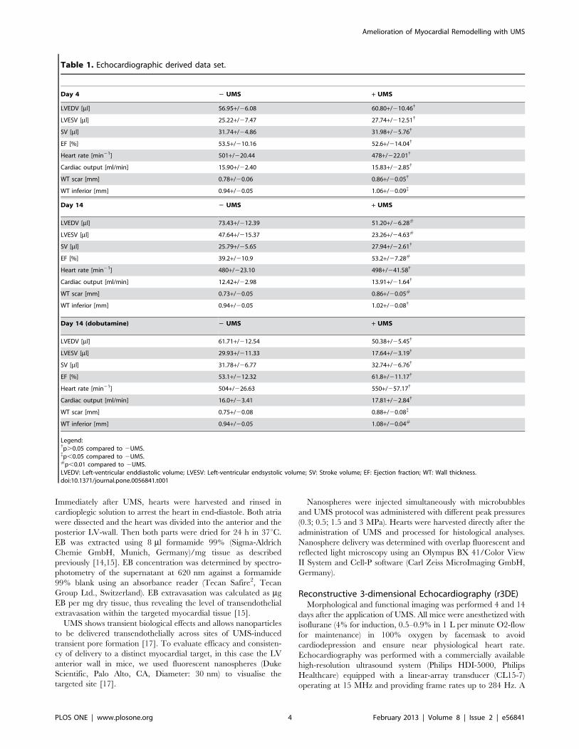
Table 1. Echocardiographic derived data set.
Day 4 2 UMS + UMS
LVEDV [ml] 56.95+/26.08 60.80+/210.46{
LVESV [ml] 25.22+/27.47 27.74+/212.51{
SV [ml] 31.74+/24.86 31.98+/25.76{
EF [%] 53.5+/210.16 52.6+/214.04{
Heart rate [min21] 501+/220.44 478+/222.01{
Cardiac output [ml/min] 15.90+/22.40 15.83+/22.85{
WT scar [mm] 0.78+/20.06 0.86+/20.05{
WT inferior [mm] 0.94+/20.05 1.06+/20.09{
Day 14 2 UMS + UMS
LVEDV [ml] 73.43+/212.39 51.20+/26.28#
LVESV [ml] 47.64+/215.37 23.26+/24.63#
SV [ml] 25.79+/25.65 27.94+/22.61{
EF [%] 39.2+/210.9 53.2+/27.28#
Heart rate [min21] 480+/223.10 498+/241.58{
Cardiac output [ml/min] 12.42+/22.98 13.91+/21.64{
WT scar [mm] 0.73+/20.05 0.86+/20.05#
WT inferior [mm] 0.94+/20.05 1.02+/20.08{
Day 14 (dobutamine) 2 UMS + UMS
LVEDV [ml] 61.71+/212.54 50.38+/25.45{
LVESV [ml] 29.93+/211.33 17.64+/23.19{
SV [ml] 31.78+/26.77 32.74+/26.76{
EF [%] 53.1+/212.32 61.8+/211.17{
Heart rate [min21] 504+/226.63 550+/257.17{
Cardiac output [ml/min] 16.0+/23.41 17.81+/22.84{
WT scar [mm] 0.75+/20.08 0.88+/20.08{
WT inferior [mm] 0.94+/20.05 1.08+/20.04#
Legend:{p.0.05 compared to 2UMS.{p,0.05 compared to 2UMS.#p,0.01 compared to 2UMS.LVEDV: Left-ventricular enddiastolic volume; LVESV: Left-ventricular endsystolic volume; SV: Stroke volume; EF: Ejection fraction; WT: Wall thickness.doi:10.1371/journal.pone.0056841.t001
Amelioration of Myocardial Remodelling with UMS
PLOS ONE | www.plosone.org 4 February 2013 | Volume 8 | Issue 2 | e56841
parasternal long-axis image was used to guide the perpendicular
angulation of the transducer for acquisition of the short-axis slices.
Then, sequenced 2D, parallel short-axis images of the left ventricle
were obtained in 500 mm steps from the aortic root towards the
apex by means of a micrometer-screw driven tripod. Ten to
fourteen short- axis segments were recorded depending on the
overall size of the left ventricle [18,19]. Parasternal short-axis
views were visually divided into six segments. Imaging was
considered adequate when the endocardial and epicardial borders
could be properly visualized in at least five segments. Cineloops of
50 frames covering minimum two heart cycles were stored digitally
and analysed off-line. In addition, we repeated the imaging
protocol with an intravenous low-dose dobutamine (10 mg/kg/min) application at day 14 [19,20].
Echocardiographic image analysis. An experienced in-
vestigator performed echocardiographic analyses blinded to
treatment strategy. End-diastolic measurements were obtained at
the peak of the R-wave, whereas end-systolic measurements were
obtained at the time of minimum internal chamber dimensions.
The acquired sequential 2D short-axis cineloops of the left
ventricle were used to measure LV-volumes at end-diastole and
end-systole and ejection fraction (EF). Myocardial compartments
were differentiated by visual assessment following established
echocardiographic criteria: [8] a) akinetic/dyskinetic wall motion
(collagenous scar): thinned, echodense wall, no systolic wall
thickening, no inotropic response to dobutamine; b) hypokinetic
wall motion (peri-infarction borderzone myocardium): normal
myocardial thickness, markedly reduced systolic wall thickening
during baseline, reduced inotropic response to dobutamine; c)
normokinetic wall motion (remote myocardium): normal myocar-
dial thickness, good systolic wall thickening, good inotropic
response to dobutamine. Due to its best correlation with the
histomorphological scar, wall motion abnormalities are reported as
the fraction of ‘‘akinetic myocardial mass’’ [19].
Reverse Transcriptase Polymerase Chain ReactionFor investigation of expression patterns we varied applied peak
pressure (0.3; 0.5; 1.5 MPa) and harvesting time (15 min, 6 h,
30 h) in mice undergoing LV-myocardial UMS. Animals with
administration of contrast agent following the insonication pro-
tocol served as controls [14]. Also, specific effects of contrast agent
only and ultrasound application without contrast agent were
investigated as control groups. After hearts were excised, all
adnexa were gently removed inclusively the left and right atria,
and finally the tissue was recovered in RNAlaterTM (Qiagen,
Ambion, Inc., Austin, Texas) and stored at 4uC. The tissue was
homogenized with a tissue-tearer and mRNA was extracted with
the PureLinkTM RNA Mini Kit (Invitrogen, Ambion, Inc., Austin,
Texas) following the manufacturers protocol. cDNA was produced
using the High-Capacity cDNA Reverse Transcription kitTM with
RNase inhibitor (Applied Biosystems, Foster City, CA, USA) as
described by the manufacturer. The PCR was performed as
a quantitative measurement with a TaqmanTM cycler and
a TaqmanTM specific real-time PCR kit (TaqManTM Gene
Expression Master Mix (Applied Biosystems, Foster City, CA,
USA)). We investigated the time course of expression patterns with
Figure 4. Global and regional left-ventricular function. (A) Mean left-ventricular ejection fraction (LV-EF) was moderately reduced four daysafter acute myocardial infarction and reperfusion (AMI/R) prior to ultrasound-mediated stimulation of microbubbles (UMS) in both groups. (B)Regional LV-function was quantified by means of reconstructive 3-dimensional echocardiography (r3DE) and is expressed as fraction of akineticmyocardial mass. UMS-treated animals (circles) demonstrated functional improvement two weeks after AMI/R, as compared to controls (squares). (C,D) Global and regional LV-function were obtained prior and during pharmacological stimulation with low-dose dobutamine on day +14. Inotropicresponse was preserved in both groups and revealed a significant increase in LV-EF and decrease in fraction of akinetic myocardial mass. Both non-invasive measures are parameters indicating preserved myocardial viability after AMI and reperfusion. In all, UMS improved LV-function after AMIR/Rwithout impact on myocardial viability.doi:10.1371/journal.pone.0056841.g004
Amelioration of Myocardial Remodelling with UMS
PLOS ONE | www.plosone.org 5 February 2013 | Volume 8 | Issue 2 | e56841

three primers: 1. vascular-endothelial growth factor-a (VEGF-a),
2. insulin-like growth factor-1 (IGF-1) and 3. caveolin-3 (Cav-3)
(Applied Biosystems, Foster City, CA, USA). The results were
calculated as 1/2 ddCt in relation to the housekeeping gene
glyceraldehyde 3-phosphate dehydrogenase (GAPDH) (Applied
Biosystems, Foster City, CA, USA) after normalization to control
mice.
Enzyme-linked Immunosorbent Assay (ELISA)To determine the protein expression of IGF-1, the myocardial
tissue was homogenized and incubated on ice for 5 min in 1 mL of
ELISA buffer containing 20 mM TRIS-HCL, 50 mM sodium
chloride, 50 mM sodium fluoride, 10 mM EDTA, 20 mM sodium
pyrophosphate, 1 mM Triton X-100 (Sigma-Aldrich, Tauf-
kirchen, Germany) in aqua bidest with protease inhibitors
(Complete Mini Tab, Roche, Mannheim, Germany) per 100 mg
tissue. The samples were incubated on ice for 10 min and
centrifuged for 15 min at 4uC at 14000 rpm. The supernatant was
used for measuring myocardial protein levels of IGF-1 (R&D
Systems, Minneapolis, USA) using a Tecan Safire2 Reader (Tecan
Group Ltd., Switzerland). We investigated the protein content
[pg/mg myocardial tissue] in: 1. control-groups without any
treatment; 2. instrumented mice with AMI/R but without UMS;
3. instrumented mice with AMI/R and with UMS; 4. UMS only.
Histomorphometric Analyses and ImmunohistochemistryTwo weeks after the UMS-application heart samples were
prepared for microtome sectioning by rinsing directly in
cardioplegic solution after harvesting. Hearts were cannulated
via the ascending aorta with a modified Langendorff-system and
retrogradely perfused with 4% paraformaldehyde (Merck KGaA,
Darmstadt, Germany) at 100 mm Hg pressure for 10 min to avoid
the collapse of the left-ventricle [7]. After tissue fixation with 4%
paraformaldehyde (Merck KGaA, Darmstadt, Germany) in 0.1 M
PBS for 12 h, hearts were incubated in 18% sucrose solution for
6 h [21]. Heart samples were snap-frozen at 280uC in Tissue-
TekTM (Sakura Fintek, Zoeterwoude, Netherlands) and 7 mmslices were prepared with a KryotomTM (Leica Microsystems
Figure 5. Histological analyses. Myocardial scar formation was determined by means of histomorphometry. (A) Representative images of shortaxis mouse heart sections stained with picrosirius-red and fast-green without (left) and with (right) UMS-treatment. The collagenous tissue is colouredred whereas myocardium is green. (B) UMS-treated mice demonstrated a significantly lower collagenous scar burden compared to control mice.doi:10.1371/journal.pone.0056841.g005
Amelioration of Myocardial Remodelling with UMS
PLOS ONE | www.plosone.org 6 February 2013 | Volume 8 | Issue 2 | e56841
GmbH, Wetzlar, Germany). The hearts were cut in transversal
direction with 500 mm distance between each stage to be able to
measure a total volume by summation of the areas of each
transversal section. Slides were stained with picrosirius-red for the
demarcation of fibrotic scar and counterstained with fast-green
[19]. Sections were fixed and photographed with an Olympus BX
41/Color View II System and Cell-P software (Carl Zeiss
MicroImaging GmbH, Germany). Quantitative histomorpho-
metric analyses of scar formation were performed as described
previously [22]. Briefly, the ratio of collagenous tissue was
analysed using three-dimensional reconstruction with image
analysis software (ImageJ; Rasband, W.S., ImageJ, U. S. National
Institutes of Health, Bethesda, Maryland, USA). Epicardial and
endocardial contours, as well as scar, borderzones and remote
regions and their epicardial courses were traced. LV-surfaces,
volumes and masses were calculated based on 3D reconstruction
of LV-geometry by method of disks [22]. For each section surfaces
and masses were calculated by multiplication of areas and
circumferential sections with the intersection interval (0.5 mm),
respectively. Summarized along the long-axis, those resulted in
myocardial volumes and surfaces. Masses were the product of
volumes and 1.05 (density of myocardial tissue). For the evaluation
of myocardial hypertrophy cross-sectional wall thickness of scar,
both adjacent borderzones, and three locations on the posterior
wall as a control region were measured (Figure 3). From these
measurements the ratio scar/borderzone was calculated by
dividing the scar thickness by the mean thickness of the
anteroseptal and anterolateral segments.
To evaluate microvascular density CD31 immunohistochemis-
try was performed. Therefore cryosections were pre-treated with
citrate for 15 min in the microwave oven, incubated for 30 min in
hydrogen peroxide 3%, and rinsed two times in PBS pH 7.4 for
5 min. Then they have been blocked with the Vectastain rat kit
(Vector Laboratories, Burlingame, CA, USA) for 15 min. CD31
staining was performed using the rat anti-mouse CD31 antibody
(PECAM-1 monoclonal rat anti-mouse, Dianova, Germany) over
night at 4uC with a dilution ratio of 1:50 in an IgG-block. As
a secondary antibody biotin was incubated for 30 min. The
antibody was detected with peroxidase using the Vectastain Rat
ABC kit (Vector Laboratories, Burlingame, CA, USA) for 30 min
and developed with diaminobenzol (DAB) and nickel for 5 min.
Microvascular density was assessed by counting the number of
CD31-positive vessels in the region of interest. Results were
collected as vessels per mm2 in the scar, both borderzones, and the
posterior wall [23].
Statistical AnalysisStatistical significance between treatment groups was tested by
means of the unpaired t-test. Comparison over time, as well as
between rest and stress, were tested with the paired Student’s t-test.
A value of p,0.05 was considered significant. Data are indicated
as mean6 SD.
Results
A total of 41 C57BL6 mice undergoing AMI/R were
randomized into two treatment groups receiving either UMS
(n= 17) or control procedure (n = 24). The functional and
morphological impacts of UMS on cardiac function after
experimental AMI/R were repetitively investigated by means of
r3DE on day 4 and 14. Echocardiographic left-ventricular
dimensions are displayed in table 1. In both treatment groups,
mice revealed a mildly decreased LV-function early after AMI/R
prior UMS. At that point of time, LV-EF values were comparable
in both treatment groups (53%610 (w/o UMS), 53%614 (UMS),
p = ns). However, by day 14, untreated mice displayed a significant
deterioration of global LV-function (39%611, p,0.01), as
compared to UMS-treated mice (53%67, p = ns).
In order to estimate regional LV-function, the akinetic mass of
LV-myocardium was quantified by means of r3DE. In parallel, on
day 4 there were no significant differences between both groups
(17%68 (w/o UMS), 16%613 (UMS), p = ns). However, by day
14 the akinetic myocardial mass of UMS-treated mice remained
stable (13%68), whereas the control group demonstrated a signif-
icant increase (27%610, p,0.001) (Figure 4 A+B). To further
investigate global and regional LV-function, mice were subjected
to low-dose dobutamine. During pharmacological stimulation two
weeks after AMI/R, no significant differences between both
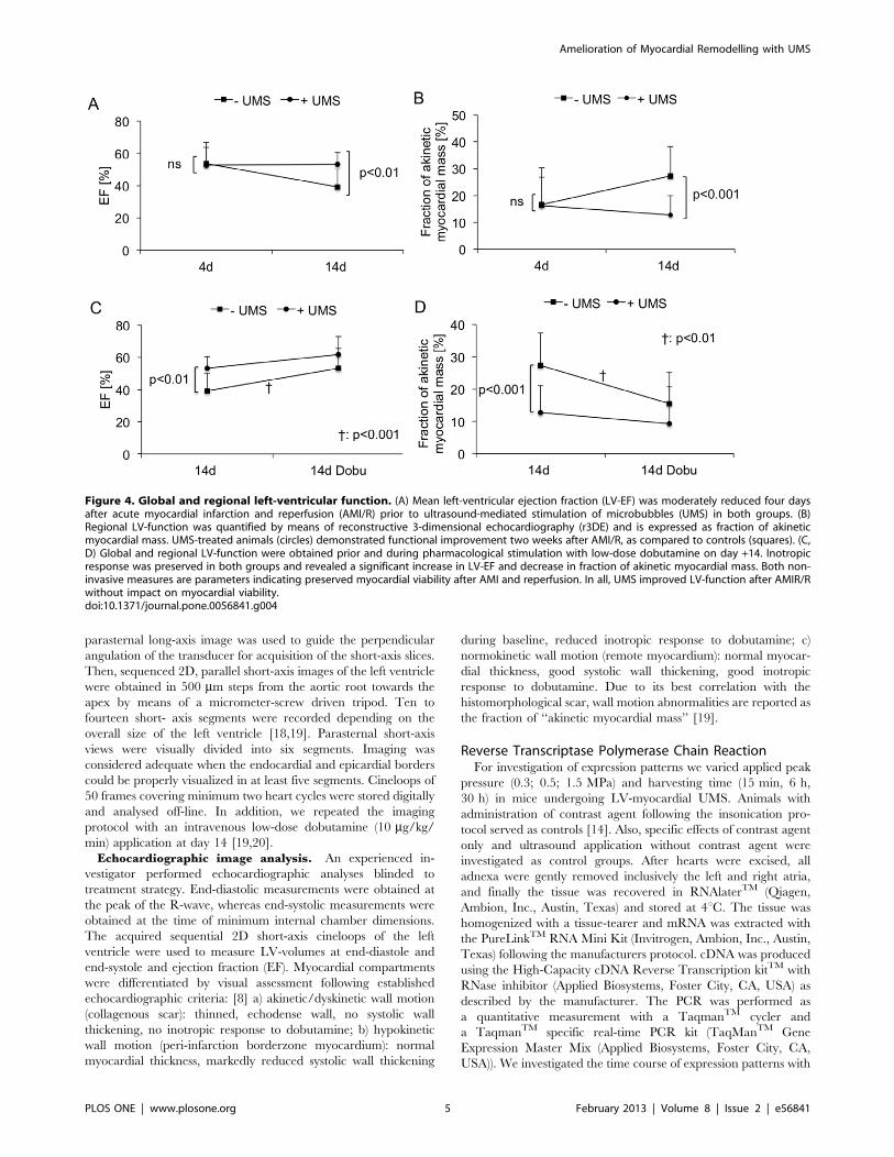
Table 2. Evaluation of hypertrophy derived fromhistomorphometric data.
2 UMS + UMS
Myocardial scar [mm] 0.72+/20.20 0.91+/20.27#
Anteroseptal left-ventricular WT[mm]
1.19+/20.26 1.30+/20.28{
Anterolateral left-ventricular WT[mm]
1.31+/20.25 1.35+/20.24{
Inferoseptal left-ventricular WT[mm]
0.83+/20.21 0.88+/20.17{
Inferior left-ventricular WT [mm] 0.96+/20.18 0.94+/20.30{
Inferolateral left-ventricular WT[mm]
1.00+/20.20 1.06+/20.21{
Ratio inferior 1.06+/20.10 0.95+/20.19#
Ratio scar/borderzone 0.59+/20.18* 0.69+/20.19{*
Heart weight [mg] 82.3+/212.2 90.7+/214.8{
Body weight [g] 20.5+/21.8 21.9+/22.3{
Normalized heart weight [mg/g] 4.01+/20.32 4.13+/20.32{
Legend:WT: Wall thickness.{p.0.05 compared to 2 UMS.#p,0.05 compared to 2 UMS.*p,0.001 compared to ‘‘ratio inferior’’ as the control region in the same group.doi:10.1371/journal.pone.0056841.t002
Figure 6. Delivery of myocardial UMS in mice. UMS-mediatedmyocardial delivery of fluorescent nanospheres was measured andquantified by fluorescence microscopy. Myocardial delivery of fluores-cent nanospheres with UMS was feasible in mice and demonstrateda dose-dependent effect.doi:10.1371/journal.pone.0056841.g006
Amelioration of Myocardial Remodelling with UMS
PLOS ONE | www.plosone.org 7 February 2013 | Volume 8 | Issue 2 | e56841
groups were observed (Figure 4 C+D). Left-ventricular ejection
fraction as a parameter for global LV-function increased during
low-dose dobutamine reflecting inotropic myocardial recruitment.
Concordant results were obtained for the quantity of akinetic
myocardial mass. During inotropic stimulation, both groups
revealed a substantial decrease of akinetic myocardial mass
(Figure 4 D). In all, UMS had no additional impact on contractile
reserve during pharmacological stimulation.
Histological analyses of scar size were concordant with
functional results. We observed a significant reduction of
myocardial scar burden in mice treated with UMS (w/o UMS:
10.9%65.8; UMS: 6.5%63.7, (p = 0.006), Figure 5). Data derived
from histomorphometric analysis demonstrate higher wall thick-
ness of scar and borderzone tissue in mice treated with UMS.
Remote regions had comparable dimensions (see table 2). Our
histological data correspond well with the echocardiographic
measurements. To our knowledge, this is the first report of
amelioration of PIR utilizing an organ-targeted, non-cell- and
non-gene-based intervention in mice.
To investigate potential mechanisms, further experiments were
conducted. We characterized the efficacy of myocardial delivery of
UMS utilizing EB and nanospheres. UMS effects were more
pronounced in the anterior LV-wall as demonstrated by a signif-
icantly higher amount of extravasated EB. This well-known effect
of UMS was shown to be dose-dependent, revealing lowest
extravasation at low emission power (0.3 MPa). In parallel, the
transendothelial distribution across microvascular structures
showed a dose-dependent increase (Figure 6). These data
demonstrate the feasibility to successfully target UMS at small
structures such as myocardium of mice.
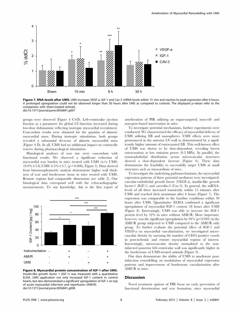
To investigate the underlying pathomechanisms, the myocardial
expression patterns of three potential mediators were investigated:
vascular-endothelial growth factor (VEGF-a), insulin-like growth
factor-1 (IGF-1) and caveolin-3 (Cav-3). In general, the mRNA-
levels of all three increased transiently within 15 minutes after
UMS and reached their maximum after 6 hours (Figure 7). The
expression was comparable to the baseline conditions within 30
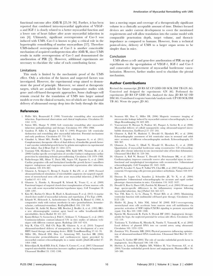
hours after UMS. Quantitative ELISA confirmed a significant
upregulation of myocardial IGF-1 content 18 hours after UMS
(Figure 8). Interestingly, UMS was able to increase the IGF-1
protein level by 52% in mice without AMI/R. More important,
however, was the significant upregulation by 94% (p,0.001) in the
AMI/R group subjected to UMS compared to the AMI/R only
group. To further evaluate the potential effect of IGF-1 and
VEGF-a on myocardial vascularization, we investigated micro-
vascular density by assessing the number of CD31-positive vessels
in post-ischemic and remote myocardial regions of interest.
Interestingly, microvascular density normalized to the non-
infarcted posterior left-ventricular wall was significantly higher in
the borderzone of UMS-treated animals (Figure 9).
Our data demonstrate the ability of UMS to ameliorate post-
infarction remodelling via modulation of myocardial expression
patterns and improvement of borderzone vascularization after
AMI/R in mice.
Discussion
Novel treatment options of PIR focus on early prevention of
functional deterioration and scar formation, since myocardial
Figure 7. RNA-levels after UMS. UMS increases VEGF-a, IGF-1 and Cav-3 mRNA-levels within 15 min and reaches its peak expression after 6 hours.A prolonged upregulation could not be observed longer than 30 hours after UMS as compared to controls. The displayed p-values refer to thecomparison with sham-treated animals.doi:10.1371/journal.pone.0056841.g007
Figure 8. Myocardial protein concentration of IGF-1 after UMS.Insulin-like growth factor 1 (IGF-1) was measured with a quantitativeELISA. UMS application not only increased IGF-1 content in controlhearts, but also demonstrated a significant upregulation of IGF-1 on topof acute myocardial infarction and reperfusion (AMI/R).doi:10.1371/journal.pone.0056841.g008
Amelioration of Myocardial Remodelling with UMS
PLOS ONE | www.plosone.org 8 February 2013 | Volume 8 | Issue 2 | e56841

damage caused by AMI have been known to be irreversible. Novel
treatment options of AMI with additional benefit to reperfusion
include cell- and gene-based strategies [6,24,25]. However, both
are based on myocardial uptake of foreign material and show
limited efficacy of transplantation and transfection, respectively
[26]. Hence, a transthoracic therapeutic option modulating
intrinsic myocardial expression patterns in PIR is of interest.
UMS is a promising non-genetic, non-cell based approach known
to influence myocardial expression patterns in rats [7]. However,
the exact mechanism of its modulatory effect has not been fully
elucidated yet. Previous studies obtained oscillation and microjet
formation of insonicated microbubbles and consecutive non-lethal
cell alteration and sterile inflammation of the targeted organ
[27,28]. However, functional data on delivery of UMS to even
smaller targets after AMI are not available. To our knowledge, this
is the first study to demonstrate feasibility and efficacy of UMS in
mice following AMI/R.
Since UMS may provide a new treatment option after AMI/R
and is able to influence PIR on top of reperfusion, we investigated
potential mechanisms of the UMS effect. Firstly, the UMS-
mediated overexpression of VEGF-a may help explain functional
improvements and amelioration of PIR [29,30]. VEGF-a plays
a substantial role in neovascularization and has been shown to
ameliorate PIR in gene- and cell-based studies [4,31,32].
Neovascularization improves myocardial function and preserves
viability of borderzone myocardium [33]. Secondly, we observed
UMS-driven overexpression of myocardial IGF-1. We demon-
strated a time-dependent, transient UMS-effect in RNA- and
protein analyses after AMI/R. In previous studies, IGF-1
displayed a critical role in PIR, improving myocardial cell survival
via PI-3K/AKT activation, which resulted in an improved
Figure 9. Microvascular density assessed by CD31 staining. (A) Microvascular density was assessed in the myocardial scar, both adjacentborderzones, and non-infarcted regions (posterior left-ventricular wall). No differences between untreated and UMS-treated animals were found withrespect to the scar tissue and the non-infarcted regions. However, the myocardial borderzone tissue of UMS-treated mice revealed a significantlyincreased microvascular density as compared to non-treated animals. (B) Representative CD31 stained histological images of scar, untreatedborderzone (2UMS), and UMS-treated borderzone (+UMS) (from left to right). CD31 positive vessels can be identified by their dark colour. Note thehigher microvascular density of the UMS-treated mice compared to the non-treated group. In contrast, scar displayed the lowest microvasculardensity.doi:10.1371/journal.pone.0056841.g009
Amelioration of Myocardial Remodelling with UMS
PLOS ONE | www.plosone.org 9 February 2013 | Volume 8 | Issue 2 | e56841
functional outcome after AMI/R [25,34–36]. Further, it has been
reported that combined intra-myocardial application of VEGF-
a and IGF-1 is closely related to a better myocardial function and
a lower rate of heart failure after acute myocardial infarction in
rats [4]. Ultimately, significant overexpression of Cav-3 was
induced with UMS. Cav-3 is known to play a critical role in the
hypertrophic remodelling of murine myocardium [37]. Therefore
UMS-induced overexpression of Cav-3 is another conceivable
mechanism of acquired cardioprotection after AMI/R, since UMS
results in stable overexpression of Cav-3 and demonstrated the
amelioration of PIR [5]. However, additional experiments are
necessary to elucidate the value of each contributing factor.
LimitationsThis study is limited by the mechanistic proof of the UMS
effect. Only a selection of the known and suspected factors was
investigated. However, the experimental setup aimed to demon-
strate the proof of principle. Moreover, we aimed at therapeutic
targets, which are available for future comparative studies with
gene- and cell-based therapeutic approaches. Some challenges will
remain crucial for the translation of UMS into larger animal
models or even the clinical scenario, two of which are: locoregional
delivery of ultrasound energy deep into the body through the skin
into a moving organ and coverage of a therapeutically significant
volume in a clinically acceptable amount of time. Distinct focused
devices are under current development to cover the mentioned
requirements and will allow translation into the canine model with
comparable penetration depth, target volume, and thoracic
impedance as compared to humans. However, from a technical
point-of-view, delivery of UMS to a larger organ seems to be
simpler than in mice.
ConclusionUMS allows a cell- and gene-free amelioration of PIR on top of
reperfusion via the up-regulation of VEGF-1, IGF-1 and Cav-3
and consecutive improvement of myocardial borderzone vascu-
larization. However, further studies need to elucidate the pivotal
mechanism.
Author Contributions
Revised the manuscript: JD RS SZ CP GDD OD SCK DM TB GN AG.
Conceived and designed the experiments: GN AG. Performed the
experiments: JD RS CP GDD AG. Analyzed the data: JD RS SZ OD
DM AG. Contributed reagents/materials/analysis tools: CP OD SCK DM
TB AG. Wrote the paper: JD AG.
References
1. Pfeffer MA, Braunwald E (1990) Ventricular remodeling after myocardial
infarction. Experimental observations and clinical implications. Circulation 81:
1161–1172.
2. Sutton MG, Sharpe N (2000) Left ventricular remodeling after myocardial
infarction: pathophysiology and therapy. Circulation 101: 2981–2988.
3. Gaudron P, Eilles C, Kugler I, Ertl G (1993) Progressive left ventricular
dysfunction and remodeling after myocardial infarction. Potential mechanisms
and early predictors. Circulation 87: 755–763.
4. Cittadini A, Monti MG, Petrillo V, Esposito G, Imparato G, et al. (2011)
Complementary therapeutic effects of dual delivery of insulin-like growth factor-
1 and vascular endothelial growth factor by gelatin microspheres in experimental
heart failure. Eur J Heart Fail 13: 1264–1274.
5. Tsutsumi YM, Horikawa YT, Jennings MM, Kidd MW, Niesman IR, et al.
(2008) Cardiac-specific overexpression of caveolin-3 induces endogenous cardiac
protection by mimicking ischemic preconditioning. Circulation 118: 1979–1988.
6. Padin-Iruegas ME, Misao Y, Davis ME, Segers VF, Esposito G, et al. (2009)
Cardiac progenitor cells and biotinylated insulin-like growth factor-1 nanofibers
improve endogenous and exogenous myocardial regeneration after infarction.
Circulation 120: 876–887.
7. Ghanem A, Steingen C, Brenig F, Funcke F, Bai ZY, et al. (2009) Focused
ultrasound-induced stimulation of microbubbles augments site-targeted engraft-
ment of mesenchymal stem cells after acute myocardial infarction. J Mol Cell
Cardiol 47: 411–418.
8. Ghanem A, Ziomka A, Krausgrill B, Schenk K, Troatz C, et al. (2009)
Functional impact of targeted closed-chest transplantation of bone marrow cells
in rats with acute myocardial ischemia/reperfusion injury. Cell Transplant 18:
1289–1297.
9. Kim SC, Boehm O, Meyer R, Hoeft A, Knufermann P, et al. (2012) A murine
closed-chest model of myocardial ischemia and reperfusion. J Vis Exp: e3896.
10. Erhardt W, Hebestedt A, Aschenbrenner G, Pichotka B, Blumel G (1984) A
comparative study with various anesthetics in mice (pentobarbitone, ketamine-
xylazine, carfentanyl-etomidate). ResExpMed(Berl) 184: 159–169.
11. Schneider M, Arditi M, Barrau MB, Brochot J, Broillet A, et al. (1995) BR1:
a new ultrasonographic contrast agent based on sulfur hexafluoride-filled
microbubbles. Invest Radiol 30: 451–457.
12. Kuntz-Hehner S, Goenechea J, Pohl C, Schlosser T, Veltmann C, et al. (2001)
Continuous-infusion contrast-enhanced US: in vitro studies of infusion tech-
niques with different contrast agents. Radiology 220: 647–654.
13. Seip R, Chin CT, Hall CS, Raju BI, Ghanem A, et al. (2010) Targeted
ultrasound-mediated delivery of nanoparticles: on the development of a new
HIFU-based therapy and imaging device. IEEE TransBiomedEng 57: 61–70.
14. Miller DL, Driscoll EM, Dou C, Armstrong WF, Lucchesi BR (2006)
Microvascular permeabilization and cardiomyocyte injury provoked by
myocardial contrast echocardiography in a canine model. JAmCollCardiol 47:
1464–1468.
15. Bekeredjian R, Kroll RD, Fein E, Tinkov S, Coester C, et al. (2007) Ultrasound
targeted microbubble destruction increases capillary permeability in hepatomas.
Ultrasound MedBiol 33: 1592–1598.
16. Swanson SD, Dou C, Miller DL (2006) Magnetic resonance imaging of
microvascular leakage induced by myocardial contrast echocardiography in rats.
Magn ResonImaging 24: 603–609.
17. Vancraeynest D, Havaux X, Pouleur AC, Pasquet A, Gerber B, et al. (2006)
Myocardial delivery of colloid nanoparticles using ultrasound-targeted micro-
bubble destruction. EurHeart J 27: 237–245.
18. Ghanem A, Roll W, Hashemi T, Dewald O, Djoufack PC, et al. (2006)
Echocardiographic assessment of left ventricular mass in neonatal and adult
mice: accuracy of different echocardiographic methods. Echocardiography 23:
900–907.
19. Ghanem A, Troatz C, Elhafi N, Dewald O, Heeschen C, et al. (2008)
Quantitation of myocardial borderzone using reconstructive 3-D echocardiog-
raphy after chronic infarction in rats: incremental value of low-dose dobutamine.
Ultrasound Med Biol 34: 559–566.
20. Ghanem A, Roll W, Bostani T, Dewald O, Fleischmann BK, et al. (2011)
Cardiomyoplasty improves contractile reserve after myocardial injury in mice -
functional and morphological investigations with reconstructive 3-dimensional
echocardiography. Cell Transplant 20: 1621–1628.
21. Roell W, Lewalter T, Sasse P, Tallini YN, Choi BR, et al. (2007) Engraftment of
connexin 43-expressing cells prevents post-infarct arrhythmia. Nature 450: 819–
824.
22. Dawson D, Lygate CA, Saunders J, Schneider JE, Ye X, et al. (2004)
Quantitative 3-dimensional echocardiography for accurate and rapid cardiac
phenotype characterization in mice. Circulation 110: 1632–1637.
23. Dewald O, Ren G, Duerr GD, Zoerlein M, Klemm C, et al. (2004) Of mice and
dogs: species-specific differences in the inflammatory response following
myocardial infarction. Am J Pathol 164: 665–677.
24. Yau TM, Kim C, Li G, Zhang Y, Weisel RD, et al. (2005) Maximizing
ventricular function with multimodal cell-based gene therapy. Circulation 112:
I123–128.
25. Haider H, Jiang S, Idris NM, Ashraf M (2008) IGF-1-overexpressing
mesenchymal stem cells accelerate bone marrow stem cell mobilization via
paracrine activation of SDF-1alpha/CXCR4 signaling to promote myocardial
repair. Circ Res 103: 1300–1308.
26. Epstein SE, Kornowski R, Fuchs S, Dvorak HF (2001) Angiogenesis therapy:
amidst the hype, the neglected potential for serious side effects. Circulation 104:
115–119.
27. Taniyama Y, Tachibana K, Hiraoka K, Namba T, Yamasaki K, et al. (2002)
Local delivery of plasmid DNA into rat carotid artery using ultrasound.
Circulation 105: 1233–1239.
28. Zarnitsyn VG, Prausnitz MR (2004) Physical parameters influencing optimiza-
tion of ultrasound-mediated DNA transfection. Ultrasound Med Biol 30: 527–
538.
29. Ferrara N, Gerber HP (2001) The role of vascular endothelial growth factor in
angiogenesis. Acta Haematol 106: 148–156.
30. Hoeben A, Landuyt B, Highley MS, Wildiers H, Van Oosterom AT, et al.
(2004) Vascular endothelial growth factor and angiogenesis. PharmacolRev 56:
549–580.
Amelioration of Myocardial Remodelling with UMS
PLOS ONE | www.plosone.org 10 February 2013 | Volume 8 | Issue 2 | e56841
31. Rosano JM, Cheheltani R, Wang B, Vora H, Kiani MF, et al. (2012) Targeted
Delivery of VEGF after a Myocardial Infarction Reduces Collagen Depositionand Improves Cardiac Function. Cardiovasc Eng Technol 3: 237–247.
32. Fujii H, Sun Z, Li SH, Wu J, Fazel S, et al. (2009) Ultrasound-targeted gene
delivery induces angiogenesis after a myocardial infarction in mice. JACCCardiovasc Imaging 2: 869–879.
33. Zhang J, Ding L, Zhao Y, Sun W, Chen B, et al. (2009) Collagen-targetingvascular endothelial growth factor improves cardiac performance after
myocardial infarction. Circulation 119: 1776–1784.
34. Fujio Y, Nguyen T, Wencker D, Kitsis RN, Walsh K (2000) Akt promotessurvival of cardiomyocytes in vitro and protects against ischemia-reperfusion
injury in mouse heart. Circulation 101: 660–667.
35. Bonni A, Brunet A, West AE, Datta SR, Takasu MA, et al. (1999) Cell survival
promoted by the Ras-MAPK signaling pathway by transcription-dependent and
-independent mechanisms. Science 286: 1358–1362.
36. Brunet A, Bonni A, Zigmond MJ, Lin MZ, Juo P, et al. (1999) Akt promotes cell
survival by phosphorylating and inhibiting a Forkhead transcription factor. Cell
96: 857–868.
37. Woodman SE, Park DS, Cohen AW, Cheung MW, Chandra M, et al. (2002)
Caveolin-3 knock-out mice develop a progressive cardiomyopathy and show
hyperactivation of the p42/44 MAPK cascade. J Biol Chem 277: 38988–38997.
Amelioration of Myocardial Remodelling with UMS
PLOS ONE | www.plosone.org 11 February 2013 | Volume 8 | Issue 2 | e56841
Related Documents