ULTRASOUND, MAGNETIC RESONANCE IMAGING (MRI), THERMODIAGNOSTIC (THERMOGRAPHY). Physical and technological bases radial methods of diagnostics with use nonionizing radiations DEPARTMENT OF ONCOLOGY AND RADIOLOGY PREPARED BY I.M.LESKIV

ULTRASOUND, MAGNETIC RESONANCE IMAGING (MRI), THERMODIAGNOSTIC (THERMOGRAPHY). Physical and technological bases radial methods of diagnostics with use.
Dec 27, 2015
Welcome message from author
This document is posted to help you gain knowledge. Please leave a comment to let me know what you think about it! Share it to your friends and learn new things together.
Transcript
ULTRASOUND, MAGNETIC RESONANCE IMAGING (MRI),
THERMODIAGNOSTIC (THERMOGRAPHY).
Physical and technological bases radial methods of
diagnostics with use nonionizing radiations
DEPARTMENT OF ONCOLOGY AND RADIOLOGY
PREPARED BY I.M.LESKIV
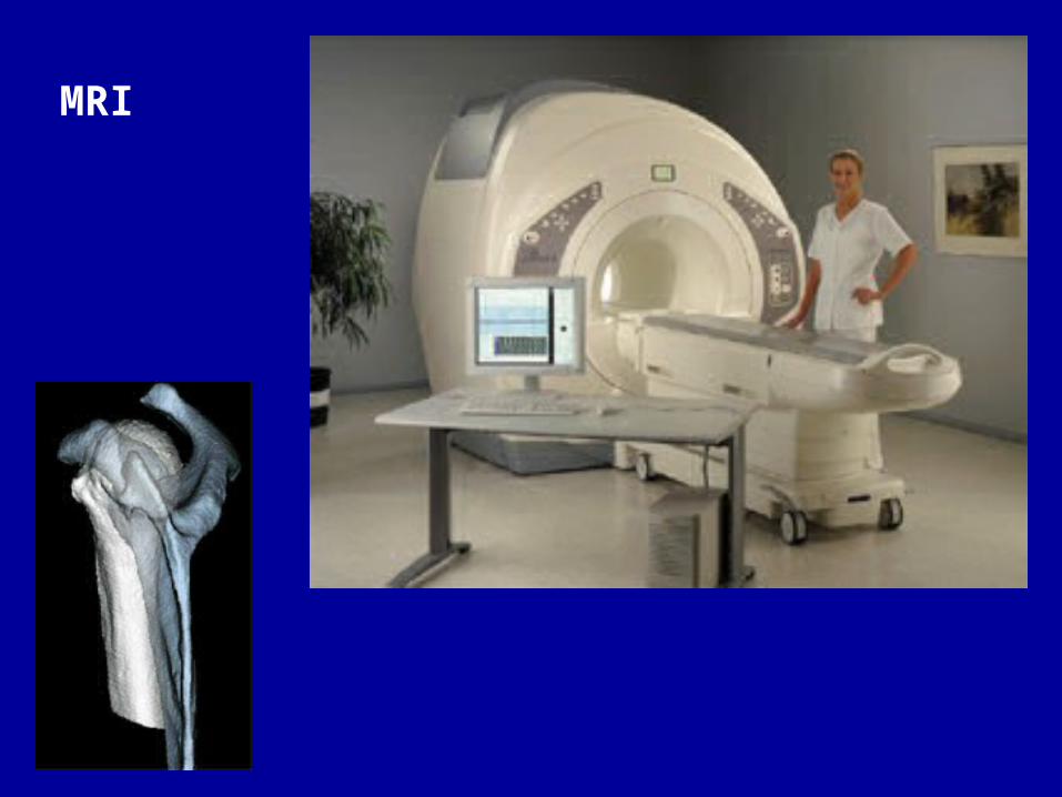
MRI
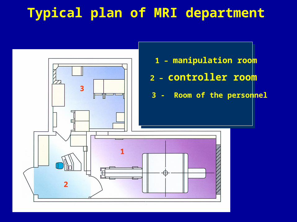
Typical plan of MRI department
1
2
3
1 – manipulation room
2 – controller room
3 - Room of the personnel

Typical plan of MRI department
Magnet and place of the operator

MRI consist: 1) a magnet; 2) a table for patient ; 3) Thermal cooler ; 4) a source of a high voltage; 5) a control panel with PS system.
1
2
4
MAGNETIC RESONANCE IMAGING (MRI)

A large cylindrical magnet with an internal diameter large enough to accept the human body provides an external magnetic field along the body axis. Gradient coils add a smaller identification field. The external magnetic field, together with the gradient field, provides a net external magnetic field, Bo. The radio frequency (RF) coil provides a force to rotate the spins away from the direction of the external magnetic field. As the nuclear spins process back toward the direction of the external magnetic field, they emit RF signals. These RF signals can be combined to form an image. Depending on the pulse sequences, the image can form maps of the proton density or can be used to form information regarding the local magnetic fields of the nuclear spins. By varying the RF pulse sequence, the image can be made up of predominantly Tl information or predominately T2 information. Tl is known as the spin lattice relaxation time. Images formed using Tl information are most heavily weighted toward proton density. T2, called the spin-spin interaction, forms images that provide information on tissue differences.

MR Signal Characteristics
• CSF: hypointense on Tl WI, hyperintense on T2WI• Grey/White Matter: Grey matter is grey and white
matter is white on T1WI and relationship is reversed on T2WI
• Most tumors are hyperintense on T2WI except Melanoma-hyperintense on Tl WI & hypointense on T2WI
• FAT—hyperintense on Tl WI, less hyperintense on T2WI• Flowing blood-Signal void
** How to Remember -Water is White in a T2 scan. Conversely, a Tl scan shows fat as being whiter.

MRI
Low resolution
High resolution

MR contrast media•
• Five to ten years ago, contrast media for MR imaging were considered completely unnecessary. Exprience has shown, however, that contrast media may increase the diagnostic unformation in several disease states. All contrast media have magnetic properties, and they change the signal intensity of the tissues where they are located. The most commonly used contrast media contain the paramagnetic metal ion gadolinium(Jd^3+). These contrast media are administered by in the body similar to water soluble X-ray contrast media.
– Contrast USED IN MRI —Gadolinium DTPA
(V.IMPORTANT)*
– No contrast used in MRCP, MR myelography etc. they
– use Heavily T2 weighted sequences
– Ischaemic Stroke detected earlier on MRI (esp. DW MRI)
– diffusion weighted imaging.
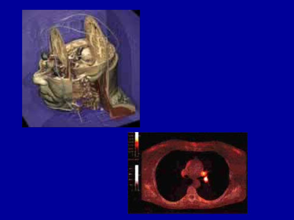
Volumetric reconstructionMRI
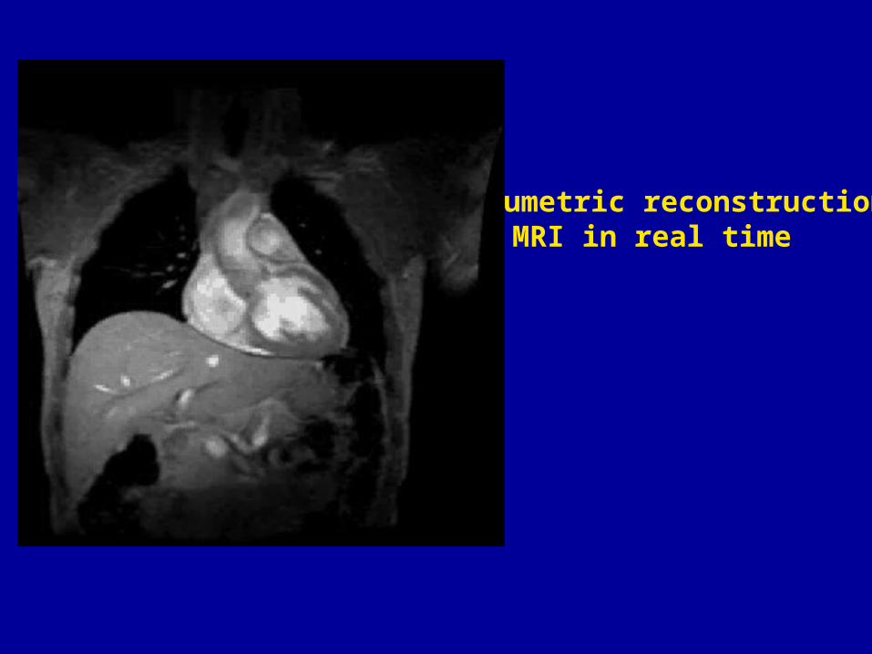
Volumetric reconstructionMRI in real time

Advantages of MRI
• High soft tissue contrast
• No ionizing radiation (Safe in pregnancy)
• Multiplanar imaging
• Better for bone marrow
• All spinal imaging- MRI, posterior-fossa—MRI.
• Doesn't use iodinated contrast so safe in iodine allergy

Contraindications and potential dangers
• No harmfull effects of the static or fluctuating magnetic field used in MR imaging, have been shown. Ferromagnetic objects are subjected to very strong mechanical foocer, however, and any ferromagnetic object having a location where motion of the object may be harmful to the patient represent on absolute contraidication to MR imaging. The most important and dangerous objects are ferromagnetic intracranial aneurysmal clips and intraocular ferromagnetic foreign objects. The main potential donger involved with these objects is serious haemorrhage. The presence of a pacemaker represents an absolute contraindication to MR imaging. The function of the pacemaker may be affected by the magnetic field,and further more, electric currents may be induced in the pacemaker electrode with possible heating of the endocardium.
• To avoid harmful heating, the maximum allowable energy transmitted to the patient is regullated by international recommendations. First trimestrer pregnancy is (by some considered) due to possible heating of the foetus. An the first trimester, the foetus. Is surrounded by a relatively large amount of amniotic fluid, and has litle capability to remove the extra heat.

Magnetic resonance spectroscopy.• MR units having a magnetic field strength of a least 1.5 tesla, may also
provide the possibility of undertaking in vivo magnetic resonance spectroscopy (MRS), that con tell the presence and relative concentration of numerous molecules or metabolites( metabolite).
• Several magnetic atomic nuclei may be used in MRS, but the two most commonly used nuclei in vivo MRS are hydrogen (1H) and phosphorous (31P).MR imaging and MR spectroscopy may be combined, c. g. By first performing (proton) MR imaging for localising purposes. An area of interest may be selected from MR images before switching to a phosphorous receiver coil for phosphorous spectoscopy. The result may be displayed as a frequency spectrum, or may also be shown as colour coding of areas in the grey scole MR image,the colours indicating the location and concentration of various phosphorous compounds such as ATR, ADP,or inorganic phosphalate. Hydrogen(proton)spectoscopy may also be done,and local concentrations of e.g. lactate indicative of ischmia may be shown. An vivo MRS thas makes it possible to acquire information on important metabolic processes in both normal and pathological tissue, and to follon functional effects of treatment .

ULTRASOUND
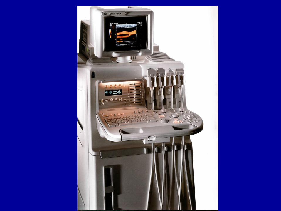

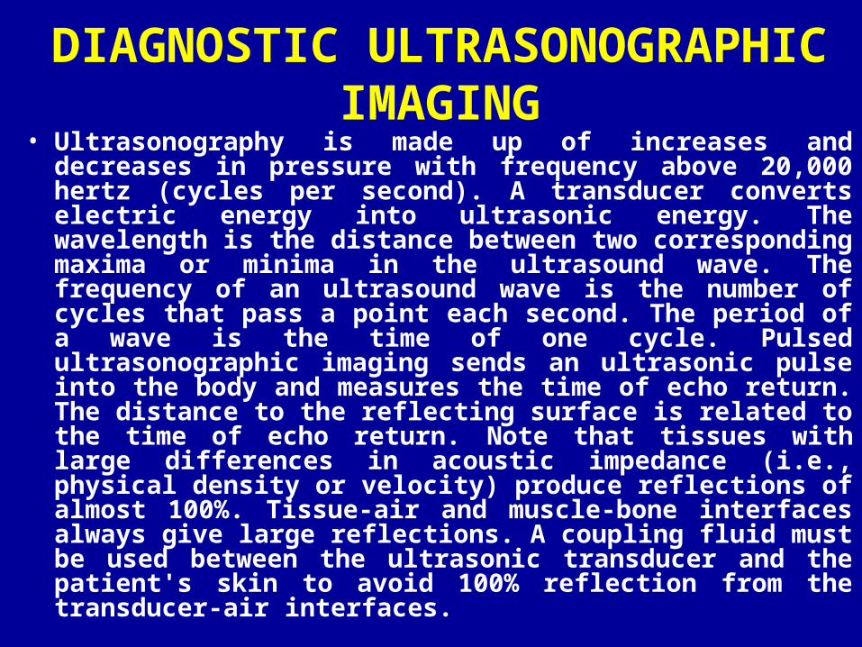
DIAGNOSTIC ULTRASONOGRAPHIC IMAGING
• Ultrasonography is made up of increases and decreases in pressure with frequency above 20,000 hertz (cycles per second). A transducer converts electric energy into ultrasonic energy. The wavelength is the distance between two corresponding maxima or minima in the ultrasound wave. The frequency of an ultrasound wave is the number of cycles that pass a point each second. The period of a wave is the time of one cycle. Pulsed ultrasonographic imaging sends an ultrasonic pulse into the body and measures the time of echo return. The distance to the reflecting surface is related to the time of echo return. Note that tissues with large differences in acoustic impedance (i.e., physical density or velocity) produce reflections of almost 100%. Tissue-air and muscle-bone interfaces always give large reflections. A coupling fluid must be used between the ultrasonic transducer and the patient's skin to avoid 100% reflection from the transducer-air interfaces.

Modes in USG
• A mode-AMPLITUDE MODEa. Ophthalmologyb. Orbital biometry
• B mode/ GRAY SCALE—For all routine applications
• M-mode - Echo cardiography• Doppleroghraphy• Duplex sonography.


Free Rotational and Slice Selection
Free Rotational and Slice Selection enables you to display any desired anatomical structure from any selected plane: horizontal, vertical and diagonal. By simply pressing a button, you choose the angle for image reconstruction. Thus, a better diagnostic confidence is achieved due to a more accurate visibility.
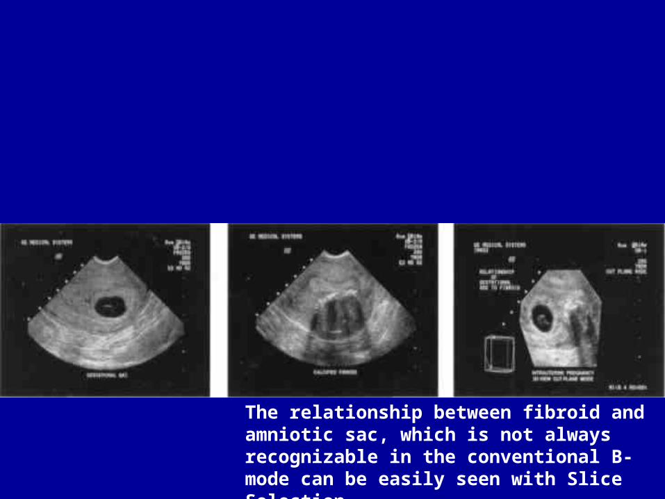
The relationship between fibroid and amniotic sac, which is not always recognizable in the conventional B-mode can be easily seen with Slice Selection
DOPPLER EFFECT
• Principle: described by Christian Johann Dopple -Frequency shift of moving object recorded
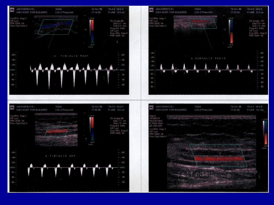
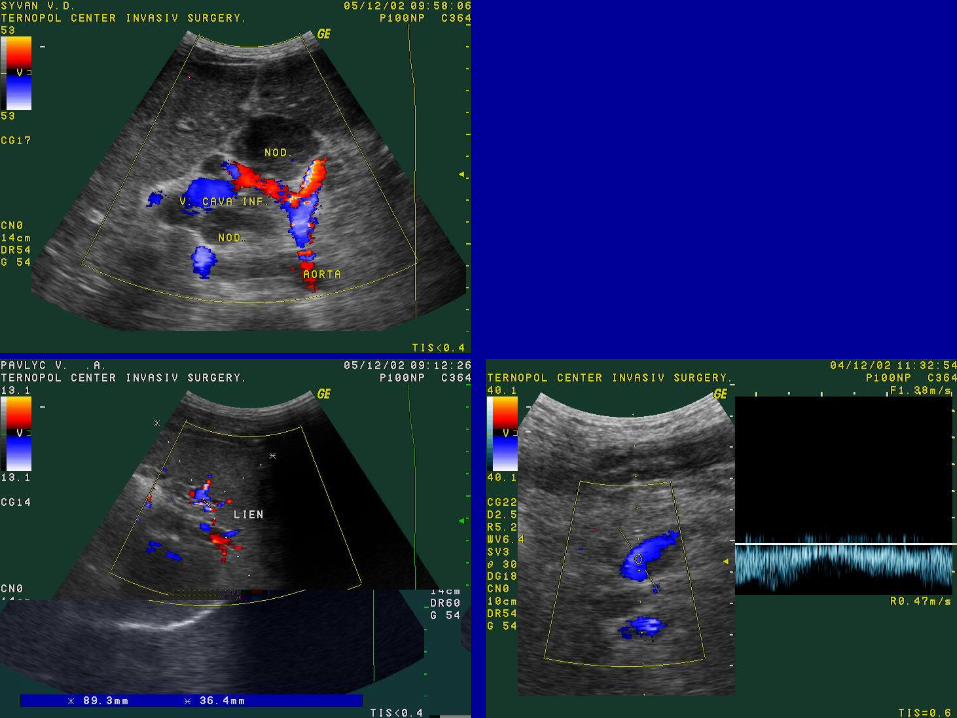
• Pulsed Doppler-gives the exact velocity waveform• Colour doppler-Direction Information-blue away from the
transducer, red is towards the transducer**• Power doppler-SIow Flow detection. Like tumoral
vascularity.• Common indications of Doppler include PVD, Carotid
atherosclerosis, Portal HT, IUGR, PIH, Renovascular HT etc.


Advantages• No radiation / portable
Disadvantage
Operator dependance
Delirious effect on small organism of USG is by acoustic cavitations.
Note—Following investigations do not use ionizing radiation:
a. Ultrasonography.
b. Thermography.
c. MRI.

Radiation hazards• X-rays used in conventional radiography and CT, as well as gamma rays and
other radionuclide emissions, are harmful. Natural radiation from the sun, radioactivity in the environment, together with atmospheric radioactivity from nuclear bombs and other man-made ionizing radiations contribute a genetic risk over which an individual doctor has no control. However, ionizing radiation for medical purposes is of several times greater magnitude than all other sources of man-made radiation and is under the control of doctors. It is their responsibility to limit the use of x-rays and other ionizing radiations to those situations where the benefit clearly outbalances the risks. Unnecessary radiation is to be deplored. The principle to be used is the so-called ALARA principle: 'as low as rea sonably achievable,. This is achieved by the use of appropriate equipment and good technique - limiting the size of the x-ray beam to the required areas, limiting the number of films to those that are necessary, keeping repeat examinations to a minimum and ensuring that the examination has not already been performed. Just as important as these factors, all of which are really the province of those who work in the x-ray department, is the avoidance of unnecessary requests for x-ray examinations, particularly those that involve high radiation exposure such as barium enema, lumbar spine x-rays and CT examinations. If possible, alternative techniques such as ultrasound or MRI should be considered.

THERMOGRAPHY
• Thermography – is the method of recording of a natural heat emission of the body of man in invisible infrared range of the electromagnetic spectrum. In present time are designed the methods of thermography in infrared, millimeter and decimeter band of wavelengths. By thermography the special thermal picture of all sites of the body is defined. In healthy man it rather constant, and changes at pathological processes. The method of thermography absolutely harmless and without any contraindications.
Technological algorithm of the work of a thermodiagnostical study
• Preparation of the room and instrumentation to examination
• Preparation of the patient for examination• Acquaintance with the case history of the patient and the
results of other examinations.• Choice of procedure of thermographic examination• Carrying out of thermographic examination• Analysis of the thermogram, comparison of the received
diagnostic information with the data of other methods of investigation
• Formulation of the results and drawing up of the examination protocol

Preparation of a workroom and instrumentation to examination
• Before the beginning of every working day the preparation of the room and instrumentation to operation is carried out which includes: checkout of the microclimate of the room, molding of fluid nitrogen in a recording thermometer, heating, check and calibration of recording thermometer.
• In the room for examination support a constant temperature in the range of 19±1°С (and for the study of dermal circulation 25±1°С) and air moisture 55-65 %.

Preparation of the patient for examination
• The examination is carried out by fasting, the repeated examinations are carried out at the same time of day, taking into account the daily temperature variations of the body
• The examination of the women is carried out on 10-12th day after the beginning of a menses
• 2 days before examination it is forbidden to apply physiotherapeutic procedures, mustard plasters, cupping glasses, irritating ointment for the narrowing or dilating of vessels, cosmetic resorts.
• 1 day before examination do not take remedies for the dilating or narrowing of vessels.
• At examination of extremities, on the eve, in the evening, it is necessary to have a bath for taking out of a cornual stratum of the skin, squamous epidermis, shortly to cut the nails, to take off ногтей a lacquer from the nails if it present.
• For the examination of the head at men it is necessary to have a shave on the eve, in evening, in order to prevent micro cuts.
• The examination is carried out in recumbent position, but it is also possible to carry out in a standing position.
Preparation of the patient for examination• Before examination the explored site of the body is denuded, then the patient adapts
to the temperature of a room during 10-15 mines., and at examination of palms and foots - 30 mines. In children the convective heat exchange is higher, than in the adults, and therefore for preventive of chills, examination is carried out at temperature 26°С. Also children are characterized by more slow thermoadaptation in comparison with the adults, which lasts near 20-25 mines. For more rapid adaptation the additional cooling of explored site by means of the fan or substances, which are quickly vaporized is applied. It enables to receive more contrast thermographic image. In the period of thermoadaptation the doctor takes the history of present illness, acquaintes to the results of other investigations, determines the indications to examination, chooses the method of examination.
• For the increasing of the efficiency of thermography the method of active thermography is applied, which is based on a different degree of response of a normal and pathologically changed tissues on physicochemical irritations. The simplest method for this purpose is the cold test. Cooling of an explored site by an aerosol of the ethanol during 10 min, or using gauze tampons, wetted with the alcohol-ethereal intermixture, enables to receive thermogram with a sharp image of zones of hyperthermia. The method of stress thermography consists in cooling of arms and forearms in the cold water (+8 - +14°С) during 0,5-2 min. Hyperglycemic test is based on intravenous introduction of the glucose in organism. The malignant tumours react to this test by increasing of temperature in affected zone on 0,7-3°С. The temperature of tumours raises also in conditions of a hyperbaric oxygenation.
The indications to application of thermography
• By the basic brunch of medicine, where the application of thermography is effective is the angiologia and oncology.
• Besides, the thermograph is indicated:• For the study of the thermo relief of a body of healthy people• For primary takeoff of the patients, who require the special
examination • For express-diagnostics of urgent conditions, including acute
inflammatory processes• For the estimation of innervations and circulation of explored site• For observing for the changes of pathological processes and the
results of their treatment.• Now it is successfully applied in such brunches of medicine, as
surgery, therapy, neurology and eye illnesses, infectious diseases, cardiology, and other
Methods of thermography
Three methods of thermographic examination are distinguished:
–а) Contact liquid-crystal thermography
–б) Remote infrared thermography
–в) Remote radio thermometry

Contact thermography• It is the method, which is based on the properties of the liquid crystals to
change the color depending on temperature. The liquid crystal is the substances, which in particular temperature range form a liquid phase, which has simultaneously properties of fluids, and crystalline solids. As the fluids, they possesses flow ability, as crystals - anisotropy. It is such original "chameleons" which are capable to change the color depending on temperature of a surface on which they are put.
• In present time the industry releases the liquid-crystal heat-indicating films, which represent a dispersion of the liquid crystals in vinyl alcohol, put on a special elastic black film, or the layer of liquid crystals between two polymer film, one of which black-colored. The skin before superimposition of the film is lubricated with Vaseline or glycerin. Such films are reusable. However, at some pathological states of the skin (combustion, frostbites, some dermal diseases), they cannot be applied. Also it is impossible to simulate a composite configuration of the body, to receive plain thermogram by them. Except the films, the special screens covered with a liquid-crystal composition are released also. During the thermography the screen comes nearer to a surface of the skin and the image appears on it.
• Despite simplicity and disposability of the method, it is necessary to note, that its opportunities are restricted because imperfection of the compound of liquid crystals and poor assortment of heat-indicating films.

• The recording thermometer consists of such basic components:
• 1) the scanning mirror;• 2) the optical system of lenses;• 3) the receiver of infrared rays;• 4) the amplifier;• 5) the computer system of collecting,
processing and reproduction of the information

Remote radiothermometry (SHF- thermometry
• The method is based on measuring of thermal super high-frequency radiation of the body of the man in centimeter and decimeter band, which show the temperature of the deep stratums of the body.
• Thus, the basic drawback of infrared thermography - receiving of the information only from the superficial layers of the body is liquidated. However, the quality of instrumentation for this method is still low, that restricts its application.
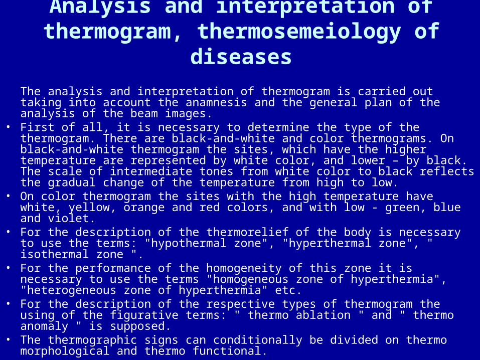
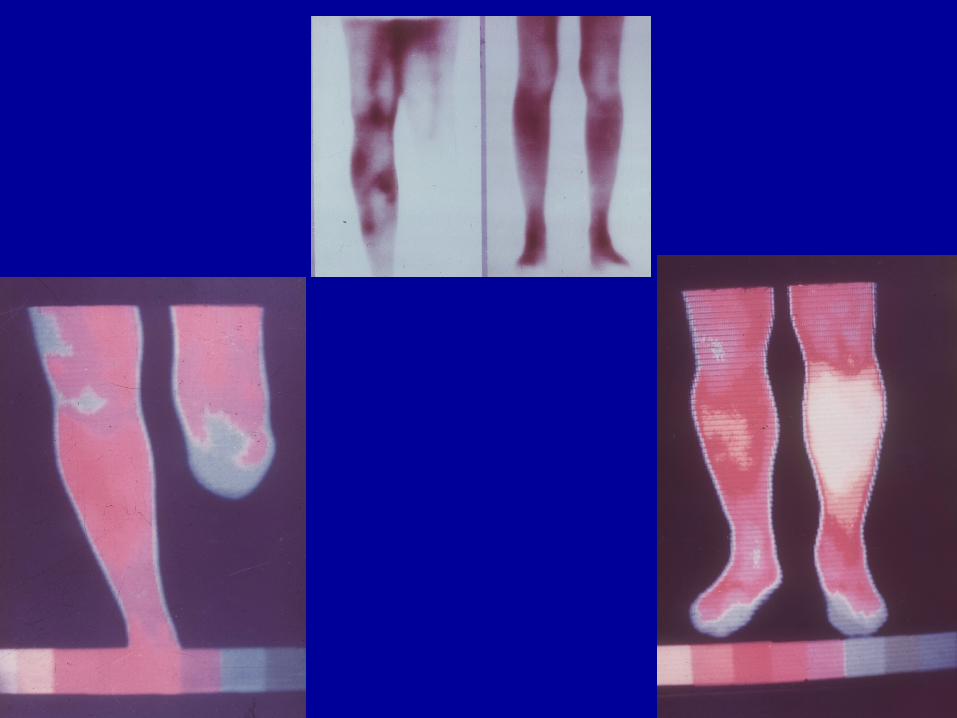
Analysis and interpretation of thermogram, thermosemeiology of diseases
The analysis and interpretation of thermogram is carried out taking into account the anamnesis and the general plan of the analysis of the beam images.
• First of all, it is necessary to determine the type of the thermogram. There are black-and-white and color thermograms. On black-and-white thermogram the sites, which have the higher temperature are represented by white color, and lower – by black. The scale of intermediate tones from white color to black reflects the gradual change of the temperature from high to low.
• On color thermogram the sites with the high temperature have white, yellow, orange and red colors, and with low - green, blue and violet.
• For the description of the thermorelief of the body is necessary to use the terms: "hypothermal zone", "hyperthermal zone", " isothermal zone ".
• For the performance of the homogeneity of this zone it is necessary to use the terms "homogeneous zone of hyperthermia", "heterogeneous zone of hyperthermia" etc.
• For the description of the respective types of thermogram the using of the figurative terms: " thermo ablation " and " thermo anomaly " is supposed.
• The thermographic signs can conditionally be divided on thermo morphological and thermo functional.
Advantages of thermography
The absolute harmlessness of the method, so it can be applied in repeated examinations, in children, pregnant women, at prophylactic examinations.
• High sensitivity of the method. A minimum temperature gradient between two points apart 1 mm is 0,1°С.
• High speed of examination. Depending on the type of thermometer the examination lasts 0,5-2 min.
• Ecological safety of instrumentation for examination.
• Opportunity of simultaneous, consecutive examination practically of all organs and systems of the man.
Drawbacks of thermography
• Low specificity of the method. Frankly, this concepts is closely connected with the qualification of the personnel. It is considered that only the specialist, who has carried out 1000-4000 thermogram and possesses the experience of the analysis and interpretation of the diagnostic information, which gains at other methods of radial diagnostics, can correctly interpret the thermograms.
• Information about thermo relief only of superficial layers of the body.
Related Documents