© 2017 Zhang et al. This work is published and licensed by Dove Medical Press Limited. The full terms of this license are available at https://www.dovepress.com/terms. php and incorporate the Creative Commons Attribution – Non Commercial (unported, v3.0) License (http://creativecommons.org/licenses/by-nc/3.0/). By accessing the work you hereby accept the Terms. Non-commercial uses of the work are permitted without any further permission from Dove Medical Press Limited, provided the work is properly attributed. For permission for commercial use of this work, please see paragraphs 4.2 and 5 of our Terms (https://www.dovepress.com/terms.php). Journal of Pain Research 2017:10 295–302 Journal of Pain Research Dovepress submit your manuscript | www.dovepress.com Dovepress 295 ORIGINAL RESEARCH open access to scientific and medical research Open Access Full Text Article http://dx.doi.org/10.2147/JPR.S127157 Ultrasound-guided alcohol neurolysis and radiofrequency ablation of painful stump neuroma: effective treatments for post-amputation pain Xin Zhang Yongming Xu Jin Zhou Shaofeng Pu Yingying Lv Yueping Chen Dongping Du Pain Management Center, Shanghai Jiao Tong University Affiliated Sixth People’s Hospital, Shanghai, People’s Republic of China Background: Post-amputation pain (PAP) is highly prevalent after limb amputation, and stump neuromas play a key role in the generation of the pain. Presently, PAP refractory to medical management is frequently treated with minimally invasive procedures guided by ultrasound, such as alcohol neurolysis and radiofrequency ablation (RFA). Objective: To record the immediate and long-term efficacy of alcohol neurolysis and RFA. We first used alcohol neurolysis and then, when necessary, we performed RFA on PAP patients. Study design: Prospective case series. Setting: Pain management center. Methods: Thirteen subjects were treated with ultrasound-guided procedures. Results: All patients were treated with neurolysis using alcohol solutions guided by ultrasound. Seven (54%) of 13 subjects achieved pain relief after 1–3 alcohol injection treatments. The remaining 6 subjects obtained pain relief after receiving 2 administrations of ultrasound-guided RFA. After a 6-month follow-up evaluation period, pain quantities were also assessed. Both stump pain (including intermittent sharp pain and continuous burning pain) and phantom pain were relieved. The frequency of intermittent sharp pain was decreased, and no complications were noted during the observation. Conclusion: The use of ultrasound guidance for alcohol injection and RFA of painful stump neuromas is a simple, radiation-free, safe, and effective procedure that provides sustained pain relief in PAP patients. In this case series, RFA was found to be an effective alternative to alcohol injection. Keywords: post-amputation pain, neuroma, ultrasound-guided, alcohol neurolysis, radiofre- quency ablation Introduction Post-amputation pain (PAP) is highly prevalent after limb amputation but remains as an extremely challenging condition to treat. 1 The loss of a body part can lead to 3 distinct descriptive sensory categories, phantom sensation, stump pain, and phantom pain. 1,2 Phantom sensation means that patients can still feel the existence of the missing limb after amputation. Usually, this type of sensation is not painful and not a clinical problem. Stump pain occurs immediately after amputation and usually is relieved after a few weeks as the wound heals. However, in some cases, persistent stump pain can occur and can be difficult to treat. Phantom pain means abnormal pain localized in the missing limb. It may be constant but has various intensities and can be described in different terms (shooting, burning, cramping, and aching). Although these 3 categories Correspondence: Dongping Du Pain Management Center, Shanghai Jiao Tong University Affiliated Sixth People’s Hospital, No. 600, Yishan Road, Shanghai, People’s Republic of China Tel +86 21 2405 8896 Fax +86 21 2405 8330 Email [email protected]

Welcome message from author
This document is posted to help you gain knowledge. Please leave a comment to let me know what you think about it! Share it to your friends and learn new things together.
Transcript
© 2017 Zhang et al. This work is published and licensed by Dove Medical Press Limited. The full terms of this license are available at https://www.dovepress.com/terms. php and incorporate the Creative Commons Attribution – Non Commercial (unported, v3.0) License (http://creativecommons.org/licenses/by-nc/3.0/). By accessing the work
you hereby accept the Terms. Non-commercial uses of the work are permitted without any further permission from Dove Medical Press Limited, provided the work is properly attributed. For permission for commercial use of this work, please see paragraphs 4.2 and 5 of our Terms (https://www.dovepress.com/terms.php).
Journal of Pain Research 2017:10 295–302
Journal of Pain Research Dovepress
submit your manuscript | www.dovepress.com
Dovepress 295
O R I G I N A L R E S E A R C H
open access to scientific and medical research
Open Access Full Text Article
http://dx.doi.org/10.2147/JPR.S127157
Ultrasound-guided alcohol neurolysis and radiofrequency ablation of painful stump neuroma: effective treatments for post-amputation pain
Xin Zhang Yongming Xu Jin Zhou Shaofeng Pu Yingying Lv Yueping Chen Dongping DuPain Management Center, Shanghai Jiao Tong University Affiliated Sixth People’s Hospital, Shanghai, People’s Republic of China
Background: Post-amputation pain (PAP) is highly prevalent after limb amputation, and stump
neuromas play a key role in the generation of the pain. Presently, PAP refractory to medical
management is frequently treated with minimally invasive procedures guided by ultrasound,
such as alcohol neurolysis and radiofrequency ablation (RFA).
Objective: To record the immediate and long-term efficacy of alcohol neurolysis and RFA.
We first used alcohol neurolysis and then, when necessary, we performed RFA on PAP patients.
Study design: Prospective case series.
Setting: Pain management center.
Methods: Thirteen subjects were treated with ultrasound-guided procedures.
Results: All patients were treated with neurolysis using alcohol solutions guided by ultrasound.
Seven (54%) of 13 subjects achieved pain relief after 1–3 alcohol injection treatments. The
remaining 6 subjects obtained pain relief after receiving 2 administrations of ultrasound-guided
RFA. After a 6-month follow-up evaluation period, pain quantities were also assessed. Both
stump pain (including intermittent sharp pain and continuous burning pain) and phantom pain
were relieved. The frequency of intermittent sharp pain was decreased, and no complications
were noted during the observation.
Conclusion: The use of ultrasound guidance for alcohol injection and RFA of painful stump
neuromas is a simple, radiation-free, safe, and effective procedure that provides sustained
pain relief in PAP patients. In this case series, RFA was found to be an effective alternative to
alcohol injection.
Keywords: post-amputation pain, neuroma, ultrasound-guided, alcohol neurolysis, radiofre-
quency ablation
IntroductionPost-amputation pain (PAP) is highly prevalent after limb amputation but remains
as an extremely challenging condition to treat.1 The loss of a body part can lead to 3
distinct descriptive sensory categories, phantom sensation, stump pain, and phantom
pain.1,2 Phantom sensation means that patients can still feel the existence of the missing
limb after amputation. Usually, this type of sensation is not painful and not a clinical
problem. Stump pain occurs immediately after amputation and usually is relieved after
a few weeks as the wound heals. However, in some cases, persistent stump pain can
occur and can be difficult to treat. Phantom pain means abnormal pain localized in the
missing limb. It may be constant but has various intensities and can be described in
different terms (shooting, burning, cramping, and aching). Although these 3 categories
Correspondence: Dongping DuPain Management Center, Shanghai Jiao Tong University Affiliated Sixth People’s Hospital, No. 600, Yishan Road, Shanghai, People’s Republic of China Tel +86 21 2405 8896 Fax +86 21 2405 8330 Email [email protected]
Journal name: Journal of Pain Research Article Designation: ORIGINAL RESEARCHYear: 2017Volume: 10Running head verso: Zhang et alRunning head recto: Ultrasound-guided treatments for post-amputation painDOI: http://dx.doi.org/10.2147/JPR.S127157

Journal of Pain Research 2017:10submit your manuscript | www.dovepress.com
Dovepress
Dovepress
296
Zhang et al
are described separately, amputees usually experience at least
one3 and, in most cases, have difficulty distinguishing one
category from another.4
The treatment of PAP is quite difficult as it has multi-
factorial mechanisms, and the pathophysiological causes
of PAP often remain unclear. The interactions between
peripheral, spinal, and supraspinal mechanisms are thought
to contribute to PAP phenomena.1 These mechanisms
include somatosensory cortical reorganization, central
sensitivity, and catastrophizing factors. Spinal reorganiza-
tion in dorsal horns, expansion of receptive fields, loss of
inhibitory interneurons, and activation of glial cells often
occur at the spinal level after a peripheral nerve injury.
Among all the peripheral mechanisms, the generation of
neuromas can lead to changes of ion channel expression,
alteration of receptor proteins, and ectopic discharges from
severed nerve endings.1 Notably, once the nerves are tran-
sected by trauma, neuromas can develop at the ends after
6–10 weeks. Neuromas may be regarded as a normal part
of the healing process, but the development of amputation
stump neuromas is a common and frequent cause of PAP.5
Injection therapy is widely used in many pain management
centers. The target of the injection is sometimes myofas-
cial tissue (at a trigger point) and sometimes the neuroma
stumps. Injected therapeutic agents can be local anesthet-
ics,6 steroids,7 chemo-denervation substances (botulinum
toxin),8 phenol,9,10 alcohol,11 etc. However, in most cases,
local injection therapy seems to be more efficacious in the
treatment of stump pain than in phantom pain. A separate,
small case series found that for patients who experienced
relief from a diagnostic local anesthetic injection, pulsed
radiofrequency ablation (RFA) was effective in relieving
PAP.12
The use of high-resolution ultrasound guidance to assist
with the injection procedure is becoming increasingly popular
because of real-time visualization of the stump neuroma in
soft tissue.13,14 Using this technique, alcohol injection11 and
RFA12,15 can be performed more easily and accurately.
However, knowledge of effective management of PAP
with alcohol neurolysis and RFA is limited, and their pro-
cedural techniques have not been standardized. The differ-
ences in outcome between alcohol injection and RFA have
not been reported. Here, we present a case series of 12
PAP patients in whom we used alcohol neurolysis first and,
when necessary, secondarily performed RFA. We tried to
obtain preliminary data on safety, efficacy, side effects, and
complications of injection therapy guided by ultrasound on
the different types of symptoms reported by PAP patients.
We are also attempting to develop a standard protocol for
alcohol neurolysis and RFA of neuroma.
MethodsSubjectsThe Institutional Review Board of Shanghai Jiaotong University
Affiliated Shanghai Sixth People’s Hospital approved the study
protocol. Informed consent for participation in the study was
obtained from each patient, and a consent form was reviewed and
signed, which included the risks, possible adverse consequences
of alcohol neurolysis and RFA. Written informed consent was
obtained for publication of this paper and accompanying images.
Twelve adult patients (7 men, 5 women; median age, 57.5 years;
range, 32–82 years) who had undergone limb amputation (upper
extremity, n = 4; lower extremity, n = 8) and presented to the
Shanghai Sixth People’s Hospital Pain Management Center with
PAP between March 2014 and April 2015 were recruited as study
subjects (Table 1). The clinical assessments for this prospective
study were performed by an experienced pain physician, who
selected study subjects with stable general condition at the
stump (no local inflammation or other acute tissue alterations).
Data recordingData regarding each patient’s pain symptoms were recorded
carefully. We divided stump pain into 2 categories. One cate-
gory, “paroxysmal” pain, was defined as intermittent sharp pain
of high intensity that had a sudden onset. The frequency of par-
oxysmal pain was recorded. The other category, “abiding” pain,
was defined as continuous burning pain of low intensity that
lasted for at least 1 hour. Each kind of stump pain was recorded
with a numerical rating scale (NRS, 10 maximum and 0 mini-
mum). Because patients often had difficulty in distinguishing
phantom pain from stump pain, we only recorded whether the
phantom pain was present (Table 1). The patients’ NRS scores
and the frequency of paroxysmal sharp pain were recorded
at 3 time points (before treatment and 2 weeks and 6 months
after the final treatment). As the frequency of paroxysmal pain
in all the patients was different, we only recorded the change
(less, equal, or more frequent than before) for the follow-up
evaluations (Table 3). During the treatment period, before each
operation, pain relief was recorded carefully. Because almost all
patients said they could not provide an accurate and comparable
NRS score during the ongoing treatment, we replaced the NRS
score with the following 4-step scale for the evaluation of pain
relief during treatments (Table 2):
Excellent – when the pain is completely resolved or
decreased by ≥75%
Good – when the pain decreased by 50%–74%
Journal of Pain Research 2017:10 submit your manuscript | www.dovepress.com
Dovepress
Dovepress
297
Ultrasound-guided treatments for post-amputation pain
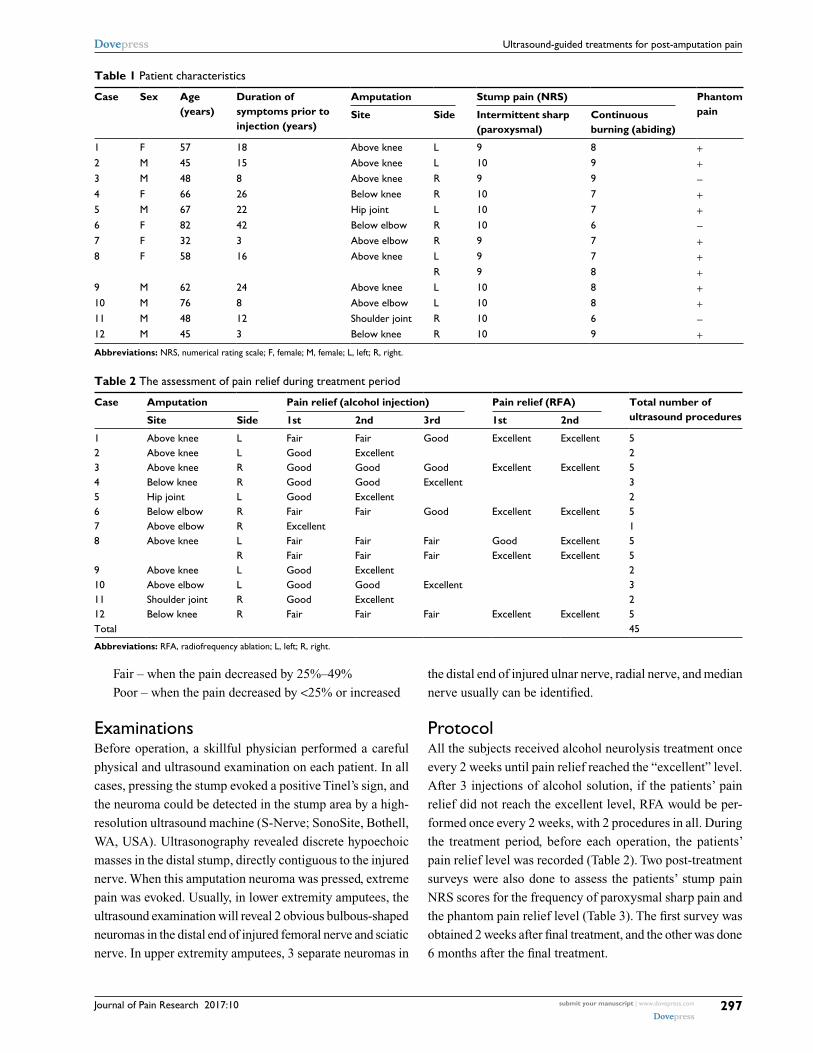
Fair – when the pain decreased by 25%–49%
Poor – when the pain decreased by <25% or increased
ExaminationsBefore operation, a skillful physician performed a careful
physical and ultrasound examination on each patient. In all
cases, pressing the stump evoked a positive Tinel’s sign, and
the neuroma could be detected in the stump area by a high-
resolution ultrasound machine (S-Nerve; SonoSite, Bothell,
WA, USA). Ultrasonography revealed discrete hypoechoic
masses in the distal stump, directly contiguous to the injured
nerve. When this amputation neuroma was pressed, extreme
pain was evoked. Usually, in lower extremity amputees, the
ultrasound examination will reveal 2 obvious bulbous-shaped
neuromas in the distal end of injured femoral nerve and sciatic
nerve. In upper extremity amputees, 3 separate neuromas in
the distal end of injured ulnar nerve, radial nerve, and median
nerve usually can be identified.
ProtocolAll the subjects received alcohol neurolysis treatment once
every 2 weeks until pain relief reached the “excellent” level.
After 3 injections of alcohol solution, if the patients’ pain
relief did not reach the excellent level, RFA would be per-
formed once every 2 weeks, with 2 procedures in all. During
the treatment period, before each operation, the patients’
pain relief level was recorded (Table 2). Two post-treatment
surveys were also done to assess the patients’ stump pain
NRS scores for the frequency of paroxysmal sharp pain and
the phantom pain relief level (Table 3). The first survey was
obtained 2 weeks after final treatment, and the other was done
6 months after the final treatment.
Table 1 Patient characteristics
Case Sex Age (years)
Duration of symptoms prior to injection (years)
Amputation Stump pain (NRS) Phantom painSite Side Intermittent sharp
(paroxysmal)Continuous burning (abiding)
1 F 57 18 Above knee L 9 8 +2 M 45 15 Above knee L 10 9 +3 M 48 8 Above knee R 9 9 −4 F 66 26 Below knee R 10 7 +5 M 67 22 Hip joint L 10 7 +6 F 82 42 Below elbow R 10 6 −7 F 32 3 Above elbow R 9 7 +8 F 58 16 Above knee L 9 7 +
R 9 8 +9 M 62 24 Above knee L 10 8 +10 M 76 8 Above elbow L 10 8 +11 M 48 12 Shoulder joint R 10 6 −12 M 45 3 Below knee R 10 9 +Abbreviations: NRS, numerical rating scale; F, female; M, female; L, left; R, right.
Table 2 The assessment of pain relief during treatment period
Case Amputation Pain relief (alcohol injection) Pain relief (RFA) Total number of ultrasound proceduresSite Side 1st 2nd 3rd 1st 2nd
1 Above knee L Fair Fair Good Excellent Excellent 52 Above knee L Good Excellent 23 Above knee R Good Good Good Excellent Excellent 54 Below knee R Good Good Excellent 35 Hip joint L Good Excellent 26 Below elbow R Fair Fair Good Excellent Excellent 57 Above elbow R Excellent 18 Above knee L Fair Fair Fair Good Excellent 5
R Fair Fair Fair Excellent Excellent 59 Above knee L Good Excellent 210 Above elbow L Good Good Excellent 311 Shoulder joint R Good Excellent 212 Below knee R Fair Fair Fair Excellent Excellent 5Total 45
Abbreviations: RFA, radiofrequency ablation; L, left; R, right.

Journal of Pain Research 2017:10submit your manuscript | www.dovepress.com
Dovepress
Dovepress
298
Zhang et al
Tab
le 3
The
ass
essm
ent
of im
med
iate
and
long
-ter
m p
ain
relie
f afte
r tr
eatm
ent
Cas
eA
mpu
tati
on
Stum
p pa
in (
NR
S)T
he c
hang
e of
fr
eque
ncy
of
paro
xysm
al p
ain
phan
tom
pa
inT
he c
hang
e of
pha
ntom
pa
inIn
term
itte
nt s
harp
(p
arox
ysm
al)
Con
tinu
ous
burn
ing
(a
bidi
ng)
Site
Side
Bef
ore
2 w
eeks
6 m
onth
sB
efor
e2
wee
ks6
mon
ths
2 w
eeks
6 m
onth
s2
wee
ks
afte
r tr
eatm
ents
6 m
onth
s af
ter
trea
tmen
ts
1A
bove
kne
eL
91
28
02
Less
Less
++−
+2
Abo
ve k
nee
L10
12
92
4Le
ssEq
ual
++−
−3
Abo
ve k
nee
R9
21
91
4Le
ssLe
ss−
n/a
n/a
4Be
low
kne
eR
102
37
13
Equa
lLe
ss++
++
5H
ip jo
int
L10
22
72
2Le
ssLe
ss++
−−
6Be
low
elb
owR
102
26
21
Less
Less
−n/
an/
a7
Abo
ve e
lbow
R9
24
73
3Le
ssLe
ss++
++
8A
bove
kne
eL
90
17
01
Less
Less
++−
+R
91
28
01
Less
Less
++9
Abo
ve k
nee
L10
02
82
3Le
ssLe
ss++
++
10A
bove
elb
owL
101
48
24
Less
Less
++++
++11
Shou
lder
join
tR
102
36
11
Less
Equa
l−
n/a
n/a
12Be
low
kne
eR
103
29
22
Less
Less
+++
+SD
0.50
6369
680.
8770
5802
0.94
7330
931.
0439
0785
0.96
0768
921.
1929
2788
Med
ian
102
27
22
Not
es: +
+, w
ith a
str
onge
r ph
anto
m p
ain;
+, w
ith a
wea
ker
phan
tom
pai
n; −
, the
pha
ntom
pai
n is
not
pre
sent
.A
bbre
viat
ions
: NR
S, n
umer
ical
rat
ing
scal
e; L
, lef
t; R
, rig
ht; n
/a, n
ot a
pplic
able
; SD
, sta
ndar
d de
viat
ion.

Journal of Pain Research 2017:10 submit your manuscript | www.dovepress.com
Dovepress
Dovepress
299
Ultrasound-guided treatments for post-amputation pain
Alcohol injectionAs described by Gruber et al,9 alcohol injection was per-
formed according to the following algorithm in each sub-
ject: patients were placed in a convenient position for the
intervention, which varied with the location of the neuroma.
After skin preparation with antiseptic solution, a linear high-
frequency ultrasound probe (6 MHz; S-Nerve; SonoSite) was
covered by a sterile plastic bag and placed on the subjects’
stump transversely (Figure 1A) to obtain a transverse axial
view (Figure 1B). The hypoechoic neuroma could be easily
detected. Then the probe was rotated vertically to reveal the
longitudinal image of the neuroma. In this image, we could
see the intact nerve tract leading to the neuroma (Figure 1C).
According to the method described by Gruber et al,9 the
analgesic drug should be injected into the nerve proximal
to the neuroma with the in-plane technique in longitudinal
image (Figure 1C). In our clinical experience, the nerve tract
proximal to the neuroma cannot be distinguished precisely in
some cases. It is known that if the injection was not performed
successfully, alcohol can damage the surrounding soft tis-
sue. Our alcohol injections have been usually performed in
the transverse image, and the injection needle was advanced
toward the neuroma body directly. When the needle penetrated
the body of the neuroma, the operator would adjust the tip
position to evoke the extreme pain that the patient could suffer
(Figure 2). After aspiration, a small amount of normal saline
(0.9% NaCl) solution would be injected to evoke the pain
and reconfirm that the needle tip was in the proper position.
When the target was confirmed, 10 mL of local anesthetic
was administered around the nerve proximal to the neuroma
with another sterile syringe under ultrasound guidance. When
the local anesthetic worked, 2–5 mL of dehydrated alcohol
solution would be injected into the neuroma body.
RFAOur RFA procedures were performed as described by Kim
et al16 with slight modification. After obtaining written
informed consent from each subject, the subject was placed
in a convenient position for the intervention, as mentioned
earlier. After skin preparation with antiseptic solution, an
ultrasound probe was placed on the subjects’ stump trans-
versely to obtain a transverse axial view (Figure 3). When
the stump neuroma was detected, slide the probe proximal
to the intact nerve pathway for ~5 mm. In this transverse
image, the response nerve diameter usually was ~4–8 mm.
After attachment to a radiofrequency (RF) generator (Baylis
Corporation, Montreal, Canada), a 10 cm RF needle with a
5 mm active tip would be advanced toward and positioned
just outside the nerve. Then, the needle was used to stimulate
the site at 0.4 mA in the sensory mode (50 Hz) to evoke the
patient’s pain. When the target was confirmed, 2 mL of the
local anesthetic solution with 10 mg of triamcinolone would
be injected through the needle. When the local anesthetic
worked, the needle would be advanced into the responding
nerve to perform RFA at 80°C for 90 seconds twice, separated
by a 60 second interval (Figure 3C).
ResultsSubject characteristics are presented in Table 1. Twelve sub-
jects (male, n = 7; female, n = 5) were enrolled. Subject 8
had a bilateral lower extremity amputation; so 13 amputation
sites are discussed. All the subjects reported 2 kinds of stump
A B
C
Figure 1 Detection of the neuromas.Notes: (A) Using ultrasound probe to scan the stump limb. (B) The transverse axial view of neuroma. (C) The longitudinal axial view of neuroma. The dotted line indicates neuroma.
Figure 2 Representative images of alcohol neurolysis to the neuromas.Notes: The injection needle was inserted into the neuroma body, and the tip was adjusted to evoke the extreme pain. The dotted line indicates neuroma.

Journal of Pain Research 2017:10submit your manuscript | www.dovepress.com
Dovepress
Dovepress
300
Zhang et al
pain (intermittent sharp pain and continuous burning pain)
and 10 of 13 (77%) had phantom pain combined with stump
pain. But almost all the patients had difficulty distinguishing
the 2 syndromes accurately.
Treatment periodWe recorded a 4-step scale for the evaluation during treat-
ments. As shown in Table 2, all the patients received 1–5
operative interventions. Seven (54%) of 13 subjects gained
pain relief ranked as excellent after 1–3 alcohol neurolysis
attempts (1 treatment session, n = 1; 2 sessions, n = 4; 3
sessions, n = 2). The remaining 6 (48%) subjects did not
achieve excellent pain relief level and then received RFA.
After administration of RFA, all the remaining subjects
reported excellent pain relief.
Final assessmentTwo post-treatment surveys of the NRS scores were obtained
from the subjects to quantify the frequency of paroxysmal
sharp pain and the level of phantom pain relief at 2 weeks and
6 months after the final treatments. Stump pain was divided
into intermittent sharp pain and continuous burning pain,
and each kind of pain has been recorded as NRS score. Two
weeks after the final treatment, the subjects had an overall
decrease in median NRS score for intermittent sharp pain
assessment from 10.0 ± 0.5 to 2.0 ± 0.9 and for continuous
burning pain assessment from 7.0 ± 1.0 to 2.0 ± 1.0. The
frequency of paroxysmal pain exhibited a large difference
among the subjects. Therefore, we only recorded the change
(less, equal, or more frequency than before treatment) for
the follow-up evaluation. Two weeks after treatments, only
1 subject (8%) reported that the frequency did not change,
and 12 (92%) subjects’ paroxysmal pain frequency was less
than before treatment (Table 3).
In the survey performed at 6 months after treatment, the
NRS scores were recorded again. The scores did not change
significantly during the follow-up period (from 2.0 ± 0.9 to
2.0 ± 0.9 for intermittent sharp pain assessment and from 2.0
± 1.0 to 2.0 ± 1.2 for continuous burning pain assessment).
Paroxysmal pain frequency at 6 months was recorded and
compared to that at 2 weeks after treatment. Compared with
2 weeks after treatments, 11 (85%) of 13 subjects’ sharp
pain frequency decreased, while 2 subjects’ (15%) frequency
remained the same (Table 3).
The characteristics of phantom pain also cannot be
quantified accurately. We used a 3-scale method (stronger
pain, ++; weaker pain, +; pain not present, −) to assess
the changes of phantom pain, but this pain recording was
complicated. Before treatment, 10 of 13 (77%) subjects had
phantom pain symptoms, whereas 2 weeks after the final
treatment, 5 (50%) of the 10 subjects were free of phantom
pain, 4 (40%) reported less pain, and only 1 (10%) reported
unchanged phantom pain. Notably, at the 6-month follow-up,
3 of 5 (60%) phantom pain-free subjects remained phantom
pain free, whereas 2 (40%) of the 5 had recrudescent phan-
tom pain, although their pain levels were lower than before
treatment. In the 4 phantom pain relief subjects, mild pain
remained at an intensity equal to the level reported 2 weeks
after the final treatment. There was only 1 subject (10%)
whose phantom pain did not change during the observa-
tion period. The 3 subjects who had no phantom pain at the
outset of the study remained free of this pain throughout the
observation period (Table 3).
DiscussionPAP is of neuropathic origin, and its treatment can be very
challenging. The underlying mechanisms for this type of
pain are multifactorial, including supraspinal-, spinal-, and
peripheral-level components.1 As indicated by multiple stud-
ies, both stump pain and phantom pain can be controlled by
peripheral nerve block to some extent.6,17 Treatments focused
on peripheral nerve might be an effective method, such as
local injection therapy, RFA, peripheral nerve stimulation,
and surgery. In this study, we used 2 methods – alcohol
neurolysis and RFA – to block the peripheral ectopic inputs
from amputation neuromas. Both procedures can relieve PAP.
Alcohol11 and phenol10 are the most commonly used
neurolytic agents for chemical ablation and have been proved
A B C
Figure 3 RF procedure.Notes: (A) The RF generator machine. (B) The transverse axial view of neuroma body. (C) RF needle was advanced to the responsible nerve 5 mm away from neuroma stalk. The dotted line indicates neuroma.Abbreviation: RF, radiofrequency.

Journal of Pain Research 2017:10 submit your manuscript | www.dovepress.com
Dovepress
Dovepress
301
Ultrasound-guided treatments for post-amputation pain
efficacious in the management of chronic neuropathic pain,
including stump neuromas. Gruber et al10 described the
technique of chemical neurolysis for neuroma in detail in
2003. They used an ultrasound probe to scan the stump limb
to gain a longitudinal image of the tumor, from which they
could see hypoechoic terminal stump neuromas continued
from the nerves of origin. Then they injected phenol into the
nerve stalk just proximal to the neuroma under an in-plane
approach. The phenol volume was ~0.3–1 mL. Lim et al11
reported using dehydrated alcohol (volume of 1.2 mL) for
injections into nerve tracts proximal to neuromas. In our
study, alcohol was injected directly into the neuroma body
other than the stalk, and we found it easier to inject the agent
into the body than into the nerve itself. In the longitudinal
view, the neuroma stalk that is in continuity with original
nerve cannot be easily identified in every patient. Normally,
the stalk is small and obscure. Therefore, injection into the
stalk is not assured. If the chemical agent is not accurately
injected into the stalk, ectopic impulses from the neuroma
may not be blocked completely and surrounding soft tissues
may be damaged. As the volume of the neuroma body is far
higher than that of the neuroma stalk, we need more of the
chemical agent to effectuate complete neurolysis. However,
the texture of neuroma bodies can be very compact, so that
even with the application of a copious amount of chemical
agent, it cannot be guaranteed that the agent will diffuse well
throughout the tumor. Hence, during the procedure we adjust
the position of the needle tip little by little until the needle
evokes the exact pain that replicates the subject’s spontaneous
events. Injecting agent into that specific area of the neuroma
can block the abnormal discharge of neuroma completely and
improve the success rate of neurolysis.
Neuroma development is a part of a normal reparative
process following peripheral nerve injury. Usually, the distal
terminal area of the injured nerve will generate a neuroma.
However, in some cases, several neuromas can grow at the
end of a nerve terminal. These neuromas can form a grape-
like cluster or gathering at the end of a single nerve fiber
(Figure 4A). In this setting, the issue of how to identify the
specific neuroma responsible for the generation of pain is
a significant problem that needs to be addressed. Use of a
nerve stimulator might be useful for this, using sensory mode
stimulation (50 Hz) at 0.4 mA to reproduce the patient’s
symptoms to help identify the target neuroma.
Ultrasound-guided RFA is also used for the treatment of
PAP. West and Wu12 reported a case series showing that for
patients who experienced relief from a diagnostic lidocaine
injection, pulsed radiofrequency (PRF) ablation was effec-
tive in relieving PAP. Kim et al16 also described this method,
placing the PRF needle on the neuroma stalk as close as
possible to the distal part of the associated nerve. However,
observations in this study suggest that some nerve tissue,
those close to the neuroma stalk, can show significant patho-
logical changes. As shown in Figure 4B and C, the nerve
tissue close to neuroma stalk shows significant swelling,
with a diameter as large as 1 cm or more (Figure 4B and C).
However, the PRF needle normally has a 5 mm active tip. It is
very difficult to use such a tiny tip to disrupt a large-diameter
nerve completely. Therefore, we revised the procedure to try
to completely block the connection between the impaired
neuroma tissue and the healthy nerve tissue. We targeted the
nerve fiber at 5 mm proximal to the neuroma stalk, which in
most cases is healthy and has a diameter of 5–8 mm. At this
point, we can block the responsible nerve input completely
to relieve PAP. In most studies, the PRF was performed at
42°C.15,16 However, in our procedures, we raised the tem-
perature to 80°C, so that we could block abnormal inputs
completely and gain great pain relief. In this case series, no
complication has been observed after PRF ablation.
PAP includes phantom sensation, stump pain, and phan-
tom pain. In our general understanding, stump pain is mainly
contributed by the peripheral mechanisms, and phantom pain
is mainly contributed by the central mechanisms. Alcohol
neurolysis and RFA are procedures that block the ectopic
peripheral nerve inputs. Theoretically, both procedures should
benefit stump pain more effectively than phantom pain. But in
reality, these 2 kinds of pain often have similar simultaneous
outbreak patterns, and patients usually cannot distinguish
A B C
Figure 4 The variation of neuromas.Notes: (A) Three neuromas origin from 1 nerve. (B and C) The nerve immediately proximal to the neuroma is pathological. The nerve is usually swollen, with its diameter as large as 1 cm or more. (B) is the transverse axial view of neuroma body, and (C) is the transverse axial view of neuroma stalk. The dotted line indicates neuroma.

Journal of Pain Research 2017:10submit your manuscript | www.dovepress.com
Dovepress
Dovepress
Journal of Pain Research
Publish your work in this journal
Submit your manuscript here: https://www.dovepress.com/journal-of-pain-research-journal
The Journal of Pain Research is an international, peer reviewed, open access, online journal that welcomes laboratory and clinical findings in the fields of pain research and the prevention and management of pain. Original research, reviews, symposium reports, hypoth-esis formation and commentaries are all considered for publication.
The manuscript management system is completely online and includes a very quick and fair peer-review system, which is all easy to use. Visit http://www.dovepress.com/testimonials.php to read real quotes from published authors.
Dovepress
302
Zhang et al
them clearly. In this case series, the 10 subjects had both
stump pain and phantom pain. They obtained great relief
of the stump pain by blocking peripheral nerve discharges
through alcohol neurolysis or RFA. Further, 9 of 10 subjects’
phantom pain also eased, though phantom pain is thought to
originate from the central nervous system. Notably, ampu-
tation of the peripheral nerves resulted in hyperexcitability
and spontaneous action potential discharge in the damaged
nerve tracts, which may be a potential source of the stump
pain, including phantom pain.18 This mechanism may help to
explain why the phantom pain was also relieved in our study.
There is also evidence from the study of Borghi et al17 to
show that peripheral nerve blocks can control phantom pain.
Studies elucidating the differences between the effects
of alcohol neurolysis and RFA on PAP are lacking. In our
study, after alcohol neurolysis, 6 of 13 subjects reported that
their pain relief did not reach the excellent level. When we
later performed RFA on these 6 subjects, they all reported
excellent pain relief. It seems that RFA can be taken as an
alternative method to treat PAP patients. Further studies
are needed to further clarify the differences between the 2
methods in more depth.
ConclusionOur case series reports data on the feasibility, safety, and
efficacy of ultrasound-guided alcohol injection and RFA
with PAP patients. We conclude that the use of ultrasound
guidance for alcohol neurolysis and RFA is a promising
tool for the treatment of PAP. Although limited by a small
sample size, our observations suggest 2 important conclu-
sions: 1) alcohol injection and RFA are safe and effective
procedures to treat PAP, including stump pain and phantom
pain; and 2) RFA might be an effective alternative method
to alcohol injection.
AcknowledgmentThis work was supported in part by the General Program of
National Natural Science Foundation of China (81370933,
81400803, and 81672237).
DisclosureThe authors report no conflicts of interest in this work.
References 1. Hsu E, Cohen SP. Postamputation pain: epidemiology, mechanisms,
and treatment. J Pain Res. 2013;6:121–136. 2. Pirowska A, Wloch T, Nowobilski R, Plaszewski M, Hocini A, Menager D.
Phantom phenomena and body scheme after limb amputation: a litera-ture review. Neurol Neurochir Pol. 2014;48(1):52–59.
3. Ephraim PL, Wegener ST, MacKenzie EJ, Dillingham TR, Pezzin LE. Phantom pain, residual limb pain, and back pain in amputees: results of a national survey. Arch Phys Med Rehabil. 2005;86(10):1910–1919.
4. Sherman RA, Sherman CJ. Prevalence and characteristics of chronic phantom limb pain among American veterans. Results of a trial survey. Am J Phys Med. 1983;62(5):227–238.
5. Henrot P, Stines J, Walter F, Martinet N, Paysant J, Blum A. Imaging of the painful lower limb stump. Radiographics. 2000;20(Spec No):S219–S235.
6. Fischler AH, Gross JB. Ultrasound-guided sciatic neuroma block for treatment of intractable stump pain. J Clin Anesth. 2007;19(8):626–628.
7. Kesikburun S, Yasar E, Dede I, Goktepe S, Tan AK. Ultrasound-guided steroid injection in the treatment of stump neuroma: pilot study. J Back Musculoskeletal Rehabil. 2014;27(3):275–279.
8. Wu H, Sultana R, Taylor KB, Szabo A. A prospective randomized double-blinded pilot study to examine the effect of botulinum toxin type A injection versus Lidocaine/Depomedrol injection on residual and phantom limb pain: initial report. Clin J Pain. 2012;28(2):108–112.
9. Gruber H, Kovacs P, Peer S, Frischhut B, Bodner G. Sonographically guided phenol injection in painful stump neuroma. AJR Am J Roent-genol. 2004;182(4):952–954.
10. Gruber H, Glodny B, Kopf H, et al. Practical experience with sonograph-ically guided phenol instillation of stump neuroma: predictors of effects, success, and outcome. AJR Am J Roentgenol. 2008;190(5):1263–1269.
11. Lim KB, Kim YS, Kim JA. Sonographically guided alcohol injection in painful stump neuroma. Ann Rehabil Med. 2012;36(3):404–408.
12. West M, Wu H. Pulsed radiofrequency ablation for residual and phantom limb pain: a case series. Pain Pract. 2010;10(5):485–491.
13. Brown MR, Farquhar-Smith P, Williams JE, Ter Haar G, deSouza NM. The use of high-intensity focused ultrasound as a novel treatment for painful conditions-a description and narrative review of the literature. Br J Anaesth. 2015;115(4):520–530.
14. Sivan M, Stoppard E. Sonographically guided phenol instillation of stump neuroma. AJR Am J Roentgenol. 2008;191(5):W208; author reply W209.
15. Restrepo-Garces CE, Marinov A, McHardy P, Faclier G, Avila A. Pulsed radiofrequency under ultrasound guidance for persistent stump-neuroma pain. Pain Pract. 2011;11(1):98–102.
16. Kim YK, Jung I, Lee CH, Kim SH, Kim JS, Yoo BW. Pulsed radiofre-quency ablation under ultrasound guidance for huge neuroma. Korean J Pain. 2014;27(3):290.
17. Borghi B, D’Addabbo M, Borghi R. Can neural blocks prevent phantom limb pain? Pain Manag. 2014;4(4):261–266.
18. Flor H, Nikolajsen L, Staehelin Jensen T. Phantom limb pain: a case of maladaptive CNS plasticity? Nat Rev Neurosci. 2006;7(11):873–881.
Related Documents